Pulse Active Transform (PAT): A Non-Invertible
Transformation with Application to ECG Biometric
Authentication
Sairul I Safie, Nurfazira H , Azavitra Z Malaysian Institute of Industrial Technology (MITEC)
University Kuala Lumpur Johor Bahru, Johor [email protected]
John J Soraghan, and Lykourgos Petropoulakis Centre for Excellence in Signal and Image Processing
University of Strathclyde, Glasgow, Scotland {j.soraghan, akis}@eee.strath.ac.uk
Abstract— This paper presents a new transformation technique called the Pulse Active transform (PAT). The PAT uses a series of harmonically related periodic triangular waveforms to decompose a signal into a finite set of pulse active features. These features incorporate the signal’s information in the pulse active domain, and which are subsequently processed for some desired application. PAT is non-invertible thus ensuring complete security of the original signal source. In this paper PAT is demonstrated on an ECG signal and used for biometric authentication. The new transformation technique is tested on 112 PTB subjects. It is shown in this paper that the new transformation has a superior performance compared to the conventional characteristic based feature extraction methods with additional security to avoid recovery of the original ECG.
Keywords- Pulse Active Transform, Electrocardiogram, Biometric Authentication
I. INTRODUCTION
Features extracted from any location of a signal represent specific information of that signal. In some cases, these locations generate distinctiveness to discriminate individuals using signals such as electrocardiograms (ECGs) [1-4]. This paper presents a new transformation technique called the Pulse Active Transform (PAT). PAT transforms a signal by considering a relationship between multiple pairs of points in a signal, and recombining this relationship to form the transform signal. Features extracted from the signal locations are separately extracted and are used to generate a new form of signal which includes unique information from different parts of the original signal. Unlike most widely used integral transformation techniques such as Fourier, Laplace or Wavelet transforms, PAT does not require any specific kernel function to transform a signal making the transformed signals hard to be inverted.
The new PAT is demonstrated for secure biometric authentication using an ECG as a signal. When algorithms are used to extract features from a biometric trait, it is important that these algorithms are not prone to invertibility attacks i.e. attacks aimed at recovering the original biometric knowing the feature vectors, algorithm and settings parameters, obtainable from the security system [5]. If this biometric information can
be reconstructed, it can no longer serve as a biometric trait and in some cases the health information embedded, as for example in ECGs, is no longer private.
The remainder of this paper is organized as follows. Section II derives the new Pulse Active transformation method (PAT). Implementation of PAT for biometric authentication is shown in section III. Section IV discusses the advantages of the fact that the PAT is non-invertible. Finally section V concludes the paper.
II. PULSEACTIVETRANSFORM
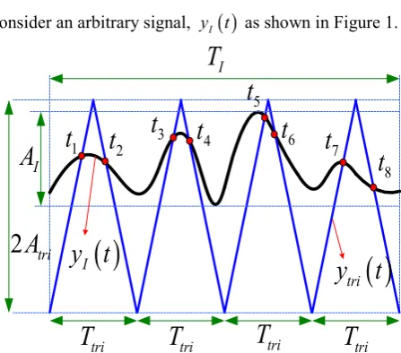
Consider an arbitrary signal, y tI
( )
as shown in Figure 1.( )
tri
y t
( )
I
y t
3
t
4
t
5
t
6
t
1
t
t
2t
78
t
tri
T
T
triT
triT
triI
T
I
A
[image:1.595.314.538.382.579.2]2
A
triFigure 1: Pulse Active Transformation waveform generation
constant parameters known as the integer value modulation factor mf, and the modulation index mi are defined as:
I f
tri T m
T
= (1)
tri i
I A m
A
= (2)
Equations (1) and (2) are used to relate the duration and amplitude between y tI
( )
and ytri( )
t ensuring that there is an integer number of periods of the triangular waveform contained withinTI. Hence mf represents the integer number of periodic triangular waves forytri( )
t used in the interval TI. For example in Figure 1, mf set equal to 4.Each period of this periodic triangular waveform intersects the underlying investigated signal at distinct locations. For each triangular wave period, the location of the first intersection from the positive slope t+ve and the location of the last intersection from the negative slope t−ve are selected as the intersection points of interest for that period. Mathematically, this is described as follows:
(2 1)
(
)
(2 1)(
)
2 1
for 1
2
ve tri tri
m m
m
t − =t+ m− T ≤t − ≤ − T (3)
(
)
2 2
2 1
for 2
m ve tri m tri
m
t =t− − T ≤t ≤mT (4)
(2m1) 2m t − t
ª º
=¬ ¼
trans
T (5)
for m=1, 2,3...mf
trans
T which is defined as the transition state vector of PAT
corresponds to specific intersection location times, as illustrated in Figure 1. To ensure intersections occur within each triangular period, Atri should be set slightly higher than
I
A . This can be achieved by ensuring mi > 1.
The PAT FPAª¬mfº¼ , is calculated by taking the
cumulative duration between two contiguous points of Ttrans
for all mf values. Mathematically, the PAT is calculated as follows:
(
2 2 1)
1 f
m
PA f m m
m
F m t t −
=
ª º= −
¬ ¼
¦
(6)for mf =1, 2,3,...,M where M is the length of the PAT
signal. From (6), the first point of the PAT signal FPA
[ ]
1 , is the duration between the intersection points of yECG( )
t and( )
triy t when a single periodic triangular wave is consider. The
next point of the PAT signal FPA
[ ]
2 is calculated when two periodic triangular waves is used to intersect the ECGcomplex. This process continues until mf reach the final
[image:2.595.322.543.138.581.2]value of M which can be fixed or set by the user.
[image:2.595.41.281.360.450.2]Figure 2: Examples of PA transformed signal
signals is normalized to 1. These signals are transformed using M= 25 and the results illustrated in Figure 2 a) to f).
PAT transforms the rate of changes between the duration and amplitudes of the investigated signals into a
unique M-dimensional vector that may be used as a specific
signature of the signal itself. Furthermore, since the PAT is non-invertible, it keeps the signal under investigation completely secure. In this paper we will investigate the validity of this suggestion by applying the PAT feature extraction process to the challenging problem of ECG biometric authentication [3].
III. PATFOR ECGAUTHENTICATION
To implement PAT for ECG biometric authentication, AI
will be replaced by AECG which is the maximum amplitude of the ECG signal, yECG
( )
t , while the duration between peaks P and T, TECG replaces TI as shown in Figure 3.( )
tri
y t
( )
ECG
y
t
3
t
t
4t
5t
61
t
t
2tri
T
T
triT
triECG
T
ECG
A
2
A
triFigure 3: PA transformed on ECG signal
We have described the reasons for selecting this ECG portion in [3]. To validate the performance of the new Pulse Active transforms on ECG biometric system, a database of 112 subjects from the Physikalisch-Technische Bundesanstalt (PTB) database is used [6]. From these 112 subjects, 98 subjects have arrhythmia beats while the rest are healthy. In this paper, biometric performance will be investigated separately between these two populations (healthy and arrhythmia). The PTB database is selected in this study because, for the recordings provided, the average time interval between any two recordings of the same subject is about 500 days [7]. These subjects have at least two different ECG recordings and the ECG sources are taken from a lead I configuration (left and right hand). For each subjects, both
recordings will undergo the Pulse Active transformation process. One of these transforms signals will be used for the training database while another will be used for the test. Each of the transforms signal in the training database is compared with all transforms signal in the test database.
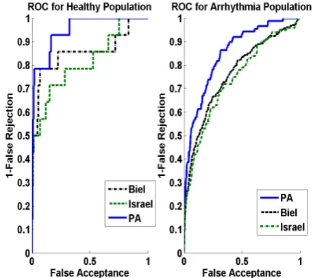
[image:3.595.58.282.324.491.2]The Euclidean distance is used as the distance measure to generate matching scores. If the transformed signals from a test and storage database are from the same subject, the matching score is labeled as a genuine score, else, the matching score is labeled as an imposter score. The score vectors are used to construct the receiver operating characteristic (ROC) curve. The ROC curve plot, is a function of the decision threshold, which plots the rate of ‘False Acceptance’ (i.e. impostor accepted as genuine) on the x-axis, against the ‘1-False Rejection’ (i.e. genuine accepted as genuine) on the y-axis. The area under an ROC curve (AUR) and Equal Error Rate (EER) are the common quantitative measure for comparing ROC curves. Their value ranges between 0 and 1. A higher value of AUR and lower value of EER indicates a system with a better performance.
Figure 4: ROC performance curve
[image:3.595.316.545.349.552.2]Table I: Biometric performance comparison values of Figure 4
Pulse Active Biel [1] Israel [2]
AUR Healthy 0.9470 0.8544 0.8136
AUR Arrhythmia 0.8513 0.7630 0.742
EER Healthy 0.1538 0.2198 0.2857
EER Arrhythmia 0.2388 0.3047 0.3101
IV. DISCUSSION
In the previous section, it was shown that PAT offers superior performance compared to other traditional characteristic based feature extraction techniques for the experimental study. This superior performance reflects the difference between amplitude and morphological shape of the transformed signal as shown in section II.
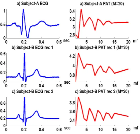
[image:4.595.60.292.371.592.2]In ECG biometric technology, it is impossible for two different ECG signals to have the exact same shape even though both ECGs may come from the same subject. This is because ECG signals are very sensitive to heart rate variability (HRV) and electrode placement. These factors will alter the temporal duration and amplitude of the ECG signals.
Figure 5: Pulse Active Transformation for on ECG signals
For example, it is known that when the heart rate increases, the total duration for all ECG complexes become shorter. Similarly, when an ECG electrode is placed at a location with thicker skin due, for example, to thicker subcutaneous layer, the signal amplitude of this ECG will be reduced. Figure 5 a) to c) illustrate different ECG signals from 2 subjects being
transformed using PAT with M = 20. As can be seen from
Figure 5 b) and c), the original and transformed signals of subject B are particularly similar between recordings 1 and 2. However it is observed that the transformed signals between subjects A and B are particularly different in term of shapes and amplitudes.
The PAT technique offers a solution to the HRV and
electrode placement problems by using the parameters mf
andmi . This is done by first detecting the fiducial points
(points of interest which correspond to peak and boundaries of the 3 major waves in an ECG signal). Two of these fiducial points are selected to start and end the PA transformation process. In this work, the peaks of P and T are selected as the two fiducial points. Fiducial point locations always vary
according to the heart rate. By fixing the value ofmf , any
changes on the heart rate (TECG reduction for example) is
compensated by the changes of the triangular period (Ttri
tends to follow the changes). Similarly, when the amplitude
ECG
A is reduced, for a fixed value of mi (in this
studymi =1.1) the amplitude of the triangular wave Atri also changes.
The values of Atri and Ttri are fundamental in determining the value of the transition state vector Ttrans. They reflect the slope of the triangular wave and determine the location where the triangular wave is to intersect the ECG signals. Although the PAT given in (6) is simple, it is impossible to reconstruct the original signal based only on the information of mf and the transform signal FPAª¬mfº¼
.
V. CONCLUSION
REFERENCES
[1] L. Biel, O. Pettersson and L. Philipson, ‘ECG analysis : a new approach in human identification’, IEEE Trans on Instrumentation and Measurement, 2001,50(3), pp.808-812.
[2] S. A. Israel, J. M. Irvine and A. Cheng, ‘ECG to identify individuals,’ Pattern Recognition, 2004, 38(1), pp. 133-142.
[3] Safie, S.I.; Soraghan, J.J.; Petropoulakis, L., "Electrocardiogram (ECG) Biometric Authentication Using Pulse Active Ratio (PAR)," Information Forensics and Security, IEEE Transactions on , vol.6, no.4, pp.1315,1322, Dec. 2011
[4] Sairul I Safie, John J Soraghan, and Lykourgos Petropoulakis, “Pulse Active Bit Feature Extractor”.18th International Conference on System, Signals and Image Processing, pp 55-58, June 16-18, 2011.
[5] A. Nagar, K. Nandakumar, A. K. Jain, "Biometric Template Transformation: A Security Analysis",Proc. of SPIE, Electronic Imaging Media Forensics and Security XII, San Jose, Jan. 2010.
[6] Goldberger AL, Amaral LAN and Glass L,. ‘PhysioBank, PhysioToolkit, and PhysioNet: Components of a New Research Resource for Complex Physiologic Signals.’, Circulation 101(23):e215-e220.