JOURNAL OFVIROLOGY, Mar. 1973,p.432-440
Copyright © 1973 American Society for Microbiology Printed in U.S.A.Vol. 11, No. 3
Physicochemical Studies
on
L-Cell Virions
J. L. NICHOLS, KRISTINA QUADE, AND R. B. LUFTIG
Departmenit oJ Microbiology and Immunology, Duke Untiversity Medical Center,
Durham, NorthCarolina 27710
Received forpublicationi21 November 1972
The L-cell virion (LCV) has been purified from supernatant fluids of mouse L
cellsgrowni insuspeinsionculture. The virion is similartoother RNAtumorviruses by several criteria: (i) thedensity of the virion is 1.16g/cm'; (ii) the virionappears as a rounded membranous particle with an outer diameter of 146.7 i 11.8 nm,
and contains knobs (7-nm diameter) overits surface; (iii) 15 polypeptides
(rang-ing in molecular weightfrom 7,000to 110,000) are detectable after electrophoresis
of virion protein in sodiumdodecyl sulfate-polyacrylamide gels; (iv) three species of RNAcan be isolated-high molecular weight (80to 88s) (50%), 7s (35%) and 4s (15%); (v) heat denaturation of the high-molecular-weight RNA yields a
heterogeneous population of molecules (20 to 35s) as wellas a 7s and 4s species.
Despite the general similarity to infectious RNA tumor viruses, LCV is
appar-ently defectiveasevidencedby the fact that it doesnotinducetumorsinanimals or
transform normal mouse cells in vitro (Kindig and Kirsten [171). The defective
nature ofthe LCV might be related to the fact that assaysfor DNA polymerase
in the virion showed only a negligible activity when compared to Rous sarcoma virus.
The presence of a C-type virus
particle
in established lines of mouse L cells was first reported by Dales andHowatson (7).Subsequent studies have shown that this L-cell virion(LCV) bandedat 1.16 to1.17 g/cm' in sucrose gradients, wasunableto inducetumors in miceor rats, and did not replicate in other cell lines (Kindig and Kirsten [17]; Kindig et al. [18]). Faras and Erikson (13)haveshownin apreliminary charac-terization of LCV RNA that thereisbotha high-molecular-weight component (82s) and a low-molecular-weight RNAcomponent (6s). Further,they
found that thehigh-molecular-weight
RNAdissociates
upon heat denaturation into a ho-mogenous RNA species with a sedimentation coefficient of approximately33s. Inthispaper we present a more detailed report on thephysico-chemicalproperties ofthe LCVRNA,as wellas
additional studies on the morphology, poly-peptide constituents, and DNA polymerase activity ofthevirion.
These studies were uindertakeni as ain initial effort toward
determining
the defectivenature of the LCV.MATERIALS AND METHODS
Viruspurification. MouseLcellswere
propa-gatedinEagleminimalessential medium (MEM) (Joklik's
Modificationi,
Grand IslandBiological
Co.)containing 5%fetal calfserum.Culturestobe
used for the isolation of virions were allowed to
grow for24h afterthe cell suspension had reached
aconcentration of 106 cells per ml.
For isolation of the virions, the cells were
re-moved from the medium by centrifugation at
10,500 X g for 10min. All subsequentprocedures
were carried out at 4 to10C. Ammonium sulfate
(360 g/liter) was added tothe supernatant fluid;
a pH of approximately 7.0 was maintained by
dropwiseaddition of 10I NaOH. The solutionwas
allowed to stand overnight. The precipitate was
removed by centrifugation at 10,500 X g for 10 min
and suspended by the addition of 0.05 M
tris-(hydroxymethyl) aminomethane (Tris)-
hydro-chloride (pH 7.2), 10-3 M
ethylenediaminetetra-acetic acid (EDTA),and 0.15 MNaCl. The crude
suspensionwascentrifugedat12,000X gfor10min
to remove anyremaining cellulardebris or
insol-ubleprotein. The virionswerethen sedimentedto
the interface ofadiscontinuousgradient
containi-ing 1 ml of60% sucroseand 2 ml of
3A%
sucrose(solutionsweremadein 0.05 MTris-hydrochloride
[pH 7.2], 10-3MEDTA,0.15 MNaCl).
Centrifuga-tion wasfor 2 h at30,000 rpm in an SW41 rotor.
V'irions were collected from the top of the 60% sucrose shelf and dialyzed against 0.05 M
Tris-hydrochloride (pH 7.2),10-3MEDTA,and0.15M NaClovernight.
The virion suspension was next subjected to
centrifugation through linear 30 to 60% sucrose
density gradients (in STE; 0.01 M
Tris-hydro-chloride [pH7.2],10-3MEDTA,0.15MNaCl)for
3 h at 30,000 rpm in an SW41 rotor. The virion
band, which sedimented about halfway
through
thegradient,was collected,dialyzed againstSTE 432on November 10, 2019 by guest
http://jvi.asm.org/
overnight, and furtherpurified by centrifugation throughlinear10%sucrose to30%Ficoll gradients
for 1 h at 30,000rpm inanSW41 rotor. The
col-lected bands were diluted fivefold in STE and
pelleted bycentrifugationin anSW50 rotos for 1 h at 40,000 rpm.
Electronmicroscopy.Themethodof
deposit-ingcarbon films on400-meshcoppergrids (Ladd)
as wellasadheringviralspecimenstothe support
filmhas beendescribed previously (21). The virus
dropletwasallowedtoadheretothesupport film
for 2 min, andthegridwasthenfloatedfacedown
on a5-mlsolutionof1.5% glutaraldehyde (Ladd)
in BUM (phosphate buffer containing 0.7 g of
Na2HPO4,
0.3gofKH2PO4,
0.4gofNaCl,2.4g ofMgSO4in 1 literof water,pH 6.7)for6min.The
grid was then stainedwith2% uranyl acetate (pH
4.2) for 2 to 4 min.
Specimens were examined in a Philips EM300
electronmicroscope. Catalase crystals were used
forcalibration(20). Countingofparticleswas
per-formed at a magnification of X 17,000, aspreviously
described (21).
Preparationofradioactive virion RNA.
32P-LCVwaspreparedby suspendingLcellsin phos-phate-freeMEMcontaining5%fetal calf serum at aconcentrationof2X 106cellsperml. After8h, carrier free 32P-orthophosphate (Schwarz/Mann)
was added to a final concentration of 5MCi/ml. Twelve hours later, the cells were removed by centrifugation (300 X g for 10 min) and
resus-pended infreshMEMwhich contained 10MCi of
32P-orthophosphateperml. After24htheculture
was diluted with anequal volume of
phosphate-free MEMcontaining10 uCiof32P-orthophosphate
perml.The cellsuspensionwasmaintained.at37C
for an additional 24 h atwhich time the virions
wereisolatedandpurified. The specificactivityof RNA isolated from the virions was 30 to 50,000 counts per min per ug.
Forthe incorporationof3H-adenosineinto LCV
RNA,cells were suspended inEagleMEM
contain-ing 10% fetal calf serum, at a concentration of
2 X 10⁶ cells per ml. The medium contained 4
,uCiof3H-adenosineper ml (NewEngland Nuclear Corp.; 14 Ci/mmol), and incorporation was
allowedtoproceed for 24 hbefore thevirionswere
isolatedandpurified.Thespecificactivityof RNA
isolatedfromthe virionswas 1,500 to 2,000counts
per min per,g.
Isolation of virion RNA. Suspensions of
virionsinSTEweremade 0.1% insodiumdodecyl sulfate (SDS) by the additionof a10%SDS
solu-tion andimmediatelyshaken withanequalvolume
ofwater-saturatedphenol. Thephenol phasewas
reextractedwithbuffer, and the RNA in the
com-bined aqueousphaseswasprecipitatedwith three
volumesof95% ethanol at -20 C overnight. The
RNA precipitate was removed by centrifugation
at 32,000 X g for 30min and washed twice with
cold95% ethanol.
Sucrose density gradient fractionation of
virion RNA. Ethanol-precipitated RNA was
briefly placed under avacuum to remove any
re-mainingethanol and thendissolved in200Mliters
ofSTE. The solutionwas layered ontopof 5 to
20%linear sucrose gradients (in STE) and
centri-fugedfor 45 minat43,000rpm inanSW50 rotor.
Twenty-drop fractions were collected from the bottom of the tube.
Denaturation of high-molecular-weight
RNA. High-molecular-weight RNA dissolved in
STE washeated at 70 C for 3minand then cooled
rapidlyinanice bucket (13).
Polyacrylamide gel electrophoresis: RNA.
RNA was subjected to electrophoresis either on
cylindrical (120mminlengthand 7mmdiameter)
orslab (250 mmby 10 mm by2 mm) 10% poly-acrylamide gels (1, 25) at10C.RNA samples ina
volume of 50to100uliters, containingafinal
con-centration of20%sucroseand0.02%bromophenol
blue, werelayeredonthe top of thegel, and
elec-trophoresiswascarriedout at acurrentof 5 mamp
per tube in the case of the cylindrical gels or40
mampin the case of the slab gels until the marker
dyeapproached thebottomofthe gel. The gel was
then fractionated into2-mm slices and each slice
wastreatedwith 1 ml of Protosol (New England
Nuclear Corp.)at37 Covernight prior to the
addi-tion of 20 ml of toluene-based scintillation fluid.
Thesampleswere countedin a Beckman scintilla-tion spectrometer.
Polyacrylamide gel electrophoresis:
pro-teins. Solutions of virion proteins (50 to 150ug)
inavolume of 100pAliters (inSTE) were combined
with 100Muliters of10 M urea containing 2% SDS
and2% ,-mercaptoethanol. Thesesolutions were placedin aboiling-waterbath for 2minand, after
cooling, 50jliters of a60% sucrose solution
con-taining0.1% bromophenolblue was added to each.
Thesamples were subjected toelectrophoresis on
7.6% or 10% polyacrylamide gels (ratioof
acryl-amide to bis-acrylamide was 37.5:1) which were
0.1M in sodiumphosphate (pH7.2), 5.8 M urea,
and contained 0.1% SDS. Electrophoresis was
carried out at room temperature at a current of
4mamp per tube for 16 to 20h.Theelectrophoresis
buffer consisted of 0.1 M sodiumphosphate (pH
7.2), 0.02 M EDTA, and 0.1% SDS.
The gels were stained for 2 h with a0.2%
solu-tion of Coomassie brilliant blue in acetic
acid-methanol-water (1:5:4) andthendestainedin 7%
acetic acid for 48 h. Spectrophotometricscans of
thestainedproteinsinthegelswereobtainedusing
aQuick-Scan apparatus (Helena Laboratories).
Molecular weight estimates ofvirion
pro-teins. Estimates ofthe virion proteinmolecular
weights using SDS-polyacrylamide gels (Shapiro
etal. [32]) wereobtained using the following
pro-teins as standards: myosin (220,000), p
hospho-rylase-a (93,000), bovine serum albumin (68,000),
pepsin (35,500), trypsin (23,300), cytochrome c
(12,380), and reovirus proteins Xl (155,000), X2
(140,000),
Al
(80,000),M2(72,000),ao2 (38,000), anda3(34,000)(33). Theproteinswerepurchasedfrom
Sigma Chemical Co. with theexception of trypsin
which was obtained from Worthington Chemical
on November 10, 2019 by guest
http://jvi.asm.org/
NICHOLS, QUADE, AND LUFTIG
Corp.Myosin was kindly provided by M. Adelman andreovirusby W. K. Joklik.
Protein and RNA determinations. Protein
determinationswere by themethod of Lowry etal.
(19) using bovine serum albumin as
standard;
RNA determinations were by the method of
Mejbaum (22) using wheat embryo ribosomal
RNA as standard (Calbiochem: E
-7'
= 203).DNA polymerase activity. DNA polymerase
wasassayed by a method essentially similar to that
described by Garapin et al. (14). Thestock
reac-tion mixturecontained: 80 pmol of
[methyl-'HJde-oxythymidine 5'triphosphate (dTTP) (9,875 counts permin per pmol; Schwarz/Mann), 200 pmol of
dTTP, 20 nmol of dATP, 20 nmol of dCTP, 20
nmol of deoxyguanosine5' triphosphate, 2pmolof
MgCl,, B-mercaptoethanol (4%), Triton X-100
(0.2%), and 20 jsmol of Tris-hydrochloride (pH
8.3). Equal volumes(0.05ml)ofthestock reaction mixture and virus suspension were mixed and
in-cubated for30min at 38C. Thereactionmixtures
were then chilled, andanequal volume of 0.1 M
sodium pyrophosphatewasadded,followedby
onie
dropof0.25% bovine serum albumin and 2.5 ml of 5% trichloroacetic acid.The precipitates were
collectedonmembranefilters (typeHA,Millipore
Corp.) and countedinatoluene-based scintillation fluid.
RESULTS
Purification of the LCV. Most purification
procedures
forRNAtumorviruses haveincludedboth
sedimentation velocity and equilibrium bandingsteps (8, 9, 13, 28). We have employed such procedures for isolating the LCV after aninitial
concentration step with ammoniumsulfate.
During the isolation and purificationprocedures
(described
inMaterials
andMethods),
LCV always
behaved
as asingle
homogeneouspopulation
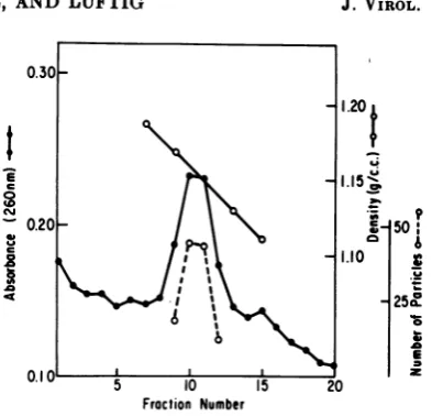
withadensity of 1.16g/cmI
(average ofthreeexperiments).
This isillustrated in Fig.1, whereitcanbeseenthat thepeak ofabsorbancy
(260
nm)
ofthe
virionsfollowingsucrosedensity
gradient
centrifugation iscoincident
with thenumbers
ofparticles observed by
electron microscopy. Asafurther indication ofpurity,
the amountofprotein
andRNA
in the virion prepa-rations wasdetermined.
Approximately
100 to 150,Ag
of purified LCV was obtained from one liter of culture medium. Electron microscope examination ofpurified preparations
indicated that at least 90% of theparticles appeared
asvirion structures. The RNA-to-protein ratio in these
preparations,
estimated on aweight
basis,
was 1:36. This ratio is in agreement with the results of Quigley et al.
(27),
which show 1.9% RNA content for avian tumor viruses. The LCV also containphospholipids (K. Quade
and J. Nichols,unpublished observations)
but their contribution to the total mass of theparticle
is notknown.Virion
polypeptides.
Acomparison
ofSDS-0.20 -50
I a, ~~~~~~~~~~~~~1.10
A
~~~~~~~~~25w!.
5 10 15 20
Fraction Number
FIG. 1. Density profile of LCV after sucrose
density gradient equilibrium centrifugation. The
30 to 60% sucrose (in STE) gradient was
centri-fuged for 25 h at 35,000/rpm in an SW50 rotor.
Twenty-drop fractions were collected, and
absorb-ancy, refractive index, and virion particle count
determinations were made on each fraction. The
viralparticles observedineach of thefractions
repre-sent an average over 10fields at anelectron
micro-scope magnification of X17 000; only those
frac-tions which showed >2 particles per field are
in-cluded.
polyacrylamide gel electrophoresis of the
poly-peptides comprising the LCV with those of a murine leukemia virus showed that the LCV
polypeptide
patternexhibits
certain similarities with the pattern for Friend leukemia virus(kindly
provided by
D.Bolognesi) (Fig.
2).
Specifically,
thelower-molecular-weight
com-ponents
(Fig.
2, arrow, andbelow)
are almostidertical,
and furtherboth
virions exhibit amultiple
number
ofhigher-molecular-weight
components. The
similarity
isespecially
of interest for the mostdensely
staining
protein
(Fig. 2,
arrow)
whichcorresponds
to thegroup-specific
antigens IV(species)
and V(inter-species) as
described by
Schafer et al.(31).
Fourteen other major
polypeptide
components were detected in the LCVby
Coomassie blue staining of the gels afterelectrophoresis.
A spectrophotometric scan of theelectrophoreti-cally resolved
polypeptides
present inpurified
virions isshown in Fig. 3.
Polypeptides
designated
1 through 15 in Fig. 3 were always
present
in quantitativelysimilar amounts in manydifferent virionpreparations. The majorpolypeptide
con-stituent (no. 12, Fig. 3) accounts for
approxi-mately 35% of the virion protein. The
poly-peptide pattern was the same if either intact virus or
phenol-isolated
proteintsoftheviruswereused as starting material for treatment with SDS, urea, and
,3-mercaptoethanol
prior
to434 J.VIROL.
on November 10, 2019 by guest
http://jvi.asm.org/
[image:3.495.266.463.61.250.2]electrophoresis. Polypeptide 11 frequently
oc-curred as a leading shoulder of the major
poly-peptide
12 (Fig. 3) but could not be completely resolvedfrom themajor componentusing differ-ent concentrations of acrylamide in the gels. For this reason, it cannot be stated with cer-tainty that it is a unique polypeptide. The molecularweights of thepolypeptidesrange from 110,000to 7,000 (Table1) and were estimatedby
comparing their rates of migrationwith thoseof marker proteins of known molecular weights (see Materials and Methods).
Virion RNA. Radioactive RNA extracted from LCV and centrifuged in sucrose density gradients showed a characteristic heterogeneous high-molecular-weight component (80 to 88s) as well as a low-molecular-weight component (Fig. 4). Approximately 50% of the total radio-activity in the RNA
preparations
was present ill the fast-sedimenting component.Electro-phoresis
of thelow-molecular-weight
component
oIn
polyacrylamide gels demonstrated thepres-ence of two distinct species ofRNA, a 7s anda
48
component. (Although an accurate s value has not been determined for this polynucleotide chain, it will be identified here as 7s R.NA, since Erikson and Erikson [Abstr.Annu. Meet. Amer. Soc. Microbiol., 1972] have shown that it is closely related in both compositionand chain lengthto78RNA molecules inotherRNAtumor viruses.) Approximately 75% of the radio-activity in the low-molecular-weightRNA
was present in the 7s species. Figure 5 shows thepolyacrylamide
gel resolutionobtained
when3H-labeled
4sand 7s virionRNAsweresubjected to electrophoresis with unfractionated 3P-RNA isolated from L cells. It canbe seen that virion 48 RNA coelectrophoresed withL-cell
4s RNA. No otherspecies of RNA, apart from the high-molecular-weightRNAand the 78and4sRNAs, could be detected in the virion.Heat denaturation of the (80 to 88s) RNA, resulted in release of RNA with asedimentation valueof approximately 358. This is a characteris-tic
feature
of the high-molecular-weightRNA
of the RNA tumor viruses (8, 12) and hasalready
been described in detail for theLCV (13). How-ever, it should be pointed out that we have
observed
some variability in differentprepara-tions: insome cases a homogeneous peak of 358 RNA resulted from denaturation, in othercases the RNA was heterogeneous and ranged from 35stoabout208 as estimated by thedistribution of radioactivity in velocity sucrose density gradients. This may be a reflection of some variability in viral
harvesting
that we have not yetdeterminied.
Wheni
denlatured fast-sediinenitinig
RNA was subjected to electrophoresis on polyacrylamideA B
0
<
FIG. 2. SLDS-polyacrylamide gel electrophoretic separation of the polypeptide8 of LCV (A) and
Friend leukemia virus (B). The arrow indicates
the major proteini, and the letter 0 indicates the
origini. The conicenttrationtofacrylamtide in the gels
was
10%y(.
The directioni ofelectrophoresis
wasfrom top tobottomtintrelationto the photograph.
on November 10, 2019 by guest
http://jvi.asm.org/
[image:4.495.306.394.75.611.2]NICHOLS, QUADE, AND LUFTIG
12
[image:5.495.59.251.177.378.2]origin ~7 11
FIG. 3. Spectrophotometric scan ofCoomassie blue-stainedLCVpolypeptides afterelectrophoretic
sepa-ration in a7.6%SDS-polyacrylamide gel.The direction ofelectrophoresis wasfromlefttorightinrelation
tothephotograph.
TABLE 1. Molecular weight estimates of LCV polypeptides after polyacrylamide gel
electrophoresis
Polypeptide number. Apparent molecular weight
1. 110,000
2. 97,000
3. 85,000
4. 76,000
5. 65,000
6. 49,000
7. 42,000
8. 40,000
9. 36,000
10.33,000
11.28,000
12.27,000
13.14,000
14.11,500
15.7,000
gels,
two distinct size classes of low-molecular-weight RNAwerereleased.
One of these (R-7s) moved slightly ahead of virion 7s RNA, the other (R-4s), coelectrophoresed with virion 4s RNA(Fig. 6).
The R-4s component accounts for about 2.5%,and
theR-7s
componentaccounts forabout 5.0%of theradioactivityin the high-molecular-weight RNA.Virion morphology. In previous electron microscope
studies
with Rauscher leukemia viruses (21), the mostsatisfactory procedure for the preparation of viral specimenswas found tobe
fixation withglutaraldehyde (1.5%), followedby
staining with uranyl acetate (2%). By this procedure the LCV appears morphologicallyidentical
to Rauscher leukemia virus, with over70%
of the particles remaining intact and ap-pearing as round, membrane-bound structures (Fig. 7A-E). A few of these particles exhibit 7-mm knobs over their surface (Fig. 7B-E). These knobs do not appear in large numbers over most of the particles (Fig. 7A) and areprobably fragileinsomedegreeas aresultof the isolation
procedure.
Such loosely bound knob structureshaverecently been observed instudies with Friend leukemiavirus(24).
A quantitative measurement of the outer
C-)
5 10 15
Fraction Number
FIG. 4. Separation offast- and
slow-sediment-ing RNA components by centrifugation in a 5 to
20% sucrose density gradient. Bacteriophage f2
(kindly provided by R. E. Webster) sedimented slightly behind the peak of the
high-molecular-weight RNA (arrow), indicating an s value of 80
to 88s for this fast-sedimenting RNA species.
Samples (25
;diters)
of eachfractionwere countedin Bray solution (6); the figures plotted represent the total radioactivity present in eachfraction.
diameter
for the virion wasobtained
using
thecatalase
crystal internal
markertechnique
(20).
The
distribution
of sizes for intactvirions
was gaussian (Fig.8),
which isrepresentative
of ahomogeneous population of particles. The averagevalueof146.7
ium
forthevirion diameter is almostidentical
with the 147-nm valueobtainied
for Rauscher leukemia virus(21).
This value is larger than the 106-nm diameter
reported
by Nermut et al.(24),
and it may be argued that theincreased
size isperhaps
due toflattening
of the virion. However,preliminary
studies (R.Luftigand K.
Culbreth,
unpublished
observations)
suggest that the smaller diameterseen
in freeze-driedpreparations
(24)
may be due to shrinkage of the virionsduring
prepara-tion. The recent report(29)
that Soule mouse436
J. VIROL.on November 10, 2019 by guest
http://jvi.asm.org/
.21
6
oI~~~~~~~~~~
a. i5S
10 20 30 40 50 60 70 s0
[image:6.495.46.244.64.260.2]Froction Number
FIG. 5. Coelectrophoresis of unifractionated 32p_
labeled L-cell RNA and 3H-virion RNA
(slow-sedimenting
component) on a10%/0
polyacrylamide slab gel. The direction of electrophoresis wasfromleft to right inrelationtothephotograph.
7
6
I
0
a.
C.)
5
4
3
2
I4
-I It
18
5 10 15 20 25 30 35 40 45
FrOction Number
FIG. 6. Coelectrophoresis of virion 4s and 7s
3H-RNA with heat-denatured,
high-molecular-weight32P-RNA ona10%polyacrylamide
cylitndri-cal gel. The direction of electrophoresis was from
left to right in relation to the photograph.
leukemia virus has a diameter of 136 nm as
determined by the independent technique of laser beat frequency spectroscopy, is more in
line with the value given inFig. 8and suggests thatonlyaminimalflatteninig,ifany,may occur
by the glutaraldehyde-uranyl acetate staining procedure.
Assay for reverse transcriptase activity.
When
reactionconditions suitable for the
demonstration
of DNApolymerase activity
in purified Rous sarcoma virus(RSV) (Prague)
were used with
LCV, only
a veryslight (2
to 3%)relative incorporation of deoxythymidine-5'-monophosphatecould bedemonstrated (Table
2).Under
the same conditions,RSV showed
exten-sive incorporation of radioactive precursor. Theaddition
ofcalf
thymusDNA
tothe
reaction mixturesdid
notsignificantly stimulatesynthesis
byLCV in comparison with RSV. Furthermore, when
LCV
and RSV were present in the same reaction mixture, essentially the full activityof
RSV was recovered, indicating that the
di-minished
LCV
polymerase activity wasprob-ably
notdue
to the presence of an inhibitor in the virionpreparation.DISCUSSION
The results
presented
in thispaper,
together with the studiesby
otherworkers (7, 13, 15, 17,18),
clearly demonstrate a close relationship between LCV and other RNA tumor viruses. Therelative
complexity of thepolypeptide
pattern, the presenceof fast- and slow-sediment-ing
RNA
species, the size, morphology, anddensity
of the virion, together with the fact that thereis
alow RNA-to-protein
ratio in thevirion, are general characteristics of oncornaviruses(2, 3,
23,
24,26,
31). On this basis,LCY
appears tobe
anexcellent
model
system forstudying
how latent tumor virions are propagated in mammalian cells.
In addition to the defective nature of the virion, there are some characteristics of LCV
which
distinguish it from other RNA tumor viruses. First, the proportion of 7s RNA relative to the high-molecular-weight RNA is much higher than in viruses such as RSV (4, 5). Also,denaturation of
the fast-sedimenting RNA from LCV releases both a 4s and 7s component, whereas only a 4s species is released fromde-naturedhigh-molecular-weight RNA from
avian-myeloblast
virus,Schmidt-Ruppin
virus or mixtures of mouse sarcoma-mouse leukemia viruses (11). Preliminary studies (J. L.Nichols,
unpublished data) indicate
that LCV 7sand
R-7s molecules are
identical
species of RNA and show slightly different electrophoretic mobilities because of different conformational states.Secondly,
assays for reversetranscriptase with LCV showedanegligible
level of activity ( < 3%), relative to that of an infectious virus, RSV (Prague). It should benoted,
however, that a slight stimulation of activity was apparent when calf thymus DNA was added to the reaction.I
11
11 II 1' III
II
II I III II It
II
II
t
I ~ ~ ~
9
on November 10, 2019 by guest
http://jvi.asm.org/
[image:6.495.50.242.323.539.2]438 NICHOLS, QUADE, AND LUFTIG J.VIROL.
A~~~~~~~~~~~
[image:7.495.78.446.80.449.2]A
7)
FIG. 7.A, Typical field ofL-cell virionsseen afterzone centrifugationon a30 to60%sucrose gradient.
Magnificationis X60,000. B-E,Selectedparticles shounngkntobsonthe viriontsurface (arrow).
Magnifica-tion is X1iS,000.
8
-P~
1467* 118 A n=34 For this reason, it ispossible
that there is areduced amount of the enzyme
present
in thevirion,
or, the LCVenzyme issufficiently
differ-6
~~~~~~~~~~ent
from that of other tumor viruses so that it_ was not
fully
active with the assay conditions0.4
~~~~~~~~~~employed.
o
~~~~~~~~~~~~In
view of the defective nature of LCV it isinteresting
to notethe recentresults ofHanafusaEE
2
~~~~~~~~~~et
al.(16),
in which an absence ofpolymerase
proteins
has been found in virions ofRSV-alpha,
a noninfectious mutant derived fromC 1200 1400 1600 1800 RSV.
Length
(A)
The recentfinidinigs
ofSchaifer
et al.(30)
indi-FIG. 8. Histogram of LGV particle dianmeters cate
onie
further way in which LCV differs fromcalibrated by the catalase crystal internal marker othermurine leukemia viruses. Ina
study
ofsixtechntique. Theaverage diameter
(t),
stanidard devi-differenit murinie
virusesitwasshownby
Ouchter-ation, and ntumberofparticlescounited (n)areiuz-di-lony
anld complemenit fixation tests, that LCVcated. does not possess either FMR or G host range
on November 10, 2019 by guest
http://jvi.asm.org/
[image:7.495.62.255.498.628.2]TABLE 2. DNA polymerase activity of LCV compared with RSV (Prague)a
Amount added Calf thymus [methyl-3H]dTMP
Virus
(pg)
DNAb incorporatedc(pmol)
LCV 28 _ 0.015
RSV (Prague) 36 _ 0.608
LCV 28 + 0.129
RSV (Prague) 36 + 18.40
LCVplus RSV (Prague) 28 pg LCV;36,ug _ 0.476
RSV (Prague)
aThevalues listed for incorporated radioactivity represent an average over at least four different
experiments, with astandard error of 20% among duplicate assays. Reaction conditions are described
in Materials andMethods.
bA
25-pug
amountof calfthymus DNA was added to thereactionmixtures whereindicated.cAbackgroundequivalent to 0.0276 pmol was found for incubated control samples, and thisvaluewas
subtracted from eachexperimental sample.
serotypes.However, knobs whicharethoughtto
correspond to virus surface glycoproteins (10),
can be found on the LCV (Fig. 1B-E). Since
type-specific antigens such as FMR or G are
thought to correspond to these surface glyco-proteins (30), the inability of LCV to
cross-react most probably indicates a different
anti-genicity for thesurfaceknob polypeptide. Studies are in progress to fully characterize
the constituent polypeptides and ribonucleate chainsofLCVandtoelucidate its morphogenesis withinLcells.
ACKNOWLEDGMENTS
Theauthorsthank Marie Waddell, Pamela Watkins,
andMollieGudger forexperttechnical assistance, and
A.Schincariol for providingasample of RSV (Prague)
as well as advice on the reverse transcriptase assay.
This work was supported by Public Health Service
grantsAI-10361 from the National Institute ofAllergy
and Infectious Diseases, and CA-11976 from the
Na-tional Cancer Institute.
LITERATURE CITED
1. Adams, J. M., P. G. N. Jeppesen, F. Sanger, and
B. G. Barrell. 1969. Nucleotide sequence from
the coat protein cistron of R17 bacteriophage
RNA. Nature (London) 223:1009-1014.
2. Bader, J. P., and T. L. Steck. 1969. Analysis of
the ribonucleic acid of murine leukemia virus.
J.Virol.4:454-459.
3. Blair, C. D., and P. H. Duesberg. 1968. Structure
of Rauscher mouse leukemia virus RNA.
Na-ture (London) 220:396-399.
4. Bishop, J. M., W. E. Levinson, N. Quintrell,
D. Sullivan,L. Fanshier, and J. Jackson. 1970.
The low molecular weight RNAs of Rous
sar-coma virus. I. The 4S RNA. Virology
42:182-195.
5. Bishop, J. M., W. E. Levinson, D. Sullivan, L.
Fanshier, N. Quintrell, and J. Jackson. 1970.
The low molecular weight RNAs of Rous
sar-comavirus. II. The 7S RNA. Virology
-42:927-937.
6. Bray, G. A. 1960. Asimpleefficient liquid
scintil-lator for countingaqueous solutions in aliquid
scintillation counter. Anal. Biochem. 1:279-285.
7. Dales, S., and A. F. Howatson. 1961. Virus-like
particles in association with L-strain cells.
Cancer Res. 21:193-197.
8. Duesberg, P. H. 1968. Physical properties of
Rous sarcoma virus RNA. Proc. Nat. Acad.
Sci. U.S.A. 60:1511-1518.
9. Duesberg, P. H., H. L. Robinson, W. S. Robin-son, R. J. Huebner, and H. C. Turner. 1968.
Proteins of Rous sarcoma virus. Virology 36:
73-86.
10. Duesberg, P. H., G. S. Martin, and P. K. Vogt.
1970. Glycoprotein components of avian and
murine RNA tumor viruses. Virology
41:631-646.
11. Erikson, E., and R. L. Erikson. 1971.
Associt-tion of 4S ribonucleic acid with oncornavirus
ribonucleic acids. J.Virol. 8:254-256.
12. Erikson, R. L. 1969. Studies on the RNA from
avianmyeloblastosisvirus. Virology37:124-131.
13. Faras, A. J., and R. L. Erikson. 1969. L-cell
virion ribonucleic acid. J. Virol. 4:31-35.
14. Garapin,A. C., J. P.McDonnel,W.Levinson, N.
Quintrell, L. Fanshier, and J. M. Bishop. 1970.
Deoxyribonucleic acid polymerase associated
with Rous sarcoma virus and avian
myelo-blastosis virus: properties of the enzyme and
itsproduct. J.Virol.6:589-598.
15. Hall,T. H., W.F.Andresen,K. K. Sanford,V. J.
Evans, andJ.W. Hartley. 1967. Virusparticles and murine leukemia virus complement-fixing
antigen in neoplastic and nonneoplastic cell
lines.Science156:85-88.
16. Hanafusa, H., D. Baltimore, D. Smoler, K. F.
Watson, A. Yaniv, and S. Spiegelman. 1972.
Absence of polymerase protein in virions of
alpha-type Rous sarcoma virus. Science 177:
1188-1191.
17. Kindig,D. A., and W.H.Kirsten. 1967.Virus-like
particles in established murine cell lines:
elec-tron microscopic observations. Science 155:
1543-1545.
18. Kindig, D.A., R.Karp,andW. H. Kirsten. 1968.
Further characterization ofL-cell virions. Proc.
Nat. Acad. Sci. U.S.A. 59:1103-1109.
19. Lowry, 0. H., N. J. Rosenbrough,A. L. Farr, and
R. J. Randall. 1951. Protein measurementwith
the Folin phenol reagent. J. Biol. Chem. 193:
265-275.
20. Luftig, R. B. 1967. An accurate measurement of
the catalase crystal period and its use as an
internal marker for electron microscopy. J.
Ultrastruct. Res. 20:91-102.
on November 10, 2019 by guest
http://jvi.asm.org/
NICHOLS, QUADE, AND LUFTIG
21. Luftig, R. B., and S. S. Kilham. 1971. An electron
microscope study of Rauscher leukemia virus. Virology 46:277-297.
22. Mejbaum, W. 1939. Uber die bestimmung Kleiner
pentosemengen insbesondere in derivaten der adenyl-sanre. Z. Physiol. Chem. 258:117-120.
23. Moroni, C. 1972. Structural proteins of Rauscher
leukemia virus and Harvey sarcoma virus.
Virology 47:1-7.
24. Nermut, M. Y., H. Frank, and W. Schafer. 1972.
Properties of mouseleukemiaviruses. III.
Elec-tron microscopic appearance as revealed after
conventional preparation techniques as well as
freeze-drying and freeze-etching. Virology 49: 345-358.
25. Nichols, J. L. 1970. Nucleotide sequencefrom the
polypeptide chain termination region ofthecoat
protein cistron in bacteriophage R17 RNA.
Nature (London) 225:147-151.
26. Nowinski, R. C., L. J. Old, W. H. Sarkar, and
D. H. Moore. 1970. Common properties of the oncogenic RNA viruses (Oncornaviruses).
Virol-ogy42:1152-1157.
27. Quigley, J. P., D. B. Rifkin, and E. Reich. 1971.
Phospholipid composition of Roussarcomavirus
host cell membranes and other enveloped RNA
viruses.Virology 46:106-116.
28. Rifkin, D. B., and E. Reich. 1971. Selective lysis
of cells transformed by Rous sarcoma virus.
Virology 45:172-181.
29. Salmeen, I., D. Gill, and L. Rimai. 1972.
Applica-tion of laser beat frequency spectroscopy to
the detection and characterization of an
onco-genic RNA virus. Biochem. Biophys. Res.
Commun. 47:1172-1178.
30. Schilfer, W., P. J. Fischinger, J. Lange, and L.
Pister. 1972. Properties of mouse leukemia
viruses. I. Characterization of various antisera
and serological identification of viral
com-ponents.Virology 47:197-209.
31. Schafer, W., J. Lange, P. J. Fischinger, H. Frank,
D. P. Bolognesi, and L. Pister. 1972. Properties of mouse leukemia viruses. II. Isolation of
viral components. Virology 47:210-228.
32. Shapiro, A. L., E. Vifnuela, and J. V. Maizel. 1967.
Molecular weight estimation of polypeptide
chainsby electrophoresis in SDS-polyacrylamide
gels. Biochem. Biophys. Res. Commun. 28:
815-820.
33. Smith, R. E., H. J. Zweerink, and W. K. Joklik.
1969. Polypeptide components of virions, top
componentandcoresof reovirustype 3.Virology
39:791-810.