The ABC's of Cell Division: Regulation
of Peptidoglycan Amidase Activity
during Cytokinesis in Escherichia coli
The Harvard community has made this
article openly available. Please share how
this access benefits you. Your story matters
Citation Yang, Desiree Choy. 2012. The ABC's of Cell Division: Regulation of Peptidoglycan Amidase Activity during Cytokinesis in Escherichia coli. Doctoral dissertation, Harvard University.
Citable link http://nrs.harvard.edu/urn-3:HUL.InstRepos:9527315
Terms of Use This article was downloaded from Harvard University’s DASH repository, and is made available under the terms and conditions applicable to Other Posted Material, as set forth at http://
© 2012 Desirée Choy Yang
Dissertation Advisor: Dr. Thomas G. Bernhardt Desirée Choy Yang
The ABC’s of Cell Division: Regulation of Peptidoglycan Amidase Activity During Cytokinesis in Escherichia coli
Abstract
The bacterial cell wall, composed of peptidoglycan (PG), is an essential component of the cell envelope. This macromolecular structure fortifies the cell membrane, determines cell shape, and helps prevent osmotic lysis. The synthesis and remodeling/recycling of this polymer is mediated by PG synthases and hydrolases, respectively. Proper control of the PG hydrolases is particularly important since misregulation of these enzymes can lead to lethal breaches in the cell wall. Surprisingly, however, the precise molecular mechanisms governing the activities of these enzymes remain poorly understood.
To help understand how PG hydrolases are regulated, I examined how their activity is controlled during cytokinesis in Escherichia coli. One important class of PG hydrolases
necessary for cell division is the LytC-type amidases (AmiA, AmiB and AmiC). These enzymes require activation by the LytM factors EnvC and NlpD. My work focused on elucidating the mechanism by which the LytM factors activate the amidases. Using a genetic enrichment strategy, I isolated amiB misregulation mutants. Interestingly, the mutations mapped to a region of AmiB found only in cell separation amidases. Structural analysis of an AmiB ortholog indicates that this region corresponds to an alpha-helical domain that appears to occlude the active site. Thus, activation of the amidases by the LytM factors likely occurs via a
Table of Contents
Title Page i
Copyright Page ii
Abstract iii
Table of Contents v
Acknowledgements vii
Dedication ix
Chapter 1: Introduction to the Gram-negative cell wall and peptidoglycan hydrolases 1
Section 1.1: The Gram-negative bacterial cell envelope and peptidoglycan 2
Section 1.2: Peptidoglycan hydrolases and their physiologic functions 8
Section 1.3: Regulation of the peptidoglycan hydrolases 16
Section 1.4: The cell division complex 21
Section 1.5: The cell division peptidoglycan hydrolases 22
Section 1.6: Dissertation overview 26
Section 1.7: References 29
Chapter 2: An ABC transporter-like complex governs cell well hydrolysis at the bacterial cytokinetic ring 39
Attributions 40
Section 2.1: Abstract 41
Section 2.2: Introduction 42
Section 2.3: Results 48
Section 2.5: Materials and methods 74
Section 2.6: References 92
Chapter 3: Conformational control of cell wall hydrolase activity at the bacterial division site 97
Attributions 98
Section 3.1: Abstract 99
Section 3.2: Introduction 100
Section 3.3: Results 103
Section 3.4: Discussion 122
Section 3.5: Materials and methods 132
Section 3.6: References 143
Chapter 4: Discussion 149
Section 4.1: Summary of results 150
Section 4.2: Future directions 153
Section 4.3: Concluding remarks 158
Acknowledgements
First and foremost, I would like to thank my PhD advisor Thomas (Tom) Bernhardt who graciously accepted me into his lab as a fifth year graduate student. I will be forever indebted to Tom for his generosity, kindness and most importantly his priceless mentorship. Tom’s guidance and support over the past two years has been instrumental in building up both my scientific abilities and confidence. It has truly been an honor and tremendous privilege to work with Tom.
I am also sincerely grateful and fortunate to have a phenomenal dissertation advisory committee of Marcia Goldberg (chair), Eric Rubin and Matt Waldor, who consistently supported me through the highs and lows of graduate school and were pivotal in helping me to achieve the level of scientific training I desired.
I would also like to thank Marcia Goldberg (chair), Simon Dove, Michael Laub and David Rudner, for agreeing to be on my defense committee and taking the time out of their busy schedules to read this dissertation.
This dissertation is dedicated to my parents and sister
Chapter 1
Introduction to the Gram-negative cell wall and
Chapter 1: Introduction to the Gram-negative cell wall and
peptidoglycan hydrolases
Section 1.1: The Gram-negative bacterial cell envelope and peptidoglycan
In Gram-negative bacteria, like Escherichia coli, the cell envelope consists of three distinct layers: an inner membrane and an outer membrane separated by the periplasm (Figure 1.1). Within the periplasmic space lies the cell wall also known as the peptidoglycan (PG) layer. As the principal load-bearing component of the bacterial cell envelope, PG is a vital
macromolecule, helping to maintain cell shape and structure as well as preventing osmotic lysis. PG, also referred to as murein, is a polysaccharide polymer made up of glycan strands consisting of repeating aminosugars N-acetyl-glucosamine (GlcNAc) and N-acetyl-muramic acid
(MurNAc), that are linked by β-1,4-glycosidic bonds. These glycan strands are covalently cross-linked to each other via short stem peptides that are attached to the MurNAc sugars (Figure 1.1 and Figure 1.2). The resulting meshwork forms a continuous structure that encases the cell. Therefore, synthesis and remodeling of PG are essential for growth and division in all cell-wall containing bacteria (1).
In order for a bacterial cell to grow and divide, it must synthesize new PG and incorporate this material into the existing matrix. De novo biosynthesis of PG begins in the cytoplasm where the precursor molecules UDP-GlcNAc and UDP-MurNAc-L-Ala-γ-D-Glu-(L
)-meso-diaminopimelic acid (DAP)-D-Ala-D-Ala (UDP-MurNAc-pentapeptide) are synthesized by
disaccharide-M G disaccharide-M G M G
M G
G M
M G M G M G
M G G M
inner membrane (IM) outer membrane (OM)
peptidoglycan (PG)
[image:12.612.182.430.71.409.2]periplasm
Figure 1.1. Schematic of the Gram-negative bacterial cell envelope. Diagram of a rod-shaped
GlcNAc MurNAc GlcNAc MurNAc GlcNAc MurNAc
L-Ala D-Glu m-A2pm
D-Ala
GlcNAc MurNAc GlcNAc MurNAc
D-Ala m-A2pm-NH2 D-Glu L-Ala m-A2pm-NH2 D-Glu L-Ala L-Ala D-Glu
GlcNAc MurNAc GlcNAc
L-Ala D-Glu m-A2pm
D-Ala m-A2pm-NH2
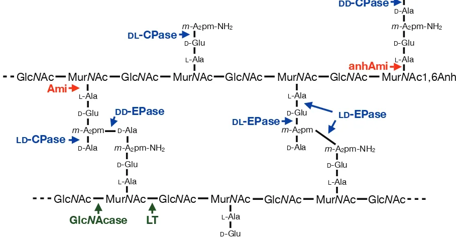
D-Glu L-Ala m-A2pm-NH2 D-Glu L-Ala D-Ala D-Ala Ami LD-CPase DD-EPase DL-CPase
DL-EPase LD-EPase
MurNAc1,6Anh GlcNAc
DD-CPase
anhAmi
[image:13.612.78.532.88.328.2]LT GlcNAcase
Figure 1.2. Structure of E. coli murein and sites of glycan, amide and peptide bond hydrolysis.
pentapeptide. Lipid II is then flipped across the inner membrane so that the disaccharide-pentapeptide moiety faces the periplasm. The identity of the flippase is currently controversial (2-4). In the periplasm, PG synthases with transglycosylase (TG) and transpeptidase (TP) activities polymerize lipid II into glycan strands and cross-link them into the existing PG meshwork via their short stem peptides (Figure 1.3) (1).
M M G
G UDP
M UDP
M G M G M G M G
M G
G M
M G M G M G
M G
G M
M
ÁLSSDVH
LQQHUPHPEUDQH 3*V\QWKDVHV
SHULSODVP
F\WRSODVP
1 2
3
4
5 6
[image:15.612.112.505.69.399.2]OLSLG, OLSLG,,
Figure 1.3. The steps of PG biosynthesis. ➀ UDP-GlcNAc is converted to UDP-MurNAc by
several reaction steps represented here by multiple black arrows. ➁ UDP-MurNAc attached to a pentapeptide side chain is then anchored in the inner membrane, generating lipid I.
➂ Attachment of GlcNAc to lipid I results in the formation of lipid II which can then be ➃
flipped across the membrane and incorporated into the existing PG meshwork via ➄
Table 1.1. Peptidoglycan synthases in E. coli.
Enzyme Gene Localization
Transglycosylase/transpeptidase PBP1a
PBP1b PBP1c
ponA (mrcA) ponB (mrcB) pbpC
Inner membrane Inner membrane Inner membrane
Transpeptidase PBP2
PBP3 pbpA (mrdA)ftsI (pbpB) Inner membrane Inner membrane
Monofunctional glycosyltransferase
MtgA mtgA Inner membrane
Section 1.2: Peptidoglycan hydrolases and their physiologic functions
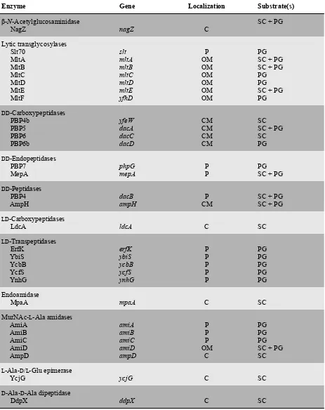
In contrast to a relatively low number of PG synthases (Table 1.1), E. coli encodes many hydrolytic enzymes with the collective capability of cleaving every amide and glycosidic linkage in PG (Table 1.2, Figure 1.2) (1, 9). These PG hydrolases are also referred to as autolysins because elevated expression of these enzymes can result in cell lysis. In total, E. coli encodes over 30 factors known or predicted to possess PG hydrolase activity that are classified into 12 different protein families (Table 1.2) (9).
Peptidoglycan hydrolases and the bonds they cleave
As previously mentioned, PG is composed of two basic linkages that are broken by PG hydrolases: glycosidic and amide bonds (Figure 1.2). PG glycosidases are enzymes that cleave within the glycan strand of murein. β-N-acetylglucosaminidases specifically cleave the GlcNAc-(1→4)-MurNAc bond whereas N-acetylmuramidases, which encompass lysozymes and lytic transglycosylases (LT), break the MurNAc-(1→4)-GlcNAc linkage. E. coli possesses only one
β-N-acetylglucosaminidase encoded by the nagZ gene (10). NagZ is not essential for cell viability, but its activity is required for cells to recycle PG. Its substrate is a cytoplasmic disaccharide intermediate of the recycling pathway (11, 12). The MurNAc-(1→4)-GlcNAc linkage can be hydrolyzed by two mechanisms: (1) hydrolysis of the glycosidic bond by
Table 1.2. Peptidoglycan hydrolases in E. coli.a
Enzyme Gene Localization Substrate(s)
β-N-Acetylglucosaminidase
NagZ nagZ C SC + PG
Lytic transglycosylases Slt70 MltA MltB MltC MltD MltE MltF slt mltA mltB mltC mltD mltE yfhD P OM OM OM OM OM OM PG SC + PG SC + PG PG PG SC + PG PG DD-Carboxypeptidases PBP4b PBP5 PBP6 PBP6b yfeW dacA dacC dacD CM CM CM CM SC SC + PG SC PG
DD-Endopeptidases
PBP7
MepA phpGmepA PP PGSC + PG
DD-Peptidases
PBP4
AmpH dacBampH CMP SC + PGSC + PG
LD-Carboxypeptidases
LdcA ldcA C SC
LD-Transpeptidases ErfK YbiS YcbB YcfS YnhG erfK ybiS ycbB ycfS ynhG P P P P P PG PG PG PG PG Endoamidase
MpaA mpaA C SC
MurNAc-L-Ala amidases
AmiA AmiB AmiC AmiD AmpD amiA amiB amiC amiD ampD P P P OM C PG PG PG SC + PG SC
L-Ala-D/L-Glu epimerase
YcjG ycjG C SC
D-Ala-D-Ala dipeptidase
DdpX ddpX C SC
indicated by their names, are either soluble or found associated with a membrane, respectively (reviewed in 9, 13). Deletion mutants lacking multiple LTs, including a sextuple mutant lacking six of the seven LTs (Slt70, MltA-E), are still viable, but grow in short chains. Surprisingly, only minor changes in the PG composition were observed for this mutant (14-16). Interestingly, however, attempts to knock-out all seven LTs have been unsuccessful, suggesting that LT activity is essential for cell growth (17).
The PG peptidases are enzymes that specifically break the amide bonds between the amino acids found in PG. These hydrolytic enzymes come in two basic flavors:
carboxypeptidases, which remove the C-terminal residue from stem peptides, or endopeptidases that cleave within the peptide. The different designations of DD-, LD- or DL-peptidase refer to the
stereochemistry of the bond cleaved by these enzymes (18). In E. coli, DD-endopeptidases
(PBP4, 7 and MepA) cleave the D-Ala-meso-A2pm cross-link formed by the transpeptidation
reaction of the HMW-PBPs (5). Interestingly, since these enzymes recognize and break the same bonds that are made by the HMW-PBPs some of them bind and are inhibited by β-lactams and thus have been classified as low molecular weight (LMW) PBPs (5, 19). In fact, except for AmpC, all the LMW-PBPs (PBP4, 4b, 5, 6, 6b, 7/8, AmpC and AmpH) in E. coli possess peptidase activity (20). In addition to the LMW-PBPs there are number of penicillin-insensitive peptidases in E. coli, including MepA, MpaA and DdpX (21-24). Finally, the N
-acetylmuramyl-L-alanine amidases are responsible for cleaving the amide bond between the N-terminal L
-alanine residue and the D-lactoyl moiety of MurNAc. E. coli possesses five known amidases:
be categorized into two distinct families, Pfam amidase_3 also known as LytC-type amidases (AmiA, AmiB and AmiC) and the amidase_2 family (AmiD and AmpD) (13, 28).
The physiologic functions of peptidoglycan hydrolases
Like E. coli, most PG-containing bacteria encode a sizable array of PG hydrolases, with several representatives from each family often being produced. Functional redundancy among these PG hydrolase families has hampered efforts to determine their specific physiological function(s) (13). However, the systematic deletion of multiple/all members in a particular PG hydrolase family is one approach that has yielded some insight (reviewed in 9, 14, 25, 29-31). These studies and deletion analyses in other organisms have implicated PG hydrolases in a diverse array of biological processes, some of which are listed in Table 1.3, including PG turnover, cell elongation in rod-shaped bacteria, cell division, cell shape determination, and contact-dependent cell lysis (reviewed in 13).
One of the most important functions for PG hydrolases is thought to be “space-maker” enzymes that break bonds in the PG meshwork to allow for the insertion of new material (13). This is largely based on theoretical considerations, as attempts to identify essential PG
Table 1.3. Examples of biological processes that employ peptidoglycan hydrolases.
Biological Function
Biological Function Functional Description and Example PG Hydrolases
Functions in bacterial cell physiology Functions in bacterial cell physiology Functions in bacterial cell physiology
Regulation of cell wall growth Removal of excess of pentapeptides in new PG by DD-carboxypeptidases
PBP5 (E. coli); PBP3 (S. pneumoniae)
PG turnover* Release of soluble PG fragments from the sacculus during growth
Lytic transglycosylases (E. coli)
Enlargement of the sacculus* Breaking of bonds to allow expansion of the sacculus during growth
Lytic transglycosylases (E. coli); LytE (B. subtilis)
Production of signaling molecules Induction of β-lactamase by PG turnover products
Lytic transglycosylases (E. coli)
Recycling of PG turnover products Cleavage turnover products to allow reuse in PG synthesis
AmpD, LdcA, NagZ (E. coli)
Cell division* Cleavage of the septum during cell division in Gram-negative species
AmiA, AmiB, AmiC (E. coli)
Cleavage of the cross-wall after division in Gram-positive species Atl (S. aureus)
Cell wall shape* Altered PG profiles can influence bacterial cell wall shape
Csd1, Csd2, Csd3 (H. pylori)
Sporulation and germination Cleavage of asymmetric septum
SpoIID, SpoIIP (B. subtilis) Spore cortex maturation
LytH (B. subtilis)
Digestion of the mother cell PG to release the endospore
LytC, CwlC, CwlH (B. subtilis)
Digestion of the spore PG during germination SleB, CwlJ (B. subtilis)
Assembly of secretion systems Specialized PG hydrolases (lytic transglycosylases) for localized PG
degradation associated with type II, type III and type IV secretion systems
VirB1 (A. tumefaciens); TraB (E. coli plasmid R721)
Pilus assembly (type IV) Specialized PG hydrolases for pilus assembly
PilT (E. coli EPEC strains)
Flagellum assembly Specialized PG hydrolases for flagellum assembly
FlgJ (E. coli)
Resuscitation of dormant cells Stimulation of cell division to exit dormant state
Table 1.3 continued.
Biological Function
Biological Function Functional Description and Example PG Hydrolases
Functions in bacterial cell physiology Functions in bacterial cell physiology Functions in bacterial cell physiology
Contact-dependent cell lysis* Secretion of autolysin effectors via type VI secretion
Tse1, Tse3 (P. aeruginosa) Autolysis in genetic
transformation Fratricide of cells S. pneumoniae: induced lysis (allolysis) of non-competent
LytA, LytC (S. pneumoniae)
Developmental lysis Lysis of cells during fruiting body formation in M. xanthus
Cannibalism in B. subtilis
Lysis of prey cells Secretion of PG hydrolases to digest peptidoglycan of prey cells
Exoenzymes of M. xanthus
Lysis of non-immune cells Plasmid-encoded bacteriocin/immunity factor
Pesticin (Y. pestis)
Biofilm formation PG hydrolases are required for initial attachment of cells to hydrophobic
surfaces
AtlE (S. epidermidis)
Pathogen-host interaction Release PG fragments recognized by the host
MreB (37, 38). B. subtilis encodes three MreB orthologs, MreB, Mbl, and MreBH, all of which have been implicated in cell elongation and rod-shape determination (39-45). Interestingly, Errington and co-workers have detected an interaction between the LytE PG hydrolase and MreBH, suggesting a role for LytE in cell wall elongation (46). Accordingly, lytE and mreBH mutants display similar cell-wall-related defects (46). Given that LytE is exported and MreBH is a cytoplasmic protein, it remains unclear how the observed LytE-MreBH interaction might facilitate proper elongation, but it has been proposed that MreBH might guide the secretion of LytE to specific locations to coordinate PG hydrolysis with the insertion of new material. Importantly, it was subsequently discovered that inactivation of the related endopeptidase CwlO was synthetically lethal with the loss of LytE function (47, 48). The terminal phenotype of the LytEˉ CwlOˉ cells was the cessation of growth, strongly suggesting that these enzymes are the postulated “space-maker” enzymes for B. subtilis cell wall expansion.
In addition to the activities of cell growth and division, recent work in Helicobacter pylori suggests that relaxation of PG cross-links by autolysins can promote helical shape (53). Helical shape is thought to be determined by three morphological features: cell elongation, curvature and twist (53). Predictions made using biophysical modeling first suggested that cell curvature and twist might be achieved by the local modification of PG cross-links along a helical path (54). The identification of four genes necessary for helical shape in H. pylori,csd1-3 (csd = cell shape determinant) and ccmA (ccm = curved cell morphology), was the first piece of
evidence to support such a model (53). Loss of these genes resulted in variable curved-rod morphologies and changes in the abundance of specific classes of cross-linked muropeptides (53). Interestingly Csd1, Csd2 and Csd3 possess LytM peptidase domains that may be
endopeptidases and/or carboxypeptidases that directly hydrolyze PG (53). In accordance with this idea, expression of a predicted catalytically impaired Csd1 variant resulted in the same curved-rod phenotype observed with the null allele of csd1 (53). These results support the model that localized changes in murein architecture by PG hydrolases can induce the helical shape characteristic of H. pylori (53).
in Pseudomonas aeruginosa to transfer lytic amidases into the periplasm of other recipient bacteria to lyse them (56).
Section 1.3: Regulation of the peptidoglycan hydrolases
While the functions of many PG hydrolases remain to be uncovered, the examples described above clearly highlight that these enzymes have diverse roles in cells ranging from promoting cell growth to cell destruction. To use PG hydrolases for constructive purposes, such as cell elongation or cell division, bacterial cells must maintain tight control over the potentially lethal hydrolytic activity of these enzymes. Although this has been appreciated for some time, very little is known about the mechanisms responsible for PG hydrolase regulation. While the details remain obscure, several general regulatory strategies have been described and/or
postulated over the years ranging from transcriptional to post-translational control mechanisms to the chemical modification of the PG substrate. Examples of each of these regulatory strategies are discussed in turn below.
Transcriptional control
during vegetative growth, cell division and motility (57-60). Moreover, PG hydrolases critical for spore-formation and germination are controlled by sporulation-specific sigma factors that spatiotemporally restrict the production of these enzymes to a specific stage during the sporulation process (61, 62).
Another major transcriptional regulator important for cell wall homeostasis is the two component-system (TCS) WalRK, which is highly conserved among low GC-containing Gram-positive bacteria and is one of the few essential TCS identified to date (63-74). The WalRK system has been most thoroughly studied in B. subtilis, Staphylococcus aureus and Streptococcus pneumoniae (47, 64, 67, 75). In these systems, phosphorylated WalR (WalR-P), the response regulator, activates expression of genes encoding cell separation PG hydrolases and represses the expression of their inhibitors (63). In B. subtilis, the sensor kinase WalK is recruited to the divisome and its kinase activity is stimulated by interactions with septal ring components (76, 77). Interestingly, the activation of WalK and its ability to phosphorylate WalR appears to require a functional division apparatus (76-78). Taken together, these findings suggest that the WalRK systems may adjust the levels of cellular PG hydrolase activity in response to changes in growth rate and division.
Post-translational control
(a) Multi-enzyme complexes
For proper cell wall assembly, PG synthesis and hydrolysis must be precisely coordinated. Therefore, it was proposed some time ago that the formation of multi-enzyme complexes, containing both PG synthases and PG hydrolases, could serve to coordinate these activities (1, 79). Indeed, several PBP-PG hydrolase interactions have been detected in E. coli using affinity chromatography (80, 81). Also, as mentioned previously, the cell wall hydrolase LytE from B. subtilis has been found to interact with a component of the cell elongation machinery, MreBH (46). Supporting evidence for such complexes also comes from work in mycobacteria, where PG synthase PBP1 was shown to interact with the PG peptidase RipA (82). Although these multi-enzyme complexes are often invoked when discussing the coordination of PG synthesis and degradation, the functional significance of PG synthase-PG hydrolase complex formation remains unclear; the phenotypic consequence of defects in synthase-hydrolase
(b) Peptidoglycan hydrolase processing
In addition to possible regulation within multi-enzyme complexes, a handful of PG hydrolases are known to require proteolytic processing for activation (13). These enzymes are synthesized and secreted as latent proenzymes and must be processed for maturation (83-87). For example, the lysostaphin-type metalloendopeptidases are inactive in their full-length form and require processing to become active (84). Additionally, the major S. aureus autolysin Atl, which facilitates daughter cell separation, is produced as a proenzyme and undergoes proteolytic processing that generates two important extracellular PG hydrolases (85). However, it remains unclear whether or not this processing event is required for Atl regulation. Interestingly, in most cases the biologically relevant peptidases responsible for these maturation events are unknown, although there are some examples where the pertinent protease has been identified (88). Identifying these proteases and determining whether or not the processing event is itself regulated will be important for understanding how proteolytic maturation might control PG hydrolase activity and promote their activation at the right time and place in the cell.
(c) Subcellular localization of peptidoglycan hydrolases
An important strategy for the spatiotemporal regulation of PG hydrolase activity is the control of enzyme localization. This is typically accomplished by the production of PG
hydrolases with accessory domains that target the factor to the desired location. For example, the E. coli amidases AmiB and AmiC possess N-terminal AMIN domains that target them to the septum to participate in the division process (89, 90). In most cases the mechanism of
the cell wall binding domains of certain PG hydrolases and polyanionic cell wall polymers, like wall teichoic acids (WTAs), has been implicated in controlling PG hydrolase localization (85). For example, Schlag and colleagues report that WTA prevents Atl from binding cell wall in S. aureus, thus reducing its overall susceptibility to Atl-mediated PG cleavage (85). Interestingly, WTA appears to be absent at the cross-wall region and local depletion is thought to result in the targeting of Atl to the equatorial surface ring where it can promote the separation of the daughter cells (85).
(d) Other mechanisms
In addition to protein-protein interactions and the control of protein localization, chemical modification of the PG layer or changes in its conformation as well as physiochemical properties are also thought to play a role in PG hydrolase regulation. Koch, et al. proposed that
topographical features of the bacterial cell wall could influence its degradation, postulating that in Gram-positive bacteria the PG bonds residing on the outer-most layers would be more stressed and therefore easier to cleave by “smart” autolysins (79, 91). In addition to physical distortion of PG, the ionic composition of the cell wall milieu may also affect enzyme activity as cell wall turnover and autolysis in B. subtilis was reported to be salt dependent (92). The proton gradient of cellular membranes in Gram-positive bacteria has also been implicated in modulating
inhibit autolysin activity (91, 93, 94). In support of this model, the cell wall of B. subtilis was shown to be acidified during respiration using pH-sensitive probes and other chemical sensing agents (94, 95). Acetylation and/or deacetylation of the PG layer can also affect its susceptibility to cleavage by PG hydrolases. For example, many Gram-negative bacteria O-acetylate the C-6 position of MurNAc and this blocks cell wall cleavage by LTs (96). However, it is not clear how the acetylation status itself might be regulated or whether acetylation or similar PG modifications are localized to specific subcellular regions to guide when and where PG hydrolases are capable of acting.
As highlighted above, our current understanding of PG hydrolase regulation is far from comprehensive. Most of the strategies identified thus far appear to point towards global
regulatory mechanisms that help determine the overall level of PG hydrolase activity in the cell (i.e. by controlling their expression). Specific mechanisms that turn a particular hydrolase “on” or “off” as needed during the processes of cell growth or division have not been described. I therefore chose to investigate the regulation of PG hydrolases required for cell division in E. coli as a model system for PG hydrolase regulation. Below I will present an overview of the cell division process and review what was known about the regulation of the cell separation amidases in E. coli prior to the start of my thesis work.
Section 1.4: The cell division complex
envelope layers simultaneously (Figure 1.1). Formation of the septal ring starts with the GTP-dependent polymerization of FtsZ, a tubulin homolog, into a ring-like structure also known as the Z-ring directly underneath the inner membrane, demarcating the future site of division (99). After formation of the Z-ring, FtsA and ZipA along with three non-essential factors ZapA, ZapB and ZapC are recruited and help to stabilize this highly dynamic structure (100-105). Once the Z-ring is formed, recruitment of other essential and non-essential downstream components occurs in an interdependent manner (Figure 1.4) (106-113). The assembly of the divisome happens in two stages, starting with the formation and stabilization of the Z-ring, which persists for about 20% of the cell cycle, followed by the simultaneous recruitment of FstQ-FtsN at about the time constriction is initiated (114).
During cytokinesis, the divisome must facilitate a number of different functions in a coordinated fashion. These activities include invagination and fission of the inner membrane, synthesis of the septal PG layer, splitting of this septal PG shared between daughter cells and finally invagination and fission of the outer membrane (98). Hydrolysis of the septal murein layer requires the activity of PG hydrolases. In E. coli, the specific autolysins required for this process have been identified and are discussed in more detail below.
Section 1.5: The cell division peptidoglycan hydrolases
In an effort to elucidate which murein hydrolases are the most critical for cell division, Heidrich and colleagues began constructing a series of mutants deleted for one or more PG hydrolase (14, 25). The results indicated that the three periplasmic, LytC-type N
Figure 1.4. Dependency pathway for septal ring assembly. The dependency pathway for assembly of the cell division apparatus is shown. Factors in black are essential and those in red are non-essential. The three main phases of the division process are indicated below the
dependency pathway.
FtsZ
FtsA ZipA
ZapA
ZapB Z-Ring
FtsEX or salt
FtsK FtsW FtsI FtsN:
Septal Ring FtsQ
FtsL FtsB
Z-Ring Assembly Septal Ring Maturation Constriction
AmiC Tol Pal AmiB
during division. As previously mentioned, amidases are hydrolytic enzymes that cleave the stem peptide from the glycan strands of PG and are thus capable of destroying peptide cross-links. Heidrich and co-workers found that mutants lacking all three amidases formed extremely long chains of cells that were unable to separate. Electron microscopy images of these cells showed that constriction and fission of the cytoplasmic membrane had occurred, but there was no evidence of any septal PG cleavage (14, 25, 31, 52). These results suggested that the amidases might be recruited to the divisome to participate directly in the division process. The subcellular localization of the amidases was therefore investigated using fluorescent protein fusions (90, 115). While both AmiB and AmiC were shown to localize to the septal ring using GFP fusions, AmiA-GFP displayed a diffuse distribution pattern throughout the periplasm indicating that it is not specifically recruited to the site of division (90). Accordingly, AmiA lacks the N-terminal septal targeting domain present in both AmiB and AmiC (Figure 1.5) (89, 90). However, although AmiA is not specifically recruited to the divisome, ΔamiBΔamiC mutants do not display a severe separation defect, implying that specific septal localization of the amidases is not required for successful cell division (115, 117, 118).
AmiA
289 aa
AmiB
445 aa
AmiC
417 aa
ss amidase
amidase AMIN
ss
amidase AMIN
ss
YgeR
251 aa
NlpD
379 aa
EnvC
419 aa
LytM LysM
ss*
LytM LysM
ss*
LytM CC
ss CC
YebA
440 aa T LysM LytM
Figure 1.5. Predicted domain structure of the E. coli LytC-type amidases and LytM factors.
Shown is a diagram depicting the predicted domain architecture of the three E. coli amidases (left) and four factors with identifiable LytM domains (right). Abbreviations: SS, signal
long chains of cells that are connected by a layer of unsplit septal PG, reminiscent of the triple amidase mutants (119). It was subsequently shown that the LytM factors are actually activators of the LytC-type amidases (118). By themselves, the amidases display relatively weak PG hydrolase activity in vitro. Interestingly, however, the LytM factors were shown to be activators of amidase activity (118). EnvC specifically activates AmiA and AmiB, while NlpD activates only AmiC (118). Although it is unclear what the remaining two LytM factors do, it is apparent that YebA and YgeR play a minor role if any during cell division (119). In total, AmiA, AmiB and AmiC along with their cognate LytM activating factors, EnvC and NlpD, constitute two partially redundant septal PG splitting pathways in E. coli (Figure 1.6). These studies shed important light on the control of PG hydrolase activation at the division site of E. coli. The goal of my thesis work has been to address two outstanding questions: (i) what components of the septal ring might control the ability of the LytM factors to activate the amidases? and (ii) how are the amidases activated by the LytM factors?
Section 1.6: Dissertation overview
Figure 1.6. Two-redundant septal PG splitting pathways in E. coli. Below is a schematic of a dividing cell. The box contains a close-up diagram of the division site highlighting the amidases and their activating, cognate LytM factor. For the purposes of this diagram the outer membrane (OM) has been omitted.
EnvC
NlpD
AmiA/B
AmiC
PG
using a number of complementary approaches, I discovered that EnvC is recruited to the septal ring by a direct protein-protein interaction with FtsX (TMD). Moreover, the ability of FtsE (NBD) to bind and hydrolyze ATP did not affect recruitment of EnvC to the divisome, but was critical for promoting cell division. These results suggest that FtsEX is an ABC transporter-like complex that utilizes the conformational change induced by ATP-binding and hydrolysis to directly modulate the ability of EnvC to activate the amidases in vivo (125). Furthermore, evidence that FtsZ and FtsE directly interact (126), provides a potential link between contraction of the Z-ring with hydrolysis of septal PG during cell division (125). In total, these findings support a novel model for how dividing bacterial cells convert the potentially dangerous activity of septal PG splitting into a more discreet and directed process (125).
Chapter 3 discusses the identification of unregulated AmiB variants that bypass the need for EnvC activation. The isolation and characterization of such mutants, which upon expression induce cells lysis, provides evidence that the LytM factors are allosterically activating the
amidases. Based on the crystal structure of AmiB from Bartonella henselae, the lytic mutations I identified all map to an alpha-helical domain that occludes the amidase active site. The results discussed here suggest that EnvC interacts with AmiB, inducing a conformational change that displaces this regulatory helix and thus allows substrate accessibility to the active site.
In Chapter 4, I summarize the major findings and implications from this work and address the impact these results have on our current understanding of how the cell wall
hydrolases are regulated in bacteria. Additionally, I propose a model for how we envision septal PG hydrolysis is controlled and coordinated during cell division. Lastly, I discuss future
directions and identify salient questions that remain unanswered in the field.
Section 1.7: References
1. Höltje JV (1998) Growth of the stress-bearing and shape-maintaining murein sacculus of Escherichia coli. Microbiol Mol Biol Rev 62:181–203.
2. Ruiz N (2008) Bioinformatics identification of MurJ (MviN) as the peptidoglycan lipid II flippase in Escherichia coli. Proc Natl Acad Sci USA 105:15553–15557.
3. Fay A, Dworkin J (2009) Bacillus subtilis homologs of MviN (MurJ), the putative
Escherichia coli lipid II flippase, are not essential for growth. J Bacteriol 191:6020–6028. 4. Fraipont C et al. (2011) The integral membrane FtsW protein and peptidoglycan synthase
PBP3 form a subcomplex in Escherichia coli. Microbiol 157:251–259.
5. Sauvage E, Kerff F, Terrak M, Ayala JA, Charlier P (2008) The penicillin-binding proteins: structure and role in peptidoglycan biosynthesis. FEMS Microbiol Rev 32:234–258.
6. Derouaux A et al. (2008) The monofunctional glycosyltransferase of Escherichia coli localizes to the cell division site and interacts with penicillin-binding protein 3, FtsW, and FtsN. J Bacteriol 190:1831–1834.
7. Spratt BG (1975) Distinct penicillin binding proteins involved in the division, elongation, and shape of Escherichia coli K12. Proc Natl Acad Sci USA 72:2999–3003.
8. Lleo MM, Canepari P, Satta G (1990) Bacterial cell shape regulation: testing of additional predictions unique to the two-competing-sites model for peptidoglycan assembly and isolation of conditional rod-shaped mutants from some wild-type cocci. J Bacteriol 172:3758–3771.
10. Cheng Q, Li H, Merdek K, Park JT (2000) Molecular characterization of the beta-N-acetylglucosaminidase of Escherichia coli and its role in cell wall recycling. J Bacteriol 182:4836–4840.
11. Park JT, Uehara T (2008) How bacteria consume their own exoskeletons (turnover and recycling of cell wall peptidoglycan). Microbiol Mol Biol Rev 72:211–27.
12. Votsch W (2000) Characterization of a beta -N-acetylglucosaminidase of Escherichia coli and Elucidation of Its Role in Muropeptide Recycling and beta -Lactamase Induction. Journal of Biological Chemistry 275:39032–39038.
13. Vollmer W, Joris B, Charlier P, Foster S (2008) Bacterial peptidoglycan (murein) hydrolases. FEMS Microbiol Rev 32:259–286.
14. Heidrich C, Ursinus A, Berger J, Schwarz H, Höltje J-V (2002) Effects of multiple deletions of murein hydrolases on viability, septum cleavage, and sensitivity to large toxic molecules in Escherichia coli. J Bacteriol 184:6093–6099.
15. Lommatzsch J, Templin MF, Kraft AR, Vollmer W, Höltje JV (1997) Outer membrane localization of murein hydrolases: MltA, a third lipoprotein lytic transglycosylase in Escherichia coli. J Bacteriol 179:5465–5470.
16. Kraft AR, Prabhu J, Ursinus A, Höltje JV (1999) Interference with murein turnover has no effect on growth but reduces beta-lactamase induction in Escherichia coli. J Bacteriol 181:7192–7198.
17. Scheurwater EM, Clarke AJ (2008) The C-terminal domain of Escherichia coli YfhD functions as a lytic transglycosylase. J Biol Chem 283:8363–8373.
18. Smith TJ, Blackman SA, Foster SJ (2000) Autolysins of Bacillus subtilis: multiple enzymes with multiple functions. Microbiol 146:249–262.
19. Goffin C, Ghuysen JM (1998) Multimodular penicillin-binding proteins: an enigmatic family of orthologs and paralogs. Microbiol Mol Biol Rev 62:1079–1093.
20. Henderson TA, Young KD, Denome SA, Elf PK (1997) AmpC and AmpH, proteins related to the class C beta-lactamases, bind penicillin and contribute to the normal morphology of Escherichia coli. J Bacteriol 179:6112–6121.
21. Iida K, Hirota Y, Schwarz U (1983) Mutants of Escherichia coli defective in penicillin-insensitive murein DD-endopeptidase. Mol Gen Genet 189:215–221.
23. Lessard IA et al. (1998) Homologs of the vancomycin resistance D-Ala-D-Ala dipeptidase VanX in Streptomyces toyocaensis, Escherichia coli and Synechocystis: attributes of catalytic efficiency, stereoselectivity and regulation with implications for function. Chem Biol 5:489–504.
24. Lessard IA, Walsh CT (1999) VanX, a bacterial D-alanyl-D-alanine dipeptidase: resistance, immunity, or survival function? Proc Natl Acad Sci USA 96:11028–11032.
25. Heidrich C et al. (2001) Involvement of N-acetylmuramyl-L-alanine amidases in cell separation and antibiotic-induced autolysis of Escherichia coli. Mol Microbiol 41:167–178. 26. Uehara T, Park JT (2007) An anhydro-N-acetylmuramyl-L-alanine amidase with broad
specificity tethered to the outer membrane of Escherichia coli. J Bacteriol 189:5634–5641. 27. Jacobs C et al. (1995) AmpD, essential for both beta-lactamase regulation and cell wall
recycling, is a novel cytosolic N-acetylmuramyl-L-alanine amidase. Mol Microbiol 15:553– 559.
28. Pennartz A, Généreux C, Parquet C, Mengin-Lecreulx D, Joris B (2009) Substrate-induced inactivation of the Escherichia coli AmiD N-acetylmuramoyl-L-alanine amidase highlights a new strategy to inhibit this class of enzyme. Antimicrob Agents Chemother 53:2991–2997. 29. Denome SA, Elf PK, Henderson TA, Nelson DE, Young KD (1999) Escherichia coli
mutants lacking all possible combinations of eight penicillin binding proteins: viability, characteristics, and implications for peptidoglycan synthesis. J Bacteriol 181:3981–3993. 30. Nelson DE, Young KD (2000) Penicillin binding protein 5 affects cell diameter, contour,
and morphology of Escherichia coli. J Bacteriol 182:1714–1721.
31. Höltje JV, Heidrich C (2001) Enzymology of elongation and constriction of the murein sacculus of Escherichia coli. Biochimie 83:103–108.
32. Goodell EW, Schwarz U (1983) Cleavage and resynthesis of peptide cross bridges in Escherichia coli murein. J Bacteriol 156:136–140.
33. Goodell EW, Schwarz U (1985) Release of cell wall peptides into culture medium by exponentially growing Escherichia coli. J Bacteriol 162:391–397.
34. Goodell EW (1985) Recycling of murein by Escherichia coli. J Bacteriol 163:305–310. 35. Pooley HM (1976) Turnover and spreading of old wall during surface growth of Bacillus
subtilis. J Bacteriol 125:1127–1138.
37. Carballido-López R, Errington J (2003) A dynamic bacterial cytoskeleton. Trends Cell Biol 13:577–583.
38. Daniel RA, Errington J (2003) Control of cell morphogenesis in bacteria: two distinct ways to make a rod-shaped cell. Cell 113:767–776.
39. Wachi M et al. (1987) Mutant isolation and molecular cloning of mre genes, which
determine cell shape, sensitivity to mecillinam, and amount of penicillin-binding proteins in Escherichia coli. J Bacteriol 169:4935–4940.
40. Wachi M, Doi M, Okada Y, Matsuhashi M (1989) New mre genes mreC and mreD, responsible for formation of the rod shape of Escherichia coli cells. J Bacteriol 171:6511– 6516.
41. van den Ent F, Amos LA, Löwe J (2001) Prokaryotic origin of the actin cytoskeleton. Nature 413:39–44.
42. Jones LJ, Carballido-López R, Errington J (2001) Control of cell shape in bacteria: helical, actin-like filaments in Bacillus subtilis. Cell 104:913–922.
43. Figge RM, Divakaruni AV, Gober JW (2004) MreB, the cell shape-determining bacterial actin homologue, co-ordinates cell wall morphogenesis in Caulobacter crescentus. Mol Microbiol 51:1321–1332.
44. Shih Y-L, Le T, Rothfield L (2003) Division site selection in Escherichia coli involves dynamic redistribution of Min proteins within coiled structures that extend between the two cell poles. Proc Natl Acad Sci USA 100:7865–7870.
45. Garner EC et al. (2011) Coupled, circumferential motions of the cell wall synthesis machinery and MreB filaments in B. subtilis. Science 333:222–225.
46. Carballido-López R et al. (2006) Actin homolog MreBH governs cell morphogenesis by localization of the cell wall hydrolase LytE. Dev Cell 11:399–409.
47. Bisicchia P et al. (2007) The essential YycFG two-component system controls cell wall metabolism in Bacillus subtilis. Mol Microbiol 65:180–200.
48. Bisicchia P et al. (2010) Peptidoglycan metabolism is controlled by the WalRK (YycFG) and PhoPR two-component systems in phosphate-limited Bacillus subtiliscells. Mol Microbiol 75:972–989.
50. Sugai M et al. (1995) Identification of endo-beta-acetylglucosaminidase and
N-acetylmuramyl-L-alanine amidase as cluster-dispersing enzymes in Staphylococcus aureus. J Bacteriol 177:1491–1496.
51. Heilmann C, Hussain M, Peters G, Gotz F (1997) Evidence for autolysin-mediated primary attachment of Staphylococcus epidermidis to a polystyrene surface. Mol Microbiol
24:1013–1024.
52. Priyadarshini R, de Pedro MA, Young KD (2007) Role of peptidoglycan amidases in the development and morphology of the division septum in Escherichia coli. J Bacteriol 189:5334–5347.
53. Sycuro LK et al. (2010) Peptidoglycan crosslinking relaxation promotes Helicobacter pylori's helical shape and stomach colonization. Cell 141:822–833.
54. Huang KC, Mukhopadhyay R, Wen B, Gitai Z, Wingreen NS (2008) Cell shape and cell-wall organization in Gram-negative bacteria. Proc Natl Acad Sci USA 105:19282–19287. 55. Uehara T, Bernhardt TG (2011) More than just lysins: peptidoglycan hydrolases tailor the
cell wall. Curr Opin Microbiol 14:698–703.
56. Russell AB et al. (2011) Type VI secretion delivers bacteriolytic effectors to target cells. Nature 475:343–347.
57. Lazarevic V, Margot P, Soldo B, Karamata D (1992) Sequencing and analysis of the
Bacillus subtilis lytRABC divergon: a regulatory unit encompassing the structural genes of the N-acetylmuramoyl-L-alanine amidase and its modifier. J Gen Microbiol 138:1949– 1961.
58. Kuroda A, Sekiguchi J (1993) High-level transcription of the major Bacillus subtilis
autolysin operon depends on expression of the sigma D gene and is affected by a sin (flaD) mutation. J Bacteriol 175:795–801.
59. Helmann JD, Márquez LM, Chamberlin MJ (1988) Cloning, sequencing, and disruption of the Bacillus subtilis sigma 28 gene. J Bacteriol 170:1568–1574.
60. Kuroda A, Sekiguchi J (1991) Molecular cloning and sequencing of a major Bacillus subtilis autolysin gene. J Bacteriol 173:7304–7312.
61. Losick R, Stragier P (1992) Crisscross regulation of cell-type-specific gene expression during development in B. subtilis. Nature 355:601–604.
63. Dubrac S, Bisicchia P, Devine KM, Msadek T (2008) A matter of life and death: cell wall homeostasis and the WalKR (YycGF) essential signal transduction pathway. Mol Microbiol 70:1307–1322.
64. Ng W-L et al. (2003) Constitutive expression of PcsB suppresses the requirement for the essential VicR (YycF) response regulator in Streptococcus pneumoniae R6. Mol Microbiol 50:1647–1663.
65. Ng W-L, Kazmierczak KM, Winkler ME (2004) Defective cell wall synthesis in
Streptococcus pneumoniae R6 depleted for the essential PcsB putative murein hydrolase or the VicR (YycF) response regulator. Mol Microbiol 53:1161–1175.
66. Fabret C, Hoch JA (1998) A two-component signal transduction system essential for growth of Bacillus subtilis: implications for anti-infective therapy. J Bacteriol 180:6375–6383. 67. Dubrac S, Boneca IG, Poupel O, Msadek T (2007) New insights into the WalK/WalR
(YycG/YycF) essential signal transduction pathway reveal a major role in controlling cell wall metabolism and biofilm formation in Staphylococcus aureus. J Bacteriol 189:8257– 8269.
68. Dubrac S, Msadek T (2004) Identification of genes controlled by the essential YycG/YycF two-component system of Staphylococcus aureus. J Bacteriol 186:1175–1181.
69. Martin PK, Li T, Sun D, Biek DP, Schmid MB (1999) Role in cell permeability of an essential two-component system in Staphylococcus aureus. J Bacteriol 181:3666–3673. 70. Senadheera MD et al. (2005) A VicRK signal transduction system in Streptococcus mutans
affects gtfBCD, gbpB, and ftf expression, biofilm formation, and genetic competence development. J Bacteriol 187:4064–4076.
71. Fukuchi K et al. (2000) The essential two-component regulatory system encoded by yycF and yycG modulates expression of the ftsAZ operon in Bacillus subtilis. Microbiol
146:1573–1583.
72. Hancock LE, Perego M (2004) Systematic inactivation and phenotypic characterization of two-component signal transduction systems of Enterococcus faecalis V583. J Bacteriol 186:7951–7958.
73. Kallipolitis BH, Ingmer H (2001) Listeria monocytogenes response regulators important for stress tolerance and pathogenesis. FEMS Microbiol Lett 204:111–115.
75. Yamamoto H et al. (2008) Post-translational control of vegetative cell separation enzymes through a direct interaction with specific inhibitor IseA in Bacillus subtilis. Mol Microbiol 70:168–182.
76. Fukushima T et al. (2010) A role for the essential YycG sensor histidine kinase in sensing cell division. Mol Microbiol 79:503–522.
77. Fukushima T, Szurmant H, Kim E, Perego M, Hoch J (2008) A sensor histidine kinase co-ordinates cell wall architecture with cell division in Bacillus subtilis. Mol Microbiol 69:621–632.
78. Pereira S, Henriques A, Pinho M, de Lencastre H, Tomasz A (2009) Evidence for a dual role of PBP1 in the cell division and cell separation of Staphylococcus aureus. Mol Microbiol 72:895–904.
79. Koch AL (1990) Additional arguments for the key role of “smart” autolysins in the enlargement of the wall of gram-negative bacteria. Res Microbiol 141:529–541.
80. Rechenberg von M, Ursinus A, Höltje JV (1996) Affinity chromatography as a means to study multienzyme complexes involved in murein synthesis. Microb Drug Resist 2:155– 157.
81. Romeis T, Höltje JV (1994) Specific interaction of penicillin-binding proteins 3 and 7/8 with soluble lytic transglycosylase in Escherichia coli. J Biol Chem 269:21603–21607. 82. Hett EC, Chao MC, Rubin EJ (2010) Interaction and modulation of two antagonistic cell
wall enzymes of mycobacteria. PLoS Pathog 6:e1001020.
83. Odintsov SG, Sabala I, Marcyjaniak M, Bochtler M (2004) Latent LytM at 1.3A resolution. J Mol Biol 335:775–785.
84. Firczuk M, Mucha A, Bochtler M (2005) Crystal structures of active LytM. J Mol Biol 354:578–590.
85. Schlag M et al. (2010) Role of staphylococcal wall teichoic acid in targeting the major autolysin Atl. Mol Microbiol 75:864–873.
86. Ruggiero A et al. (2010) Structure and functional regulation of RipA, a mycobacterial enzyme essential for daughter cell separation. Structure 18:1184–1190.
88. Kessler E, Safrin M, Gustin JK, Ohman DE (1998) Elastase and the LasA protease of Pseudomonas aeruginosa are secreted with their propeptides. J Biol Chem 273:30225– 30231.
89. de Souza RF, Anantharaman V, de Souza SJ, Aravind L, Gueiros-Filho FJ (2008) AMIN domains have a predicted role in localization of diverse periplasmic protein complexes. Bioinformatics 24:2423–2426.
90. Bernhardt TG, de Boer PAJ (2003) The Escherichia coli amidase AmiC is a periplasmic septal ring component exported via the twin-arginine transport pathway. Mol Microbiol 48:1171–1182.
91. Koch AL, Kirchner G, Doyle RJ, Burdett ID (1985) How does a Bacillus split its septum right down the middle? Ann Inst Pasteur Microbiol 136A:91–98.
92. Cheung HY, Freese E (1985) Monovalent cations enable cell wall turnover of the turnover-deficient lyt-15 mutant of Bacillus subtilis. J Bacteriol 161:1222–1225.
93. Jolliffe LK, Doyle RJ, Streips UN (1981) The energized membrane and cellular autolysis in Bacillus subtilis. Cell 25:753–763.
94. Kemper MA, Urrutia MM, Beveridge TJ, Koch AL, Doyle RJ (1993) Proton motive force may regulate cell wall-associated enzymes of Bacillus subtilis. J Bacteriol 175:5690–5696. 95. Calamita HG, Ehringer WD, Koch AL, Doyle RJ (2001) Evidence that the cell wall of
Bacillus subtilis is protonated during respiration. Proc Natl Acad Sci USA 98:15260–15263. 96. Moynihan PJ, Clarke AJ (2011) O-Acetylated peptidoglycan: Controlling the activity of
bacterial autolysins and lytic enzymes of innate immune systems. Int J Biochem Cell Biol 43:1655–1659.
97. Blaauwen den T, de Pedro MA, Nguyen-Distèche M, Ayala JA (2008) Morphogenesis of rod-shaped sacculi. FEMS Microbiol Rev 32:321–344.
98. de Boer PA (2010) Advances in understanding E. coli cell fission. Current Opin Microbiol 13:730–737.
99. Bi EF, Lutkenhaus J (1991) FtsZ ring structure associated with division in Escherichia coli. Nature 354:161–164.
101. Durand-Heredia JM, Yu HH, De Carlo S, Lesser CF, Janakiraman A (2011) Identification and Characterization of ZapC, a Stabilizer of the FtsZ Ring in Escherichia coli. J Bacteriol 193:1405–1413.
102. Ebersbach G, Galli E, Møller-Jensen J, Löwe J, Gerdes K (2008) Novel coiled-coil cell division factor ZapB stimulates Z ring assembly and cell division. Mol Microbiol 68:720– 735.
103. Galli E, Gerdes K (2010) Spatial resolution of two bacterial cell division proteins: ZapA recruits ZapB to the inner face of the Z-ring. Mol Microbiol 76:1514–1526.
104. Hale CA et al. (2011) Identification of Escherichia coli ZapC (YcbW) as a component of the division apparatus that binds and bundles FtsZ polymers. J Bacteriol 193:1393–1404. 105. Pichoff S, Lutkenhaus J (2002) Unique and overlapping roles for ZipA and FtsA in septal
ring assembly in Escherichia coli. EMBO J 21:685–693.
106. Addinall SG, Cao C, Lutkenhaus J (1997) FtsN, a late recruit to the septum in Escherichia coli. Mol Microbiol 25:303–309.
107. Chen JC, Beckwith J (2001) FtsQ, FtsL and FtsI require FtsK, but not FtsN, for co-localization with FtsZ during Escherichia coli cell division. Mol Microbiol 42:395–413. 108. Hale CA, de Boer PA (1999) Recruitment of ZipA to the septal ring of Escherichia coli is
dependent on FtsZ and independent of FtsA. J Bacteriol 181:167–176.
109. Hale CA, de Boer PAJ (2002) ZipA is required for recruitment of FtsK, FtsQ, FtsL, and FtsN to the septal ring in Escherichia coli. J Bacteriol 184:2552–2556.
110. Mercer KLN, Weiss DS (2002) The Escherichia coli cell division protein FtsW is required to recruit its cognate transpeptidase, FtsI (PBP3), to the division site. J Bacteriol 184:904– 912.
111. Weiss DS, Chen JC, Ghigo JM, Boyd D, Beckwith J (1999) Localization of FtsI (PBP3) to the septal ring requires its membrane anchor, the Z ring, FtsA, FtsQ, and FtsL. J Bacteriol 181:508–520.
112. Goehring NW, Beckwith J (2005) Diverse paths to midcell: assembly of the bacterial cell division machinery. Curr Biol 15:R514–26.
114. Aarsman MEG et al. (2005) Maturation of the Escherichia coli divisome occurs in two steps. Mol Microbiol 55:1631–1645.
115. Peters NT, Dinh T, Bernhardt TG (2011) A Fail-Safe Mechanism in the Septal Ring Assembly Pathway Generated by the Sequential Recruitment of Cell Separation Amidases and Their Activators. J Bacteriol 193:4973–4983.
116. Finn RD et al. (2008) The Pfam protein families database. Nucleic Acids Res 36:D281–288. 117. Chung HS et al. (2009) Rapid β-lactam-induced lysis requires successful assembly of the
cell division machinery. Proc Natl Acad Sci USA 106:21872–21877.
118. Uehara T, Parzych KR, Dinh T, Bernhardt TG (2010) Daughter cell separation is controlled by cytokinetic ring-activated cell wall hydrolysis. EMBO J 29:1412–1422.
119. Uehara T, Dinh T, Bernhardt TG (2009) LytM-domain factors are required for daughter cell separation and rapid ampicillin-induced lysis in Escherichia coli. J Bacteriol 191:5094– 5107.
120. Browder H, Zygmunt W, Young J, Tavormina P (1965) Lysostaphin: Enzymatic Mode of Action. Biochem Biophys Res Commun 19:383–389.
121. Bernhardt TG, de Boer PAJ (2004) Screening for synthetic lethal mutants in Escherichia coli and identification of EnvC (YibP) as a periplasmic septal ring factor with murein hydrolase activity. Mol Microbiol 52:1255–1269.
122. Paradis-Bleau C et al. (2010) Lipoprotein cofactors located in the outer membrane activate bacterial cell wall polymerases. Cell 143:1110–1120.
123. Schmidt KL et al. (2004) A predicted ABC transporter, FtsEX, is needed for cell division in Escherichia coli. J Bacteriol 186:785–793.
124. Rees DC, Johnson E, Lewinson O (2009) ABC transporters: the power to change. Nat Rev Mol Cell Biol 10:218–227.
125. Yang DC et al. (2011) An ATP-binding cassette transporter-like complex governs cell-wall hydrolysis at the bacterial cytokinetic ring. Proc Natl Acad Sci USA 108:E1052–60.
Chapter 2
An ABC transporter-like complex governs cell well
Attributions
The work presented in this chapter was a collaborative effort among all the listed authors.
Monica Markovski performed the initial screen that identified both envC and ftsEX as being
synthetically lethal with the loss of PBP1b. Nick Peters verified these results by generating the
spot dilution images (Figure 2.3), which additionally illustrated other shared synthetic lethal
mutant phenotypes of ftsEX and envC. He also performed the localization experiments looking
at the dependency of EnvC on FtsEX for recruitment to the septal ring (Figure 2.6) and did
Western blot analysis on these samples showing that there was no appreciable difference in
EnvC-mCherry accumulation in FtsEX+ versus FtsEX- cells (Figure 2.7). Katherine Parzych did
the experiments confirming that ΔenvC and ΔftsEX phenocopy each other in a ΔnlpD strain
background (Figure 2.4). She also determined the stability of WT EnvC in FtsEX- cells (Figure
2.5A). Desirée Yang performed the fractionation experiments determining the subcellular
localization of EnvC in WT and FtsEX- cells (Figure 2.5B). Additionally, she did all the
interaction studies including the bacterial two-hybrid assay and purified protein pull-downs
(Figure 2.8). Desirée Yang also performed all the localization experiments looking at EnvC
recruitment or lack thereof in the Loop1FtsX-deletion derivatives as well as the ATPase-defective
FtsE mutants (Figures 2.9 - 2.12). Finally, the measurements represented in Tables 2.1 and 2.2
where performed by Desirée Yang. Thomas Bernhardt composed the manuscript and all
Chapter 2: An ABC transporter-like complex governs cell
wall hydrolysis at the bacterial cytokinetic ring
Desirée C. Yang1, Nick T. Peters1✝, Katherine R. Parzych1✝, Tsuyoshi Uehara1,Monica
Markovski1, and Thomas G. Bernhardt1
1Department of Microbiology and Immunobiology, Harvard Medical School, Boston, MA 02115
✝These authors contributed equally to this work; reprinted with permission from PNAS
Section 2.1: Abstract
ABC transporters are ubiquitous membrane protein complexes that move substrates
across membranes. They do so using ATP-induced conformational changes in their nucleotide
binding domains (NBDs) to alter the conformation of the transport cavity formed by their
transmembrane domains (TMDs). In Escherichia coli, an ABC transporter-like complex
composed of FtsE (NBD) and FtsX (TMD) has long been known to be important for cytokinesis,
but its role in the process has remained mysterious. Here we identify FtsEX as a regulator of cell
wall hydrolysis at the division site. Cell wall material synthesized by the division machinery is
initially shared by daughter cells and must be split by hydrolytic enzymes called amidases to
drive daughter cell separation. We recently showed that the amidases require activation at the
cytokinetic ring by proteins with LytM domains, of which EnvC is the most critical. In this
report, we demonstrate that the FtsEX directly recruits EnvC to the septum via an interaction
between EnvC and a periplasmic loop of FtsX. Importantly, we also show that FtsEX variants
predicted to be ATPase defective still recruit EnvC to the septum but fail to promote cell
hydrolysis in the cytoplasm. Since FtsE has been reported to interact with the tubulin-like FtsZ
protein, this provides a potential mechanism for coupling amidase activity with the contraction of
the FtsZ cytoskeletal ring.
Section 2.2: Introduction
Cytokinesis in Escherichia coli and other bacteria is mediated by a ring-shaped
multi-protein machine called the “septal ring” or “divisome” (1). Assembly of this machine begins
with the polymerization of the tubulin-like FtsZ protein into a ring-like structure just underneath
the cell membrane at the prospective site of division (2). Several FtsZ-binding proteins have
been identified in E. coli (FtsA, ZipA, ZapA, and ZapC). Along with the ZapA-binding protein
ZapB, they appear to play partially redundant roles in the formation and stabilization of the
Z-ring structure (3-10). Once formed, the Z-Z-ring promotes septal Z-ring assembly by facilitating the
recruitment of the remaining essential and non-essential division proteins to the division site
according to a mostly linear dependency pathway (1).
Because the functions of many of its components are ill-defined, the mechanism(s) by
which the septal ring promotes cell constriction remain largely mysterious (1). One of the most
enigmatic division factors has been the ABC-transporter-like complex formed by FtsE and FtsX
(FtsEX) (11). ABC transporters are integral membrane protein complexes that use the energy of
ATP hydrolysis to transport substrates across membranes (12). They are typically composed of
two polytopic transmembrane domains (TMDs) and a pair of cytosolic nucleotide binding
domains (NBDs) often called ATP-binding cassettes (ABCs). Structural studies of complete
response to nucleotide binding and hydrolysis (12). The NBD subunits interconvert between an
open and a closed conformation at the membrane surface during an ATP hydrolysis cycle (12).
These conformational changes are, in turn, transmitted to the TMD subunits to promote
transitions between outward-facing and inward-facing conformations of the central cavity
formed by the TMDs (12). Transport is thus promoted by alternating access of the
substrate-binding site to opposing sides of the membrane.
The role of the FtsEX complex in cell division has remained unclear for some time (13,
14). FtsE is the NBD component of the complex and FtsX is the TMD component (15) (Figure
2.1A-B). Both factors localize to the septal ring and are conditionally essential for cell division
(11). In medium of low osmotic strength, such as LB without added NaCl, cells lacking FtsEX
display a lethal division defect (11, 15). They form smooth filaments that assemble Z-rings, but
these structures are compromised because they fail to recruit FtsK and other downstream
division factors (11). This phenotype along with the observation that FtsE and FtsX interact with
several different division proteins (16, 17) has lead to the idea that one important function of
FtsEX is to stabilize the septal ring structure (18-20). Consistent with this idea, the division
defect of FtsEX- cells can be partially suppressed by the overproduction of other division
proteins (FtsZ, FtsN, or FtsP) (18). Increasing the osmolarity of the medium and lowering the
growth temperature also restores division function to FtsEX- mutants (11, 15, 18, 19). The
mechanisms by which high osmolarity or division protein overproduction bypass the FtsEX
requirement for cell division are not known. However, these growth conditions presumably
allow essential division proteins downstream of FtsEX in the recruitment hierarchy to assemble
X IM periplasm
cytoplasm
E NBD TMD
E X
N loop1
C
FtsX (352aa)
EnvC
(419aa) N C
CC domain LytM
A
B
[image:53.612.219.387.68.326.2]C
Figure 2.1. Domain structure of FtsEX and EnvC. (A) Diagram of the FtsEX ABC system.
sites of constriction in FtsEX- cells grown in LB with 1% NaCl (11). Over the years, many
functions for FtsEX have been proposed, most of them assuming that it must transport a substrate
(20) (and references therein). In this report, we present evidence that FtsEX may not be a
transporter at all but is instead an important regulator of cell wall turnover at the division site.
Most bacteria surround themselves with a polysaccharide cell wall matrix called
peptidoglycan (PG) (21). This meshwork is essential for cellular integrity and is composed of
glycan strands connected to one another by crosslinks between attached peptide moieties (21)
(Figure 2.2). During cytokinesis in Gram-negative bacteria, septal PG is synthesized by the
divisome (1). This material is thought to be initially shared by the developing daughter cells and
must be split to facilitate outer membrane constriction and daughter cell separation (1) (Figure
2.2). Septal PG splitting is mediated by the periplasmic PG amidases, AmiA, AmiB, and AmiC
(22). Amidases are PG hydrolases that break crosslinks in the PG meshwork by cleaving bonds
that link stem peptides to the glycan strands. Mutants lacking amidase activity complete inner
membrane constriction and fusion. However, they fail to split septal PG and form long chains of
cells connected by shared layers of PG and a partially constricted outer membrane layer (22, 23).
The amidases must be tightly controlled to prevent them from creating lesions in the cell
wall that can result in cell lysis. Part of this regulation appears to rely on the fact that the PG
amidases alone are weakly active enzymes (24). To efficiently hydrolyze PG, they require
activation by EnvC and NlpD (24), divisome-associated proteins with LytM domains (Pfam,
Peptidase_M23) (25, 26). EnvC specifically activates AmiA and AmiB while NlpD specifically
activates AmiC (24). Accordingly, mutants lacking both EnvC and NlpD have a chaining
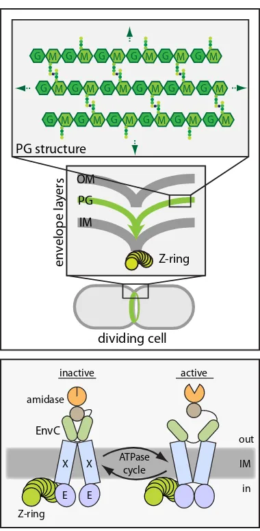
Figure 2.2. Coordinated envelope constriction in Gram-negative bacteria and model for regulated PG hydrolase activity at the division site. (Upper) Diagrams showing a dividing cell with an assembled cytokinetic ring apparatus (green) (bottom diagram), a close-up diagram of the division site highlighting the coordinated constriction of the envelope layers (IM, inner membrane; OM, outer membrane; PG, peptidoglycan; Z-ring, FtsZ cytoskeletal ring) (middle diagram), and a diagram of the PG chemical structure (G, N-acetylglucosamine; M, N
-acetylmuramic acid) (top diagram). Colored dots in the top diagram represent attached peptides. The PG structure continues in all directions to envelop the cell (green arrows). (Lower) A
schematic diagram of a putative FtsEX–EnvC–amidase complex at the Z-ring. We propose that conformational changes in FtsEX induced by FtsE-mediated ATP hydrolysis are transmitted to EnvC to control its ability to activate the amidases so that they can cleave the septal PG (not drawn). Z-ring X E EnvC amidase IM E X inactive ATPase cycle active in out M
G G M G M G M G M G M
M
G G M G M G M G M G M
M
G G M G M G M G M G M


![Figure 2.12. EnvC localization in cells producing FtsE* variants. Cells of DY18(attλTD80) [ΔftsEX ΔenvC (Plac::envC-mCherry)] harboring the integrated expression constructs (attHKDY166) [Plac::ftsE(K41M)X-GFP] (A) or (attHKDY168) [Plac::ftsE(E163Q)X-GFP] (B) were grown and visualized as described in the legend for Figure 2.9.](https://thumb-us.123doks.com/thumbv2/123dok_us/7916325.190897/76.612.137.474.69.433/localization-producing-harboring-integrated-expression-constructs-visualized-described.webp)
