0022-538X/07/$08.00⫹0 doi:10.1128/JVI.02244-06
Copyright © 2007, American Society for Microbiology. All Rights Reserved.
Epstein-Barr Virus BHRF1 Micro- and Stable RNAs during
Latency III and after Induction of Replication
䌤
Li Xing
1,2and Elliott Kieff
1,2*
Department of Medicine and Department of Microbiology and Molecular Genetics, Harvard Medical School,1and
The Channing Laboratory, Brigham and Women’s Hospital, 181 Longwood Avenue,2Boston, Massachusetts 02115
Received 12 October 2006/Accepted 5 July 2007
Epstein-Barr virus (EBV) microRNAs miR-BHRF1-1, -2, and -3 have been detected in latency III-infected lymphoblasts, where they are encoded within EBNA transcripts (X. Cai, A. Schafer, S. Lu, J. P. Bilello, R. C. Desrosiers, R. Edwards, N. Raab-Traub, and B. R. Cullen, PLoS Pathog. 2:e23, 2006). In latency III-infected lymphoblasts, we have also identified a stable 1.3-kb RNA, which begins 3ⴕ to miR-BHRF1-1, includes the BHRF1 open reading frame, and ends near miR-BHRF1-2. This 1.3-kb RNA is the residue of Drosha cleavage of the BHRF1 microRNAs from EBNA transcripts. Early after induction of EBV replication in latency I-infected Akata lymphoblasts, BHRF1 spliced 1.4-kb mRNA accumulated along with low levels of miR-BHRF1-2 and -3 and a 0.9-kb Drosha or miR-miR-BHRF1-2 cleavage product of BHRF1 mRNA. The turning on of latency III infection at 48 to 72 h after induction of EBV replication was associated with higher miR-BHRF1-1, -2, and -3 levels; accumulation of the 1.3-kb RNA residue in the nucleus; abundant BHRF1 spliced 1.4-kb mRNA in the cytoplasm; and more abundant 0.9-kb mRNA cleavage product in the cytoplasm. These findings implicate miR-BHRF1-2 in 3ⴕcleavage of BHRF1 mRNA in the cytoplasm and Drosha in cleavage of latency III EBNA and EBV replication-associated BHRF1 transcripts in the nucleus.
In primary human infection, Epstein-Barr virus (EBV) rep-licates in the oropharyngeal epithelium (60) and establishes latency III infection in B lymphocytes (48, 62, 67). During latency III infection, the EBV Cp or Wp EBNA promoters drive expression of six nuclear antigen proteins (EBNA2, EBNALP, EBNA3A, EBNA3B, EBNA3C, and EBNA1) from a single alternatively spliced transcript (37, 54). The latency III primary EBNA transcripts include many open reading frames (ORFs) expressed in EBV replication and are the likely source of the 3 BHRF1 micro-RNAs (miRNAs), which are encoded in an intron of most EBNA RNAs (11, 50). In latency III infection, EBV also expresses three integral membrane protein (LMP1, LMP2A, and LMP2B)-encoding mRNAs, two small RNAs (EBER1 and -2), BamHI A rightward transcripts (BARTs) (7, 15, 22, 37, 54, 56, 58), and 24 BART miRNAs (11, 29, 50). Latency III EBV gene expression causes continuous cell proliferation, which results in vitro in lymphoblastoid cell lines (LCLs) and in vivo in lymphoproliferative diseases (37, 54). Only BART miRNAs are detected in latency I- or II-infected cells, in which EBNA1 is the only EBNA expressed, from a promoter downstream of BHRF1. However, latency III-associated proteins are also detected with EBV replication in epithelial cells in vivo (68) or late in EBV replication in latency I-infected Burkitt’s lymphoma (BL) cells (72).
miRNAs are small non-protein-coding 20- to 25-nucleotide (nt) single-strand RNAs, which negatively control protein ex-pression, by inhibiting translation or cleaving of mRNA (2, 6). Most miRNAs are processed in the cell nucleus from RNA polymerase II capped and polyadenylated RNAs by the RNase
III enzyme Drosha to release 70-nt RNA hairpin pre-miRNA (6, 10, 39, 40). Pre-miRNAs are exported to the cytoplasm by exportin 5 (44, 70). In the cytoplasm, pre-miRNA can be cleaved by the RNase III enzyme Dicer (33) in association with TRBP (17) to generate 22-nt mature miRNAs (21). Mature miRNAs can be incorporated into RNA-induced silencing complexes (RISC) and can direct RISC to complementary mRNA targets (6). The targets of the EBV miRNAs are not known, although miR-BART2 may cleave EBV DNA poly-merase (BALF5) mRNA (11, 26, 50).
The experiments reported here investigate EBV miR-BHRF1-1, -2, and -3, which are encoded within introns of EBNA transcripts and are expressed in latency III-infected lymphoblasts, but not in latency I-infected BL or latency II-associated nasopharyngeal carcinoma (NPC) cells (Fig. 1A) (11, 50). miR-BHRF1-1, -2, and -3 are likely to be Drosha-cleaved products of EBNA introns. BHRF1 is an antiapoptotic Bcl-2 homologue, which is expressed early in EBV replication (31). Although RNAs that initiate upstream of the BHRF1 promoter and include the BHRF1 ORF are detected in latency III-infected lymphoblasts (3, 49, 52, 58), BHRF1 monoclonal antibody (MAb) rarely detects BHRF1 protein until early in EBV replication, when BHRF1 abundantly accumulates (49). miR-BHRF1-1 overlaps with the BHRF1 mRNA transcrip-tional start site and is therefore not encoded in BHRF1 mRNA, whereas miR-BHRF1-2 and -3 are potentially en-coded in the BHRF1 mRNA 3⬘-untranslated sequence and may therefore be expressed from early times in EBV replica-tion (3, 19, 50, 52).
MATERIALS AND METHODS
Cell culture and antibodies.B95-8 (46, 59), IB4 (65), recently derived LCLs, NPC C666-1 (18), and EBV-infected or uninfected BJAB (25), BL41 (14), and Akata (63, 64) cells were maintained in RPMI 1640 medium (Gibco-BRL) supplemented with 10% or 20% Fetal Plex animal serum complex (Gemini). C15 * Corresponding author. Mailing address: Brigham and Women’s
Hospital, Harvard Medical School, Channing Laboratory, 181 Long-wood Ave., Boston, MA 02115-5804. Phone: (617) 525-4252. Fax: (617) 525-4251. E-mail: [email protected].
䌤Published ahead of print on 11 July 2007.
9967
on November 8, 2019 by guest
http://jvi.asm.org/
and C17 nasopharyngeal tumors were passaged in nude mice as xenografts (9). MAbs PE2 (71), A10 (45), and 3E8 (42) are specific to EBNA2, EBNA3C, and BHRF1, respectively.
Western blot analysis.Cells were harvested at indicated times after virus replication induction and lysed in 2⫻sodium dodecyl sulfate-polyacrylamide gel electrophoresis loading buffer. Cell extracts were separated by 10% sodium
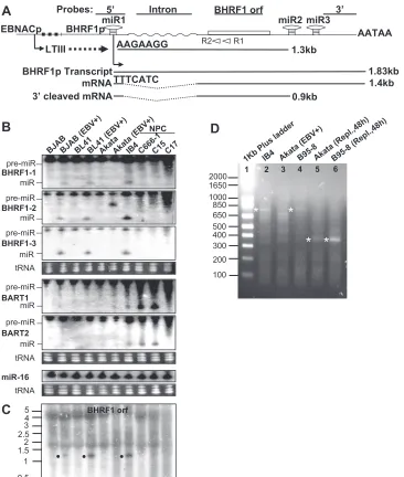
[image:2.585.111.477.65.497.2]dodecyl sulfate-polyacrylamide gel electrophoresis and transferred to nitrocellu-lose membranes (Bio-Rad). Following incubation in blocking buffer (phosphate-buffered saline containing 0.1% Tween 20 and 5% nonfat milk), membranes were incubated with diluted primary MAbs overnight at 4°C with gentle shaking. Following washes and secondary antibody incubation, specific binding was visu-alized by ECL enhanced chemiluminescence (Amersham Biosciences). FIG. 1. miR-BHRF1-1, -2, and -3 and an unspliced 1.3-kb BHRF1 RNA are putative Drosha cleavage products from a latency III EBNA promoter transcript intron. (A) Schematic diagram showing the early virus replication BHRF1 promoter (BHRF1p) and 1.83-kb primary transcript, the 1.4-kb spliced BHRF1 mRNA, the far upstream latency III EBNA promoters (EBNACp) that transcribe 110 kb of the 170-kb EBV genome during latency III infection, the probes used in Northern blots (5⬘, intron, ORF, and 3⬘), the AATAA poly(A) addition signal, and the positions of the primary miR-BHRF1-1 (miR1), miR-BHRF1-2 (miR3), and miR-BHRF1-3 (miR3) genes. 5⬘-RACE-determined sequences of early replication-associated 1.4-kb BHRF1 mRNA and 1.3-kb latency III-associated RNA are shown. Two open arrows represent primers Racer1 (R1) and Racer2 (R2) used in 5⬘-RACE. (B) Northern analysis of selected EBV miRNAs in total RNA from indicated cell lines and tumors. Positions of miRNAs (miR) and pre-miRNAs (pre-miR) are indicated. EBV⫹, EBV infected. Ethidium bromide-stained tRNAs are included as loading controls. (C) Northern analysis of BHRF1 RNAs with the32P-labeled BHRF1 ORF probe detects the 1.3-kb EBNA transcript putative
Drosha cleavage product. The sizes of Millennium RNA size makers (Ambion) are shown on the left in kb. (D) 5⬘-RACE analysis of BHRF1 RNA from indicated cells in latent infection or after induction of virus replication at 48 h (Repl.,48h). Reverse transcription was primed with Racer1 primer. The resulting cDNAs were poly(dC) tailed and amplified by PCR. PCR products from latency III IB4 (lane 2), latency I Akata (lane 3), latency III B95-8 (lane 4), and 48-h virus replication-induced Akata (lane 5) and B95-8 (lane 6) cells were separated in 1.5% agarose gel. PCR products were purified, cloned, and sequenced. Sequencing-confirmed EBV-specific PCR products are labeled with asterisks on the left of the lanes. The sizes of the 1-kb Plus ladder (Invitrogen) are shown on the left in bp.
on November 8, 2019 by guest
http://jvi.asm.org/
RNA preparation and Northern analysis.Total RNA was prepared using TRIzol reagent (Invitrogen). Cytoplasmic and nuclear RNAs were fractionated from Akata cells at 48 h after virus replication induction by immunoglobulin G (IgG) cross-linking. Briefly, cells were resuspended in 4 volumes buffer A (10 mM HEPES [pH 7.9], 10 mM KCl, 1.5 mM MgCl2) for 30 min on ice, Dounce
homogenized, and spun at 2,000 rpm at 4°C for 5 min. The supernatant was extracted with TRIzol reagent to isolate cytoplasmic RNA. The buffer A-washed pellet was resuspended in 3 volumes of buffer B (20 mM HEPES [pH 7.9], 20% glycerol, 420 mM NaCl, 1,5 mM MgCl2, 0.2 mM EDTA), incubated for 30 min
on ice, and spun at 12,000 rpm for 20 min at 4°C. The separated supernatant was extracted with TRIzol reagent to isolate nuclear RNA. For miRNA or mRNA detection, 30g total RNA per lane was separated in 15% polyacrylamide gel or in denaturing 1% agarose–2.2 M formaldehyde gel, transferred onto GeneScreen Plus hybridization transfer membranes (Perkin-Elmer), immobilized by baking at 80°C for 2 h, and hybridized with32P-labeled RNA probes at 37°C overnight or 32P-labeled DNA probes at 42°C overnight.32P-labeled RNA probes specific for
each miRNA were prepared by in vitro transcription using the mirVana miRNA probe construction kit (Ambion) and primers (Table 1).32P-labeled 5⬘, intron,
ORF, and 3⬘EBV BHRF1 DNA probes were prepared by random primer DNA labeling (Invitrogen) using BHRF1 5⬘-untranslated, intron, ORF, and 3⬘ -un-translated sequences as templates. Membranes were hybridized in Ultrahyb-Oligo hybridization buffer (Ambion). The signals in Northern blots were de-tected and quantified using PhosphorImager software (Amersham Biosciences).
Induction of EBV replication.Virus replication was induced in latency I Akata cells or latency III B95-8 cells. About 2⫻106to 5⫻106Akata cells were treated
with 50g per ml mouse anti-human IgG (Dako Cyto, Japan) at 37°C for 4 h. Fresh medium was added to keep the cells at 5⫻105cells per ml until harvest.
EBV replication in B95-8 cells was induced by adding 4-hydroxytamoxifen to latency III-infected B95-8 LCL cells that stably express an RNA encoding a 4-hydroxytamoxifen-dependent fusion of the EBV immediately-early gene cod-ing for BZLF1 (Zta) with a mutant estrogen receptor (34). Cell surface EBV
gp350 late gene expression was assayed with MAb 72A1 as an indicator of late virus replication (53, 66).
5ⴕ-RACE.Rapid amplification of cDNA 5⬘ ends (5⬘-RACE) was done as instructed by the manufacturer (Invitrogen). Briefly, the cDNAs were initially synthesized from 2g of total RNAs by SuperScript reverse transcriptase using a BHRF1 ORF-specific Racer1 primer (Table 1 and Fig. 1A). After RNase H treatment, polydeoxycytosines were added to the 3⬘ends of the cDNA by ter-minal deoxynucleotide transferase. PCR was done with nested Racer2 primer (Table 1 and Fig. 1A) and universal primer (Gibco-BRL) in a total volume of 50
l withTaqDNA polymerase (Invitrogen) at 95°C for 2 min with 35 cycles at 94°C for 50 s, 50°C for 50 s, and 72°C for 1.5 min, followed by extension at 72°C for 10 min. The amplicons were analyzed by 1.5% agarose gel electrophoresis, gel purified, cloned into pCR4-TOPO (Invitrogen), and sequenced. The same se-quence was obtained from at least four individual clones for each EBV specific PCR product.
Nucleotide sequence accession number. The DNA sequence of the EBV Akata strain BHRF1 gene has been deposited in GenBank under accession no. EF192979.
RESULTS
MiR-BHRF1-1, -2, and -3 are expressed at high level in latency III B cells, whereas miR-BART1 and -2 are expressed at high level in NPC cells.Northern hybridization was done with32P-labeled miRNA probes to validate our detection of
EBV miRNAs. MiR-BHRF1-1, -2, and -3 were clearly de-tected in latency III EBV-infected BJAB, BL41, B95-8, IB4, and recently derived LCLs, but not in latency II NPC C666-1 (18), C15 cells (9), or latency I-infected Akata BL cells (Fig. 1B) (data not shown). Cell miR-16 blots are included as load-ing controls (38). In contrast, miR-BART1 and -2 were ex-pressed at lower levels in latency III-infected B cells and la-tency I-infected Akata BL cells than in lala-tency II-infected NPC C666-1 and C15 cells (Fig. 1B), in accord with previous results (11, 16, 27, 36, 50, 56, 61). EBV miRNAs were not detected in NPC C17 cells, presumably related to the low EBV genome presence and expression (9). Thus, high-level miR-BHRF1-1, -2, and -3 are tightly associated with latency III, as opposed to latency I and II (11) (Table 2).
Latency III is associated with expression of a stable, un-spliced, 1.3-kb BHRF1 ORF-containing RNA.To further ana-lyze BHRF1 latency III-associated RNAs, Northern hybridiza-tion was done with32P-labeled 5⬘, intron, BHRF1 ORF, and 3⬘
EBV DNA probes (Fig. 1A and Table 1). Northern analysis of total RNA detected an unspliced, 1.3-kb BHRF1 RNA in latency III-infected BJAB, BL41, and IB4 cells with BHRF1 ORF, 5⬘, and intron probes, but not with the 3⬘probe (Fig. 1A and C and Table 2) (data not shown). This RNA was not detected in uninfected BJAB and BL41 BL cells, EBV latency I-infected Akata cells, or latency II-infected NPC C666-1, C15, or C17 cells (Fig. 1A and C) (data not shown), indicating that the 1.3-kb BHRF1 RNA is associated with miR-BHRF1-1, -2, and -3 expression and EBV latency III but not latency I or II infection.
[image:3.585.42.283.75.345.2]To further characterize the BHRF1 1.3-kb RNA detected in Northern blots, the 5⬘sequence of the BHRF1 RNAs from IB4 and B95-8 cells was obtained by 5⬘-RACE analysis using la-tency I Akata cell RNA as a negative control. A 0.75-kbp PCR product was amplified from BHRF1 1.3-kb RNA from IB4 and B95-8 cells (Fig. 1D). Four 0.75-kbp PCR product clones were sequenced. The 1.3-kb BHRF1 RNA began with 5⬘ -AAGAAGGG-3⬘(GenBank accession no. EF192979) (Fig. 1A and Table 2), which is the predicted 3⬘Drosha cutting site for
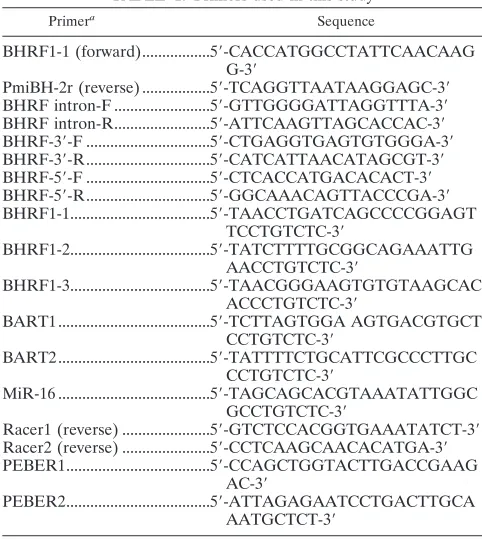
TABLE 1. Primers used in this study
Primera Sequence
BHRF1-1 (forward)...5⬘-CACCATGGCCTATTCAACAAG G-3⬘
PmiBH-2r (reverse) ...5⬘-TCAGGTTAATAAGGAGC-3⬘ BHRF intron-F ...5⬘-GTTGGGGATTAGGTTTA-3⬘ BHRF intron-R...5⬘-ATTCAAGTTAGCACCAC-3⬘ BHRF-3⬘-F ...5⬘-CTGAGGTGAGTGTGGGA-3⬘ BHRF-3⬘-R...5⬘-CATCATTAACATAGCGT-3⬘ BHRF-5⬘-F ...5⬘-CTCACCATGACACACT-3⬘ BHRF-5⬘-R...5⬘-GGCAAACAGTTACCCGA-3⬘ BHRF1-1...5⬘-TAACCTGATCAGCCCCGGAGT
TCCTGTCTC-3⬘
BHRF1-2...5⬘-TATCTTTTGCGGCAGAAATTG AACCTGTCTC-3⬘
BHRF1-3...5⬘-TAACGGGAAGTGTGTAAGCAC ACCCTGTCTC-3⬘
BART1 ...5⬘-TCTTAGTGGA AGTGACGTGCT CCTGTCTC-3⬘
BART2 ...5⬘-TATTTTCTGCATTCGCCCTTGC CCTGTCTC-3⬘
MiR-16 ...5⬘-TAGCAGCACGTAAATATTGGC GCCTGTCTC-3⬘
Racer1 (reverse) ...5⬘-GTCTCCACGGTGAAATATCT-3⬘ Racer2 (reverse) ...5⬘-CCTCAAGCAACACATGA-3⬘ PEBER1...5⬘-CCAGCTGGTACTTGACCGAAG
AC-3⬘
PEBER2...5⬘-ATTAGAGAATCCTGACTTGCA AATGCTCT-3⬘
aPrimer pairs BHRF1-1/PmiBH-2r, BHRF intron-F/BHRF intron-R,
BHRF-3⬘-F/BHRF-3⬘-R, and BHRF-5⬘-F/BHRF-5⬘-R were used in PCR to amplify EBV genomic DNA fragments from EBV-infected Akata cells for preparation of
32P-labeled probes specific to the BHRF1 ORF, intron, and 3⬘- and 5⬘
-untrans-lated regions, respectively. Oligonucleotides BHRF1-1, BHRF1-2, BHRF1-3, BART1, BART2, and MiR-16 were used as templates to produce32P-labeled
RNA probes specific for miR-BHRF1-1, miR-BHRF1-2, miR-BHRF1-3,
miR-BART1, miR-BART2, and miR-16, respectively.
on November 8, 2019 by guest
http://jvi.asm.org/
miR-BHRF1-1 (50). Since a Drosha cleavage 5⬘ of miR-BHRF1-2 would result in a 1,312-nt RNA versus 1,430 nt for cleavage 5⬘of miR-BHRF1-3, the size of the RNA, detection with BHRF1 intron probe, and a specific inability to detect the 1.3-kb RNA with the BHRF1 3⬘probe are evidence that the 1.3-kb BHRF1 RNA extends from 3⬘of miR-BHRF1-1 to 5⬘of miR-BHRF1-2. No EBV-specific PCR product was detected with RNAs from latency I-infected Akata cells (Fig. 1D) (se-quence data not shown). These data indicate that latency III EBV infection is associated with the accumulation of miR-BHRF1-1, -2, and -3 and of a stable unspliced BHRF1 1.3-kb RNA and support a model that these RNAs are Drosha cleav-age products of EBNA transcripts or introns.
BHRF1 RNAs after induction of EBV replication in latency I-infected Akata cells.To test whether EBV BHRF1 miRNAs or 1.3-kb RNAs are also expressed as a consequence of early replication-associated BHRF1 mRNA expression or latency III EBNA expression late in EBV replication in latency I-in-fected Akata cells (72), EBV replication was induced in latency I-infected Akata cells (63, 64). Cell surface EBV gp350 late gene expression was assayed with MAb 72A1 as an indicator of late virus replication (53, 66). EBV gp350 expression was de-tected as early as 12 h, increased at 24, and peaked at 48 h, when 40% of Akata cells were gp350 positive by microscopy (data not shown).
Early EBV replication-associated BHRF1 protein (Fig. 2B) and spliced and polyadenylated BHRF1 1.4-kb mRNA (Fig. 3 and Table 2) were detected by 12 h after induction of EBV replication. BHRF1 1.4-kb mRNA [including 100-nt poly(A)
tail] increased somewhat at 24 to 72 h and persisted at high levels (Fig. 3, ORF and 3⬘probe). BHRF1 protein increased at 24 and 48 h and somewhat thereafter (Fig. 2B). As expected, BHRF1 mRNA hybridized to the ORF and 3⬘probes but not the intron probe (Fig. 1A and 3 and Table 2). An EBV-specific 0.32-kbp PCR product with the beginning sequence 5⬘-TTTC ATC-3⬘ (Fig. 1A and Table 2) was amplified by 5⬘-RACE analysis of RNA from induced Akata and B95-8 cells at 48 h (Fig. 1D, lanes 5 and 6). Similar products were not detected from latency I Akata or latency III B95-8 cell RNAs without induction of virus replication (Fig. 1D, lanes 3 and 4). Four clones of the 0.32-kbp 5⬘-RACE product were sequenced. The sequence start site was at nt 41501 in the EBV genome (GenBank accession no. AJ507799), 9 nt 5⬘ to nt 41510, the previous start site determined by S1 analysis (19), and 25 nt 3⬘ to the end of the BHRF1 TATTA sequence (TATA box).
[image:4.585.42.543.82.357.2]At 12 h to 48 h, a small amount of 0.9-kb BHRF1 3⬘-cleaved mRNA was detected by BHRF1 ORF and 5⬘probes but not by intron or 3⬘ probes (Fig. 3B and Table 2) (data not shown). Intron probe detected the 1.3-kb latency III RNA and a non-specific 1.0-kb RNA (Fig. 3A). The 0.9-kb BHRF1 RNA sig-nificantly increased in abundance at 48 to 72 h (Fig. 3B). As previously described (72), late-replication-onset latency III-associated EBNA2, EBNA3C, and EBNA1 expression were detected at 24 to 48 h and were abundant at 72 h (Fig. 2B). In parallel with latency III EBNA expression, pre-miR-BHRF1-1, -2, and -3 were readily detected at 48 h and were abundant by 72 h (Fig. 2A). By 72 h, miR-BHRF1-1, -2, and -3 (Fig. 2A) and 1.3-kb Drosha-cleaved EBNA intron RNA were abundant
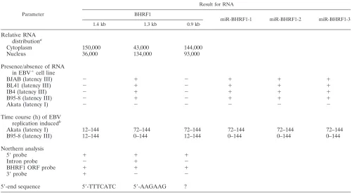
TABLE 2. BHRF1 RNAs in EBV-infected cells
Parameter
Result for RNA
BHRF1
miR-BHRF1-1 miR-BHRF1-2 miR-BHRF1-3
1.4 kb 1.3 kb 0.9 kb
Relative RNA distributiona
Cytoplasm 150,000 43,000 144,000
Nucleus 36,000 134,000 93,000
Presence/absence of RNA in EBV⫹cell line
BJAB (latency III) ⫺ ⫹ ⫺ ⫹ ⫹ ⫹
BL41 (latency III) ⫺ ⫹ ⫺ ⫹ ⫹ ⫹
IB4 (latency III) ⫺ ⫹ ⫺ ⫹ ⫹ ⫹
B95-8 (latency III) ⫺ ⫹ ⫺ ⫹ ⫹ ⫹
Akata (latency I) ⫺ ⫺ ⫺ ⫺ ⫺ ⫺
Time course (h) of EBV replication inducedb
Akata (latency I) 12–144 72–144 72–144 72–144 72–144 72–144
B95-8 (latency III) 12–144 0–144 12–144 0–144 0–144 0–144
Northern analysis
5⬘probe ⫹ ⫹ ⫹
Intron probe ⫺ ⫹ ⫺
BHRF1 ORF probe ⫹ ⫹ ⫹
3⬘probe ⫹ ⫺ ⫺
5⬘-end sequence 5⬘-TTTCATC 5⬘-AAGAAG ?
aRelative RNA distribution in cytoplasm and nucleus was determined from RNA in Fig. 3D. The EBER distributions in the mock and IgG cross-linked RNA
preparations were 43,000 and 40,000 in cytoplasm and 577,000 and 608,000 in nucleus.
bTime course of high-level expression after virus replication induction.
on November 8, 2019 by guest
http://jvi.asm.org/
(Fig. 3). The 1.3-kb RNA hybridized to 5⬘, intron, and ORF probes but not to the 3⬘probe, consistent with cleavage 5⬘to miR-BHRF1-2 or -3 (Fig. 3) (data not shown). The 1.3-kb RNA was identical in size to the latency III-associated EBNA intron RNA, which is Drosha cleaved 3⬘to miR-BHRF1-1 and 5⬘to miR-BHRF1-2, versus the 1.43 kb expected from Drosha cleavage 5⬘to miR-BHRF1-3. Thus, activation of latency III EBNA transcription is the likely basis for the high-level miR-BHRF1-1, -2, and -3 and 1.3-kb residual EBNA intron RNA late in Akata cells (Fig. 3 and Table 2).
As miR-BHRF1-1, -2, and -3 became abundant at 48 to 72 h (Fig. 2A), the 0.9-kb ORF-positive and 3⬘-truncated RNA also became more abundant (Fig. 3B and Table 2). Thus, the 0.9-kb RNA has the size and sequence content consistent with cleav-age of BHRF1 mRNA 3⬘to miR-BHRF1-2 by Drosha in the nucleus or by miR-BHRF1-2 in the cytoplasm.
To further clarify the genesis of 0.9-kb BHRF1 3⬘-truncated RNA, cytoplasmic and nuclear RNAs were isolated from
EBV-infected Akata cells at 48 h after virus replication induc-tion, and RNAs were analyzed by Northern hybridization with
32P-labeled BHRF1 ORF probe. As a control, the isolated
RNAs were separated in 5% polyacrylamide gel and probed with 32P-labeled DNA oligonucleotide probes specific for
EBV-encoded EBER1 and EBER2 small nuclear RNAs (Ta-ble 1) (5, 24, 32, 41). As shown in Fig. 3D, EBERs were in the nuclear RNA fraction from Akata cells before and 48 h after induction of virus replication. Furthermore, the latency III EBNA-associated 1.3-kb unspliced and putatively Drosha-cleaved BHRF1 RNA was threefold more abundant in the 48-h nuclear versus cytoplasmic RNA (Fig. 3D and Table 2). Moreover, BHRF1 1.4-kb mRNA was fourfold more abundant in 48-h cytoplasmic versus nuclear RNA. Importantly, the 0.9-kb spliced and 3⬘-truncated BHRF1 RNA was 1.6-fold more abundant in the cytoplasm than in the nucleus, consistent with this RNA being mostly a product of miR-BHRF1-2 cleav-age of BHRF1 mRNA in the cytoplasm, as postulated (38).
miR-BART1 and -2 expression did not change following induction of EBV replication in latency I-infected Akata cells (Fig. 2B).
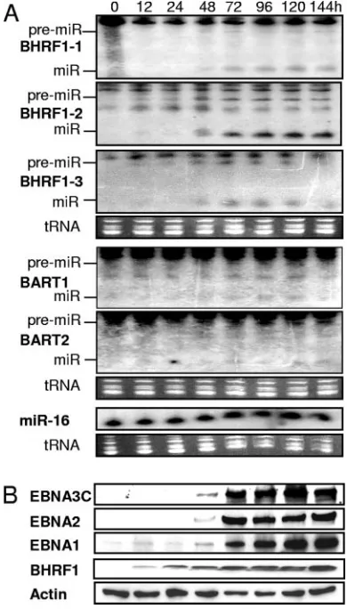
EBV replication in latency III-infected B95-8 cells does not result in significantly increased miR-BHRF1-1, -2, or -3 or 1.3-kb RNA levels.To evaluate the effects of preexistent la-tency III EBV infection on EBV replication-associated RNA levels, EBV replication was induced in B95-8 cells (34). Sur-face gp350 expression increased at 12 to 24 h and peaked at 48 h, when 50% of B95-8 cells were gp350 positive by micro-scopic evaluation (data not shown). In contrast to latency I-in-fected Akata cells, EBNA proteins (Fig. 4B), pre-miR- and miR-BHRF1-1, -2, and -3, and 1.3-kb residual Drosha-cleaved RNA barely changed at any time following induction of repli-cation in latency III-infected B95-8 cells (Fig. 4A) (data not shown). BHRF1 mRNA and protein increased significantly by 12 h and were at high levels thereafter (Fig. 4B and C). BHRF1 0.9-kb RNA was also detected at a constant level by BHRF1 ORF and 5⬘probes but not by intron and 3⬘probes from 12 h onward (Fig. 4C and Table 1) (data not shown). These data indicate that EBV replication in preexisting latency III-in-fected cells is associated with little change in EBNA, miR-BHRF1-1, -2, or -3, or 1.3-kb RNA expression, early turn on of BHRF1 1.4-kb mRNA and 0.9-kb cleavage product, and a constant ratio of BHRF1 mRNA and cleavage product throughout replication.
DISCUSSION
[image:5.585.65.260.70.412.2]In sum, these data confirm that high-level miR-BHRF1-1, -2, and -3 are a characteristic of EBV latency III infection and EBNA transcription (11) and further indicate that EBNA in-tron cleavage by Drosha to produce miR-BHRF1-1, -2, and -3 also results in a residual stable 1.3-kb RNA. This RNA begins with 5⬘-AAGAAGGG-3⬘, which is the predicted sequence (50) 3⬘ to the miR-BHRF1-1 cleavage site and therefore differs from the early replication BHRF1 1.4-kb mRNA, which we now find begins 35 nt 5⬘ to the 1.3-kb RNA. Both RNAs hybridized to the BHRF1 5⬘and ORF probes indicated in Fig. 1 and differ in that the 1.3-kb RNA hybridizes to the intron probe but not to the 3⬘ probe, whereas the 1.4-kb mRNA hybridizes to the 3⬘probe but not to the intron probe. By size,
FIG. 2. Induction of EBV replication in latency I-infected Akata cells results in late replication-associated latency III EBNA proteins and miR-BHRF1-1, -2, and -3. (A) Northern analysis of EBV miRNAs using total RNAs extracted at the indicated time points after EBV-infected Akata cell-IgG cross-linking. miRNA (miR) and pre-miRNA (pre-miR) are indicated. (B) Time course of latency III EBNA and BHRF1 protein expression after EBV-infected Akata cell IgG cross-linking. At the indicated times after induction of virus replication, Western blot analysis was done with MAbs specific to the indicated proteins.
on November 8, 2019 by guest
http://jvi.asm.org/
the 1.3-kb RNA is most likely Drosha cleaved at the beginning of miR-BHRF1-2, since that would yield a 1.312-nt RNA ver-sus 1.430 nt for Drosha cleavage at the beginning of miR-BHRF1-3. The defined length of the 1.3-kb RNA is also evi-dence that at least two miRNAs are cleaved from the primary EBNA transcript, which is much longer and variable in length (37). Moreover, latency III RNAs and proteins and high-level miR-BHRF1-1, -2, and -3 and 1.3-kb RNAs accumulate late in replication in latency I-infected Akata cells (72) and the 1.3-kb RNA localizes to the nucleus (Fig. 3D and Table 2), as ex-pected for a stable product of Drosha cleavage. In stability, the 1.3-kb BHRF1 RNA is reminiscent of the stable truncated RNA fragment identified from Drosha processing of cell pri-mary miR-30 (10).
At 12 h after induction of EBV replication in latency I-in-fected Akata cells, miR-BHRF1, -2, and -3 were detected along with moderately high levels of 1.4-kb BHRF1 mRNA (43, 72) and small amounts of 0.9-kb BHRF1 ORF-positive, intron-negative, 3⬘-truncated mRNA. Since the BHRF1 early promoter initiates transcription within miR-BHRF1-1-encod-ing DNA, the expression of miR-BHRF1-2 and -3 in the ab-sence of miR-BHRF1-1 indicates that miR-BHRF1-2 and -3 are processed by Drosha cleavage of an RNA initiated by the BHRF1 early promoter. Since spliced RNAs are usually ex-ported, Drosha cleavage of a nonspliced BHRF1 transcript might have been anticipated and would have resulted in a 1.3-kb intron probe-positive RNA. The absence of 1.3-kb RNA and the presence of a 0.9-kb ORF-positive, spliced, and 3⬘ -truncated RNA at 12 to 24 h (Fig. 3 and Table 2) are consistent
with the possibility that the 0.9-kb RNA is a miR-BHRF1-2 cytoplasmic cleavage product of the 1.4-kb BHRF1 spliced mRNA.
The 0.9-kb BHRF1 spliced and 3⬘-truncated RNA increased in abundance at 48 to 72 h (Fig. 3B), concomitant with latency III protein expression, miR-BHRF1-1 expression, increased miR-BHRF1-2 and -3 expression, and unspliced 1.3-kb resid-ual Drosha-cleaved RNA expression. miR-BHRF1-1 expres-sion implicates latency III EBNA transcripts for the increased miR-BHRF1 RNAs. Latency III EBNA transcription and Dro-sha cleavage of EBNA RNA late in virus replication in Akata cells are the likely origin of high-level miR-BHRF1-1, -2, and -3 and unspliced 1.3-kb BHRF1 RNA. The concomitant in-crease in latency III miR-BHRF1-2 and 0.9-kb spliced and 3⬘-truncated RNA and the cytoplasmic localization of the 0.9-kb RNA at 48 h (Fig. 3 and Table 2) clearly implicate miR-BHRF1-2 cleavage in the cytoplasm in the genesis of the 0.9-kb RNA, although increased Drosha cleavage of spliced BHRF1 mRNA in the nucleus has not been fully excluded.
miRNAs can mediate target mRNA cleavage of fully or partially complementary target sequences (4, 73). EBV miR-BART2 is implicated in cleaving its complement in the BALF5 mRNA 3⬘-untranslated sequence (26, 50). In Caenorhabditis
elegans development, let-7 miRNA cleaves partially
comple-mentary lin-41 target mRNA and lin-4 miRNA cleaves par-tially complementary lin-14and lin-28 (4). Cytoplasmic heli-cases such as Dicer (28) and Gemin3 (69), a component of RISC (47), may unwind target RNAs and enable miRNA bind-ing to fully or partially complementary targets. In stable latency
FIG. 3. Time course of BHRF1 RNA expression in EBV-infected Akata cells after IgG cross-linking. Shown are the BHRF1 intron probe (A), ORF probe (B), and 3⬘-untranslated region probe (C), as depicted in Fig. 1. (D) Subcellular localization of 1.4-kb BHRF1 mRNA, 1.3-kb unspliced, cleaved BHRF1 RNA, and 0.9-kb spliced, cleaved BHRF1 RNA. Cytoplasmic (Cyto) and nuclear (Nuc) RNAs were separately isolated from EBV-infected Akata cells at 48 h after mock (⫺) or IgG (⫹) cross-linking. The sizes of Millennium RNA markers (Ambion) are shown on the right in kb. EBV BHRF1 RNAs and EBERs are indicated by size in kb and bp, respectively. NS, nonspecific 1.0-kb cell RNA.
on November 8, 2019 by guest
http://jvi.asm.org/
III infection in LCLs, Drosha cleavage of EBNA transcript derived potential BHRF1-encoding RNA within the nucleus and miR-BHRF1-2-mediated cleavage of BHRF1 RNA in the cytoplasm may protect against the potential excessive survival effects of BHRF1 expression.
The high-level expression of miR-BHRF1-1, -2, and -3 and of intron-containing 1.3-kb BHRF1 RNA in latency III in LCLs and late in replication suggests that these RNAs may be important for B-lymphocyte survival during stable latency III infection (1, 35), during EBV replication, or in the face of T-cell immune attack, in vivo.
Other herpesviruses such as Kaposi’s sarcoma-associated herpesvirus (KSHV) (13, 29, 51, 57), mouse gammaherpesvirus 68 (MHV68) (51), human cytomegalovirus (51), herpes sim-plex virus type 1 (HSV-1) (20, 30), and Marek’s disease virus (MDV) (8) also encode miRNAs. KSHV encodes 12 distinct miRNAs from a 4.5-kb genome segment, directly upstream of KSHV latency-associated ORFs 71, 72, and 73 (12, 55). These miRNAs are expressed in latently infected cells and are largely unaffected by induction of virus replication (13). MHV68 en-codes nine miRNAs within a 6-kb region that enen-codes eight
small RNAs. These miRNAs may be processed from RNA polymerase III transcripts (51). Human cytomegalovirus miRNA genes are spread through the genome (51). MDV encodes eight miRNAs, five of which flank themeqoncogene. Three map to the latency-associated transcript (LAT) and may contribute to MDV-induced cell transformation (8). HSV-1 LAT miRNA is surprisingly similar to EBV BHRF1 miRNAs in being produced from a stable intron in latent infection and late in replication (23). HSV-1 miR-LAT down-regulates transforming growth factorand SMAD3 expression and has antiapoptotic effects (30). EBV miR-BHRF1-2 and -3 RNAs may have opposite effects in down-regulating BHRF1 expres-sion.
ACKNOWLEDGMENTS
We thank Nancy Raab-Traub from the University of North Carolina at Chapel Hill for providing NPC tumor C15 and C17 and for helpful comments; Mei-Ying Liu and Jen-Yang Chen from National Taiwan University for generously providing MAb 3E8; and Ellen Cahir-Mc-Farland, Eric Johannsen, Fred Wang, Bo Zhao, and Kenneth Kaye for comments.
FIG. 4. Induction of EBV replication in latency III-infected B95-8 cells does not significantly affect miR-BHRF1-1, -2, or -3 or latency III EBNA protein expression but results in virus replication-associated 1.4-kb BHRF1 mRNA and spliced, 3⬘-truncated 0.9-kb BHRF1 RNA expression. (A) Northern analysis of selected EBV miRNAs using total RNA from B95-8 cells following induction of virus replication. miRNA (miR) and pre-miRNA (pre-miR) are indicated. (B) EBNA and BHRF1 protein expression in B95-8 cells following induction of virus replication. Western blot analysis was done with indicated antibodies and whole-cell extracts prepared at the indicated time points after induction of virus replication. (C) Time course of BHRF1 RNA expression in latency III-infected B95-8 cells after EBV replication induction. Ethidium bromide-stained rRNAs are included as an RNA loading control. The 0.9-kb and 1.4-kb RNA positions are indicated. The sizes of Millennium RNA markers (Ambion) are shown on the right in kb.
on November 8, 2019 by guest
http://jvi.asm.org/
This research was supported by grant CA47006 from the National Cancer Institute of the USPHS.
REFERENCES
1.Alfieri, C., M. Birkenbach, and E. Kieff.1991. Early events in Epstein-Barr virus infection of human B lymphocytes. Virology181:595–608.
2.Ambros, V.2004. The functions of animal microRNAs. Nature431:350–355. 3.Austin, P. J., E. Flemington, C. N. Yandava, J. L. Strominger, and S. H. Speck.1988. Complex transcription of the Epstein-Barr virus BamHI frag-ment H rightward open reading frame 1 (BHRF1) in latently and lytically infected B lymphocytes. Proc. Natl. Acad. Sci. USA85:3678–3682. 4.Bagga, S., J. Bracht, S. Hunter, K. Massirer, J. Holtz, R. Eachus, and A. E.
Pasquinelli.2005. Regulation by let-7 and lin-4 miRNAs results in target mRNA degradation. Cell122:553–563.
5.Barletta, J. M., D. W. Kingma, Y. Ling, P. Charache, R. B. Mann, and R. F. Ambinder.1993. Rapid in situ hybridization for the diagnosis of latent Epstein-Barr virus infection. Mol. Cell Probes7:105–109.
6.Bartel, D. P.2004. MicroRNAs: genomics, biogenesis, mechanism, and func-tion. Cell116:281–297.
7.Brooks, L. A., A. L. Lear, L. S. Young, and A. B. Rickinson.1993. Transcripts from the Epstein-Barr virus BamHI A fragment are detectable in all three forms of virus latency. J. Virol.67:3182–3190.
8.Burnside, J., E. Bernberg, A. Anderson, C. Lu, B. C. Meyers, P. J. Green, N. Jain, G. Isaacs, and R. W. Morgan.2006. Marek’s disease virus encodes microRNAs that map tomeqand the latency-associated transcript. J. Virol.
80:8778–8786.
9.Busson, P., G. Ganem, P. Flores, F. Mugneret, B. Clausse, B. Caillou, K. Braham, H. Wakasugi, M. Lipinski, and T. Tursz.1988. Establishment and characterization of three transplantable EBV-containing nasopharyngeal carcinomas. Int. J. Cancer42:599–606.
10.Cai, X., C. H. Hagedorn, and B. R. Cullen.2004. Human microRNAs are processed from capped, polyadenylated transcripts that can also function as mRNAs. RNA10:1957–1966.
11.Cai, X., A. Schafer, S. Lu, J. P. Bilello, R. C. Desrosiers, R. Edwards, N. Raab-Traub, and B. R. Cullen.2006. Epstein-Barr virus microRNAs are evolutionarily conserved and differentially expressed. PLoS Pathog.2:e23. 12.Cai, X., and B. R. Cullen.2006. Transcriptional origin of Kaposi’s
sarcoma-associated herpesvirus microRNAs. J. Virol.80:2234–2242.
13.Cai, X., S. Lu, Z. Zhang, C. M. Gonzalez, B. Damania, and B. R. Cullen.
2005. Kaposi’s sarcoma-associated herpesvirus expresses an array of viral microRNAs in latently infected cells. Proc. Natl. Acad. Sci. USA102:5570– 5575.
14.Carter, K. L., E. Cahir-McFarland, and E. Kieff.2002. Epstein-Barr virus-induced changes in B-lymphocyte gene expression J. Virol.76:10427–10436. 15.Chen, H., J. Huang, F. Y. Wu, G. Liao, L. Hutt-Fletcher, and S. D. Hayward.
2005. Regulation of expression of the Epstein-Barr virus BamHI-A right-ward transcripts. J. Virol.79:1724–1733.
16.Chen, H. L., M. M. Lung, J. S. Sham, D. T. Choy, B. E. Griffin, and M. H. Ng.1992. Transcription of BamHI-A region of the EBV genome in NPC tissues and B cells. Virology191:193–201.
17.Chendrimada, T. P., R. I. Gregory, E. Kumaraswamy, J. Norman, N. Cooch, K. Nishikura, and R. Shiekhattar.2005. TRBP recruits the Dicer complex to Ago2 for microRNA processing and gene silencing Nature436:740–744. 18.Cheung, S. T., D. P. Huang, A. B. Hui, K. W. Lo, C. W. Ko, Y. S. Tsang, N.
Wong, B. M. Whitney, and J. C. Lee.1999. Nasopharyngeal carcinoma cell line (C666-1) consistently harbouring Epstein-Barr virus. Int. J. Cancer83:
121–126.
19.Cox, M. A., J. Leahy, and J. M. Hardwick.1990. An enhancer within the divergent promoter of Epstein-Barr virus responds synergistically to the R and Z transactivators J. Virol.64:313–321.
20.Cui, C., A. Griffiths, G. Li, L. M. Silva, M. F. Kramer, T. Gaasterland, X.-J. Wang, and D. M. Coen.2006. Prediction and identification of herpes simplex virus 1-encoded microRNAs. J. Virol.80:5499–5508.
21.Cullen, B. R.2004. Transcription and processing of human microRNA pre-cursors. Mol. Cell16:861–865.
22.de Jesus, O., P. R. Smith, L. C. Spender, C. Elgueta Karstegl, H. H. Niller, D. Huang, and P. J. Farrell.2003. Updated Epstein-Barr virus (EBV) DNA sequence and analysis of a promoter for the BART (CST, BARF0) RNAs of EBV. J. Gen. Virol.84:1443–1450.
23.Dobson, A. T., F. Sederati, G. Devi-Rao, W. M. Flanagan, M. J. Farrell, J. G. Stevens, E. K. Wagner, and L. T. Feldman. 1989. Identification of the latency-associated transcript promoter by expression of rabbit beta-globin mRNA in mouse sensory nerve ganglia latently infected with a recombinant herpes simplex virus. J. Virol.63:3844–3851.
24.Fok, V., K. Friend, and J. A. Steitz.2006. Epstein-Barr virus noncoding RNAs are confined to the nucleus, whereas their partner, the human La protein, undergoes nucleocytoplasmic shuttling. J. Cell Biol.173:319–325. 25.Fresen, K. O., and H. zur Hausen.1977. Transient induction of a nuclear
antigen unrelated to Epstein-Barr nuclear antigen in cells of two human B-lymphoma lines converted by Epstein-Barr virus. Proc. Natl. Acad. Sci. USA74:363–366.
26.Furnari, F. B., M. D. Adams, and J. S. Pagano. 1993. Unconventional
processing of the 3⬘ termini of the Epstein-Barr virus DNA polymerase mRNA. Proc. Natl. Acad. Sci. USA90:378–382.
27.Gilligan, K., H. Sato, P. Rajadurai, P. Busson, L. Young, A. Rickinson, T. Tursz, and N. Raab-Traub.1990. Novel transcription from the Epstein-Barr virus terminalEcoRI fragment, DIJhet, in a nasopharyngeal carcinoma. J. Virol.64:4948–4956.
28.Grishok, A., A. E. Pasquinelli, D. Conte, N. Li, S. Parrish, I. Ha, D. L. Baillie, A. Fire, G. Ruvkun, and C. C. Mello.2001. Genes and mechanisms related to RNA interference regulate expression of the small temporal RNAs that control C. elegans developmental timing Cell106:23–34.
29.Grundhoff, A., C. S. Sullivan, and D. Ganem.2006. A combined computa-tional and microarray-based approach identifies novel microRNAs encoded by human gamma-herpesviruses. RNA12:733–750.
30.Gupta, A., J. J. Gartner, P. Sethupathy, A. G. Hatzigeorgiou, and N. W. Fraser.2006. Anti-apoptotic function of a microRNA encoded by the HSV-1 latency-associated transcript. Nature442:82–85.
31.Henderson, S., D. Huen, M. Rowe, C. Dawson, G. Johnson, and A. Rickin-son.1993. Epstein-Barr virus-coded BHRF1 protein, a viral homologue of Bcl-2, protects human B cells from programmed cell death. Proc. Natl. Acad. Sci. USA90:8479–8483.
32.Howe, J. G., and J. A. Steitz. 1986. Localization of Epstein-Barr virus-encoded small RNAs by in situ hybridization. Proc. Natl. Acad. Sci. USA
83:9006–9010.
33.Hutvagner, G., J. McLachlan, A. E. Pasquinelli, E. Balint, T. Tuschl, and P. D. Zamore.2001. A cellular function for the RNA-interference enzyme Dicer in the maturation of the let-7 small temporal RNA. Science293:834– 838.
34.Johannsen, E., M. Luftig, M. R. Chase, S. Weicksel, E. Cahir-McFarland, D. Illanes, D. Sarracino, and E. Kieff.2004. Proteins of purified Epstein-Barr virus. Proc. Natl. Acad. Sci. USA101:16286–16291.
35.Kaiser, C., G. Laux, D. Eick, N. Jochner, G. W. Bornkamm, and B. Kempkes.
1999. The proto-oncogene c-mycis a direct target gene of Epstein-Barr virus nuclear antigen 2. J. Virol.73:4481–4484.
36.Karran, L., Y. Gao, P. R. Smith, and B. E. Griffin.1992. Expression of a family of complementary-strand transcripts in Epstein-Barr virus-infected cells Proc. Natl. Acad. Sci. USA89:8058–8062.
37.Kieff, E., and A. B. Rickinson.2007. Epstein-Barr virus and its replication, p. 2603–2654.InD. M. Knipe and P. M. Howley (ed.), Fields virology, 5th ed., vol. 2. Lippincott Williams and Wilkins, Philadelphia, PA.
38.Lagos-Quintana, M., R. Rauhut, W. Lendeckel, and T. Tuschl.2001. Iden-tification of novel genes coding for small expressed RNAs. Science294:853– 858.
39.Lee, Y., C. Ahn, J. Han, H. Choi, J. Kim, J. Yim, J. Lee, P. Provost, O. Radmark, S. Kim, and V. N. Kim.2003. The nuclear RNase III Drosha initiates microRNA processing. Nature425:415–419.
40.Lee, Y., M. Kim, J. Han, K. H. Yeom, S. Lee, S. H. Baek, and V. N. Kim.2004. MicroRNA genes are transcribed by RNA polymerase II. EMBO J.23:4051– 4060.
41.Lerner, M. R., N. C. Andrews, G. Miller, and J. A. Steitz.1981. Two small RNAs encoded by Epstein-Barr virus and complexed with protein are pre-cipitated by antibodies from patients with systemic lupus erythematosus. Proc. Natl. Acad. Sci. USA78:805–809.
42.Liu, M. Y., Y. Y. Shih, S. P. Chou, C. J. Chen, T. S. Sheen, C. S. Yang, and J. Y. Chen.1998. Antibody against the Epstein-Barr virus BHRF1 protein, a homologue of Bcl-2, in patients with nasopharyngeal carcinoma. J. Med. Virol.56:179–185.
43.Lu, C. C., Y. Y. Jeng, C. H. Tsai, M. Y. Liu, S. W. Yeh, T. Y. Hsu, and M. R. Chen.2006. Genome-wide transcription program and expression of the Rta responsive gene of Epstein-Barr virus. Virology345:358–372.
44.Lund, E., S. Guttinger, A. Calado, J. E. Dahlberg, and U. Kutay.2004. Nuclear export of microRNA precursors. Science303:95–98.
45.Maunders, M. J., L. Petti, and M. Rowe.1994. Precipitation of the Epstein-Barr virus protein EBNA 2 by an EBNA 3c-specific monoclonal antibody. J. Gen. Virol.75:769–778.
46.Miller, G., and M. Lipman.1973. Release of infectious Epstein-Barr virus by transformed marmoset leukocytes. Proc. Natl. Acad. Sci. USA70:190–194. 47.Murashov, A. K., V. Chintalgattu, R. R. Islamov, T. E. Lever, E. S. Pak, P. L. Sierpinski, L. C. Katwa, and M. R. Van Scott.2007. RNAi pathway is functional in peripheral nerve axons. FASEB J.21:656–670.
48.Niedobitek, G., S. Hamilton-Dutoit, H. Herbst, T. Finn, M. Vetner, G. Pall-esen, and H. Stein.1989. Identification of Epstein-Barr virus-infected cells in tonsils of acute infectious mononucleosis by in situ hybridization. Hum. Pathol.20:796–799.
49.Pearson, G. R., J. Luka, L. Petti, J. Sample, M. Birkenbach, D. Braun, and E. Kieff.1987. Identification of an Epstein-Barr virus early gene encoding a second component of the restricted early antigen complex Virology160:151– 161.
50.Pfeffer, S., M. Zavolan, F. A. Grasser, M. Chien, J. J. Russo, J. Ju, B. John, A. J. Enright, D. Marks, C. Sander, and T. Tuschl.2004. Identification of virus-encoded microRNAs. Science304:734–736.
51.Pfeffer, S., A. Sewer, M. Lagos-Quintana, R. Sheridan, C. Sander, F. A. Grasser, L. F. van Dyk, C. K. Ho, S. Shuman, M. Chien, J. J. Russo, J. Ju,
on November 8, 2019 by guest
http://jvi.asm.org/
G. Randall, B. D. Lindenbach, C. M. Rice, V. Simon, D. D. Ho, M. Zavolan, and T. Tuschl.2005. Identification of microRNAs of the herpesvirus family. Nat. Methods2:269–276.
52.Pfitzner, A. J., E. C. Tsai, J. L. Strominger, and S. H. Speck.1987. Isolation and characterization of cDNA clones corresponding to transcripts from the BamHI H and F regions of the Epstein-Barr virus genome. J. Virol.61:2902– 2909.
53.Qualtiere, L. F., J. F. Decoteau, and M. Hassan Nasr-el-Din.1987. Epitope mapping of the major Epstein-Barr virus outer envelope glycoprotein gp350/ 220. J. Gen. Virol.68:535–543.
54.Rickinson, A. B., and E. Kieff.2007. Epstein-Barr virus, p. 2655–2700.In
D. M. Knipe and P. M. Howley (ed.), Fields virology, 5th ed., vol. 2. Lip-pincott Williams and Wilkins, Philadelphia, PA.
55.Russo, J. J., R. A. Bohenzky, M. C. Chien, J. Chen, M. Yan, D. Maddalena, J. P. Parry, D. Peruzzi, I. S. Edelman, Y. Chang, and P. S. Moore.1996. Nucleotide sequence of the Kaposi sarcoma-associated herpesvirus (HHV8). Proc. Natl. Acad. Sci. USA93:14862–14867.
56.Sadler, R. H., and N. Raab-Traub.1995. Structural analyses of the Epstein-Barr virusBamHI A transcripts. J. Virol.69:1132–1141.
57.Samols, M. A., J. Hu, R. L. Skalsky, and R. Renne.2005. Cloning and identification of a microRNA cluster within the latency-associated region of Kaposi’s sarcoma-associated herpesvirus. J. Virol.79:9301–9305. 58.Sample, J., and E. Kieff.1990. Transcription of the Epstein-Barr virus
ge-nome during latency in growth-transformed lymphocytes. J. Virol.64:1667– 1674.
59.Shaw, J. E., R. G. Petit, and K. Leung.1987. Growth of B95-8 cells and expression of Epstein-Barr virus lytic phase in serum-free medium. J. Virol.
61:4033–4037.
60.Sixbey, J. W., J. G. Nedrud, N. Raab-Traub, R. A. Hanes, and J. S. Pagano.
1984. Epstein-Barr virus replication in oropharyngeal epithelial cells. N. Engl. J. Med.310:1225–1230.
61.Smith, P. R., O. de Jesus, D. Turner, M. Hollyoake, C. E. Karstegl, B. E. Griffin, L. Karran, Y. Wang, S. D. Hayward, and P. J. Farrell.2000. Struc-ture and coding content of CST (BART) family RNAs of Epstein-Barr virus. J. Virol.74:3082–3092.
62.Souza, T. A., B. D. Stollar, J. L. Sullivan, K. Luzuriaga, and D. A. Thorley-Lawson.2005. Peripheral B cells latently infected with Epstein-Barr virus
display molecular hallmarks of classical antigen-selected memory B cells. Proc. Natl. Acad. Sci. USA102:18093–18098.
63.Takada, K.1984. Cross-linking of cell surface immunoglobulins induces Epstein-Barr virus in Burkitt lymphoma lines. Int. J. Cancer33:27–32. 64.Takada, K., K. Horinouchi, Y. Ono, T. Aya, T. Osato, M. Takahashi, and S.
Hayasaka.1991. An Epstein-Barr virus-producer line Akata: establishment of the cell line and analysis of viral DNA. Virus Genes5:147–156. 65.Takakuwa, T., W. J. Luo, M. F. Ham, N. Wada, and K. Aozasa.2005.
Identification of Epstein-Barr virus integrated sites in lymphoblastoid cell line (IB4). Virus Res.108:133–138.
66.Thorley-Lawson, D. A., and K. Geilinger. 1980. Monoclonal antibodies against the major glycoprotein (gp350/220) of Epstein-Barr virus neutralize infectivity. Proc. Natl. Acad. Sci. USA77:5307–5311.
67.Tierney, R. J., N. Steven, L. S. Young, and A. B. Rickinson.1994. Epstein-Barr virus latency in blood mononuclear cells: analysis of viral gene tran-scription during primary infection and in the carrier state. J. Virol.68:
7374–7385.
68.Webster-Cyriaque, J., J. Middeldorp, and N. Raab-Traub.2000. Hairy leu-koplakia: an unusual combination of transforming and permissive Epstein-Barr virus infections. J. Virol.74:7610–7618.
69.Yan, X., J.-F. Mouillet, Q. Ou, and Y. Sadovsky.2003. A novel domain within the DEAD-box protein DP103 is essential for transcriptional repression and helicase activity. Mol. Cell. Biol.23:414–423.
70.Yi, R., Y. Qin, I. G. Macara, and B. R. Cullen.2003. Exportin-5 mediates the nuclear export of pre-microRNAs and short hairpin RNAs. Genes Dev.
17:3011–3016.
71.Young, L., C. Alfieri, K. Hennessy, H. Evans, C. O’Hara, K. C. Anderson, J. Ritz, R. S. Shapiro, A. Rickinson, E. Kieff, et al.1989. Expression of Epstein-Barr virus transformation-associated genes in tissues of patients with EBV lymphoproliferative disease. N. Engl. J. Med.321:1080–1085.
72.Yuan, J., E. Cahir-McFarland, B. Zhao, and E. Kieff.2006. Virus and cell RNAs expressed during Epstein-Barr virus replication. J. Virol.80:2548– 2565.
73.Zeng, Y., R. Yi, and B. R. Cullen.2003. MicroRNAs and small interfering RNAs can inhibit mRNA expression by similar mechanisms. Proc. Natl. Acad. Sci. USA100:9779–9784.