Environmental Sampling of
Burkholderia pseudomallei
Direk Limmathurotsakul1,2*, David A. B. Dance3,4, Vanaporn Wuthiekanun2, Mirjam Kaestli5,
Mark Mayo5, Jeffrey Warner6, David M. Wagner7, Apichai Tuanyok8, Heiman Wertheim4,9, Tan Yoke
Cheng10, Chiranjay Mukhopadhyay11, Savithiri Puthucheary12, Nicholas P. J. Day2,4, Ivo Steinmetz13, Bart J. Currie5, Sharon J. Peacock2,14,15*
1Department of Tropical Hygiene, Faculty of Tropical Medicine, Mahidol University, Bangkok, Thailand,2Mahidol-Oxford Tropical Medicine Research Unit, Faculty of Tropical Medicine, Mahidol University, Bangkok, Thailand,3Wellcome Trust-Mahosot Hospital-Oxford Tropical Medicine Research Collaboration, Microbiology Laboratory, Mahosot Hospital, Vientiane, Lao People’s Democratic Republic,4Centre for Clinical Vaccinology and Tropical Medicine, Nuffield Department of Clinical Medicine, University of Oxford, Oxford, United Kingdom,5Menzies School of Health Research and Northern Territory Clinical School, Royal Darwin Hospital, Darwin, Northern Territory, Australia,6Microbiology and Immunology, James Cook University, Townsville, Australia,7Center for Microbial Genetics and Genomics, Northern Arizona University, Flagstaff, Arizona, United States of America,8Department of Biological Sciences, Northern Arizona University, Flagstaff, Arizona, United States of America,
9Oxford University Clinical Research Unit, Hanoi, Vietnam,10DSO National Laboratories, Singapore, Singapore,11Department of Microbiology, Kasturba Medical College, Manipal University, Manipal, Karnataka, India,12Research and Evaluation Department, Duke-NUS Graduate Medical School Singapore, Singapore,13Friedrich Loeffler Institute of Medical Microbiology, Ernst Moritz Arndt University of Greifswald, Greifswald, Germany,14Department of Medicine, Cambridge University, Addenbrooke’s Hospital, Cambridge, United Kingdom,15Department of Microbiology and Immunology, Faculty of Tropical Medicine, Mahidol University, Bangkok, Thailand
Abstract
Background:Burkholderia pseudomallei, a Tier 1 Select Agent and the cause of melioidosis, is a Gram-negative bacillus present in the environment in many tropical countries. Defining the global pattern ofB. pseudomalleidistribution underpins efforts to prevent infection, and is dependent upon robust environmental sampling methodology. Our objective was to review the literature on the detection of environmentalB. pseudomallei, update the risk map for melioidosis, and propose international consensus guidelines for soil sampling.
Methods/Principal Findings: An international working party (Detection of Environmental Burkholderia pseudomallei
Working Party (DEBWorP)) was formed during the VIth World Melioidosis Congress in 2010. PubMed (January 1912 to December 2011) was searched using the following MeSH terms:pseudomallei or melioidosis. Bibliographies were hand-searched for secondary references. The reported geographical distribution ofB. pseudomallei in the environment was mapped and categorized as definite, probable, or possible. The methodology used for detecting environmental B. pseudomalleiwas extracted and collated. We found that global coverage was patchy, with a lack of studies in many areas where melioidosis is suspected to occur. The sampling strategies and bacterial identification methods used were highly variable, and not all were robust. We developed consensus guidelines with the goals of reducing the probability of false-negative results, and the provision of affordable and ‘low-tech’ methodology that is applicable in both developed and developing countries.
Conclusions/Significance:The proposed consensus guidelines provide the basis for the development of an accurate and comprehensive global map of environmentalB. pseudomallei.
Citation:Limmathurotsakul D, Dance DAB, Wuthiekanun V, Kaestli M, Mayo M, et al. (2013) Systematic Review and Consensus Guidelines for Environmental Sampling ofBurkholderia pseudomallei. PLoS Negl Trop Dis 7(3): e2105. doi:10.1371/journal.pntd.0002105
Editor:Jeremy Farrar, Oxford University Clinical Research Unit, Viet Nam
ReceivedJune 26, 2012;AcceptedJanuary 28, 2013;PublishedMarch 21, 2013
Copyright:ß2013 Limmathurotsakul et al. This is an open-access article distributed under the terms of the Creative Commons Attribution License, which permits unrestricted use, distribution, and reproduction in any medium, provided the original author and source are credited.
Funding:This study was funded by the Wellcome Trust. DL is supported by the Wellcome Trust (090219/Z/09/Z). SJP is supported by the NIHR Cambridge Biomedical Research Centre. The funders had no role in study design, data collection and analysis, decision to publish, or preparation of the manuscript.
Competing Interests:The authors have declared that no competing interests exist.
* E-mail: direk@tropmedres.ac (DL); sjp97@medschl.cam.ac.uk (SJP)
Introduction
Melioidosis, a community-acquired infectious disease caused by the environmental Gram-negative bacillusBurkholderia pseudomallei, was first described in Burma in 1912 [1]. To date, most cases have been reported from northeast Thailand where it is the third most common cause of death due to infectious diseases after HIV/AIDS and tuberculosis [2], and from Darwin in northern Australia
B. pseudomalleimay be dismissed as a culture contaminant [5], or misidentified by standard identification methods including API20NE and automated bacterial identification systems [6,7].
Humans acquire melioidosis following contact withB. pseudo-mallei in the environment. A number of epidemiological and animal studies have indicated that melioidosis is not contagious, and that disease is acquired following skin inoculation, inhalation or ingestion ofB. pseudomallei[8]. Defining the global distribution of environmentalB. pseudomalleiis important for the development of a risk map for melioidosis, since this provides the geographical setting for preventive measures as well as raising awareness of this disease among healthcare workers in affected areas. Environmen-tal sampling can be used to identify areas where people are at risk even before cases are recognized. For example, the first environmental survey around Vientiane City (the capital of Lao PDR) in 1998 demonstrated the presence ofB. pseudomalleiprior to the recognition of human disease [9]. This drove an effort to identifyB. pseudomalleifrom clinical specimens, with the first case of melioidosis being identified in 1999 [10], which has been followed by the identification of more than 560 culture-positive melioidosis patients in the past 12 years.
Environmental surveys have provided evidence for the presence of environmentalB. pseudomalleiin geographically defined regions within numerous countries in southeast Asia, Australia, Papua New Guinea, parts of South America and elsewhere [4]. Although this has provided valuable information, these studies lacked standardization in almost all aspects of study design and conduct. Whilst methodological variability has no effect in the event that the result is positive, poor sampling methods may give rise to false negative results and inappropriate reassurances regarding the absence of risk [11]. The information generated to date has also been piecemeal and provides a very incomplete global risk map, with vast regions of the world completely unmapped, including Indonesia, India, Africa, North America and most of South America. In addition, questions extending beyond risk, such asB. pseudomalleipersistence and bacterial load in soil over time, during different weather conditions and in neighboring regions of the
same or adjacent countries cannot be addressed unless the methodology is standardized. Ideally, the sampling technique should be relatively simple and detection of B. pseudomallei performed at low cost across the world. However, no protocol or standard operating procedure (SOP) is currently available for investigators to download and use.
Recognising these problems, our objectives were to form a working party of individuals with experience in the detection of environmental B. pseudomallei, to use this body to develop consensus guidelines on sampling study design and conduct, to make these freely available to the scientific community, and to facilitate their uptake worldwide by ensuring affordability and simplicity of methodology.
Methods
Literature Review
Search strategy and study selection. PubMed (January 1912 to January 2011) was searched using the following MeSH terms: melioidosis and pseudomallei. The search was limited to studies published in English and French. The predetermined eligibility criterion for inclusion was a study conducted to detectB. pseudomalleiin the environment. Titles and abstracts were screened for relevance, and bibliographies from selected studies hand-searched for secondary references. Database searching was performed and selected by DL and reviewed by DABD and SJP. Data extraction. A data extraction form to record the methodology used to detect environmental B. pseudomallei and study findings was developed and piloted with a subset of the first 20 eligible studies prior to development of a final version (Text S1). In brief, the data extracted related to geographical location, study design, type of sample taken (soil or water), depth of sampling (for soil sampling), amount of soil (in gram) or water (in ml) collected, number of samples collected, the proportion of positive samples, and the methods used to detect and identifyB. pseudomallei. Data from all studies included in the final review were extracted by DL, reviewed by DABD and SJP, and any disagreement resolved by discussion.
Definitions. The presence of environmentalB. pseudomalleiin each country was categorized as being (i) definite, (ii) probable, or (iii) possible (Table 1). ‘Definite’ was defined by the detection ofB. pseudomalleifrom the environment using culture or a specific PCR forB. pseudomalleiwith or without evidence of melioidosis having been acquired in that country. ‘Probable’ was defined when no reports were identified in the published literature of environmental sampling but clinical reports indicated in-country disease acqui-sition. This drew on data from the most recent reviews of the distribution of human melioidosis [4,12]. ‘Possible’ was defined as the detection ofB. pseudomalleifrom the environment using culture or PCR methodology that did not include a confirmatory test for B. pseudomallei in a setting that lacked evidence of melioidosis having been acquired in that area/country. This included several countries where the detection of environmentalB. pseudomalleiwas reported prior to the description of the highly relatedBurkholderia thailandensisas a separate species in 1998 [13–21]. Prior to this,B. thailandensis was referred to as ‘non-pathogenic’ or ‘arabinose-positive’B. pseudomallei[22].B. pseudomalleiandB. thailandensisare indistinguishable on the basis of colony morphology, antimicrobial susceptibility pattern and many biochemical tests (arabinose assimilation being an important exception) [22,23]. A few early studies inoculated suspected B. pseudomallei colonies or environ-mental samples into an animal model to isolate the organism or determine virulence. This would be predicted to distinguish betweenB. pseudomalleiand non-virulentBurkholderiaspp. [22], and Author Summary
Melioidosis is a serious infectious disease caused by the Tier 1 selected agent and Gram-negative environmental saprophyte, Burkholderia pseudomallei. The organism is commonly found in soil and water in melioidosis endemic areas. Infection in humans occurs following bacterial inoculation, inhalation or ingestion. There is a striking lack of accurate information on the global risk of melioidosis, something that could be determined from the global distribution of environmental B. pseudomallei. Soil sam-pling to detect the presence ofB. pseudomalleihas been
was accepted as ‘definite’ evidence of B. pseudomallei. The global map showing the distribution ofB. pseudomalleiwas generated by ArcGIS (10.0, Redlands, CA)
Recommendations
Forming the working party. The Detection of Environ-mental Burkholderia pseudomallei Working Party (DEBWorP) was formed during the VIth World Melioidosis Congress held in
Townsville, Australia in December 2010. Following an announce-ment of the initiative, interested individuals were identified, the consortium formed, and email used to communicate with its members.
Development of consensus on the detection of B. pseudomalleiin soil. A questionnaire was formulated by four investigators (DL, DABD, BC and SJP) based on areas of variation in practice relating to study design and methodology for the detection ofB. pseudomallei in soil (Text S2). This was sent to all members of DEBWorP. Answers and comments were collated, and areas of common and variant practice identified. A second questionnaire was developed to cover areas of variant practice, which was again sent to all members. Recommendations on best practice were reached based on a combination of information from both questionnaires, and circulated to the working party members for final approval. The recommendations did not include study design and methodology for detection ofB. pseudomalleiin air or water, or quantitation ofB. pseudomalleiin soil.
Results and Discussion
Literature Review
The search terms used identified 2,218 articles, 62 of which remained after screening of titles and abstracts (Figure 1). These were retrieved and the full text reviewed. An additional 10 articles were identified from the bibliography of the 62 articles which had been missed during the primary search either because they did not have an informative title or abstract (n = 4) [14,19,24,25], or were not listed on PubMed (n = 6) [26–31]. Three review articles without additional information on primary environmental
sam-pling were excluded [32–34]. Eight articles described more information on previous environmental sampling studies and were included [11,29,30,35–39]. Therefore, 69 articles reporting 61 environmental studies for the presence ofB. pseudomalleipublished between 1912 and 2011 were included in the review (Table 1 and Table S1).
A total of 50/61 (82%) environmental studies reported the detection of environmentalB. pseudomalleiidentified using culture and/or a PCR specific for B. pseudomallei (Table S1). Strains collected in France [14], Burkina Faso [40], Madagascar [14], and Niger [40] were later confirmed asB. pseudomallei by genotyping [41]. Another 7/61 studies reported the detection of environmen-talB. pseudomalleiusing culture and/or PCR, but did not exclude the possibility that isolates were other, non-pathogenic environ-mental Burkholderia spp. [13,15–18,20,21,38]. Only 3/61 studies (one from Kenya and two from Australia) reported negative environmental surveys forB. pseudomallei [25,42,43], and a study from the USA in 1977 identified a B. pseudomallei-like organism which was later identified asB. oklahomensis[44,45].
[image:3.612.62.555.78.286.2]Global distribution of environmental B. pseudo-mallei. There was ‘definite’ evidence for the presence of environmental B. pseudomallei in 17 countries (Table 1 & Figure 2). Eight were either in southeast Asia (Cambodia, Lao PDR, Malaysia, Singapore, Thailand and Vietnam) or Oceania (Australia and Papua New Guinea), with the remainder (n = 9) being Brazil [46,47], Burkina Faso [40], China [31,48,49], France [14], Iran [50], Madagascar [14], Niger [40], Sri Lanka [51] and Taiwan [52–55]. The area sampled within each country was nearly always limited (Table S1). In France, soil culture positive for B. pseudomalleiwere initially reported in the ‘Jardin des Plantes’ in Paris after an outbreak of animal melioidosis, which was thought to have originated from a panda imported from China, but the organism was subsequently reported to have been detected in soil throughout the country [14,29,30]. Although one clinical and one environmental strain isolated in France were later confirmed asB. pseudomalleiby genotyping [41], there was insufficient information given about the identification of B. pseudomallei isolated from multiple soil samples collected from across France to be entirely
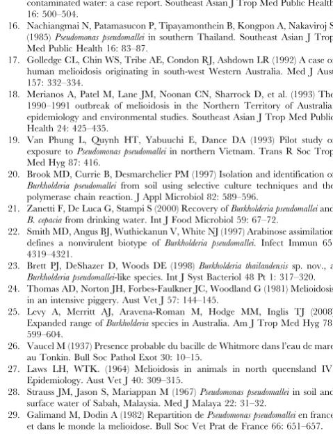
Table 1.Global distribution of environmentalB. pseudomallei.
Level of
evidence Definition Countries
Definite (1) Organism isolated from soil or water with adequate identification by culture or a B. pseudomallei-specific PCR, and (2) Evidence for melioidosis having been acquired in that country
Asia (Cambodia [98], China [31,48,49], Iran [50], Lao PDR [9,63], Malaysia [28,58,68,77], Singapore [78,99], Sri Lanka [51], Taiwan [52–55], Thailand [11,15,16,37,39,57,60,64– 66,84,85,100,101] and Vietnam [19,26,102,103]), Oceania (Australia,
[17,18,20,24,25,27,38,42,59,61,62,71–75,104] and Papua New Guinea [67]), Africa (Burkina Faso [40], Madagascar,[14], Niger [40]), Europe (France [14,29,30])*, and, South America (Brazil [14,46,47])
Probable (1) No report identified ofB. pseudomalleiisolation from soil or water, and (2) Evidence for melioidosis having been acquired in that country
Asia (Bangladesh, Brunei, Egypt, India, Indonesia, Myanmar, Pakistan, Philippines and Saudi Arabia), Ocenia (Fiji), Africa (Chad, Gambia, Kenya, Nigeria, Sierra Leone, South Africa and Uganda), Central America (Costa Rica, El Salvador, Honduras, Mexico and Panama), South America (Colombia, Ecuador, Puerto Rico and Venezuela), Europe (Turkey), and Others (Aruba, Guadeloupe, Guam, Mauritius, Martinique, New Caledonia, Puerto Rico) [4,12]
Possible (1) Organism isolated from soil or water that was considered to beB. pseudomallei, but (2) identification process not sufficient to exclude other, non-pathogenic environmentalBurkholderia spp. such asB. thailandensis, and 2) No evidence for melioidosis having been acquired in that country
Coˆte d’Ivoire [14], Haiti [14], Italy [21] and Peru [14]
*In France, soil culture positive forB. pseudomalleiwas initially reported in the ‘Jardin des Plantes’ in Paris after an outbreak of animal melioidosis which was thought to have originated from a panda imported from China, and the organism was subsequently reported to have been detected in soil throughout the country [14,29,30]. There is no evidence to suggest its continuing presence.
sure of their identity, and they are not available for further testing [14,29,30]. Importation followed by environmental treatment to eradicateB. pseudomalleiwill result in a change in classification, but it is unclear from the literature whether B. pseudomalleihas been eradicated in France. A further 34 countries were assigned to the ‘probable’ category based on clinical evidence of indigenous melioidosis but lack of environmental studies. Two studies described the molecular identification or genotyping of environ-mental B. pseudomallei isolates from Ecuador, Kenya and Venezuela [41,56], but no environmental sampling studies positive for B. pseudomallei were identified for these countries in the published literature. A total of 4 countries including Coˆte d’Ivoire [14], Haiti [14], Italy [21] and Peru [14] were assigned to the ‘possible’ category (Table 1 & Figure 2) based on inadequate bacterial confirmation of putative environmental B. pseudomallei combined with a lack of evidence for indigenous melioidosis.
Sampling strategies used for the detection of environmental B. pseudomallei. Published sampling strate-gies for the detection of environmentalB. pseudomalleiare shown in Table 2. Sampling was performed in both dry and wet seasons,
and sampling duration ranged from 1 day to 3 years [57]. A consistent difference in positivity rates between the wet and dry season was not established. Three studies found a higher positivity rate in the wet season [35,58,59], and two studies reported a higher positivity rate in the dry season [20,60]. A recent study found that, in a given region, most areas had higher positivity in the wet season but some had a higher positivity in the dry season, which suggested that other factors such as the presence of animals or land use also contribute to differences in positivity rates between wet and dry seasons [61].
Of 61 studies, 55 evaluated the presence ofB. pseudomalleiin soil, and 35 in water. The majority of studies chose sampling sites on an ad hocbasis. Of 54 studies with information about land use for the sampling site, 20 were conducted in rice fields and 35 in other areas including animal pens, residential areas around the homes of cases, forests, scrubland, and agricultural fields containing other crops. Most studies collected a low number of samples (2 to 7) per study site, and did not provide a detailed description of the sampling design or strategy, sample size calculation or distance between sampling points within each site. Three articles described
Figure 1. Flow diagram showing study selection.
random selection of the study site in a given area using GPS, and provided a detailed sampling strategy [61–63]. The largest number of samples collected from a single site was 100, in which samples were collected using a fixed interval grid [63–65]. Soil sampling depth ranged from surface to 90 cm. The weight of each soil sample collected ranged from 2 to 1,000 grams [18,43].
Methods of B. pseudomallei detection in soil. The methodology used to detect B. pseudomalleiin soil samples has 2 main stages: (i) bacterial extraction, and (ii) detection methods using culture or PCR (or historically, animal inoculation) (Table 3). The process of bacterial extraction involves the addition of a solution to the soil, mixing with various degrees of homogeniza-tion, and a period of settling prior to removal of the supernatant. The solution used has varied between distilled water, normal saline, detergent solution [66], or enrichment media, with a variable soil to solution ratio (wt/vol) ranging from 2:1 to 1:10 [49,57]. The method used to mix the soil and solution has varied between manual shaking, vortexing or use of an orbital shaker. The time period used to mix the solution has varied from less than 1 minute to 48 hours [61,67], and the time for soil sedimentation after mixing from 5 minutes to 24 hours [66,67]. The volume of fluid used for culture has varied from 0.5 to 10 ml of supernatant [60,67], or the spun deposit of 80 ml of supernatant [35]. The volume used for DNA extraction prior to PCR has varied from 3 ml of supernatant [53], the deposit of 20 ml of supernatant
[image:5.612.60.554.60.366.2][61,62], or direct extraction from different weight of soil [39,52,54]. The volume used for guinea-pig or hamster inocula-tion has varied from 1 to 2 ml [57,68].
The most common detection method has been culture using selective media (n = 46). Most protocols used a selective enrich-ment broth (n = 44), with a variable specimen to medium ratio (vol/vol) ranging from 1:1 to 1:20 [67]. The broth used varied and included tryptone soya broth plus crystal violet (5 mg/l) and colistin (20 or 50 mg/l) (CVCB or Ashdown broth) [69], and L-threonine buffered salt solution (TBSS or Galimand and Dodin broth) [14] with or without colistin (20 or 50 mg/l). Culture of bacterial extraction solution on selective agar plates was described in 15 studies, and Ashdown agar was commonly used [69]. The volume of fluid inoculated onto each agar plate varied from 10 to 400ml [9,64]. Temperature of incubation varied between 30 and 42uC [19,67]. The overall efficiency of different techniques at each stage has not been adequately compared. In eight studies using both culture and PCR, the positivity rate forB. pseudomalleiwas higher by PCR than by culture [20,39,51–55,62,64,70].
Methods used to detect B. pseudomallei in water. The methodology used to detectB. pseudomalleiin water samples has 2 main stages: (i) bacterial concentration, and (ii) detection methods using culture or animal inoculation. The volume of each water sample collected ranged from 1 to 5,000 ml [24,59,71–74]. The method used for bacterial concentration has varied between
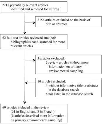
Figure 2. Global map showing the distribution ofB. pseudomallei.Definitions of definite, probable and possible presence of environmentalB. pseudomalleiare described in Table 1.1represents ‘Jardin des Plantes’ in Paris where soil cultures positive forB. pseudomalleiwere initially reported after an outbreak of melioidosis, which was thought to have originated from a panda imported from China [14].2represents Bologna, Italy, whereB.
pseudomalleiin tap water (6 out of 85 specimens) was reported in 2000 [21]. However, confirmation ofB. pseudomalleiby specific identification methods was not reported.3represents Chittering, southwest Western Australia, whereB. pseudomalleiwas isolated and confirmed from a single soil specimen in 1980, following the outbreak of melioidosis in animals [17,38]. There has been no evidence of environmentalB. pseudomallei in southwest Western Australia since then.
filtration, centrifugation [43], or precipitation with potassium alum [27]. Filter pore size has varied from 0.20, 0.22 or 0.45mm [18,21,25,42,73–76]. The volume of fluid used for direct culture was 50 ml, from which the bacteria were extracted either by centrifugation [43], or using potassium alum [27]. The volume used for guinea-pig or hamster inoculation has varied from 1 to 2 ml [24,58,68,71,77,78]. The first evidence ofB. pseudomalleiin water came from a study published in 1937 which involved immersion of a guinea pig in water following scarification of its abdomen, following which B. pseudomallei was isolated from its blood [26]. The relative sensitivity of detection using culture versus animal inoculation has not been reported.
Methods used to detectB. pseudomalleiin air. There are no studies in PubMED that report air sampling forB. pseudomallei. An MSc thesis written by Kinoshita contains details of the culture ofB. pseudomalleifrom air at the Hong Kong oceanarium in 1989, 1993 and 1995 [31]. The sampling technique used was to hold an agar plate at about shoulder level to oncoming winds during a typhoon. Kinoshita repeated air sampling by collecting 171 typhoon samples between 1999 and 2002, but all were culture negative forB. pseudomallei[31].
Recommendations on the Detection ofB. pseudomalleiin Soil
All 16 members of DEBWorP agreed that the first recommen-dations would focus on soil sampling alone, and that there was not
enough evidence for recommendations to be made on the detection of B. pseudomallei in water and air. All members completed the original version of the questionnaire about variations in study design and methodology for the qualitative detection ofB. pseudomalleiin soil (Text S2). A second iteration was developed after identifying additional issues that could not be resolved without further consultation. All 16 members completed the second version, after which consensus recommendations were developed, sent to all members for comments, and agreed upon. Specific recommendations are shown in Table 2–4, the basis for which is described below.
[image:6.612.63.555.76.428.2]Choice of sampling site and strategy. The most appro-priate sampling strategy will depend on the objectives of the study, and whether any information is already available for the geographical area to be sampled (Table 2). For pilot studies that are conducted to identify B. pseudomallei in the environment in areas where sampling has not been performed previously, investigators should gather any available information about possible or definite melioidosis cases in the locality, and sampling site selection should target their residence or work place. In the absence of such information, a less targeted approach will be required in which GIS (geographic information system) software is used to support the random identification of several pilot locations in a given region or country. For large environmental surveys in areas where B. pseudomallei is known to be present in the environment, selection of sampling sites using GIS software is
Table 2.Published and recommended sampling strategies for the isolation ofB. pseudomalleifrom soil.
Sampling strategy Published sampling strategy Consensus guideline
Sample size calculation Not stated and often low sample size Sample size calculation should be presented and should correspond with the aims of the study
Sampling site selection Variable, including random site selection and practical considerations (e.g. sampling at points along a main road)
For pilot studies that are conducted to identify environmentalB. pseudomalleiin areas where sampling has not been done previously, choose sites most likely to be positive based on available information such as areas around households or working fields of melioidosis patients. If such information is unavailable, use the GIS program to randomly select sampling sites
For large environmental surveys in areas whereB. pseudomalleiis known to be present in the environment, use the GIS program to randomly select sites across the designated region
Sampling points per site Ranged from 2 to 100 points per field Use a fixed interval sampling grid
To determine presence ofB. pseudomalleiin one field (around 50650 sq meters), 100 points per site
To determine presence or distribution ofB. pseudomallei in a wider area, number of points per site and number of sites should be calculated based on geo-statistical sample size calculation which should provide the confidence level required
Distance between sampling point within a sampling site
1 to 5 meters, or not reported If no prior information available forB. pseudomallei distribution in test area, take samples at a distance of 2.5 to 5 meters apart
If prior information is available, samples should be taken at an optimal distance based on geo-statistical sample size calculation
Soil sampling depth Ranged from 0 to 90 cm of depth 30 cm depth
Weight of soil sample per sampling point Ranged from 2 to 1,000 gram of soil 10 gram of soil (put into universal tube)
Temperature during transportation of sample to laboratory
Variable, including room temperature and refrigerated temperature
At ambient temperature and away from direct sunlight or heat source
Process soil samples as soon as possible
also recommended. Within a given location (study site), we recommend the use of a fixed interval grid based on its simplicity and the need for standardization.
Number of samples. Taking an insufficient number of soil samples from a designated sampling site runs the risk of a false
[image:7.612.62.557.78.399.2]negative result [11]. This may be due to insensitive detection methods, or because saprophytic bacteria exist in aggregates and can give rise to hot spots and intervening areas that are negative for a specific bacterium. This has been shown to be the case forB. pseudomallei[11]. Because of this, random sampling methods using
Table 3.Published and recommended methodologies for the isolation ofB. pseudomalleifrom soil.
Methodologies Published methods Consensus guideline
B. pseudomalleiextraction solution Distilled water, normal saline, detergents or enrichment media
Threonine-basal salt plus colistin 50 mg/L (TBSS-C50 broth)
Ashdown broth containing colistin and crystal violet is an alternative
Ratio of soil and extraction solution (wt/wt)
Ranged from 2:1 to 1:10 1:1 (10 gram of soil to 10 ml of TBSS-C50 or Ashdown broth)
Extraction method Manual shaking, vortexing or orbital shaker Vortexing for 30 seconds
Manual mixing of soil is an alternative option, and may be required if sample is compacted
Techniques for detection of B. pseudomallei
Culture, PCR or animal inoculation Culture (PCR could be added as an additional technique if available)
Protocol for culture Variable, including direct culture on solid media and quantitation, and qualitative methods relying on broth enrichment
Incubate the specimen (universal tube with 10 gram of soil plus 10 ml TBSS-C50 or Ashdown broth) for 48 hours
Temperature of incubator Variable, ranged from 37 to 42uC 40uC is recommended, and 37–42uC is an alternative option
Protocol for sub-culture Variable Subculture 10mL of supernatant onto an Ashdown agar plate,
and streak to achieve single colonies
Incubate plate and examine every 24 hours for 7 days
Identification ofB. pseudomallei Variable, including basic microbiological tests (which include typical colony morphology, Gram stain, positive oxidase test, inability to assimilate arabinose, resistance to gentamicin and colistin with susceptibility to co-amoxiclav) and biochemical kits (including API20NE [105] and Vitek) with or without additional confirmatory tests (specific latex agglutination test [89], or a specific PCR assay [62,75,91,93])
Basic microbiological tests (which include typical colony morphology, Gram stain, positive oxidase test, inability to assimilate arabinose, resistance to gentamicin and colistin with susceptibility to co-amoxiclav) is mandatory plus at least one confirmatory test (API20NE, Vitek system, specific latex agglutination test [89] or a specific PCR assay [62,75,91,93], unless latex test or PCR assay was used during screening)
Specific latex agglutination test [89], or a specific PCR assay [62,75,91,93] can be used a screening test
doi:10.1371/journal.pntd.0002105.t003
Table 4.Publishing the findings of studies conducted to isolateB. pseudomalleifrom soil.
Reporting the findings Published findings Consensus guideline
After publication, deposit raw data to website
Variably reported After publication, raw data can be deposited to website www.melioidosis.info at the discretion of PI and sponsor of each study
GPS location of study site Variably reported After publication, GPS data can be deposited to website www.melioidosis.info at the discretion of PI and sponsor of each study taking account of issues of anonymity.
Positivity rate in each study site and pattern of positivity in each study site
Variably reported Describe in the manuscript if available.
After publication, details of results can be deposited to website www.melioidosis.info at the discretion of PI and sponsor of each study
Soil type and history of land use Variably reported Describe the current land use in the manuscript, together with the history of land use if available
Describe the soil texture using previously described method such as ribbon test [97].
After publication, details of results can be deposited to website www.melioidosis.info at the discretion of PI and sponsor of each study
Sampling time and weather at sampling time point (e.g. rainfall, season)
Variably reported Describe in the manuscript.
After publication, details of results can be deposited to website www.melioidosis.info at the discretion of PI and sponsor of each study
[image:7.612.62.557.506.720.2]a low sample size may be associated with a low power of detection and a high false negative (type II error) rate [79]. This can be avoided by increasing the number of samples taken [11]. Based on statistical considerations, to determine the presence of B. pseudomallei in an area of around 50650 sq meters, a minimum of 100 sampling points is suggested. This is strongly supported by a recent study in Lao PDR in which one field was deemed positive based on only 1 out of 100 positive sampling points [63].
If a region is already known or highly suspected to be positive for B. pseudomallei, an alternative approach is to use adaptive sampling in which a pilot study is performed in a defined experimental area in which a number of random points (e.g. 20) are sampled. If any are positive forB. pseudomallei, this confirms the presence of the organism and is sufficient to define this as an area of risk for humans and livestock. If all samples are negative, a second round of sampling is done in which 100 samples are taken from the same site using a fixed interval grid. To determine the presence or distribution of B. pseudomallei in a wider area, the number of samples taken per site and the number of sites investigated could be calculated based on a geo-statistical sample size calculation [80,81].
Distance between samples. The presence of hot spots for a specific bacterium in the environment leads to an effect described by the term ‘spatial autocorrelation’, which influences the distance required between each sampling point. What this means in practice is that sampling points adjacent to each other are more likely to yield the same result (e.g. a sample next to a negative sample is likely to be negative) [11]. The distance over which counts of a given environmental bacterium are related (range of spatial autocorrelation) can be defined using a geostatistical tool called the semivariogram [80]. Ideally, the effect of spatial autocorrelation would be factored in to the sampling strategy for B. pseudomallei, but this value is likely to be influenced by physicochemical soil parameters and vegetation [82], and vary between and possibly within countries. Therefore, it is not practical to define this prior to formal sampling in most settings. Studies in Thailand suggest that the distance between samples should be between 2.5 and 5 m apart [11], although it is uncertain whether this applies elsewhere. Given the paucity of data on the optimal distance between samples we suggest that sampling be performed 2.5 to 5 m apart, accepting that this is somewhat arbitrary. The optimal sampling distance specific to the study region could be subsequently estimated based on the results of pilot study data for 100 sampling points for one or more sites [80]. Soil sampling: quantity, sampling depth and transport to the laboratory. We recommend a depth for soil sampling of 30 cm. This is based on published evidence that the proportion of samples that are culture positive for B. pseudomallei is higher at 30 cm than at a shallower depth, but comparable to samples taken deeper than 30 cm [35,47,53,60,62,83,84]. The quantity of soil collected per sample has varied markedly in published studies, and there is no evidence that collecting a greater weight of soil is associated with a higher sensitivity. We suggest taking a weight of 10 grams per sample based on practicality and ease of method-ology [85]. As there is evidence showing that survival of B. pseudomallei is decreased at low temperatures [86], soil samples should be kept at ambient temperature (24 to 32uC) and away from direct sunlight or heat source during transportation to the laboratory. The specimen should be processed as soon as possible. Extraction of bacteria from soil, and detection and identification of B. pseudomallei. We recommend the use of culture as the standard method for environmentalB. pseudomallei detection in the context of global mapping efforts on the basis of simplicity, specificity and low cost (Table 3). The optimal ratio of
soil to extraction solution, mixing technique and sedimentation time are not known. Selective broths have been compared in both laboratory [87] and field settings [20,60]. We proposed that each 10 gram soil sample be placed into a universal tube, mixed with 10 ml of enrichment medium (either TBSS with colistin 50 mg/l (TBSS-C50) or Ashdown broth), vortexed for 30 seconds, and incubated at 40uC in air for 48 hours. Based on scientific evidence and agreement of the working party, TBSS-C50 is recommended as the primary enrichment medium with Ashdown broth as an alternative. A volume of 10ml of the upper layer of enrichment medium should be streaked to achieve single colonies onto a whole Ashdown agar plate, incubated at 40uC in air and examined every 24 hours for 7 days. This incubation temperature was chosen based on evidence that it allows growth ofB. pseudomallei[88], but is inhibitory to some other soil flora (personal observation by DABD and VW). However, incubation at 37uC is acceptable in the event that resources are not available to incubate at 40uC. Subculture of 10ml is based on experience in Thailand and represents a balance between detection of B. pseudomallei and limiting the bioburden of other flora that grow on the agar plate. Subculture of higher volumes (100ml) may be associated with a higher yield although there currently is no published evidence to support this.
Several steps of the method recommended here (direct culture of 10 gram of soil in 10 ml of TBSS-C50 and subculture onto Ashdown agar) are based on methods in widespread use in Australia [62,75]. Furthermore, the sensitivity of our recom-mended method was recently compared to a more laborious method which has been used extensively in Thailand [85]. The latter involves collection of 100 gram of soil which is mixed with 100 ml of distilled water, left to settle overnight, and the upper layer of water removed for culture on Ashdown agar and in TBSS-C50. In the comparative study, 94 out of 200 soil samples were culture positive for B. pseudomallei [85]. Yield was not different between the two methods (70/94 vs. 79/94 respectively; p = 0.15), supporting the use of our currently recommended method.
Identification of B. pseudomallei. Any colony with a colony morphology suggestive ofB. pseudomallei can be tested by basic microbiological tests (typical colony morphology on Ash-down agar, Gram stain, positive oxidase test, inability to assimilate arabinose, resistant to gentamicin and colistin, susceptible to co-amoxiclav) followed by confirmatory tests (specific latex aggluti-nation test [89], a specific PCR assay [53,55,62,75,90–95], or validated identification kits such as API20NE or Vitek system). The API20NE database does not include a profile of B. thailandensis, which give results that are similar to those for B. pseudomallei except that B. thailandensis is positive for arabinose assimilation. For rapid evaluation, a specific latex agglutination [89,96] or PCR assay [53,55,62,75,90–95] could be used as a screening test, followed by basic microbiological tests to complete the identification process.
downloadable protocols describing methodology for soil sampling and culture, including details of each reagent and test used (Text S3). The recommended protocols have been successfully used in Thailand [85], although further evaluation of these is required in different countries.
Although the methodology presented here aims to reduce the risk of false negative sampling surveys, this is unlikely to be perfect. As a result, a single negative sampling survey does not represent definite evidence that the site is free ofB. pseudomallei, although it would be predicted to reflect a region of much lower risk compared with a positive site. The need to undertake further sampling requires consideration of risk-benefit. There is also considerable scope to improve on the methodology described here, including improvement in the sensitivity of culture which could include the development of media that are even more selective for B. pseudomalleiin soil, and ultimately the development of easy-to-use and accurate diagnostic kits for environmental sampling. Our recommendations will be updated in the future as and when new information or knowledge becomes available.
Concluding comments. Our knowledge of the global distribution ofB. pseudomalleiis incomplete, and the methodology to determine the presence of this organism in the environment has not been standardized and is liable to false negativity (if insufficient samples are taken or inappropriate techniques are used), and false positivity (if methods are not adequate to exclude related Burkholderia species). We have provided consensus guidelines on strategies and methodologies to determine the presence of B. pseudomalleiin soil that are simple and applicable in settings with limited resources. To develop a complete risk map of melioidosis, our working party aims to support and promote environmental studies on a global scale, supported by a website (www.melioidosis.
info) with downloadable protocols and a mechanism for data collection and sharing.
Supporting Information
[image:9.612.313.553.422.739.2]Table S1 Characteristics of studies included in the review. (DOC)
Table S2 PRISMA checklist. (DOC)
Text S1 Data extraction form for studies that determined the presence ofBurkholderia pseudomalleiin the environment.
(DOC)
Text S2 Questionnaire on the detection of environmental Burkholderia pseudomallei.
(DOC)
Text S3 Standard Operating Procedure (SOP): simplified method for the isolation ofBurkholderia pseudomalleifrom soil. (DOC)
Acknowledgments
We thank Prapass Wannapinij, Nuttapol Panachuenwongsakul, Thatsanun Ngernseng, Wirichada Pan-ngum, Jem Chalk and Dean Sherwood for development of the website (www.melioidosis.info).
Author Contributions
Conceived and designed the experiments: DL DABD BJC SJP. Performed the experiments: DL DABD. Analyzed the data: DL DABD BJC SJP. Contributed reagents/materials/analysis tools: DL DABD VW MK MM JW DMW AT HW TYC CM SP NPJD IS BJC SJP. Wrote the paper: DL DABD BJC SJP.
References
1. Whitmore A, Krishnaswami CS (1912) An account of the discovery of a hitherto undescribed infective disease occuring among the population of Rangoon. Indian Med Gazette 47: 262–267.
2. Limmathurotsakul D, Wongratanacheewin S, Teerawattanasook N, Wongsu-van G, Chaisuksant S, et al. (2010) Increasing incidence of human melioidosis in Northeast Thailand. Am J Trop Med Hyg 82: 1113–1117.
3. Currie BJ, Fisher DA, Howard DM, Burrow JN, Selvanayagam S, et al. (2000) The epidemiology of melioidosis in Australia and Papua New Guinea. Acta Trop 74: 121–127.
4. Currie BJ, Dance DA, Cheng AC (2008) The global distribution ofBurkholderia pseudomalleiand melioidosis: an update. Trans R Soc Trop Med Hyg 102 Suppl 1: S1–4.
5. John TJ, Jesudason MV, Lalitha MK, Ganesh A, Mohandas V, et al. (1996) Melioidosis in India: the tip of the iceberg? Indian J Med Res 103: 62–65. 6. Weissert C, Dollenmaier G, Rafeiner P, Riehm J, Schultze D (2009)Burkholderia
pseudomalleimisidentified by automated system. Emerg Infect Dis 15: 1799– 1801.
7. Deepak RN, Crawley B, Phang E (2008)Burkholderia pseudomalleiidentification: a comparison between the API 20NE and VITEK2GN systems. Trans R Soc Trop Med Hyg 102 Suppl 1: S42–44.
8. Cheng AC, Currie BJ (2005) Melioidosis: epidemiology, pathophysiology, and management. Clin Microbiol Rev 18: 383–416.
9. Wuthiekanun V, Mayxay M, Chierakul W, Phetsouvanh R, Cheng AC, et al. (2005) Detection ofBurkholderia pseudomallei in soil within the Lao People’s Democratic Republic. J Clin Microbiol 43: 923–924.
10. Phetsouvanh R, Phongmany S, Newton P, Mayxay M, Ramsay A, et al. (2001) Melioidosis and Pandora’s box in the Lao People’s Democratic Republic. Clin Infect Dis 32: 653–654.
11. Limmathurotsakul D, Wuthiekanun V, Chantratita N, Wongsuvan G, Amornchai P, et al. (2010)Burkholderia pseudomalleiis spatially distributed in soil in northeast Thailand. PLoS Negl Trop Dis 4: e694.
12. Wertheim HFL (2012) Melioidosis. Atlas of Human Infectious Diseases. 1st edition. Wiley-Blackwell. pp. 75.
13. Leclerc H, Sureau P (1956) [Research on bacteriophages of theWhitmore bacillus in stagnant waters of Hanoi]. Bull Soc Pathol Exot Filiales 49: 874–882. 14. Galimand M, Dodin A (1982) [Focus on melioidosis throughout the world].
Bull Soc Pathol Exot Filiales 75: 375–383.
15. Achana V, Silpapojakul K, Thininta W, Kalnaowakul S (1985) Acute Pseudomonas pseudomallei pneumonia and septicemia following aspiration of
contaminated water: a case report. Southeast Asian J Trop Med Public Health 16: 500–504.
16. Nachiangmai N, Patamasucon P, Tipayamonthein B, Kongpon A, Nakaviroj S (1985)Pseudomonas pseudomalleiin southern Thailand. Southeast Asian J Trop Med Public Health 16: 83–87.
17. Golledge CL, Chin WS, Tribe AE, Condon RJ, Ashdown LR (1992) A case of human melioidosis originating in south-west Western Australia. Med J Aust 157: 332–334.
18. Merianos A, Patel M, Lane JM, Noonan CN, Sharrock D, et al. (1993) The 1990–1991 outbreak of melioidosis in the Northern Territory of Australia: epidemiology and environmental studies. Southeast Asian J Trop Med Public Health 24: 425–435.
19. Van Phung L, Quynh HT, Yabuuchi E, Dance DA (1993) Pilot study of exposure toPseudomonas pseudomalleiin northern Vietnam. Trans R Soc Trop Med Hyg 87: 416.
20. Brook MD, Currie B, Desmarchelier PM (1997) Isolation and identification of Burkholderia pseudomallei from soil using selective culture techniques and the polymerase chain reaction. J Appl Microbiol 82: 589–596.
21. Zanetti F, De Luca G, Stampi S (2000) Recovery ofBurkholderia pseudomalleiand B. cepaciafrom drinking water. Int J Food Microbiol 59: 67–72.
22. Smith MD, Angus BJ, Wuthiekanun V, White NJ (1997) Arabinose assimilation defines a nonvirulent biotype of Burkholderia pseudomallei. Infect Immun 65: 4319–4321.
23. Brett PJ, DeShazer D, Woods DE (1998)Burkholderia thailandensissp. nov., a Burkholderia pseudomallei-like species. Int J Syst Bacteriol 48 Pt 1: 317–320. 24. Thomas AD, Norton JH, Forbes-Faulkner JC, Woodland G (1981) Melioidosis
in an intensive piggery. Aust Vet J 57: 144–145.
25. Levy A, Merritt AJ, Aravena-Roman M, Hodge MM, Inglis TJ (2008) Expanded range ofBurkholderiaspecies in Australia. Am J Trop Med Hyg 78: 599–604.
26. Vaucel M (1937) Presence probable du bacille de Whitmore dans l’eau de mare au Tonkin. Bull Soc Pathol Exot 30: 10–15.
27. Laws LH, WTK. (1964) Melioidosis in animals in north queensland IV. Epidemiology. Aust Vet J 40: 309–315.
28. Strauss JM, Jason S, Mariappan M (1967)Pseudomonas pseudomalleiin soil and surface water of Sabah, Malaysia. Med J Malaya 22: 31–32.
30. Mollaret H (1988) L’affaire du jardin des plantes. Medecine et Maladies Infectieuses: 643–654.
31. Kinoshita R (2003) Epidemiology of melioidosis in an oceanarium: a clinical, environmental and molecular study. University of Hong Kong. Available: http://hub.hku.hk/handle/10722/30476 Accessed 20 Oct 2012.
32. Dodin A, Galimand M (1986) [Origin, course and recession of an infectious disease, melioidosis, in temperate countries]. Arch Inst Pasteur Tunis 63: 69– 73.
33. Inglis TJ, O’Reilly L, Merritt AJ, Levy A, Heath CH (2011) The aftermath of the Western Australian melioidosis outbreak. Am J Trop Med Hyg 84: 851– 857.
34. Dance DA (2000) Ecology of Burkholderia pseudomallei and the interactions between environmentalBurkholderiaspp. and human-animal hosts. Acta Trop 74: 159–168.
35. Thomas AD, Forbes-Faulkner J, Parker M (1979) Isolation of Pseudomonas pseudomalleifrom clay layers at defined depths. Am J Epidemiol 110: 515–521. 36. Wuthiekanun V, Smith MD, Dance DA, Walsh AL, Pitt TL, et al. (1996) Biochemical characteristics of clinical and environmental isolates ofBurkholderia pseudomallei. J Med Microbiol 45: 408–412.
37. Trakulsomboon S, Vuddhakul V, Tharavichitkul P, Na-Gnam N, Suputta-mongkol Y, et al. (1999) Epidemiology of arabinose assimilation inBurkholderia pseudomalleiisolated from patients and soil in Thailand. Southeast Asian J Trop Med Public Health 30: 756–759.
38. Currie B, Smith-Vaughan H, Golledge C, Buller N, Sriprakash KS, et al. (1994)Pseudomonas pseudomalleiisolates collected over 25 years from a non-tropical endemic focus show clonality on the basis of ribotyping. Epidemiol Infect 113: 307–312.
39. Trung TT, Hetzer A, Gohler A, Topfstedt E, Wuthiekanun V, et al. (2011) Highly sensitive direct detection and quantification ofBurkholderia pseudomallei bacteria in environmental soil samples by using real-time PCR. Appl Environ Microbiol 77: 6486–6494.
40. Dodin A, Ferry R (1974) [Epidemiological studies of the bacillus of Whitmore in Africa]. Bull Soc Pathol Exot Filiales 67: 121–126.
41. Godoy D, Randle G, Simpson AJ, Aanensen DM, Pitt TL, et al. (2003) Multilocus sequence typing and evolutionary relationships among the causative agents of melioidosis and glanders, Burkholderia pseudomalleiand Burkholderia mallei. J Clin Microbiol 41: 2068–2079.
42. Ketterer PJ, Webster WR, Shield J, Arthur RJ, Blackall PJ, et al. (1986) Melioidosis in intensive piggeries in south eastern Queensland. Aust Vet J 63: 146–149.
43. Batchelor BI, Paul J, Trakulsomboon S, Mgongo M, Dance DA (1994) Melioidosis survey in Kenya. Trans R Soc Trop Med Hyg 88: 181. 44. McCormick JB, Weaver RE, Hayes PS, Boyce JM, Feldman RA (1977) Wound
infection by an indigenousPseudomonas pseudomallei-like organism isolated from the soil: case report and epidemiologic study. J Infect Dis 135: 103–107. 45. Glass MB, Steigerwalt AG, Jordan JG, Wilkins PP, Gee JE (2006) Burkholderia
oklahomensis sp. nov., aBurkholderia pseudomallei-like species formerly known as the Oklahoma strain ofPseudomonas pseudomallei. Int J Syst Evol Microbiol 56: 2171–2176.
46. Rolim DB, Vilar DC, Sousa AQ, Miralles IS, de Oliveira DC, et al. (2005) Melioidosis, northeastern Brazil. Emerg Infect Dis 11: 1458–1460. 47. Rolim DB, Rocha MF, Brilhante RS, Cordeiro RA, Leitao NP, Jr., et al. (2009)
Environmental isolates ofBurkholderia pseudomalleiin Ceara State, northeastern Brazil. Appl Environ Microbiol 75: 1215–1218.
48. Yang S, Tong S, Lu Z (1995) Geographical distribution of Pseudomonas pseudomalleiin China. Southeast Asian J Trop Med Public Health 26: 636–638. 49. Ma G, Zheng D, Cai Q, Yuan Z (2010) Prevalence ofBurkholderia pseudomalleiin
Guangxi, China. Epidemiol Infect 138: 37–39.
50. Pourtaghva M, Machoun A, Dodin A (1975) [Demonstration ofPseudomonas pseudomallei(Whitmore’s bacillus) in the mud of Iranian ricefields (author’s transl)]. Bull Soc Pathol Exot Filiales 68: 367–370.
51. Inglis TJ, Merritt A, Montgomery J, Jayasinghe I, Thevanesam V, et al. (2008) Deployable laboratory response to emergence of melioidosis in central Sri Lanka. J Clin Microbiol 46: 3479–3481.
52. Chen YS, Lin HH, Mu JJ, Chiang CS, Chen CH, et al. (2010) Distribution of melioidosis cases and viable Burkholderia pseudomallei in soil: evidence for emerging melioidosis in Taiwan. J Clin Microbiol 48: 1432–1434. 53. Kao CM, Chen SC, Chen YS, Lin HM, Chen YL (2003) Detection of
Burkholderia pseudomalleiin rice fields with PCR-based technique. Folia Microbiol (Praha) 48: 521–524.
54. Su HP, Yang HW, Chen YL, Ferng TL, Chou YL, et al. (2007) Prevalence of melioidosis in the Er-Ren River Basin, Taiwan: implications for transmission. J Clin Microbiol 45: 2599–2603.
55. Lin HH, Chen YS, Li YC, Tseng IL, Hsieh TH, et al. (2011)Burkholderia multivoransacts as an antagonist against the growth ofBurkholderia pseudomalleiin soil. Microbiol Immunol 55: 616–624.
56. Tomaso H, Pitt TL, Landt O, Al Dahouk S, Scholz HC, et al. (2005) Rapid presumptive identification ofBurkholderia pseudomalleiwith real-time PCR assays using fluorescent hybridization probes. Mol Cell Probes 19: 9–20.
57. Finkelstein RA, Atthasampunna P, Chulasamaya M (2000) Pseudomonas (Burkholderia) pseudomalleiin Thailand, 1964–1967: geographic distribution of the organism, attempts to identify cases of active infection, and presence of antibody in representative sera. Am J Trop Med Hyg 62: 232–239.
58. Strauss JM, Ellison DW, Gan E, Jason S, Marcarelli JL, et al. (1969) Melioidosis in Malaysia. IV. Intensive ecological study of Carey Island, Selangor, forPseudomonas pseudomallei. Med J Malaya 24: 94–100.
59. Baker A, Tahani D, Gardiner C, Bristow KL, Greenhill AR, et al. (2011) Groundwater seeps facilitate exposure toBurkholderia pseudomallei. Appl Environ Microbiol 77: 7243–7246.
60. Wuthiekanun V, Smith MD, Dance DA, White NJ (1995) Isolation of Pseudomonas pseudomalleifrom soil in north-eastern Thailand. Trans R Soc Trop Med Hyg 89: 41–43.
61. Kaestli M, Mayo M, Harrington G, Ward L, Watt F, et al. (2009) Landscape changes influence the occurrence of the melioidosis bacteriumBurkholderia pseudomalleiin soil in northern Australia. PLoS Negl Trop Dis 3: e364. 62. Kaestli M, Mayo M, Harrington G, Watt F, Hill J, et al. (2007) Sensitive and
specific molecular detection ofBurkholderia pseudomallei, the causative agent of melioidosis, in the soil of tropical northern Australia. Appl Environ Microbiol 73: 6891–6897.
63. Rattanavong S, Wuthiekanun V, Langla S, Amornchai P, Sirisouk J, et al. (2011) Randomized soil survey of the distribution ofBurkholderia pseudomalleiin rice fields in Laos. Appl Environ Microbiol 77: 532–536.
64. Chantratita N, Wuthiekanun V, Limmathurotsakul D, Vesaratchavest M, Thanwisai A, et al. (2008) Genetic diversity and microevolution ofBurkholderia pseudomalleiin the environment. PLoS Negl Trop Dis 2: e182.
65. Wuthiekanun V, Limmathurotsakul D, Chantratita N, Feil EJ, Day NP, et al. (2009)Burkholderia Pseudomallei is genetically diverse in agricultural land in Northeast Thailand. PLoS Negl Trop Dis 3: e496.
66. Trung TT, Hetzer A, Topfstedt E, Gohler A, Limmathurotsakul D, et al. (2011) Improved culture-based detection and quantification ofBurkholderia pseudomalleifrom soil. Trans R Soc Trop Med Hyg 105: 346–351. 67. Warner JM, Pelowa DB, Gal D, Rai G, Mayo M, et al. (2008) The
epidemiology of melioidosis in the Balimo region of Papua New Guinea. Epidemiol Infect 136: 965–971.
68. Ellison DW, Baker HJ, Mariappan M (1969) Melioidosis in Malaysia. I. A method for isolation ofPseudomonas pseudomalleifrom soil and surface water. Am J Trop Med Hyg 18: 694–697.
69. Ashdown LR (1979) An improved screening technique for isolation of Pseudomonas pseudomalleifrom clinical specimens. Pathology 11: 293–297. 70. Inglis TJ, Levy A, Merritt AJ, Hodge M, McDonald R, et al. (2009) Melioidosis
risk in a tropical industrial environment. Am J Trop Med Hyg 80: 78–84. 71. Thomas AD (1977) The isolation ofPseudomonas pseudomalleifrom soil in North
Queensland. Aust Vet J 53: 408.
72. Inglis TJ, Garrow SC, Henderson M, Clair A, Sampson J, et al. (2000) Burkholderia pseudomalleitraced to water treatment plant in Australia. Emerg Infect Dis 6: 56–59.
73. Currie BJ, Mayo M, Anstey NM, Donohoe P, Haase A, et al. (2001) A cluster of melioidosis cases from an endemic region is clonal and is linked to the water supply using molecular typing ofBurkholderia pseudomalleiisolates. Am J Trop Med Hyg 65: 177–179.
74. Mayo M, Kaesti M, Harrington G, Cheng AC, Ward L, et al. (2011) Burkholderia pseudomallei in unchlorinated domestic bore water, Tropical Northern Australia. Emerging infectious diseases 17: 1283–1285.
75. Inglis TJ, Foster NF, Gal D, Powell K, Mayo M, et al. (2004) Preliminary report on the northern Australian melioidosis environmental surveillance project. Epidemiol Infect 132: 813–820.
76. Draper AD, Mayo M, Harrington G, Karp D, Yinfoo D, et al. (2010) Association of the melioidosis agent Burkholderia pseudomallei with water parameters in rural water supplies in Northern Australia. Appl Environ Microbiol 76: 5305–5307.
77. Strauss JM, Groves MG, Mariappan M, Ellison DW (1969) Melioidosis in Malaysia. II. Distribution ofPseudomonas pseudomalleiin soil and surface water. Am J Trop Med Hyg 18: 698–702.
78. Thin RN, Groves M, Rapmund G, Mariappan M (1971) Pseudomonas pseudomalleiin the surface water of Singapore. Singapore Med J 12: 181–182. 79. Klironomos JN, Rillig MC, Allen MF (1999) Designing belowground field
experiments with the help of semi-variance and power analyses. Appl Soil Ecol 12: 227–238.
80. Cressie N (1993) Statistics for Spatial Data. Wiley-Interscience.
81. Modis K, Papaodysseus K (2008) Theoretical estimation of the critical sampling size for homogeneous ore bodies with small nugget effect. Math Geol 38: 12.
82. Kaestli M, Schmid M, Mayo M, Rothballer M, Harrington G, et al. (2011) Out of the ground: aerial and exotic habitats of the melioidosis bacterium Burkholderia pseudomalleiin grasses in Australia. Environ Microbiol 14: 2058– 2070.
83. U’Ren J M, Hornstra H, Pearson T, Schupp JM, Leadem B, et al. (2007) Fine-scale genetic diversity amongBurkholderia pseudomalleisoil isolates in northeast Thailand. Appl Environ Microbiol 73: 6678–6681.
84. Palasatien S, Lertsirivorakul R, Royros P, Wongratanacheewin S, Sermswan RW (2008) Soil physicochemical properties related to the presence of Burkholderia pseudomallei. Trans R Soc Trop Med Hyg 102 Suppl 1: S5–9. 85. Limmathurotsakul D, Wuthiekanun V, Amornchai P, Wongsuwan G, Day NP,
86. Tong S, Yang S, Lu Z, He W (1996) Laboratory investigation of ecological factors influencing the environmental presence of Burkholderia pseudomallei. Microbiol Immunol 40: 451–453.
87. Ashdown LR, Clarke SG (1992) Evaluation of Culture Techniques for Isolation ofPseudomonas pseudomalleifrom Soil. Appl Environ Microbiol 58: 4011–4015. 88. Chen YS, Chen SC, Kao CM, Chen YL (2003) Effects of soil pH, temperature and water content on the growth ofBurkholderia pseudomallei. Folia Microbiol (Praha) 48: 253–256.
89. Wuthiekanun V, Anuntagool N, White NJ, Sirisinha S (2002) Short report: a rapid method for the differentiation ofBurkholderia pseudomalleiandBurkholderia thailandensis. Am J Trop Med Hyg 66: 759–761.
90. Kunakorn M, Markham RB (1995) Clinically practical seminested PCR for Burkholderia pseudomalleiquantitated by enzyme immunoassay with and without solution hybridization. J Clin Microbiol 33: 2131–2135.
91. U’Ren JM, Van Ert MN, Schupp JM, Easterday WR, Simonson TS, et al. (2005) Use of a real-time PCR TaqMan assay for rapid identification and differentiation ofBurkholderia pseudomalleiandBurkholderia mallei. J Clin Microbiol 43: 5771–5774.
92. Merritt A, Inglis TJ, Chidlow G, Harnett G (2006) PCR-based identification of Burkholderia pseudomallei. Rev Inst Med Trop Sao Paulo 48: 239–244. 93. Dharakul T, Tassaneetrithep B, Trakulsomboon S, Songsivilai S (1999)
Phylogenetic analysis of Ara+and Ara2Burkholderia pseudomalleiisolates and development of a multiplex PCR procedure for rapid discrimination between the two biotypes. J Clin Microbiol 37: 1906–1912.
94. Payne GW, Vandamme P, Morgan SH, Lipuma JJ, Coenye T, et al. (2005) Development of a recA gene-based identification approach for the entire Burkholderiagenus. Applied and environmental microbiology 71: 3917–3927. 95. Novak RT, Glass MB, Gee JE, Gal D, Mayo MJ, et al. (2006) Development
and evaluation of a real-time PCR assay targeting the type III secretion system ofBurkholderia pseudomallei. J Clin Microbiol 44: 85–90.
96. Steinmetz I, Reganzerowski A, Brenneke B, Haussler S, Simpson A, et al. (1999) Rapid identification ofBurkholderia pseudomallei by latex agglutination based on an exopolysaccharide-specific monoclonal antibody. J Clin Microbiol 37: 225–228.
97. United States Deparment of Agriculture (USDA) (2001) Soil Quality Test Kit Guide. Available: http://soils.usda.gov/sqi/assessment/files/test_kit_ complete.pdf. Accessed 20 Oct 2011.
98. Wuthiekanun V, Pheaktra N, Putchhat H, Sin L, Sen B, et al. (2008) Burkholderia pseudomalleiantibodies in children, Cambodia. Emerg Infect Dis 14: 301–303.
99. Yap EH, Thong TW, Tan AL, Yeo M, Tan HC, et al. (1995) Comparison of Pseudomonas pseudomallei from humans, animals, soil and water by restriction endonuclease analysis. Singapore Med J 36: 60–62.
100. Smith MD, Wuthiekanun V, Walsh AL, White NJ (1995) Quantitative recovery ofBurkholderia pseudomalleifrom soil in Thailand. Trans R Soc Trop Med Hyg 89: 488–490.
101. Vuddhakul V, Tharavichitkul P, Na-Ngam N, Jitsurong S, Kunthawa B, et al. (1999) Epidemiology ofBurkholderia pseudomalleiin Thailand. Am J Trop Med Hyg 60: 458–461.
102. Chambon L (1955) Isolation of Whitmore’s bacillus from external environ-ment. Ann Inst Pasteur (Paris) 89: 229–235.
103. Parry CM, Wuthiekanun V, Hoa NT, Diep TS, Thao LT, et al. (1999) Melioidosis in Southern Vietnam: clinical surveillance and environmental sampling. Clin Infect Dis 29: 1323–1326.
104. Ashdown LR (1979) Nosocomial infection due toPseudomonas pseudomallei: two cases and an epidemiologic study. Rev Infect Dis 1: 891–894.
![Figure 2. Global map showing the distribution of B. pseudomalleimethods was not reported.after an outbreak of melioidosis, which was thought to have originated from a panda imported from China [14].pseudomalleipseudomallei](https://thumb-us.123doks.com/thumbv2/123dok_us/155347.32491/5.612.60.554.60.366/distribution-pseudomalleimethods-reported-outbreak-melioidosis-originated-imported-pseudomalleipseudomallei.webp)