SAMHD1 Restricts HIV-1 Cell-to-Cell Transmission and Limits
Immune Detection in Monocyte-Derived Dendritic Cells
Isabel Puigdomènech, Nicoletta Casartelli, Françoise Porrot, Olivier Schwartz
Institut Pasteur, Unité Virus et Immunité, Département de Virologie, and CNRS, URA3015, Paris, France
SAMHD1 is a viral restriction factor expressed in dendritic cells and other cells, inhibiting infection by cell-free human immu-nodeficiency virus type 1 (HIV-1) particles. SAMHD1 depletes the intracellular pool of deoxynucleoside triphosphates, thus im-pairing HIV-1 reverse transcription and productive infection in noncycling cells. The Vpx protein from HIV-2 or simian immu-nodeficiency virus (SIVsm/SIVmac) antagonizes the effect of SAMHD1 by triggering its degradation. A large part of HIV-1 spread occurs through direct contacts between infected cells and bystander target cells. Here, we asked whether SAMHD1 im-pairs direct HIV-1 transmission from infected T lymphocytes to monocyte-derived dendritic cells (MDDCs). HIV-1-infected lymphocytes were cocultivated with MDDCs that have been pretreated or not with Vpx or with small interfering RNA against SAMHD1. We show that in the cocultures, SAMHD1 significantly inhibits productive cell-to-cell transmission to target MDDCs and prevents the type I interferon response and expression of the interferon-stimulated gene MxA. Therefore, SAMHD1, by con-trolling the sensitivity of MDDCs to HIV-1 infection during intercellular contacts, impacts their ability to sense the virus and to trigger an innate immune response.
D
endritic cells (DCs) are professional antigen-presenting cellslinking innate and adaptive immune responses. DCs gener-ally recognize pathogens in the periphery and then mature and migrate to lymphoid tissues to elicit a response. This process in-volves expression of costimulatory molecules and production of type I interferons (IFNs) and cytokines. IFN secretion induces numerous interferon-stimulated genes (ISGs) that help control
viral replication and activates immunity (1).
Viruses use multiple mechanisms to avoid innate sensing, cy-tokine production, antiviral activity of ISGs, and restriction
fac-tors (1–3). For HIV-1, the proteins Vif and Vpu counteract the
effects of the restriction factors APOBEC and tetherin. APOBEC proteins induce G-to-A hypermutations in the nascent viral DNA during reverse transcription, while tetherin blocks viral release
(2). Other primate lentiviruses (human immunodeficiency virus
type 2 [HIV-2] and simian immunodeficiency virus [SIV]) pos-sess an additional protein, Vpx, whose function has recently been
deciphered (4–8). Vpx facilitates replication of HIV-2 and some
SIV in myeloid cells but is dispensable in cycling lymphocytes (4).
Vpx triggers the destruction of an early-acting restriction factor
and promotes synthesis of viral DNA in nondividing cells (6). This
restriction factor is active against not only HIV-2 and SIV but also retroviruses like HIV-1 that lack Vpx.
Monocyte-derived DCs (MDDCs) express receptors allowing HIV-1 capture and entry and efficiently transmit the virus to
ac-tivated CD4⫹T cells but are poorly sensitive to productive HIV-1
infection. However, intracellular delivery of Vpx to MDDCs, through treatment with nonreplicative SIV particles carrying Vpx,
dramatically enhances HIV-1 infection (6,9). Vpx acts by
induc-ing the nuclear degradation of SAMHD1, a cellular protein found
in various cell types, including myeloid cells (5, 7,10,11) and
CD4⫹ T lymphocytes (12–15). SAMHD1 is a deoxynucleoside
triphosphohydrolase that cleaves deoxynucleotide triphosphates
(dNTPs) (16,17). SAMHD1 is primarily localized in the nucleus
and depletes the pool of intracellular nucleotides in noncycling
cells (11,18). In myeloid cells, in the presence of SAMHD1, the
low levels of dNTPs are not sufficient to allow potent and rapid
HIV-1 replication, but minimal viral growth can be achieved (11,
18). SAMHD1 also restricts HIV-1 reverse transcription in
quies-cent CD4⫹T cells (12–15). Before being identified as an
anti-HIV-1 restriction factor, SAMHD1 was reported to be deficient in individuals with Aircaidi-Goutières syndrome (AGS), an autoim-mune disease mimicking signs of congenital viral infection with
spontaneous production of type I IFNs (19). Monocytes from
AGS patients with mutated SAMHD1 are sensitive to HIV-1 (20).
The low sensitivity of MDDCs to productive HIV-1 infection has important consequences on virus sensing and type I IFN pro-duction by these cells. In the presence of Vpx, HIV-1-infected MDDCs readily mature and release type I IFN, revealing a cryptic
mechanism of HIV-1 recognition (20–22). Similarly, an HIV-1
strain modified to package SIV Vpx efficiently replicates in
MDDCs and induces a potent type I IFN response (23). These
observations raised the hypothesis that SAMHD1, in addition to impairing HIV-1 replication, may also influence the triggering of an immune response in myeloid MDDCs.
Most, if not all, of the studies regarding the sensitivity of MDDCs to HIV-1 and the impact of SAMHD1 have been
per-formed using cell-free virions (7,21). However, HIV-1 replication
occurs efficiently through cell-to-cell contacts (24–26). In
lym-phocytes, these contacts lead to the formation of virological syn-apses, which are cohesive supramolecular structures allowing rapid transfer of budding viruses to new target cells. The passage of HIV-1 occurs between lymphocytes but also between other cell types. The virus is efficiently transmitted from infected
macro-phages to T cells, across transient adhesive contacts (27). MDDCs
capture cell-free virions and transmit the virus to CD4⫹T cells, a
Received14 September 2012Accepted17 December 2012
Published ahead of print26 December 2012
Address correspondence to Olivier Schwartz, schwartz@pasteur.fr.
Copyright © 2013, American Society for Microbiology. All Rights Reserved.
doi:10.1128/JVI.02514-12
on November 7, 2019 by guest
http://jvi.asm.org/
phenomenon improved by DC maturation (28, 29). HIV-1 spreads from virus-containing MDDCs that are not necessarily productively infected to T cells via a so-called infectious synapse
(30–32). Much less is known about the ability of infected
lympho-cytes to propagate the infection to MDDCs. HIV-1-infected lym-phocytes form abnormal immunological synapses with
antigen-presenting cells (33–35), likely altering the function and fate of
lymphocytes. Contacts between infected T cells and MDDCs
in-duce effective viral capture (28), but subsequent events leading to
productive infection of MDDCs have not been thoroughly char-acterized.
From the standpoint of innate immunity, HIV-1-infected cells are more potent inducers of type I IFN than virions. For example, plasmacytoid dendritic cells (pDCs) rapidly detect HIV-1-in-fected cells, mainly through Toll-like receptor 7 (TLR7), leading
to type I IFN secretion (36). How MDDCs react and produce IFN
when they encounter infected lymphocytes is less well defined. Here, we asked whether HIV-1 can be transmitted from in-fected lymphocytes to cocultivated MDDCs and examined the effect of SAMHD1 on viral transmission and triggering of the type I IFN response.
MATERIALS AND METHODS
Cells, viruses, and reagents.Buffy coats from human healthy donors were obtained from the Etablissment Français du Sang. Monocytes were iso-lated using a CD14⫹selection kit (Miltenyi Biotech). MDDCs were gen-erated by culturing monocytes with 50 ng ml⫺1interleukin-4 (IL-4) and
10 ng ml⫺1granulocyte-macrophage colony-stimulating factor for 5 days. Purified CD4⫹T cells were stimulated with phytohemagglutinin (1g ml⫺1) for 24 h and cultured with IL-2 (50 U ml⫺1). MT4 cells and cells of
the HL116 cell line were grown as described previously (36).
NL4-3 and NLAD8 HIV-1 strains and the primary isolate 132W (37) were produced in MT4 cells. Vesicular stomatitis virus G glycoprotein (VSV-G)-pseudotyped viruses and green fluorescent protein (GFP)-ex-pressing lentiviral particles (LV-GFPs) were generated as described previ-ously (38). SIV type 3 (SIV-3)-positive wild-type and the⌬Vpx plasmids (a kind gift from Monsef Benkirane) were used to produce virion-like particles (VLPs) as described previously (7, 39). Influenza virus A (FLUAV; A/PR/8/34) was from Charles River Laboratories and used at 10 hemagglutination units (HAU) ml⫺1, unless otherwise specified. The
re-verse transcriptase inhibitor nevirapine (NVP) (used at a final concentra-tion of 6.25g ml⫺1) and the integrase inhibitor raltegravir (used at a final
concentration of 1M ml⫺1) were from the NIH AIDS Reagents
Pro-gram.
siRNA transfection.A pool of small interfering RNA (siRNA) against SAMHD1 (L-013950-01; Dharmacon) and, as a negative control, siRNA against dynamin-2 (control siRNA [siCTR]; L-004007-00; Dharmacon) were used. MDDCs (5⫻105) were transfected with 100 nM siRNA using
the HiPerFect reagent (Qiagen) in 12-well plates, with two rounds of transfection as described previously (7). SAMHD1 silencing was assessed by flow cytometry and immunoblotting 24 h later. At that time, MDDCs were transduced with LV-GFPs (100 ng ml⫺1). Two days after
transduc-tion, the level of GFP-expressing cells was checked by flow cytometry. Infection of MDDCs with cell-free HIV-1 or by coculture with in-fected T cells.MDDCs were seeded in flat-bottomed 96-well plates at 1⫻ 106MDDCs per well. MT4 or primary CD4⫹T cells were infected with HIV(VSV) 2 days before the start of the coculture. Donor T cells were stained with 5-chloromethylfluorescein diacetate (CMFDA; Invitrogen) as described previously (36). MT4 and CD4⫹T cells were then coculti-vated with MDDCs at a 1:2 ratio (T cells/MDDCs). Noninfected T cells were used as negative controls. Cocultures were performed for 48, 72, or 96 h. Cell-free NL4-3, NLAD8, and 132W virions and HIV(VSV) pseu-dotypes were used at 100 ng ml⫺1of p24. When stated, VLPs were added
to MDDCs 2 h before HIV-1 infection and maintained during the exper-iment. Shaking and transwell experiments were performed as described previously (26).
Immunoblotting.Lysates from MDDCs were analyzed for expression of SAMHD1 and-actin as described previously (7). The polyclonal anti-SAMHD1 antibody (Abcam) and the monoclonal anti--actin (clone AC-15; Sigma) were used at 1/5,000.
Flow cytometry.To stain SAMHD1, a nuclear protein, MDDCs were fixed with phosphonoformic acid (4%) for 10 min, followed by permea-bilization with phosphate-buffered saline–azide and Triton X-100 0.5% for 20 min. Cells were stained either with the mouse monoclonal IgG1 anti-SAMHD1 antibody (clone I19-18) coupled with Alexa 488 or with a rabbit polyclonal anti-SAMHD1 (12586-1-AP; Proteintech) for 20 min at room temperature. I19-18 antibody was generated by the Institut Pasteur antibody production core facility, using a recombinant SAMHD1– gluta-thioneS-transferase protein as an antigen. It was selected for its ability to recognize SAMHD1 in Western blot and flow cytometry assays. Double Gag and MxA stainings were performed with the Gag KC57 (Beckman and Coulter) and MxA (clone MN143, provided by Otto Haller) antibod-ies as described previously (36). The anti-nucleoprotein (anti-NP) FLUAV monoclonal antibody (ATCC HB-65) was used to monitor infec-tion of MDDCs by flow cytometry. Samples were analyzed with a Canto II fluorescence-activated cell sorter from Becton, Dickinson.
Measurement of IFN release.Type I IFN secretion was quantified using a reporter cell line, HL116, that carries the luciferase gene under the control of the IFN-inducible 6-16 promoter, as described previously (36). IFN levels are expressed as the equivalent of the alpha 2a IFN (IFN-␣2a) concentration (in picomolar).
Statistical analysis.Statistical analysis was performed using the Wil-coxon matched-pairs test, except for the assays whose results are pre-sented inFig. 2Dand4D(Mann-Whitney test) andFig. 2E(unpairedt
test).
RESULTS
Vpx enhances HIV-1 transmission from T cells to MDDCs.We asked whether preincubation of MDDCs with VLPs enhances their sensitivity to infection via HIV-1-infected cells, as is the case of cell-free HIV-1 infection. Exposure of MDDCs to VLPs strongly reduced the levels of SAMHD1, as observed by flow
cy-tometry and Western blotting (Fig. 1A). Depending on the
do-nors, 80 to 100% of the DC population displayed low to
undetect-able levels of SAMHD1 (Fig. 1and not shown). As expected, the
VLPs dramatically increased the efficiency of MDDC transduction with a GFP-encoding lentiviral vector (LV-GFP) (mean, 16-fold
increase of GFP-positive [Gag⫹] cells with five independent
do-nors) (Fig. 1B).
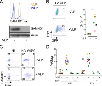
We then evaluated the effect of Vpx on MDDC infection by cell-free HIV-1. MDDCs were incubated for 2 h with or without VLPs before exposure to HIV(VSV), a VSV-pseudotyped HIV-1. The extent of infection was measured by flow cytometry, at 48 and 72 h postinfection, by performing double HIV-1 Gag and
SAMHD1 staining (Fig. 1C). The results of one representative
experiment are shownFig. 1C, and the mean values obtained with
eight independent donors are shown inFig. 1D. Without VLPs,
the efficiency of infection was low, with 5 and 10% of cells being
Gag⫹at 48 and 72 h, respectively. The Gag signal corresponded to
newly synthesized viral proteins, since it was no longer detected when MDDCs were treated with the reverse transcriptase
inhibi-tor NVP (Fig. 1D). As previously reported (7), VLPs strongly
in-creased the number of infected cells (Fig. 1CandD). Interestingly,
the Gag-expressing cells were mostly found within the
SAMHD1-low population (Fig. 1C), confirming that SAMHD1 prevents
ef-ficient HIV-1 infection in MDDCs.
on November 7, 2019 by guest
http://jvi.asm.org/
We next asked whether HIV-1 cell-to-cell transmission to MDDCs could overcome the antiviral effect of SAMHD1. We set up a coculture system, in which HIV-1-infected T cells are mixed
with target MDDCs (Fig. 2A). We used a T cell line (MT4) or
autologous primary CD4⫹T cells as donors. The T cells were
infected 2 days before the coculture, in order to get a significant proportion of HIV-1-producing cells. HIV-infected T cells were stained with the CMFDA tracker before coculture, to distinguish donor and target cells. MDDCs were incubated or not with VLPs 2 h before addition of donor cells. Infection of MDDCs was mea-sured at 48 and 72 h postcoculture by double Gag and SAMHD1
staining (Fig. 2BandC). In this coculture system, in the absence of
Vpx (⫺VLP), MDDCs were poorly sensitive to infection. One
experiment and the mean values of independent experiments with
9 donors are shown inFig. 2B. Even though MT4 cells were highly
infected at the beginning of the coculture (40 to 80% of Gag⫹cells;
not shown), only 8 and 12% of MDDCs became infected at 48 and
72 h, respectively (Fig. 2B). The Gag signal in MDDCs was
signif-icantly reduced in the presence of NVP, indicating that it mostly corresponded to productive infection and not to capture of
in-coming viral material (Fig. 2BandC).
Interestingly, addition of VLPs to MDDCs decreased SAMHD1 levels and significantly enhanced the appearance of
Gag-expressing MDDCs (Fig. 2B). Similar results were obtained
with HIV-1-infected primary autologous CD4⫹T cells (with 10 to
20% of Gag⫹cells; not shown) used as donors (Fig. 2C).
A large part of HIV-1-infected MT4 cells died during the co-culture, as assessed by calculating the percentage of donor cells in the coculture at 72 h (not shown). This death was likely due to a viral cytopathic effect, since noninfected MT4 cells remained
de-tectable. Primary infected CD4⫹T cells, which are less prone to
HIV-1-induced cytopathic effects, at least during the 3 days of the experiment, were still present in the coculture after 72 h (not shown).
Noteworthy, VLPs devoid of Vpx (VLP⌬Vpx) did not
down-regulate SAMHD1 (not shown), nor did they enhance HIV-1
in-fection (Fig. 2D), confirming that Vpx is required to mediate these
effects.
Numerous clusters of infected lymphocytes and MDDCs were observed in the coculture (not shown). Moreover, the separation of donor T cells and target MDDCs in transwell chambers or ap-plication of a gentle and continuous shaking to the cocultures to
FIG 1Vpx degrades SAMHD1 and enhances cell-free HIV-1 infection of MDDCs. (A) Levels of SAMHD1 expression in MDDCs treated or not with VLPs. MDDCs were analyzed for SAMHD1 expression by flow cytometry (top) and Western blotting (bottom). Actin staining was used as a control of gel loading. The anti-SAMHD1 monoclonal antibody I19-18 was used. Similar results were obtained with a rabbit polyclonal anti-SAMHD1 antibody (not shown). (B) Effect of VLPs on MDDC sensitivity to lentiviral transduction. MDDCs were treated or not with VLPs for 2 h and transduced with LV-GFPs (100 ng ml⫺1of p24). The percentages of GFP-expressing cells were measured 48 h later. (Left) Results of one representative experiment; (right) mean of the results obtained with five independent donors. Cells from each donor are represented by a different symbol. (C and D) Effect of VLPs on MDDC sensitivity to cell-free HIV-1 infection. MDDCs were treated or not with VLPs for 2 h and infected with HIV(VSV) (100 ng ml⫺1of p24). Levels of SAMHD1 and Gag were measured 48 or 72 h later. (C) Results of one representative experiment at 48 h postinfection. Numbers represent the percentage of cells in each quadrant. NI, noninfected. (D) Quantifi-cation of the percentage of Gag expression in cells from eight different donors after 48 and 72 h of infection. NVP was used to inhibit productive HIV-1 infection. *, significant differences (P⬍0.05, Wilcoxon matched-pairs test) between untreated and VLP-treated samples.
Puigdomènech et al.
on November 7, 2019 by guest
http://jvi.asm.org/
[image:3.585.113.472.68.371.2]avoid long contacts between cells (26) significantly decreased the
percentage of infected MDDCs (Fig. 2E), strongly suggesting that
most of the virus was transmitted through cell-to-cell contacts. Altogether, these results show that HIV-1 cell-to-cell transmis-sion to MDDCs is less efficient in the absence of Vpx, suggesting that contacts with infected cells do not overcome HIV-1 restric-tion in MDDCs.
Vpx induces sensing of HIV-1-infected T cells by MDDCs.In
contrast to pDCs (21,36,40), MDDCs do not produce detectable
levels of type I IFN when exposed to cell-free HIV-1. However, the addition of Vpx in MDDCs, by allowing HIV-1 infection,
pro-motes IFN release (21). We previously reported that HIV-infected
T cells are much more potent inducers of type I IFN than free
virions in pDCs (36). We asked whether SAMHD1 might prevent
recognition of cell-free HIV-1 and HIV-1-infected T cells by MDDCs.
We first examined the impact of VLPs on the sensing of virions. MDDCs that were initially analyzed for their sensitivity to
HIV-(VSV) (Fig. 1D) were further stained for MxA, an
interferon-stim-ulated gene (36, 41). Flow cytometry indicated that MxA was
strongly induced by virions only in the presence of VLPs (Fig. 3A
andB). Expression of MxA was induced in both infected and
FIG 2Vpx enhances HIV-1 transmission from T cells to MDDCs. (A) Representation of the experimental system. HIV-1-infected donor lymphocytes (either MT4 or autologous CD4⫹T cells) were labeled with the cell tracker CMFDA and cocultured with target MDDCs preincubated or not with VLPs for 2 h. Coculture was analyzed for Gag and SAMHD1 expression at 48 and 72 h. FACS, fluorescence-activated cell sorting. (B) Effect of VLPs on MDDC sensitivity to coculture with HIV-infected MT4 cells. Results of one representative experiment (for the 48-h time point) are shown. Dot plots show the percentages of SAMHD1 and Gag within target MDDCs. *, significant differences (P⬍0.05). (C) Compilation of the percentage of Gag expression in cells from eight different donors after 48 and 72 h of infection. Cocultures were performed with MT4 cells (left) or primary CD4⫹T cells (right). NVP was used to inhibit productive HIV-1 transmission from T cells to MDDCs. *, significant differences (P⬍0.05, Wilcoxon matched-pairs test) between untreated and VLP-treated samples. (D) VLPs lacking Vpx do not enhance sensitivity to HIV-1 infection. MDDCs were treated with VLPs carrying Vpx (VLP) or devoid of Vpx (VLP⌬Vpx) for 2 h or left untreated (⫺VLP). MDDCs were then cocultivated with HIV-infected MT4 cells. Percentages of Gag-expressing MDDCs were measured at 72 h postinfection. The mean⫾SD of at least 5 independent experiments is shown. *, significant differences (P⬍0.05). (E) Analysis of viral transmission between HIV-1-infected MT4 cells and target MDDCs. Cocultures of MT4 cells and MDDCs either were kept static (left) or were gently shaken to avoid long contacts (middle). Donor and target cells were also placed in a transwell chamber (right) in which donor MT4 cells and recipient MDDCs were separated by a virus-permeable membrane. Percentages of Gag-expressing MDDCs were measured at 72 h postinfection. The mean⫾SD of 3 independent experiments is shown. *, significant differences (P⬍0.05).
on November 7, 2019 by guest
http://jvi.asm.org/
[image:4.585.111.475.67.455.2]bystander MDDCs (Fig. 3A), demonstrating a paracrine effect. Furthermore, MxA induction correlated with a release of type I
IFN in supernatants (Fig. 3B, right). NVP not only blocked HIV-1
infection but also prevented MxA expression and IFN release
(Fig. 3B). VLPs also increased infection of the X4 strain NL4-3, the R5 strain NLAD8, and the primary dual-tropic 132W isolate (Fig. 3C), indicating that the enhancing effect of Vpx is not limited to VSV-pseudotyped viruses. These non-VSV-pseudotyped
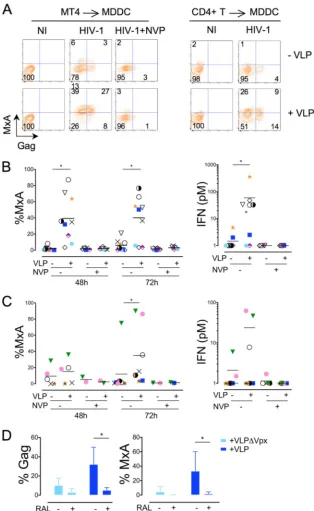
FIG 3Vpx induces sensing of cell-free HIV-1 in MDDCs. Effect of VLPs on the sensing of cell-free HIV-1 by MDDCs. Cells were treated or not with VLPs for 2 h and infected with HIV(VSV) (100 ng ml⫺1of p24), as described in the legend toFig. 1. Levels of the interferon-stimulated protein MxA and Gag were quantified by flow cytometry at 72 h. (A) FLUAV, a strong type I IFN inducer, was used as a positive control of MxA induction. (B) Compilation of the percentage of MDDCs expressing MxA at 48 and 72 h after HIV(VSV) infection. Cells from eight independent donors were analyzed (left). Levels of type I IFN released in supernatants were also measured (right). For each donor, the highest value of type I IFN secreted (at 48 h or 72 h) is shown. (C) MDDCs were treated with VLPs for 2 h (⫹VLP) or left untreated (⫺VLP). MDDCs were then infected with various HIV-1 isolates: the X4 strain NL4-3, the dual-tropic primary isolate 132W (100 ng ml⫺1of p24), and the R5 strain NLAD8 (300 ng ml⫺1of p24). HIV-1 devoid of viral envelope glycoprotein (⌬Env; 100 ng ml⫺1of p24) was used as a control of HIV-1 infection. Percentages of cells expressing Gag (left) and MxA (middle) and levels of type I IFN released in the supernatant (right) were measured at 72 h postinfection. The mean⫾SD of two independent experiments performed with two different donors is shown. (D) FLUAV sensing by MDDCs. Cells were treated or not with VLPs for 2 h and infected with FLUAV (10 HAU ml⫺1). Levels of MxA and type I IFN released were measured at 48 h. VLPs did not enhance FLUAV detection, even when a suboptimal dose of FLUAV was used (not shown). *, significant differences (P⬍0.05, Wilcoxon matched-pairs test) between untreated and VLP-treated samples.
Puigdomènech et al.
on November 7, 2019 by guest
http://jvi.asm.org/
[image:5.585.134.448.66.548.2]HIV-1 isolates similarly triggered an IFN response, but only in the
presence of Vpx (Fig. 3C). Of note, cells from two out of eight
independent donors did not release detectable IFN in response to cell-free HIV-1, even though VLPs enhanced infection. FLUAV, a well-characterized IFN inducer, was used as a positive control. As
expected (42), FLUAV productively infected MDDCs, as assessed
by flow cytometry after staining with an anti-NP antibody (not shown). FLUAV induced similar levels of MxA and type I IFN,
irrespective of Vpx (Fig. 3D).
We then determined how VLPs affected sensing of HIV-1-in-fected T cells by MDDCs. Without VLPs, HIV-inHIV-1-in-fected MT4 cells
did not trigger a potent immune response in MDDCs (Fig. 4Aand
B). Addition of VLPs induced MxA expression in MDDCs and
IFN release in the cocultures (Fig. 4AandB). HIV-1-infected
au-tologous CD4⫹T cells also induced MxA and IFN in the presence
of VLPs (Fig. 4AandC); however, induction was at levels lower
than those for MT4 cells. For example, MxA was significantly up-regulated at the 48-h time point with MT4 cells and only at 72 h with primary lymphocytes. NVP inhibited infection of MDDCs (Fig. 2) and sensing of infected T cells (Fig. 4). The integrase in-hibitor raltegravir also inhibited productive infection of MDDCs cultivated with infected MT4 cells and impaired MxA expression
in MDDCs (Fig. 4D).
Therefore, MDDCs do not spontaneously trigger an IFN re-sponse when they encounter HIV-1-infected T cells. The addition of Vpx overcomes restriction to HIV-1 infection and promotes sensing of HIV-1-infected T cells.
SAMHD1 prevents HIV-1 transmission and reduces sensing of HIV-1-infected T cells in MDDCs.To further study the role of SAMHD1 in HIV-1 infection and sensing in MDDCs, we silenced this protein with siRNA. As previously reported, transfection of siRNA against SAMHD1 (and not that of a control siRNA, siCTR)
(7) induced SAMHD1 silencing, with 40 to 60% decreases in the
levels of the protein, depending on the donors, being observed by
flow cytometry and Western blotting (Fig. 5A). Thus, the silencing
procedure decreases SAMHD1 levels, but the degradation that it induces is less potent than the degradation induced by Vpx (com-pareFig. 1Aand5A). As expected, in SAMHD1-silenced cells, there was a strong increase of transduction with LV-GFPs (Fig. 5B). Infection of MDDCs with free HIV(VSV) was also
sig-nificantly augmented (Fig. 5CandD). The MxA protein was
in-duced upon HIV-1 infection in SAMHD1-silenced cells (Fig. 5C
andD). MxA induction was significant but less marked than that
in VLP-exposed MDDCs (Fig. 3). We hypothesize that the
resid-ual levels of SAMHD1 may partly protect MDDCs against HIV-1 infection and sensing.
SAMHD1 silencing also facilitated infection of MDDCs
cocul-tivated with HIV-1-infected T cells (Fig. 6). A significant 2-fold
increase in the proportion of Gag⫹MDDCs was detected. Again,
this augmentation was less marked than that in VLP-exposed MDDCs but was associated with viral sensing and expression of
the MxA protein in MDDCs (Fig. 6C).
We only occasionally detected type I IFN release in the super-natants of SAMHD1-silenced MDDCs exposed to cell-free HIV-(VSV) or cocultivated with HIV-infected lymphocytes (not shown). This might be due to the relatively low MDDC concen-tration in cultures after siRNA transfection, a procedure that can be harmful for the cells.
MT4 cells are better HIV-1 transmitters and trigger a stronger
innate response than primary CD4⫹T cells in MDDCs (Fig. 2and
4). We asked whether there is a correlation between the levels of
Gag and the induction of MxA in MDDCs. Plotting of these two parameters, irrespective of any VLP treatment or any SAMHD1 silencing, demonstrated a correlation, with a threshold of innate detection at about 20% of Gag-positive MDDCs (not shown). This correlation was visible with both HIV-1 cell-free particles and infected cells (not shown).
Therefore, siRNA transfection allows a partial but significant reduction of SAMHD1 levels in MDDCs. SAMHD1-silenced cells are more sensitive to infection by virions or by coculture with infected lymphocytes. Moreover, in silenced cells, infection trig-gers innate sensing of the virus, as assessed by MxA expression. This phenomenon is more pronounced with HIV-1-infected T cells than with free virions. High levels of productive infection in MDDCs are required to induce sensing. Altogether, our results highlight a role for SAMHD1 in controlling HIV-1 immune rec-ognition by MDDCs.
DISCUSSION
Myeloid DCs are poorly permissive to infection by cell-free HIV-1. In these noncycling cells, viral reverse transcription occurs
slowly and almost covertly (43), because of a limited availability of
intracellular dNTPs (11,18). The recently identified
deoxynucleo-side triphosphohydrolase SAMHD1 cleaves dNTPs and thus acts
as an antiviral protein in MDDCs and other myeloid cells (11,16).
We show here that MDDCs are to a large extent resistant to infec-tion when they encounter HIV-1-infected cells. Mixing infected lymphocytes with MDDCs led to a modest infection, with less than 10% of MDDCs expressing Gag 2 or 3 days after the begin-ning of the coculture. This restriction of infection is in large part mediated by SAMHD1. SAMHD1 levels are not modified in MDDCs cocultivated with infected lymphocytes. Moreover, addi-tion of SIV particles carrying the lentiviral protein Vpx, which degrades SAMHD1, or silencing of SAMHD1 with siRNAs signif-icantly enhanced productive infection of MDDCs. Our results strongly suggest that contacts with HIV-1-infected lymphocytes do not affect SAMHD1 activity in MDDCs and thus do not lead to an increase of the intracellular dNTP pool.
Direct intercellular transmission of HIV-1 is far more efficient
than infection with free virions (24,26). The high multiplicity of
infection generated during the formation of virological synapses may allow escape from antiviral immune responses and
therapeu-tics (37, 44). The antiviral activity of some restriction factors is
saturable. For instance, Trim5a, which targets incoming lentiviral
capsids, does not prevent cell-to-cell transmission (45). In
con-trast, tetherin, which sequesters budding virions at the cell surface,
inhibits this mode of viral spread (46,47). Given that SAMHD1
degrades cellular dNTPs, it is not unexpected to observe that the enzyme inhibits HIV-1 cell-to-cell transmission. Moreover, SAMHD1 binds RNA, and this binding inhibits its enzymatic
ac-tivity (48, 49). Recent enzymatic analysis also showed that
SAMHD1 does not hydrolyze ribonucleoside triphosphates (rNTPs) or single-stranded DNA, double-stranded DNA, or RNA
(16). These assays were carried out with a recombinant proteinin
vitroand do not formally rule out the possibility that the enzyme may target additional viral or cellular nucleic acids within the cell.
Furthermore, coculture of MDDCs with infected CD4⫹T cells
could have affected the expression or function of SAMHD1, but our results demonstrate that this is not the case. It might have also been conceivable that during coculture and viral cell-to-cell
on November 7, 2019 by guest
http://jvi.asm.org/
mission, dNTPs carried over from T cells to MDDCs might have overcome SAMHD1 restriction. Again, this is apparently not the case. Altogether, our results show that SAMHD1, due to its orig-inal mode of action, is not saturable and remains operative during HIV-1 intercellular spread.
Silencing of SAMHD1 significantly relieved the block to HIV-1 intercellular transmission, although less efficiently than the addi-tion of Vpx to MDDCs. This may be due to the incomplete silenc-ing achieved by siRNA, which led to residual levels of SAMHD1. It is also possible that Vpx exerts additional, SAMHD1-independent
FIG 4Vpx induces sensing of HIV-1-infected T cells by MDDCs. Effect of VLPs on the sensing of HIV-1-infected lymphocytes by MDDCs. The latter were treated or not with VLPs for 2 h and exposed to HIV-1-infected MT4 cells (left) or autologous CD4⫹T cells (right) as described in the legend toFig. 2. Levels of the interferon-stimulated protein MxA and Gag were quantified by flow cytometry at 48 and 72 h. (A) Results of representative experiments are shown. (B and C) Compilation of the levels of MxA (left) and IFN (right) released in supernatants in MDDCs (from eight independent donors) cocultured with either HIV-1-infected MT4 cells (B) or HIV-1-infected autologous CD4⫹T cells (C) for 48 and 72 h. NVP was used to inhibit productive HIV-1 transmission from T cells to MDDCs. *, significant differences (P⬍0.05, Wilcoxon matched-pairs test) between untreated and VLP-treated samples. (D) MDDCs were treated or not with VLPs for 2 h and exposed to HIV-1-infected MT4 cells in the absence or presence of raltegravir (RAL; 1M). Percentages of Gag and MxA expressing cells were quantified by flow cytometry at 72 h on gated MDDCs. A compilation of the levels of Gag (left) and MxA (right) in 4 independent experiments is shown. *,P⬍0.05.
Puigdomènech et al.
on November 7, 2019 by guest
http://jvi.asm.org/
[image:7.585.134.448.65.576.2]effects to facilitate infection (50). Vpx has been reported to bind and partially counteract another cellular protein, APOBEC3A,
which may inhibit HIV-1 infection in myeloid cells (51,52). It will
be of interest to assess whether SAMHD1 exerts its antiviral activ-ity in coordination with other enzymes regulating the dNTP pool or the metabolism of nucleic acids.
During chronic HIV infection, circulating myeloid DC
num-bers are reduced (53). Moreover, the frequency of infected
MD-DCs in HIV-1-positive individuals is low, probably 10- to 100-fold
lower than that of infected CD4 T cells (29). The reasons
explain-ing this moderate susceptibility are multiple and include low levels of HIV receptors, a rapid and extensive degradation of internal-ized HIV, and as demonstrated here, the presence of the restric-tion factor SAMHD1, which may prevent HIV-1 cell-to-cell
trans-missionin vivo.
Why does HIV-1 not possess a protein with anti-SAMHD1 activity, in contrast to HIV-2 and SIV? HIV-1 encodes a reverse
transcriptase with a lowKm(54) that binds to dNTPs with a
par-ticularly high affinity. This property contributes to the low levels of reverse transcription in noncycling myeloid cells, in which
dNTPs are scarce (4, 11, 18). The HIV-2 reverse transcriptase
probably has a lower affinity for dNTPs (50), which may be
detri-mental for this virus in noncycling cells. It has been reported that
DCs are less susceptible to HIV-2 than to HIV-1, when the
infec-tion is performed with cell-free virions (55). It will be worth
ex-amining whether HIV-2 more efficiently infects MDDCs by cell-to-cell transmission and, if so, to evaluate the impact of Vpx on this phenomenon.
The lack of Vpx in HIV-1 has also been proposed to allow
avoiding an immune response in MDDCs (21). Indeed, MDDCs
infected with HIV-1 produced type I IFN and upregulated
CD86 only in the presence of Vpx (21,23). Our results extend
these observations. We show here that in the absence of Vpx, neither free virions nor HIV-1-infected cells trigger detectable IFN release by MDDCs, although very modest effects can rarely be observed in some donors. However, addition of VLPs pro-moted sensing of HIV-1, assessed by measuring type I IFN production and MxA expression. MxA is widely used as a
sur-rogate marker of IFN production (41). MxA is induced by type
I IFN and the closely related type III IFN but not by type II IFN. It will be worth determining whether type III IFN and other cytokines are also induced upon sensing of HIV-1 by MDDCs. Interestingly, HIV-1-infected T cells are more potent inducers
of IFN than free virions (compareFig. 3and4), in line with our
previous results obtained in other hematopoietic cell types
(peripheral blood mononuclear cells and pDCs) (36). Future
FIG 5Effect of SAMHD1 silencing on cell-free HIV-1 infection and sensing by MDDCs. Levels of SAMHD1 in MDDCs transfected with siRNA against SAMHD1 (si SAMHD1) or with a control siRNA (si CTR). (A) Analysis was performed by flow cytometry (top) and Western blotting (bottom). Actin staining was used as a control of gel loading. (B) Effect of SAMHD1 silencing on MDDC sensitivity to lentiviral transduction. Control or SAMHD1-silenced MDDCs were transduced with LV-GFPs (100 ng ml⫺1of p24). Percentages of GFP-positive cells were measured 48 h later. (Left) Results of one representative experiment; (right) the mean of the results obtained with five independent donors. (C) Effect of SAMHD1 silencing on infection and sensing of cell-free HIV-1 by MDDCs. Control or SAMHD1-silenced MDDCs were infected with HIV(VSV) (100 ng ml⫺1of p24). Levels of the interferon-stimulated protein MxA and Gag were quantified by flow cytometry. Results of one representative experiment at 72 h are shown. The percentages of Gag- and MxA-positive cells are indicated in the dot plots. (D) Compilation of the levels of Gag (left) and MxA (right) expression in MDDCs from five independent donors. Stainings were performed at either 72 or 96 h postinfection, depending on the donors. The HIV-1 inhibitor NVP was used when indicated. *, significant differences (P⬍0.05, Wilcoxon matched-pairs test).
on November 7, 2019 by guest
http://jvi.asm.org/
[image:8.585.113.475.66.357.2]studies will help determine which viral or cellular components allow potent sensing of infected cells.
The reverse transcriptase inhibitor NVP prevented MDDC re-sponsiveness, indicating that incoming viral RNA is not sufficient to induce IFN production. The integrase inhibitor raltegravir also inhibited sensing of HIV-infected cells. This result suggested that the response was triggered postintegration, consistent with the
findings of Manel et al. (21) and Sunseri et al. (23), obtained with
cell-free viral particles. This result also suggests that incomplete reverse transcripts, which are known to trigger an inflammatory
response in CD4⫹T cells (56), are poorly sensed in MDDCs. It will
be worthwhile to test various viral mutants to further characterize which steps of the viral life cycle, after reverse transcription and integration, mediate the sensing of infection in MDDCs. In par-ticular, when the infection is performed with cell-free HIV-1, sensing involves binding of cyclophilin A to newly synthesized
Gag proteins (21). It is tempting to speculate that additional
path-ways of sensing may be triggered upon cell-to-cell contacts. Silencing of SAMHD1 promoted HIV-1 recognition, demon-strating that this restriction factor negatively regulates virus-in-duced immune responses in MDDCs. In line with our data, monocytes from individuals with Aicardi-Goutieres syndrome are highly susceptible to HIV-1 and produce proinflammatory
cytokines upon infection (20). Therefore, the restriction of HIV-1
infection by SAMHD1 directly impacts the triggering of an innate immune response in myeloid MDDCs. These cells have a central role in linking innate and adaptive responses. The type of innate stimuli impacts the outcome of the adaptive response, including
polarization of CD4⫹T cells, antibody shaping, and establishment
of memory (57,58). Furthermore, HIV-1, by avoiding efficient
productive infection of MDDCs through preservation of SAMHD1, may also control the array of presented or cross-pre-sented viral antigens, resulting in qualitatively or quantitatively
different CD8⫹and CD4⫹responses (57,59).
It is noticeable that there were some variations in the type I IFN/MxA responses between donors, even though the levels of virus infection showed less variation. We observed that there is a significant correlation between the levels of Gag expression and the percentage of MxA-positive cells (data not shown), and this may explain why some donors elicited clear responses to infection, whereas others did not. It is also likely that the extent of the innate response may vary between individuals. It will be of interest to analyze serial samples from the same donors, to determine if they behave consistently.
In contrast to myeloid DCs, pDCs efficiently and rapidly detect HIV-1, mainly through TLR7, leading to type I IFN secretion
in-dependently of productive infection (36,60). It will be important
to study whether pDCs, which are noncycling cells, express SAMHD1 and, if so, what are the possible effects on IFN produc-tion. Various cell types with different mechanisms are thus in-volved in the recognition of HIV-1 virions and infected cells. Overall, our results show that in myeloid DCs, SAMHD1 controls not only HIV-1 replication but also the immune response of the host.
ACKNOWLEDGMENTS
We thank members of the Virus and Immunity Unit for discussions and Monsef Benkirane and Diana Ayinde for critical readings of the manu-script.
Work in our lab is supported by grants from the Agence Nationale de Recherche sur le SIDA (ANRS), SIDACTION, the AREVA Foundation, the Vaccine Research Institute, the Labex IBEID program, the FP7 pro-gram HIT Hidden HIV (Health-F3-2012-305762), and the Institut Pas-teur. I.P. is supported by the Marie Curie Intra-European Fellowship for Career Development (FP-7-PEOPLE-2011-IEF).
I.P., N.C., and F.P. performed research and analyzed data. I.P., N.C., and O.S. designed the research and wrote the paper. O.S. supervised the study.
We declare no competing financial interest.
FIG 6Role of SAMHD1 during HIV-1 transmission from T cells to MDDCs. (A) Schematic representation of the experimental system. MDDCs were treated with siRNA against SAMHD1 or with a control siRNA (si CTR) 48 h before the start of the coculture. MDDCs were then mixed with CMFDA-labeled HIV-1-infected T cells (either MT4 or autologous CD4⫹T cells). MDDCs were analyzed for levels of Gag and MxA after 72 and 96 h of coculture. (B) Results of one representative experiment of HIV-1-infected MT4 cells cocultured with MDDCs are shown. Numbers indicate the percentages of Gag- and MxA-expressing cells in MDDCs at 72 h. (C) Compilation of the percentage of Gag (left) and MxA (right) in MDDCs cocultured with infected T cells (MT4 or autologous CD4⫹ T cells). The HIV-1 inhibitor NVP was used when indicated. *, significant differences (P⬍0.05, Wilcoxon matched-pairs test) between SAMHD1- and CTR-silenced MDDCs.
Puigdomènech et al.
on November 7, 2019 by guest
http://jvi.asm.org/
[image:9.585.134.451.65.265.2]REFERENCES
1.Hughes R, Towers G, Noursadeghi M.2012. Innate immune interferon responses to human immunodeficiency virus-1 infection. Rev. Med. Vi-rol.22:257–266.
2.Neil SJ, Zang T, Bieniasz PD.2008. Tetherin inhibits retrovirus release and is antagonized by HIV-1 Vpu. Nature451:425– 430.
3.Sheehy AM, Gaddis NC, Choi JD, Malim MH. 2002. Isolation of a human gene that inhibits HIV-1 infection and is suppressed by the viral Vif protein. Nature418:646 – 650.
4.Ayinde D, Casartelli N, Schwartz O.2012. Restricting HIV the SAMHD1 way: through nucleotide starvation. Nat. Rev. Microbiol.10:675– 680. 5.Hrecka K, Hao C, Gierszewska M, Swanson SK, Kesik-Brodacka M,
Srivastava S, Florens L, Washburn MP, Skowronski J.2011. Vpx relieves inhibition of HIV-1 infection of macrophages mediated by the SAMHD1 protein. Nature474:658 – 661.
6.Kaushik R, Zhu X, Stranska R, Wu Y, Stevenson M.2009. A cellular restriction dictates the permissivity of nondividing monocytes/ macrophages to lentivirus and gammaretrovirus infection. Cell Host Mi-crobe6:68 – 80.
7.Laguette N, Sobhian B, Casartelli N, Ringeard M, Chable-Bessia C, Segeral E, Yatim A, Emiliani S, Schwartz O, Benkirane M. 2011. SAMHD1 is the dendritic- and myeloid-cell-specific HIV-1 restriction factor counteracted by Vpx. Nature474:654 – 657.
8.Lim ES, Fregoso OI, McCoy CO, Matsen FA, Malik HS, Emerman M.
2012. The ability of primate lentiviruses to degrade the monocyte restric-tion factor SAMHD1 preceded the birth of the viral accessory protein Vpx. Cell Host Microbe11:194 –204.
9.Sharova N, Wu Y, Zhu X, Stranska R, Kaushik R, Sharkey M, Stevenson M.2008. Primate lentiviral Vpx commandeers DDB1 to counteract a macrophage restriction. PLoS Pathog.4:e1000057. doi:10.1371/journal .ppat.1000057.
10. Brandariz-Nunez A, Valle-Casuso JC, White TE, Laguette N, Benkirane M, Brojatsch J, Diaz-Griffero F.2012. Role of SAMHD1 nuclear local-ization in restriction of HIV-1 and SIVmac. Retrovirology9:49. doi:10 .1186/1742-4690-9-49.
11. Lahouassa H, Daddacha W, Hofmann H, Ayinde D, Logue EC, Dragin L, Bloch N, Maudet C, Bertrand M, Gramberg T, Pancino G, Priet S, Canard B, Laguette N, Benkirane M, Transy C, Landau NR, Kim B, Margottin-Goguet F.2012. SAMHD1 restricts the replication of human immunodeficiency virus type 1 by depleting the intracellular pool of de-oxynucleoside triphosphates. Nat. Immunol.13:223–228.
12. Baldauf HM, Pan X, Erikson E, Schmidt S, Daddacha W, Burggraf M, Schenkova K, Ambiel I, Wabnitz G, Gramberg T, Panitz S, Flory E, Landau NR, Sertel S, Rutsch F, Lasitschka F, Kim B, Konig R, Fackler OT, Keppler OT.2012. SAMHD1 restricts HIV-1 infection in resting CD4(⫹) T cells. Nat. Med.18:1682–1689.
13. Descours B, Cribier A, Chable-Bessia C, Ayinde D, Rice G, Crow Y, Yatim A, Schwartz O, Laguette N, Benkirane M.2012. SAMHD1 re-stricts HIV-1 reverse transcription in quiescent CD4⫹T-cells. Retrovirol-ogy9:87. doi:10.1186/1742-4690-9-87.
14. Wu L.2012. SAMHD1: a new contributor to HIV-1 restriction in resting CD4⫹T-cells. Retrovirology9:88. doi:10.1186/1742-4690-9-88. 15. Yan N, Lieberman J.2012. SAMHD1 does it again, now in resting T cells.
Nat. Med.18:1611–1612.
16. Goldstone DC, Ennis-Adeniran V, Hedden JJ, Groom HC, Rice GI, Christodoulou E, Walker PA, Kelly G, Haire LF, Yap MW, de Carvalho LP, Stoye JP, Crow YJ, Taylor IA, Webb M.2011. HIV-1 restriction factor SAMHD1 is a deoxynucleoside triphosphate triphosphohydrolase. Nature480:379 –382.
17. Powell RD, Holland PJ, Hollis T, Perrino FW.2011. Aicardi-Goutieres syndrome gene and HIV-1 restriction factor SAMHD1 is a dGTP-regulated deoxynucleotide triphosphohydrolase. J. Biol. Chem.286: 43596 – 43600.
18. Kim B, Nguyen LA, Daddacha W, Hollenbaugh JA.2012. Tight inter-play among SAMHD1 protein level, cellular dNTP levels, and HIV-1 pro-viral DNA synthesis kinetics in human primary monocyte-derived mac-rophages. J. Biol. Chem.287:21570 –21574.
19. Rice GI, Bond J, Asipu A, Brunette RL, Manfield IW, Carr IM, Fuller JC, Jackson RM, Lamb T, Briggs TA, Ali M, Gornall H, Couthard LR, Aeby A, Attard-Montalto SP, Bertini E, Bodemer C, Brockmann K, Brueton LA, Corry PC, Desguerre I, Fazzi E, Cazorla AG, Gener B, Hamel BC, Heiberg A, Hunter M, van der Knaap MS, Kumar R, Lagae
L, Landrieu PG, Lourenco CM, Marom D, McDermott MF, van der Merwe W, Orcesi S, Prendiville JS, Rasmussen M, Shalev SA, Soler DM, Shinawi M, Spiegel R, Tan TY, Vanderver A, Wakeling EL, Wassmer E, Whittaker E, Lebon P, Stetson DB, Bonthron DT, Crow YJ. 2009. Mutations involved in Aicardi-Goutieres syndrome implicate SAMHD1 as regulator of the innate immune response. Nat. Genet.41:829 – 832. 20. Berger A, Sommer AF, Zwarg J, Hamdorf M, Welzel K, Esly N, Panitz
S, Reuter A, Ramos I, Jatiani A, Mulder LC, Fernandez-Sesma A, Rutsch F, Simon V, Konig R, Flory E.2011. SAMHD1-deficient CD14⫹ cells from individuals with Aicardi-Goutieres syndrome are highly suscep-tible to HIV-1 infection. PLoS Pathog.7:e1002425. doi:10.1371/journal .ppat.1002425.
21. Manel N, Hogstad B, Wang Y, Levy DE, Unutmaz D, Littman DR.2010. A cryptic sensor for HIV-1 activates antiviral innate immunity in dendritic cells. Nature467:214 –217.
22. Pertel T, Reinhard C, Luban J.2011. Vpx rescues HIV-1 transduction of dendritic cells from the antiviral state established by type 1 interferon. Retrovirology8:49. doi:10.1186/1742-4690-8-49.
23. Sunseri N, O’Brien M, Bhardwaj N, Landau NR.2011. Human immu-nodeficiency virus type 1 modified to package simian immuimmu-nodeficiency virus Vpx efficiently infects macrophages and dendritic cells. J. Virol.85: 6263– 6274.
24. Chen P, Hubner W, Spinelli MA, Chen BK.2007. Predominant mode of human immunodeficiency virus transfer between T cells is mediated by sustained Env-dependent neutralization-resistant virological synapses. J. Virol.81:12582–12595.
25. Jolly C, Kashefi K, Hollinshead M, Sattentau QJ.2004. HIV-1 cell to cell transfer across an Env-induced, actin-dependent synapse. J. Exp. Med.
199:283–293.
26. Sourisseau M, Sol-Foulon N, Porrot F, Blanchet F, Schwartz O.2007. Inefficient human immunodeficiency virus replication in mobile lympho-cytes. J. Virol.81:1000 –1012.
27. Groot F, Welsch S, Sattentau QJ.2008. Efficient HIV-1 transmission from macrophages to T cells across transient virological synapses. Blood
111:4660 – 4663.
28. Izquierdo-Useros N, Esteban O, Rodriguez-Plata MT, Erkizia I, Prado JG, Blanco J, Garcia-Parajo MF, Martinez-Picado J. 2011. Dynamic imaging of cell-free and cell-associated viral capture in mature dendritic cells. Traffic12:1702–1713.
29. Wu L, KewalRamani VN.2006. Dendritic-cell interactions with HIV: infection and viral dissemination. Nat. Rev. Immunol.6:859 – 868. 30. Blanchet F, Moris A, Mitchell JP, Piguet V.2011. A look at HIV journey:
from dendritic cells to infection spread in CD4(⫹) T cells. Curr. Opin. HIV AIDS6:391–397.
31. McDonald D, Wu L, Bohks SM, KewalRamani VN, Unutmaz D, Hope TJ.2003. Recruitment of HIV and its receptors to dendritic cell-T cell junctions. Science300:1295–1297.
32. Petit C, Buseyne F, Boccaccio C, Abastado JP, Heard JM, Schwartz O.
2001. Nef is required for efficient HIV-1 replication in cocultures of den-dritic cells and lymphocytes. Virology286:225–236.
33. Arhel N, Lehmann M, Clauss K, Nienhaus GU, Piguet V, Kirchhoff F.
2009. The inability to disrupt the immunological synapse between in-fected human T cells and APCs distinguishes HIV-1 from most other primate lentiviruses. J. Clin. Invest.119:2965–2975.
34. Fackler OT, Alcover A, Schwartz O.2007. Modulation of the immuno-logical synapse: a key to HIV-1 pathogenesis? Nat. Rev. Immunol.7:310 – 317.
35. Thoulouze MI, Sol-Foulon N, Blanchet F, Dautry-Varsat A, Schwartz O, Alcover A.2006. Human immunodeficiency virus type 1 infection impairs the formation of the immunological synapse. Immunity24:547– 561.
36. Lepelley A, Louis S, Sourisseau M, Law HK, Pothlichet J, Schilte C, Chaperot L, Plumas J, Randall RE, Si-Tahar M, Mammano F, Albert ML, Schwartz O.2011. Innate sensing of HIV-infected cells. PLoS Pathog.
7:e1001284. doi:10.1371/journal.ppat.1001284.
37. Vendrame D, Sourisseau M, Perrin V, Schwartz O, Mammano F.2009. Partial inhibition of human immunodeficiency virus replication by type I interferons: impact of cell-to-cell viral transfer. J. Virol.83:10527–10537. 38. Roesch F, Meziane O, Kula A, Nisole S, Porrot F, Anderson I, Mam-mano F, Fassati A, Marcello A, Benkirane M, Schwartz O.2012. Hy-perthermia stimulates HIV-1 replication. PLoS Pathog.8:e1002792. doi: 10.1371/journal.ppat.1002792.
39. Goujon C, Jarrosson-Wuilleme L, Bernaud J, Rigal D, Darlix JL,
on November 7, 2019 by guest
http://jvi.asm.org/
marelli A.2006. With a little help from a friend: increasing HIV transduc-tion of monocyte-derived dendritic cells with virion-like particles of SIV-(MAC). Gene Ther.13:991–994.
40. Fonteneau JF, Larsson M, Beignon AS, McKenna K, Dasilva I, Amara A, Liu YJ, Lifson JD, Littman DR, Bhardwaj N.2004. Human immu-nodeficiency virus type 1 activates plasmacytoid dendritic cells and con-comitantly induces the bystander maturation of myeloid dendritic cells. J. Virol.78:5223–5232.
41. Haller O, Kochs G.2011. Human MxA protein: an interferon-induced dynamin-like GTPase with broad antiviral activity. J. Interferon Cytokine Res.31:79 – 87.
42. Cella M, Salio M, Sakakibara Y, Langen H, Julkunen I, Lanzavecchia A.
1999. Maturation, activation, and protection of dendritic cells induced by double-stranded RNA. J. Exp. Med.189:821– 829.
43. Nobile C, Petit C, Moris A, Skrabal K, Abastado JP, Mammano F, Schwartz O.2005. Covert human immunodeficiency virus replication in dendritic cells and in DC-SIGN-expressing cells promotes long-term transmission to lymphocytes. J. Virol.79:5386 –5399.
44. Sigal A, Kim JT, Balazs AB, Dekel E, Mayo A, Milo R, Baltimore D.
2011. Cell-to-cell spread of HIV permits ongoing replication despite an-tiretroviral therapy. Nature477:95–98.
45. Richardson MW, Carroll RG, Stremlau M, Korokhov N, Humeau LM, Silvestri G, Sodroski J, Riley JL.2008. Mode of transmission affects the sensitivity of human immunodeficiency virus type 1 to restriction by rhe-sus TRIM5alpha. J. Virol.82:11117–11128.
46. Casartelli N, Sourisseau M, Feldmann J, Guivel-Benhassine F, Mallet A, Marcelin AG, Guatelli J, Schwartz O.2010. Tetherin restricts productive HIV-1 cell-to-cell transmission. PLoS Pathog.6:e1000955. doi:10.1371 /journal.ppat.1000955.
47. Kuhl BD, Sloan RD, Donahue DA, Bar-Magen T, Liang C, Wainberg MA.2010. Tetherin restricts direct cell-to-cell infection of HIV-1. Retro-virology7:115. doi:10.1186/1742-4690-7-115.
48. Goncalves A, Karayel E, Rice GI, Bennett KL, Crow YJ, Superti-Furga G, Burckstummer T.2012. SAMHD1 is a nucleic-acid binding protein that is mislocalized due to Aicardi-Goutieres syndrome-associated muta-tions. Hum. Mutat.33:1116 –1122.
49. White TE, Brandariz-Nunez A, Carlos Valle-Casuso J, Amie S, Nguyen L, Kim B, Brojatsch J, Diaz-Griffero F. 13 November2012. Contribution of SAM and HD domains to retroviral restriction mediated by human
SAMHD1. Virology doi:10.1016/j.virol.2012.10.029. [Epub ahead of print.]
50.Fujita M, Nomaguchi M, Adachi A, Otsuka M. 2012. SAMHD1-dependent and -inSAMHD1-dependent functions of HIV-2/SIV Vpx protein. Front. Microbiol.3:297. doi:10.3389/fmicb.2012.00297.
51. Berger A, Munk C, Schweizer M, Cichutek K, Schule S, Flory E.2010. Interaction of Vpx and apolipoprotein B mRNA-editing catalytic poly-peptide 3 family member A (APOBEC3A) correlates with efficient lenti-virus infection of monocytes. J. Biol. Chem.285:12248 –12254. 52. Berger G, Durand S, Fargier G, Nguyen XN, Cordeil S, Bouaziz S,
Muriaux D, Darlix JL, Cimarelli A.2011. APOBEC3A is a specific inhib-itor of the early phases of HIV-1 infection in myeloid cells. PLoS Pathog.
7:e1002221. doi:10.1371/journal.ppat.1002221.
53. Servet C, Zitvogel L, Hosmalin A.2002. Dendritic cells in innate immune responses against HIV. Curr. Mol. Med.2:739 –756.
54. Diamond TL, Roshal M, Jamburuthugoda VK, Reynolds HM, Merriam AR, Lee KY, Balakrishnan M, Bambara RA, Planelles V, Dewhurst S, Kim B.2004. Macrophage tropism of HIV-1 depends on efficient cellular dNTP utilization by reverse transcriptase. J. Biol. Chem.279:51545– 51553.
55. Duvall MG, Lore K, Blaak H, Ambrozak DA, Adams WC, Santos K, Geldmacher C, Mascola JR, McMichael AJ, Jaye A, Whittle HC, Row-land-Jones SL, Koup RA.2007. Dendritic cells are less susceptible to human immunodeficiency virus type 2 (HIV-2) infection than to HIV-1 infection. J. Virol.81:13486 –13498.
56. Doitsh G, Cavrois M, Lassen KG, Zepeda O, Yang Z, Santiago ML, Hebbeler AM, Greene WC.2010. Abortive HIV infection mediates CD4 T cell depletion and inflammation in human lymphoid tissue. Cell143: 789 – 801.
57. Manel N, Littman DR.2011. Hiding in plain sight: how HIV evades innate immune responses. Cell147:271–274.
58. Steinman RM, Hemmi H.2006. Dendritic cells: translating innate to adaptive immunity. Curr. Top. Microbiol. Immunol.311:17–58. 59. Buseyne F, Le Gall S, Boccaccio C, Abastado JP, Lifson JD, Arthur LO,
Riviere Y, Heard JM, Schwartz O.2001. MHC-I-restricted presentation of HIV-1 virion antigens without viral replication. Nat. Med.7:344 –349. 60. Beignon AS, McKenna K, Skoberne M, Manches O, DaSilva I, Ka-vanagh DG, Larsson M, Gorelick RJ, Lifson JD, Bhardwaj N. 2005. Endocytosis of HIV-1 activates plasmacytoid dendritic cells via Toll-like receptor-viral RNA interactions. J. Clin. Invest.115:3265–3275. Puigdomènech et al.