Sindbis Virus Can Exploit a Host Antiviral Protein To Evade Immune
Surveillance
Xinlu Wang,aMelody M. H. Li,bJing Zhao,cShenglan Li,aMargaret R. MacDonald,bCharles M. Rice,bXiang Gao,cGuangxia Gaoa CAS Key Laboratory for Infection and Immunity, Institute of Biophysics, Chinese Academy of Sciences, Chaoyang District, Beijing, Chinaa; The Rockefeller University, New York, New York, USAb; Model Animal Research Center, Nanjing University, Nanjing, Chinac
ABSTRACT
Viral infection induces production of type I interferons (IFNs), which stimulate the expression of a variety of antiviral factors to inhibit viral replication. To establish effective infection, viruses need to develop strategies to evade the immune responses. A neurovirulent Sindbis virus strain with neuroinvasive properties (SVNI) causes lethal encephalitis in mice, and its replication in cultured cells is inhibited by the zinc finger antiviral protein (ZAP), a host factor that specifically inhibits the replication of cer-tain viruses by binding to the viral mRNAs, repressing the translation of target mRNA, and promoting the degradation of target mRNA. We report here that murine embryonic fibroblast cells from ZAP knockout mice supported more efficient SVNI replica-tion than wild-type cells. SVNI infecreplica-tion of 10-day-old suckling mice led to reduced survival in the knockout mice. Unexpectedly, however, SVNI infection of 23-day-old weanling mice, whose immune system is more developed than that of the suckling mice, resulted in significantly improved survival in ZAP knockout mice. Further analyses revealed that in the weanling knockout mice, SVNI replicated more efficiently in lymphoid tissues at early times postinfection and induced higher levels of IFN production, which restricted viral spread to the central nervous system. Blocking IFN activity through the use of receptor-neutralizing anti-bodies rendered knockout mice more sensitive to SVNI infection than wild-type mice. These results uncover a mechanism by which SVNI exploits a host antiviral factor to evade innate immune surveillance.
IMPORTANCE
Sindbis virus, a prototypic member of theAlphavirusgenus, has been used to study the pathogenesis of acute viral encephalitis
in mice for many years. How the virus evades immune surveillance to establish effective infection is largely unknown. ZAP is a host antiviral factor that potently inhibits Sindbis virus replication in cell culture. We show here that infection of ZAP knockout suckling mice with an SVNI led to faster disease progression. However, SVNI infection of weanling mice led to slower disease progression in knockout mice. Further analyses revealed that in weanling knockout mice, SVNI replicated more efficiently in lymphoid tissues at early times postinfection and induced higher levels of interferon production, which restricted viral spread to the central nervous system. These results uncover a mechanism by which SVNI exploits a host antiviral factor to evade innate immune surveillance and allow enhanced neuroinvasion.
V
iral infection induces the production of type I interferons (IFNs), which inhibit viral replication through a variety of mechanisms (1,2). To establish effective infection, viruses need to develop strategies to evade the immune responses. Sindbis virus is a prototypic member of theAlphavirus genus, whose members include viruses such as eastern, western, and Venezuelan equine encephalitis viruses that can cause fatal encephalitis in humans and equines (3). Sindbis virus infection of mice has been used as a model to study the pathogenesis of acute viral encephalitis for many years (4). After peripheral inoculation, localized replication leads to viremia, with subsequent spread to various tissues (5). The outcome of infection is both age and virus strain dependent. Some strains of the virus display neuroinvasive properties and after peripheral inoculation can spread through the blood to infect the central nervous system, while others are neurovirulent and cause disease only after direct inoculation into the brain. Simi-larly, age affects the outcome; some strains spread from the pe-riphery and cause fatal encephalomyelitis in suckling mice but not in weanling mice (6). A strain of Sindbis virus that is both neuro-invasive and neurovirulent (SVNI), adapted from extensive pas-saging in mouse brains, can reach the brain and cause lethal en-cephalitis in weanling mice after peripheral inoculation (7,8).Sindbis virus infection induces type I IFN production in a
manner dependent on RIG-I, MDA5, or PKR (9,10). Type I IFN plays important roles in controlling Sindbis virus infection, as the absence of type I IFN signaling results in an otherwise avirulent virus gaining the ability to propagate, disseminate, and become rapidly fatal (11). Multiple IFN-stimulated genes have been re-ported to act as antiviral factors against Sindbis virus (12,13).
ZAP is an IFN-stimulated host factor that specifically inhibits the replication of certain viruses in cell culture, including both RNA and DNA viruses such as Sindbis virus (14), Ebola virus (15), human immunodeficiency virus 1 (16), and hepatitis B virus (17). ZAP is not a universal antiviral factor; some viruses replicate nor-mally in ZAP-expressing cells (14). Whether a virus is susceptible to ZAP seems to be determined by the presence of specific
se-Received26 July 2016 Accepted29 August 2016
Accepted manuscript posted online31 August 2016
CitationWang X, Li MMH, Zhao J, Li S, MacDonald MR, Rice CM, Gao X, Gao G. 2016. Sindbis virus can exploit a host antiviral protein to evade immune surveillance. J Virol 90:10247–10258.doi:10.1128/JVI.01487-16. Editor:M. S. Diamond, Washington University School of Medicine
Address correspondence to Guangxia Gao, gaogx@moon.ibp.ac.cn.
Copyright © 2016, American Society for Microbiology. All Rights Reserved.
on November 7, 2019 by guest
http://jvi.asm.org/
quences in the viral mRNAs (18,19). ZAP specifically binds to target viral mRNA and inhibits its expression by repressing the translation and/or promoting the degradation of the mRNA (16, 20). In addition to the antiviral activity, some cellular functions of ZAP have been reported. ZAP promotes the degradation of TRAILR4 transcript and thus increases the sensitivity of cells to TRAIL-induced apoptosis (21). ZAP is also involved in regulating microRNA-mediated gene silencing (22,23).
ZAP efficiently inhibits the replication of Sindbis virus in cell culture (14), and the antiviral activity of ZAP requires the expres-sion of other IFN-stimulated genes (ISGs) (24). Recently, Kozaki et al. reported that infection of 10-day-old suckling ZAP knockout (KO) mice with a Sindbis virus strain that is neurovirulent but does not cause lethal encephalitis resulted in elevated viral titers in the brain (25). In the present study, we show that consistent with their results, infection of suckling mice with SVNI caused faster animal death in the knockout mice. However, SVNI infection of 23-day-old weanling mice resulted in better survival of the
knock-out mice. We show that in ZAP knockknock-out weanling mice the viral replication in lymphoid tissues is enhanced but the viral replica-tion in the brain is considerably restricted. Further analyses re-vealed that enhanced viral replication in the lymphoid tissues in-duced higher levels of type I IFNs, which protected the central nervous system from viral infection in the knockout mice.
MATERIALS AND METHODS
Reagents.All of the antibodies for fluorescence-activated cell sorter (FACS) assays, including anti-CD3, anti-CD19, anti-NK1.1, anti-CD4, anti-CD8, and anti-CD69, were purchased from eBioscience.
Generation of ZAP knockout mice.A fragment of theZc3hav1(ZAP) gene was retrieved first. To construct the targeting vector, the first loxP-Neo-loxP cassette was inserted into the left arm of exon 2 and deleted with Cre recombinase to leave just one loxP site. The second loxP/FRT-Neo-FRT/loxP cassette then was inserted into the right arm of exon 2 (Fig. 1). Linearized targeting vector was electroporated into embryonic stem (ES) cells from 129 mice (Jackson Laboratories). After G418 selection and mini-Southern blot analyses, positive ES cell clones were identified. Pos-FIG 1Construction of ZAP knockout mice. (A) Schematic representation of the procedures utilized to generate ZAP knockout mice. LoxP sequences were added to both sides of the second exon to obtain the LoxP-anchored conditional knockout mice. The conditional knockout mice were crossed with EIIa-Cre transgenic mice to generate complete ZAP knockout mice in which exon 2 of the Zc3hav1 gene was deleted. (B) Genomic DNA was prepared from ZAP⫹/⫹, ZAP⫺/⫺, and
ZAP⫹/⫺mice, and the genotype was analyzed by PCR. Two sets of primers were used to amplify the region. (Top) Products obtained using primers 1 and 2, which
amplify a 0.5-kb fragment from the complete knockout allele. (Middle) Products obtained using primers 2 and 3, which amplify a 0.25-kb fragment only from the wild-type allele. (Bottom) GAPDH was amplified as a control. (C) Total RNA was isolated from ZAP⫹/⫹and ZAP⫺/⫺MEF cells. ZAP mRNA was analyzed
by RT-PCR. The expected amplicon sizes are 546 bp (wild type) and 410 bp (knockout). GAPDH mRNA was amplified by RT-PCR as a control for RNA integrity. Wang et al.
on November 7, 2019 by guest
http://jvi.asm.org/
itive clones were injected into C57BL/6J blastocysts, which were then uter-ine transferred to pseudopregnant recipients to produce 129:C57BL/6J chimeras. By crossing the chimeric mice with C57BL/6J mice, ZAPloxP/⫹
mutant mice carrying one conditional mutant form were obtained. In order to generate conventional ZAP knockout mice, the ZAPloxP/⫹mice
were crossed with EIIa-Cre transgenic mice (Jackson Laboratories) to delete the second exon of the ZAP gene and backcrossed into the C57BL/6J background for eight generations. The ZAP knockout mice and wild-type mice used in this study were obtained by crossing the ZAP⫹/⫺
mice.
Genotyping of mice.The genotypes of ZAP knockout mice were an-alyzed by PCR using mouse tail genomic DNA as the template. For ZAP knockout mice, the 0.5-kb PCR product (using primer 1 and primer 2) was detected. For wild-type mice, the 0.25-kb product (using primer 3 and primer 2) was detected (Fig. 1B).
That ZAP knockout was successful was also verified by analysis of mRNA by reverse transcription-PCR (RT-PCR). PCR using primers ZAP-Sp and ZAP-Ap, flanking the 2nd exon ofZc3hav1mRNA, produced a 546-bp PCR product for the wild-type mice and a 410-bp product for the ZAP knockout mice (Fig. 1C).
The following primers were used: primer 1, 5=-GGCTGGATTCAAGG
ACTCAGCTC; primer 2, 5=-CAGCCATGTGGACCCAAATTCTTC; primer
3, 5=-AGCGACCCTTTCTTCATGCCTGAG; GAPDH-Sp for genomic PCR,
5=-AAGCTCACTGGCATGGCCTT; GAPDH-Ap for genomic PCR, 5=
-GAGGCCATGTAGGCCATGAG; ZAP-Sp, 5=-ATGACGGATCCCGAGGT
ATTCTGTT; and ZAP-Ap, 5=-GCAGTTGCCCCGGGTGAAGTGCTCA.
Plasmid construction.pSVNI is an infectious clone of SVNI that has been reported previously (7). Plasmid encoding SVNI-nsP3-nLuc, an SVNI virus carrying the NanoLuc-luciferase (nLuc) reporter, was con-structed by in-frame insertion of the nLuc coding sequence (Promega) into the SpeI site within the nsP3-coding region of pSVNI.
Cell culture.All cell lines were maintained at 37°C in humidified chambers containing 5% CO2. Baby hamster kidney cells (BHK-21)
(ATCC) used for SVNI production and viral titer measurement were cul-tured in Dulbecco’s modified Eagle’s medium (DMEM) (Invitrogen) sup-plemented with 10% fetal bovine serum (FBS) (Gibco). Mouse embryonic fibroblast (MEF) cells from wild-type and ZAP knockout mice were pre-pared from day 13.5 embryos and cultured in DMEM supplemented with 10% FBS. Peritoneal macrophages were harvested from the peritoneal cavity. In brief, 1⫻phosphate-buffered saline (PBS) containing 2% FBS was injected into the peritoneal cavity. After three rounds of aspiration and injection with a syringe, the peritoneal fluid was collected. Cells were washed 3 times with PBS and seeded in RPMI 1640 (Invitrogen) supple-mented with 10% FBS. After 1 to 2 h, the unattached cells were washed away and the remaining cells were cultured overnight for further use.
Virus production, titration, and infection.SVNI and the reporter virus SVNI-nsP3-nLuc were produced and titrated essentially as previ-ously reported (14). Briefly, the infectious clones pSVNI and pSVNI-nsP3-nluc were linearized with XhoI andin vitrotranscribed into RNA with SP6 RNA polymerase (Promega) in the presence of a Cap analog (Promega). The transcripts were transfected into BHK-21 cells using Lipofectamine 2000 (Invitrogen) by following the manufacturer’s in-structions. At 24 h posttransfection, the virus in the medium was har-vested and stored at⫺80°C.
Virus stocks and virus samples were titrated in duplicate by infection of BHK-21 cells at serial dilutions in DMEM supplemented with 1% FBS. At 1 h postinfection, cells were covered with DMEM overlay containing 1.2% agarose and 2% FBS. Plaques were enumerated at 1 to 2 days postin-fection.
MEF cells were seeded at 3⫻105cells per well in a six-well plate the
day prior to infection. The cells were infected with the virus for 1 h at a multiplicity of infection (MOI) of 0.01 in DMEM supplemented with 1% FBS. Cells were washed twice with 1⫻PBS and cultured in DMEM with 2% FBS. At 48 h postinfection, supernatants were collected and titrated in duplicate on BHK-21 cells. Macrophage cells were seeded at 3⫻105per
35-mm dish the day prior to infection. Cells were infected with SVNI or SVNI-nsP3-nluc at an MOI of 5. At various time points postinfection, cells were lysed for total RNA isolation or luciferase activity assay.
Mouse infection, tissue collection, and nLuc activity analysis. Twen-ty-three-day-old mice were challenged with 13,000 PFU of SVNI by intra-peritoneal injection or 3,000 PFU of SVNI by intracranial injection. Ten-day-old mice were challenged intraperitoneally with 3,000 PFU of SVNI. Eight-week-old mice were challenged intraperitoneally with 39,000 PFU of SVNI. Disease progression and mortality were monitored daily. Blood was collected by retro-orbital bleeding for viral titer determination and IFN assays. To measure the viral titers in the central nervous system, animals were perfused with PBS. The brain and spinal cord were then extracted, weighed, frozen, and thawed twice, followed by homogeniza-tion in PBS. Serial 10-fold diluhomogeniza-tions of each homogenate were assayed on BHK-21 cells to determine viral titers. For SVNI-nsP3-nLuc-infected mice, tissues were homogenized in 1⫻passive lysis buffer (Promega) prior to luciferase activity assay by following the protocol for the Nano-Glo luciferase assay system (Promega).
To administer the neutralizing antibody against type I IFN receptor 1 (IFNAR1) (clone MAR1-5A3; BioXcell), 23-day-old mice were treated by intraperitoneal injection with the antibody at the following time points (and doses): day⫺1 (500g), day 0 (500g), and day 2 (250g). Mock-infected animals received an equivalent volume of PBS. On day 0, mice were challenged with SVNI (13,000 PFU per animal) by intraperitoneal injection. Disease progression and mortality were monitored daily.
Real-time PCR.Total RNA from tissues or cells was extracted using TRIzol reagent (Invitrogen) by following the manufacturer’s instructions, followed by reverse transcription with murine leukemia virus (MLV) re-verse transcriptase using random primers. The levels of mRNAs encoding ZAP, ISG15, and IFI27 were measured by SYBR green real-time PCR in a Rotor-gene 6000 (Corbett Life Science) using the following program: (i) 50°C for 2 min, 1 cycle; (ii) 95°C for 5 min, 1 cycle; (iii) 95°C for 15 s, 60°C for 30 s, and 72°C for 30 s, 40 cycles; and (iv) 72°C for 10 min, 1 cycle. The level of mRNA encoding glyceraldehyde-3-phosphate dehydrogenase (GAPDH) served as an internal control. The primer sequences are the
following: qmZAP F, 5=-ATATCGAGCGGGCCTATTGTG-3=; qmZAP
R, 5=-GCTCTCATGCCCATACTGAGT-3=; qmISG15 F, 5=-GGTGTCCG
TGACTAACTCCAT-3=; qmISG15 R, 5=-TGGAAAGGGTAAGACCGTC
CT-3=; qmIFI27 F, 5=-GCTTGTTGGGAACCCTGTTTG-3=; qmIFI27 R,
5=-GGATGGCATTTGTTGATGTGGAG-3=; qmGAPDH F, 5=-CAACAG
CAACTCCCACTCTTC-3=; and qmGAPDH R, 5=-GGTCCAGGGTTTC
TTACTCCTT-3=.
Cytokine measurement.Mouse serum levels of IFN-␣and IFN- were determined using enzyme-linked immunosorbent assay (ELISA) kits from eBioscience and PBL Assay Science, respectively. All measure-ments were performed by following the manufacturer’s protocol.
Flow cytometry analyses.After harvest, the spleen was transferred to a 6-cm dish containing 3 to 5 ml of ice-cold FACS buffer (2%, vol/vol, FBS in 1⫻Dulbecco’s PBS) and teased into a single-cell suspension by pressing with the plunger of a 5-ml syringe in a cell strainer (BD). Cells were collected by centrifuging the suspension for 5 min (350⫻g) at 4°C. Red blood cells were removed with red blood cell lysis buffer (BioLegend) by following the manufacturer’s instructions. About 1 million cells were stained with labeled antibodies and analyzed with a BD FACSCalibur analyzer.
Statistical analyses.All statistical analyses were performed in Prism (GraphPad Software). Means between two groups were compared using the two-tailedttest. Kaplan-Meier survival curves were compared using the log-rank (Mantel-Cox) test. APvalue of⬍0.05 was considered signif-icant.
Ethics statement.All mice were housed under specific-pathogen-free conditions in the animal care facilities at the Institute of Biophysics, Chi-nese Academy of Sciences. All animal experiments were performed in strict accordance with the Guide for the Care and Use of Laboratory An-imals issued by the Ministry of Science and Technology of the People’s
on November 7, 2019 by guest
http://jvi.asm.org/
Republic of China (www.lascn.net/Item/14.aspx). The project and proto-col were approved by the Institutional Laboratory Animal Care and Use Committee (license no. DWSWAQ[ABSL-2]201303).
RESULTS
ZAP knockout increases the sensitivity of suckling mice but de-creases the sensitivity of weanling mice to SVNI-induced
dis-ease progression.To study the role of ZAP in viral infectionin
vivo, ZAP knockout mice were generated. loxP sequences were first inserted to flank the second exon of the ZAP-encoding
Zc3hav1gene by gene targeting (Fig. 1A). The mice were then
crossed with transgenic mice expressing EIIa-Cre to generate mice with complete deletion of the second exon (see Materials and Methods for details). Since the protein sequences encoded by the second exon are critical for target RNA binding and deletion of this exon results in a frameshift in the subsequent coding se-quence, functional ZAP protein should be lost in the knockout mice. Genotypes were confirmed by PCR, as well as by analysis of the resulting mRNA (Fig. 1BandC). No obvious abnormal gross morphological and behavioral characteristics were observed in the ZAP knockout mice.
We first analyzed the antiviral activity of endogenous ZAP against SVNI in cell culture. Mouse embryonic fibroblast (MEF) cells were prepared from littermate wild-type and knockout mice and infected with SVNI. At 48 h postinfection, viral titers in the
culture supernatants were measured. ZAP-deficient cells sup-ported better virus replication than the wild-type cells (Fig. 2A).
We next infected mice with SVNI by intraperitoneal injection and analyzed virus-induced disease progression. Recently, Kozaki et al. reported that infection of 10-day-old suckling mice with a Sindbis virus strain that is neurovirulent but does not cause lethal encephalitis resulted in elevated viral titers in the brain of ZAP KO mice (25). To test whether SVNI and the Sindbis strain used in their study behave differently, we infected 10-day-old mice with SVNI. Consistent with their results, ZAP knockout mice died faster than wild-type mice (Fig. 2B), indicating that knockout of ZAP rendered the suckling mice more sensitive to Sindbis infec-tion. Since the SVNI used here is both neuroinvasive and neuro-virulent and causes lethal encephalitis in weanling mice, we used the virus to infect 23-day-old weanling mice, whose immune sys-tem is more developed than that of the suckling mice. Given the above-described results, we expected that the weanling knockout mice should also die faster from the viral infection. Surprisingly, however, the average survival time of the knockout mice was sig-nificantly longer than that of the wild-type mice (Fig. 2C). Nota-bly, disease progression correlated well with the damage to the central nervous system, with symptoms such as limb paralysis observed for all animals before they succumbed to the infection. To probe the role of ZAP in SVNI infection in the adult mice, 8-week-old mice were infected with the virus. No symptoms were FIG 2Differential susceptibility to SVNI-mediated disease progression was observed in the ZAP knockout suckling and weanling mice. (A) MEF cells isolated from wild-type (WT) or ZAP knockout (KO) mice were infected with SVNI at an MOI of 1. At 48 h postinfection, culture supernatants were collected and titrated on permissive BHK-21 cells. Data presented are means⫾SD from three independent experiments. **,P⬍0.01. (B) Mice (n⫽14 per genotype; 10 days old) were infected with SVNI by intraperitoneal injection, and survival of the mice was monitored daily. Mantel-Cox testing showed a statistically significant difference between genotypes (P⬍0.01). Data are combined from two independent experiments. (C) Mice (n⫽34 per genotype; 23 days old) were infected with SVNI by intraperitoneal injection, and survival of the mice was monitored daily for 12 days. Mantel-Cox testing showed a statistically significant difference between genotypes (P⬍0.001). Data are combined from four independent experiments. (D) Mice (n⫽14 per genotype; 8 weeks old) were infected with SVNI by intraperitoneal injection, and survival of the mice was monitored daily for 20 days. Mantel-Cox testing showed no statistically significant difference between genotypes (P⫽1). Data presented are combined from two independent experiments.
Wang et al.
on November 7, 2019 by guest
http://jvi.asm.org/
[image:4.585.136.450.65.331.2]observed in either ZAP knockout or wild-type mice even at 20 days postinfection (Fig. 2D).
ZAP knockout results in increased SVNI replication in pe-ripheral tissues but reduced viral replication in the central
ner-vous system of weanling mice.To understand the phenotypic
differences between ZAP knockout and wild-type mice of differ-ent ages, we first analyzed SVNI titers in the serum and cdiffer-entral nervous system. The 10-day-old mice were infected by intraperi-toneal injection. At 12 h postinfection, serum viral titers were measured, and at 2 days postinfection brain viral titers were mea-sured. The data showed that both serum and brain viral titers in the knockout mice were significantly higher than those in the wild-type mice (Fig. 3A). The difference in the viral titers in the
central nervous system is consistent with the difference in the survival of these mice (Fig. 2B).
The 23-day-old mice were infected with SVNI, and serum viral titers were measured at various time points. At 2 or 5 days postin-fection, the mice were sacrificed and viral titers in the central ner-vous system were measured. Data showed that in both wild-type and ZAP knockout mice, serum viral titers reached high levels at 12 h and 24 h postinfection and dropped more than 10-fold at 36 h postinfection (Fig. 3B). At 48 h postinfection, when the virus is expected to move to the central nervous system (8), serum viral titers were below the detection limit (data not shown). Compari-son of the serum viral titers in the knockout and wild-type mice revealed that at 12 h and 24 h postinfection, the viral titers in the FIG 3SVNI infection of ZAP knockout weanling mice resulted in increased serum viral titers but decreased viral titers in the central nervous system. Mice were infected with SVNI by intraperitoneal injection. At the time points indicated, samples were collected, homogenized in PBS, and clarified by centrifugation. SVNI titers were measured on BHK-21 cells. (A) Mice (n⫽4 per genotype; 10 days old) were infected with SVNI (3,000 PFU per mouse). (B to D) Mice (23 days old; n⫽5 per genotype [B],n⫽8 per genotype [C], andn⫽4 per genotype [D]) were infected with SVNI (13,000 PFU per mouse). (E and F) Mice (n⫽8 per genotype; 8 weeks old) were infected with SVNI (39,000 PFU per mouse). *,P⬍0.05; **P⬍0.01; ***,P⬍0.001; ND, not detected.
on November 7, 2019 by guest
http://jvi.asm.org/
[image:5.585.135.453.64.507.2]knockout mice were about 10-fold higher than those in the wild-type mice, but at 36 h postinfection the titers were not significantly different (Fig. 3B). In contrast to the serum viral titers early after infection, at 2 and 5 days postinfection, the viral titers in the brain and spinal cord of the weanling knockout mice were much lower than the viral titers in the wild-type mice (Fig. 3CandD). Again, the difference in the viral titers in the central nervous system is consistent with the difference in the survival of these mice.
For 8 week-old mice, higher viral titers were detected in the serum of ZAP knockout mice at 24 h postinfection (Fig. 3E). How-ever, no virus was detected in the brain at 2 or 5 days postinfection (Fig. 3F). These results indicate that although the virus replicated better in the periphery in the adult knockout mice, they failed to enter the brain, which is consistent with the above-described sur-vival phenotype (Fig. 2D).
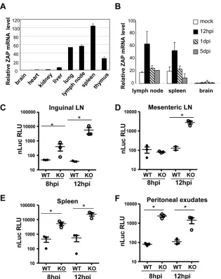
It has been documented that Sindbis virus initially replicates in the CD11c⫺CD11b⫹cells in the draining lymph nodes and spleen (11,26). In addition, ZAP is mainly expressed in lymphoid tissues, including spleen, lymph nodes, and thymus (Fig. 4A), and SVNI infection further upregulates ZAP expression (Fig. 4B). Thus, we analyzed the replication of the virus in lymphoid tissues early after infection of 23-day-old mice. For more sensitive measurement of the viral replication in these tissues, a replication-competent re-porter SVNI virus expressing nano-luciferase (SVNI-nsP3-nLuc) was constructed. Increased virus replication is expected to result in increased expression of the reporter. The results showed that in all lymphoid tissues examined, which included inguinal and mes-enteric lymph nodes, spleen, and peritoneal exudates, the reporter was expressed significantly better in the knockout mice than in the wild-type mice (Fig. 4CtoF). The difference in the reporter virus FIG 4SVNI replication was higher in the peripheral lymphoid tissues of the ZAP knockout mice than in the wild-type mice. (A) Wild-type mice (n⫽2 per genotype; 23 days old) were sacrificed and the indicated fresh tissues were collected for extraction of total RNA. ZAP levels were quantified by RT-quantitative PCR (qPCR) and normalized to GAPDH mRNA levels. Data presented are mean⫾standard deviation (SD) ZAP levels in the two animals. (B) Wild-type mice (n⫽3 per genotype; 23 days old) were mock treated or infected with SVNI by intraperitoneal injection. Fresh tissues were collected at the time points indicated for extraction of total RNA. ZAP levels were quantified by RT-qPCR and normalized to GAPDH mRNA levels. Data presented are mean⫾SD ZAP levels in three animals. (C to F) Mice (n⫽3 per genotype; 23 days old) were infected with a replication-competent reporter virus, SVNI-nsP3-nLuc. At the time points indicated, samples were collected, homogenized in lysis buffer, and clarified by centrifugation. The reporter expression in the lysate was measured with a luminometer. LN, lymph node. *,P⬍0.05.
Wang et al.
on November 7, 2019 by guest
http://jvi.asm.org/
[image:6.585.136.451.64.462.2]expression levels in the lymphoid tissues early after infection is consistent with the difference in the early serum viral titers be-tween the knockout and wild-type mice. Collectively, these results demonstrate that compared to the wild-type mice, ZAP knockout weanling mice support higher SVNI replication levels in the lym-phoid tissues early after infection, but that viral spread to or rep-lication within the central nervous system is restricted.
Higher viral titers in the periphery of ZAP knockout mice
result in higher-level immune activation.To determine whether
ZAP knockout affects the immune responses to SVNI infection, we analyzed distributions of the splenocyte subsets of both 10-day-old and 23-10-day-old mice with or without SVNI infection. In naive mice, no obvious difference between wild-type and ZAP knockout mice was observed in the total number of white blood cells (data not shown) or in the distributions of splenic lymphoid subsets (Fig. 5and6). Upon SVNI infection, the percentage of CD69⫹cells increased significantly (Fig. 5and6). CD69 expres-sion is rapidly induced in activated T and B cells, neutrophils, and NK cells (27,28). Increased percentages of CD69⫹cells indicated that SVNI infection activated the immune system. In both 10-day-old and 23-day-10-day-old mice, higher levels of CD69⫹cells were ob-served in ZAP knockout mice (Fig. 5and6), which were in parallel with the higher serum viral titers in these mice (Fig. 3AandB). These results suggest that ZAP knockout generally does not affect the immune system in naive mice or activation of the immune system upon SVNI infection.
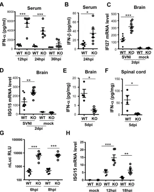
SVNI induces higher IFN levels in the peripheral tissues and higher ISG levels in the brain of weanling ZAP knockout mice.
Viral infection induces the production of type I IFNs, which play important roles in determining the outcome of the infection. We next analyzed the levels of IFN-␣and IFN-at various time points postinfection. Without SVNI infection, the serum levels of IFN-␣ and IFN-were not detectable (data not shown). In both knock-out and wild-type weanling mice, the highest levels of serum IFN-␣were detected at 12 h postinfection, and the IFN-␣levels dropped thereafter over time (Fig. 7A). At 12 h and 24 h postin-fection, the serum levels of IFN-␣in the knockout mice were significantly higher than those in the wild-type mice (Fig. 7A). In comparison, at 36 h postinfection, the serum IFN-␣levels were not significantly different (Fig. 7A). Compared with IFN-␣levels, IFN-levels were much lower in both types of mice (compareFig. 7AandB). Nonetheless, IFN-levels in the serum of knockout mice were still significantly higher than those in the wild-type mice (Fig. 7B). These results demonstrate that the serum levels of IFN-␣paralleled the serum viral titers early after infection, con-sistent with the results previously reported by others (11,29–34). These results also suggest that IFN-␣is a major type I IFN induced by SVNI infection.
We next analyzed IFN-␣levels in the central nervous system. At 2 days postinfection, IFN-␣levels in the brain from either ZAP knockout or wild-type mice were below the detection limit of the FIG 5Analyses of immune cells of 10-day-old wild-type and ZAP knockout
mice. Ten-day-old mice (n⫽4 per genotype) were mock treated or infected with SVNI by intraperitoneal injection. At 2 days postinfection, splenic cells were collected. (A) The relative proportions of T and B lymphocytes, NK cells, and activated cells were analyzed by staining with CD3, CD19, anti-NK1.1, and anti-CD69 antibodies, respectively. (B) CD3-positive cells were further analyzed using antibodies to CD4 and CD8.
FIG 6Analyses of immune cells of 23-day-old wild-type and ZAP knockout mice. Twenty-three-day-old mice (n⫽4 per genotype) were mock treated or infected with SVNI by intraperitoneal injection. At 2 days postinfection, splenic cells were collected. (A) The relative proportions of T and B lym-phocytes, NK cells, and activated immune cells were analyzed by staining with anti-CD3, anti-CD19, anti-NK1.1, and anti-CD69 antibodies, respec-tively. (B) CD3-positive cells were further analyzed using antibodies to CD4 and CD8.
on November 7, 2019 by guest
http://jvi.asm.org/
[image:7.585.42.546.67.356.2]ELISA. Thus, we measured the mRNA levels of two representative ISGs, ISG15 and IFI27, to determine to what extent the cells in the brain have been exposed to type I IFN. The results showed that the brain mRNA levels of both ISG15 and IFI27 were higher in the knockout mice (Fig. 7CandD), implying greater exposure to IFN in the brains of knockout animals. At this same time point, how-ever, the viral titers in the brain were lower in the knockout mice (Fig. 3C), suggesting that ISG expression limits viral spread to or replication in the brain. At 5 days postinfection, when the brain IFN-␣protein levels could be easily detected by the ELISA, the IFN-␣levels in both brain and spinal cord were lower in the knockout mice than in the wild-type mice (Fig. 7E andF), in parallel with the viral titers (Fig. 3D). This suggests that at this time point IFN-␣levels are a consequence of viral replication levels and that IFN is locally produced.
IFN-␣is induced mostly in cells of lymphoid origin, whereas IFN-is produced by most cell types (35). It has been reported that upon infection by intraperitoneal injection the virus first pri-marily comes in contact with peritoneal macrophages and macro-phage-derived dendritic cells (36), and that macrophage-like cells may serve as important early IFN-␣producers after alphavirus infection (26). To further probe the role of ZAP in controlling SVNI infection and the subsequent production of IFN-␣, macro-phages were isolated from peritoneal exudates and infected with the SVNI-nsP3-nLuc reporter virus. The results indicated that the reporter virus was expressed at more than 10-fold higher levels in the cells isolated from the knockout mice than in the cells from the wild-type mice (Fig. 7G). We attempted to measure IFN-␣protein levels induced by the viral infection in the culture supernatants of the cells, but the levels were too low to be detected by ELISA. As a FIG 7SVNI induces higher IFN levels in the peripheral tissues and higher ISG levels in the brain of weanling ZAP knockout mice early postinfection. (A and B) Mice (23 days old) were infected with SVNI by intraperitoneal injection, and blood was collected by retro-orbital bleeding at the time points indicated. Serum levels of IFN-␣(A) (n⫽5 per genotype) and IFN-(B) (n⫽6 per genotype) were measured by ELISA. (C and D) Mice (n⫽8 per genotype for SVNI infection; n⫽4 for mock infection; 23 days old) were infected with SVNI by intraperitoneal injection. At 2 days postinfection, the brain was collected for RT-qPCR analysis of IFI27 (C) and ISG15 (D) mRNA levels. (E and F) Mice (n⫽4 per genotype) were infected with SVNI by intraperitoneal injection. At 5 days postinfection, brain (E) and spinal cord (F) were collected and homogenized in PBS, and levels of IFN-␣were measured by ELISA. (G and H) Macrophage cells were prepared from peritoneal exudates (n⫽6 mice per genotype; 23 days old) and infected with SVNI-nsP3-nLuc reporter virus. At the time points indicated, cells were lysed and subjected to measurement of luciferase activity (G) or ISG15 mRNA levels (H). *,P⬍0.05; **,P⬍0.01; ***,P⬍0.001.
Wang et al.
on November 7, 2019 by guest
http://jvi.asm.org/
[image:8.585.137.451.64.455.2]surrogate, we again used ISG15 mRNA levels as an indicator of the type I IFN level. As expected, viral infection induced ISG15 ex-pression, and the virus-induced ISG15 levels were much higher in the macrophages isolated from the knockout mice than in cells from wild-type mice (Fig. 7H). Collectively, these results define ZAP’s important role in the modulation of SVNI replication in macrophages and thereby in limiting virus-induced production of IFN-␣in the host.
Blocking of the type I IFN receptors renders weanling ZAP knockout mice more sensitive to viral infection than the
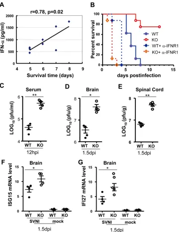
wild-type mice.The above-described results strongly suggest that SVNI
replication in the lymphoid tissues of weanling mice induces the production of IFN-␣, which restricts subsequent viral infection of the central nervous system. To substantiate this notion, we ana-lyzed the relationship between serum IFN-␣levels and disease progression in the wild-type mice. Indeed, the serum IFN-␣level was statistically proportional to the survival time of the mice (Fig. 8A), with animals having the highest IFN-␣levels also surviving the longest. To further prove that virus-induced IFN-␣plays a critical role in the protection of the central nervous system, we used a neutralizing antibody against type I IFN receptor 1 (IFNAR1) to block type I IFN action. Mice were infected with SVNI, with or without administration of the neutralizing anti-body before and during infection. Blocking of type I IFN receptor increased the susceptibility of both wild-type and knockout mice to SVNI-induced mortality (Fig. 8B), further demonstrating the importance of type I IFN in restricting SVNI infection. Consistent with the above-described results, in the absence of the antibody, knockout mice had increased survival after SVNI infection com-pared to the wild-type mice (Fig. 8B). In contrast, in the presence of the neutralizing antibody, ZAP knockout mice died much faster than the wild-type mice (Fig. 8B). To correlate the survival time and viral titers, the viral titers in the serum early after infection and in the central nervous system before animal death were measured. In the presence of the antibody, the viral titers in both serum and the central nervous system were significantly higher in the knock-out mice than in the wild-type mice (Fig. 8CtoE). The higher viral titers in the knockout mice are likely a result of the lack of restric-tion of SVNI replicarestric-tion by the basal levels of ZAP. Analysis of the mRNA levels of ISG15 and IFI27 in the brain revealed that they were higher in ZAP knockout mice (Fig. 8FandG), which paral-leled the viral titers (Fig. 8DandE), suggesting that their expres-sion was induced by the virus in the brain. These results are con-sistent with the notion that the extended survival time of ZAP knockout mice was a result of higher serum IFN-␣levels.
To further substantiate this notion, 23-day-old mice were in-fected by direct intracranial injection, and the brain viral titers were measured. The results showed that the brain viral titers of knockout mice were higher than those in the wild-type mice (Fig. 9A). Consistent with this finding, the knockout mice died faster than the wild-type mice (Fig. 9B). The differences in the brain virus titers and survival times between knockout and wild-type mice were statistically significant but the magnitudes were rela-tively small. This could be accounted for by the fact that ZAP is expressed at a very low level in the brain (Fig. 4A).
DISCUSSION
Type I IFNs play very important roles in innate immunity against viral infections. Viruses have evolved multiple mechanisms to an-tagonize or evade IFN-mediated immunity. The most commonly
reported mechanism is that the virus encodes antagonists to block the induction or to counteract the effectors of IFNs (37,38). Here, we provide evidence showing that instead of antagonizing the an-tiviral activity of ZAP, SVNI exploits ZAP in weanling mice to limit its replication in lymphoid tissues and minimize IFN induc-tion in order to evade immune surveillance for enhanced dissem-ination to the central nervous system.
When the mice were infected by intraperitoneal injection, SVNI replicated better in the lymphoid tissues of the weanling ZAP knockout mice than the wild-type mice. However, the viral titers in the central nervous system were much lower in the knock-out mice (Fig. 3). These results imply that some protective mech-anisms exist in the ZAP knockout mice. SVNI infection led to ISG15 upregulation in the lymphoid tissues of knockout mice early postinfection (Fig. 7H), and type I IFN receptor blockade abrogated ZAP-mediated protection from viral encephalitis (Fig. 8B), indicating that the protection was mediated by type I IFNs. When the mice were infected by intracranial injection, which is expected to bypass the peripheral IFN response, ZAP knockout mice died faster than wild-type mice (Fig. 9), further suggesting the protective function of peripheral IFNs induced by SVNI intra-peritoneal infection.
The antiviral activity of type I IFNs is mediated primarily by ISGs. In the presence of intact type I IFN signaling, ZAP knockout allowed macrophages and lymphoid tissues to support SVNI rep-lication to levels 10-fold higher than that seen in the wild-type tissues, indicating that ZAP plays a major role in IFN inhibition of viral replication. Nonetheless, abrogation of IFN signaling still dramatically compromises the survival of both the wild-type and knockout mice (Fig. 8B), indicating that additional antiviral mechanisms other than ZAP are in place to control SVNI replica-tion. Indeed, antiviral factors such as viperin and ISG15 have been reported to act against Sindbis virus (13,39,40).
SVNI infection led to higher mRNA levels of IFI27 and ISG15 in the brains of the knockout mice than those of the wild-type mice at 2 days postinfection (Fig. 7CandD), although IFN-␣ protein was below the detection limit. The higher levels of these ISGs are expected to better protect the brain from viral infection. Indeed, the brain viral titers of the same group of animals were much lower in the knockout mice than in the wild-type mice (Fig. 3C). We propose that the elevated ISG levels in the brain at 2 days postinfection result from higher IFN production in the peripheral tissues. Although it has been reported that the IFN protein cannot easily cross the blood-brain barrier (BBB) (41), immune cells in-fected by SVNI (dendritic cells and macrophages) could infiltrate the brain and produce IFN locally. In addition, the effects of high IFN levels and virus infection on the blood-brain barrier perme-ability are not well established, which could also play a role. At 5 days postinfection, IFN-␣protein could be easily detected in the central nervous system and the levels were much lower in the knockout mice than in the wild-type mice (Fig. 7EandF). The IFN levels paralleled the viral titers in the central nervous system at this time point, suggesting that the IFN was produced locally as a result of viral infection. Collectively, these results support the notion that in the knockout mice the virus replicated better in the periph-eral tissues and induced higher levels of type I IFN, which re-stricted viral infection in the central nervous system due to higher levels of ISG expression. Nevertheless, it is also possible that the protective role of increased IFN-␣levels in the knockout mice is at the interface of the periphery and the central nervous system,
on November 7, 2019 by guest
http://jvi.asm.org/
fecting the as-yet poorly understood process of Sindbis virus neu-roinvasion. For example, it has been reported that upon intranasal VSV instillation, early IFN response in the glomerular layer of the olfactory bulb is critically required to prevent viral spread over the entire CNS and thus confers survival (41). The mechanism by which increased early IFN-␣levels lead to better control of Sindbis virus neuroinvasive disease is an interesting area for future inves-tigation.
SVNI infection of suckling mice caused faster animal death in the knockout mice (Fig. 2B). However, SVNI infection of weanling mice resulted in better survival of the knockout mice (Fig. 2C). This difference could be caused by a few possibilities. Sindbis virus is known to be very sensitive to IFN (11). How-ever, it has been reported that in neonatal mice, although the serum IFN level is relatively high, the virus titer is much higher than that in the adult mice, implying that the IFN system of FIG 8Type I IFN receptor blockade renders ZAP knockout mice more sensitive to SVNI infection than the wild-type mice. (A) Wild-type mice (n⫽8; 23 days old) were infected with SVNI by intraperitoneal injection. At 12 h postinfection, blood was collected by retro-orbital bleeding, and serum levels of IFN-␣were measured. Disease progression and animal survival were monitored daily. The relationship between survival time and IFN-␣levels was plotted (P⬍0.05). (B) Wild-type and ZAP knockout mice (n⫽8 mice per group; 23 days old) were mock treated or treated with blocking antibody against IFNAR1 by intraperitoneal injection (at day⫺1 and 0 and 2 days relative to infection), followed by SVNI infection by intraperitoneal injection. Disease progression and animal survival were monitored daily. Data presented are representative of two independent experiments.P⬍0.001 for WT versus KO and for WT plus␣-IFNR1 versus KO plus
␣-IFNR1. (C to G) Mice (23 days old) were infected with SVNI. At days⫺1 and 0 of infection, mice were mock treated or treated with a blocking antibody against IFNAR1 (n⫽4 per group). At the times indicated, viral titers in the serum (C), brain (D), or spinal cord (E) were determined. ISG15 (F) and IFI27 (G) mRNA levels in the brain were measured by RT-qPCR. *,P⬍0.05; **,P⬍0.01.
Wang et al.
on November 7, 2019 by guest
http://jvi.asm.org/
[image:10.585.113.476.64.532.2]neonates is not very effective and does not play a major role in controlling viral replication (11,29). Second, permeability of the BBB is a critical factor that controls Sindbis virus invasion of mouse brain (42). In newborn and suckling mice, some non-neuroinvasive strains of Sindbis virus induce fatal encephalitis after intracranial or intraperitoneal inoculation, whereas in weanling mice the virus induces fatal encephalitis only after intracranial inoculation (43, 44). This age-dependent resis-tance to neuroinvasion often coincides with the maturation of the BBB (44). It is conceivable that, in weanling mice with mature BBB and relatively low serum viral titers, only a small number of the virions in the blood can enter the brain, so the virus would be controlled by the IFN in the brain. In suckling mice, which lack a developed BBB (43,44), virus in the periph-ery would have much easier access to the brain, and the rela-tively high level of the virus in the brain would not be con-trolled by the relatively low level of IFN. Further investigation is needed to better understand the age-dependent phenotypes. There is increasing evidence indicating that this is a widely used strategy for viruses to exploit host antiviral factors to evade im-mune surveillance. For example, North American eastern equine encephalitis virus, also an alphavirus, was demonstrated to make use of host microRNAs that target the viral genome to limit its replication in myeloid-lineage cells to avoid the induction of type I IFN (45). In another example, HIV-1 uses the cellular factor Trex1, which degrades the viral DNA, to evade immune detection (46). ZAP-mediated evasion of the immune surveillance by SVNI is conceptually similar to but mechanistically different from these reported processes.
In summary, we report here that in weanling mice SVNI exploits the IFN-inducible host antiviral factor ZAP to evade type I IFN-mediated innate immune surveillance. ZAP has been reported to inhibit the replication of other viruses, such as Ebola virus and HIV-1. It would be interesting to investigate the roles of ZAP in the inhibition of these viruses by IFNin vitro
andin vivo.
FUNDING INFORMATION
This work, including the efforts of Guangxia Gao, was funded by National Natural Science Foundation of China (NSFC) (81530066). This work, including the efforts of Xinlu Wang, was funded by National Natural Science Foundation of China (NSFC) (81471965). This work, including the efforts of Guangxia Gao, was funded by Ministry of Science and Technology of the People’s Republic of China (MOST) (973 Program 2012CB910203).
The funders had no role in study design, data collection and interpreta-tion, or the decision to submit the work for publication.
REFERENCES
1.Schneider WM, Chevillotte MD, Rice CM.2014. Interferon-stimulated genes: a complex web of host defenses. Annu Rev Immunol32:513–545.
http://dx.doi.org/10.1146/annurev-immunol-032713-120231.
2.Stetson DB, Medzhitov R.2006. Type I interferons in host defense. Immunity25:373–381.http://dx.doi.org/10.1016/j.immuni.2006.08.007. 3.Ryman KD, Klimstra WB.2008. Host responses to alphavirus infec-tion. Immunol Rev225:27– 45.http://dx.doi.org/10.1111/j.1600-065X .2008.00670.x.
4.Griffin DE, Mendoza QP.1986. Identification of the inflammatory cells present in the central nervous system of normal and mast cell-deficient mice during Sindbis virus encephalitis. Cell Immunol97:454 – 459.http: //dx.doi.org/10.1016/0008-8749(86)90414-4.
5.Griffin DE.1989. Molecular pathogenesis of Sindbis virus encephalitis in experimental animals. Adv Virus Res36:255–271.http://dx.doi.org/10 .1016/S0065-3527(08)60587-4.
6.Tucker PC, Strauss EG, Kuhn RJ, Strauss JH, Griffin DE.1993. Viral determinants of age-dependent virulence of Sindbis virus for mice. J Virol 67:4605– 4610.
7.Dubuisson J, Lustig S, Ruggli N, Akov Y, Rice CM. 1997. Genetic determinants of Sindbis virus neuroinvasiveness. J Virol71:2636 –2646. 8.Lustig S, Halevy M, Ben-Nathan D, Akov Y.1992. A novel variant of
Sindbis virus is both neurovirulent and neuroinvasive in adult mice. Arch Virol122:237–248.http://dx.doi.org/10.1007/BF01317186.
9.Burke CW, Gardner CL, Steffan JJ, Ryman KD, Klimstra WB.2009. Characteristics of alpha/beta interferon induction after infection of mu-rine fibroblasts with wild-type and mutant alphaviruses. Virology395: 121–132.http://dx.doi.org/10.1016/j.virol.2009.08.039.
10. Akhrymuk I, Frolov I, Frolova EI.2016. Both RIG-I and MDA5 detect alphavirus replication in concentration-dependent mode. Virology487: 230 –241.http://dx.doi.org/10.1016/j.virol.2015.09.023.
FIG 9ZAP knockout mice die faster than wild-type mice when infected with SVNI by intracranial injection. (A) Mice (23 days old,n⫽8 per genotype) were infected with SVNI by intracranial injection. At the time points indicated, brain samples were collected and viral titers were measured. *,P⬍0.01. (B) Mice (n⫽28 per genotype, 23 days old) were infected with SVNI by intracranial injection, and animal survival was monitored daily for 6 days. Mantel-Cox testing showed a statistically significant difference between genotypes (P⬍0.01). Data presented are combined from two independent experiments.
on November 7, 2019 by guest
http://jvi.asm.org/
[image:11.585.146.452.65.238.2]11. Ryman KD, Klimstra WB, Nguyen KB, Biron CA, Johnston RE.2000. Alpha/beta interferon protects adult mice from fatal Sindbis virus infec-tion and is an important determinant of cell and tissue tropism. J Virol 74:3366 –3378.http://dx.doi.org/10.1128/JVI.74.7.3366-3378.2000. 12. Schoggins JW, MacDuff DA, Imanaka N, Gainey MD, Shrestha B,
Eitson JL, Mar KB, Richardson RB, Ratushny AV, Litvak V, Dabelic R, Manicassamy B, Aitchison JD, Aderem A, Elliott RM, Garcia-Sastre A, Racaniello V, Snijder EJ, Yokoyama WM, Diamond MS, Virgin HW, Rice CM.2014. Pan-viral specificity of IFN-induced genes reveals new roles for cGAS in innate immunity. Nature505:691– 695.
13. Zhang Y, Burke CW, Ryman KD, Klimstra WB.2007. Identification and characterization of interferon-induced proteins that inhibit alphavirus repli-cation. J Virol81:11246 –11255.http://dx.doi.org/10.1128/JVI.01282-07. 14. Bick MJ, Carroll JW, Gao G, Goff SP, Rice CM, MacDonald MR.2003.
Expression of the zinc-finger antiviral protein inhibits alphavirus replica-tion. J Virol77:11555–11562.http://dx.doi.org/10.1128/JVI.77.21.11555 -11562.2003.
15. Muller S, Moller P, Bick MJ, Wurr S, Becker S, Gunther S, Kummerer BM.2007. Inhibition of filovirus replication by the zinc finger antiviral protein. J Virol81:2391–2400.http://dx.doi.org/10.1128/JVI.01601-06. 16. Zhu Y, Chen G, Lv F, Wang X, Ji X, Xu Y, Sun J, Wu L, Zheng YT, Gao
G.2011. Zinc-finger antiviral protein inhibits HIV-1 infection by selectively targeting multiply spliced viral mRNAs for degradation. Proc Natl Acad Sci U S A108:15834 –15839.http://dx.doi.org/10.1073/pnas.1101676108. 17. Mao R, Nie H, Cai D, Zhang J, Liu H, Yan R, Cuconati A, Block TM,
Guo JT, Guo H.2013. Inhibition of hepatitis B virus replication by the host zinc finger antiviral protein. PLoS Pathog9:e1003494.http://dx.doi .org/10.1371/journal.ppat.1003494.
18. Guo X, Carroll JW, Macdonald MR, Goff SP, Gao G.2004. The zinc finger antiviral protein directly binds to specific viral mRNAs through the CCCH zinc finger motifs. J Virol78:12781–12787.http://dx.doi.org/10 .1128/JVI.78.23.12781-12787.2004.
19. Zhu Y, Gao G.2008. ZAP-mediated mRNA degradation. RNA Biol5:65– 67.http://dx.doi.org/10.4161/rna.5.2.6044.
20. Zhu Y, Wang X, Goff SP, Gao G.2012. Translational repression precedes and is required for ZAP-mediated mRNA decay. EMBO J31:4236 – 4246.
http://dx.doi.org/10.1038/emboj.2012.271.
21. Todorova T, Bock FJ, Chang P.2014. PARP13 regulates cellular mRNA post-transcriptionally and functions as a pro-apoptotic factor by destabi-lizing TRAILR4 transcript. Nat Commun5:5362.http://dx.doi.org/10 .1038/ncomms6362.
22. Leung AK, Vyas S, Rood JE, Bhutkar A, Sharp PA, Chang P.2011. Poly(ADP-ribose) regulates stress responses and microRNA activity in the cytoplasm. Mol Cell42:489 – 499.http://dx.doi.org/10.1016/j.molcel.2011 .04.015.
23. Seo GJ, Kincaid RP, Phanaksri T, Burke JM, Pare JM, Cox JE, Hsiang TY, Krug RM, Sullivan CS.2013. Reciprocal inhibition between intra-cellular antiviral signaling and the RNAi machinery in mammalian cells. Cell Host Microbe14:435– 445.http://dx.doi.org/10.1016/j.chom.2013 .09.002.
24. Karki S, Li MM, Schoggins JW, Tian S, Rice CM, MacDonald MR.2012. Multiple interferon stimulated genes synergize with the zinc finger antivi-ral protein to mediate anti-alphavirus activity. PLoS One7:e37398.http: //dx.doi.org/10.1371/journal.pone.0037398.
25. Kozaki T, Takahama M, Misawa T, Matsuura Y, Akira S, Saitoh T.2015. Role of zinc-finger antiviral protein in host defense against Sindbis virus. Int Immunol27:357–364.http://dx.doi.org/10.1093/intimm/dxv010. 26. Burke CW.2009. Ph.D. thesis. The influence of cell type on alphavirus
type I interferon induction. Louisiana State University, Shreveport, LA, USA.
27. Marzio R, Mauel J, Betz-Corradin S.1999. CD69 and regulation of the immune function. Immunopharmacol Immunotoxicol21:565–582.http: //dx.doi.org/10.3109/08923979909007126.
28. Ziegler SF, Ramsdell F, Hjerrild KA, Armitage RJ, Grabstein KH, Hennen KB, Farrah T, Fanslow WC, Shevach EM, Alderson MR.
1993. Molecular characterization of the early activation antigen CD69: a type II membrane glycoprotein related to a family of natural killer cell activation antigens. Eur J Immunol23:1643–1648.http://dx.doi.org /10.1002/eji.1830230737.
29. Klimstra WB, Ryman KD, Bernard KA, Nguyen KB, Biron CA, John-ston RE.1999. Infection of neonatal mice with Sindbis virus results in a systemic inflammatory response syndrome. J Virol73:10387–10398. 30. Reinarz AB, Broome MG, Sagik BP.1971. Age-dependent resistance of
mice to Sindbis virus infection: viral replication as a function of host age. Infect Immun3:268 –273.
31. Sherman LA, Griffin DE.1990. Pathogenesis of encephalitis induced in newborn mice by virulent and avirulent strains of Sindbis virus. J Virol 64:2041–2046.
32. Trgovcich J, Aronson JF, Eldridge JC, Johnston RE.1999. TNFalpha, interferon, and stress response induction as a function of age-related sus-ceptibility to fatal Sindbis virus infection of mice. Virology263:339 –348.
http://dx.doi.org/10.1006/viro.1999.9913.
33. Trgovcich J, Aronson JF, Johnston RE.1996. Fatal Sindbis virus infection of neonatal mice in the absence of encephalitis. Virology224:73– 83.http: //dx.doi.org/10.1006/viro.1996.0508.
34. Vilcek J.1964. Production of interferon by newborn and adult mice in-fected with Sindbis virus. Virology22:651– 652.http://dx.doi.org/10.1016 /0042-6822(64)90091-1.
35. Stark GR, Kerr IM, Williams BR, Silverman RH, Schreiber RD.1998. How cells respond to interferons. Annu Rev Biochem67:227–264.http: //dx.doi.org/10.1146/annurev.biochem.67.1.227.
36. Mahanty S, Gupta M, Paragas J, Bray M, Ahmed R, Rollin PE.2003. Protection from lethal infection is determined by innate immune re-sponses in a mouse model of Ebola virus infection. Virology312:415– 424.
http://dx.doi.org/10.1016/S0042-6822(03)00233-2.
37. Malim MH, Bieniasz PD.2012. HIV restriction factors and mechanisms of evasion. Cold Spring Harb Perspect Med2:a006940.
38. Wang BX, Fish EN.2012. The yin and yang of viruses and interferons. Trends Immunol33:190 –197.http://dx.doi.org/10.1016/j.it.2012.01.004. 39. Lenschow DJ, Giannakopoulos NV, Gunn LJ, Johnston C, O’Guin AK, Schmidt RE, Levine B, Virgin HWT. 2005. Identification of interferon-stimulated gene 15 as an antiviral molecule during Sindbis virus infection in vivo. J Virol79:13974 –13983.http://dx.doi.org/10 .1128/JVI.79.22.13974-13983.2005.
40. Lenschow DJ, Lai C, Frias-Staheli N, Giannakopoulos NV, Lutz A, Wolff T, Osiak A, Levine B, Schmidt RE, Garcia-Sastre A, Leib DA, Pekosz A, Knobeloch KP, Horak I, Virgin HWT.2007. IFN-stimulated gene 15 functions as a critical antiviral molecule against influenza, herpes, and Sindbis viruses. Proc Natl Acad Sci U S A104:1371–1376.http://dx .doi.org/10.1073/pnas.0607038104.
41. Detje CN, Meyer T, Schmidt H, Kreuz D, Rose JK, Bechmann I, Prinz M, Kalinke U.2009. Local type I IFN receptor signaling protects against virus spread within the central nervous system. J Immunol182:2297– 2304.http://dx.doi.org/10.4049/jimmunol.0800596.
42. Lustig S, Halevy M, Ben-Nathan D, Rice CM, Kobiler D.1999. The role of host immunocompetence in neuroinvasion of Sindbis virus. Arch Virol 144:1159 –1171.http://dx.doi.org/10.1007/s007050050576.
43. Johnson RT.1965. Virus invasion of the central nervous system: a study of Sindbis virus infection in the mouse using fluorescent antibody. Am J Pathol46:929 –943.
44. Kobiler D, Lustig S, Shapira S (ed). 2001. Blood brain barrier: drug delivery and brain pathology, 1st ed. Springer, New York, NY.
45. Trobaugh DW, Gardner CL, Sun C, Haddow AD, Wang E, Chapnik E, Mildner A, Weaver SC, Ryman KD, Klimstra WB.2014. RNA viruses can hijack vertebrate microRNAs to suppress innate immunity. Nature 506:245–248.
46. Luban J.2012. Innate immune sensing of HIV-1 by dendritic cells. Cell Host Microbe12:408 – 418.http://dx.doi.org/10.1016/j.chom.2012.10 .002.
Wang et al.