Inhibition of the Membrane Attack Complex by Dengue Virus NS1
through Interaction with Vitronectin and Terminal Complement
Proteins
Jonas Nascimento Conde,aEmiliana Mandarano da Silva,aDiego Allonso,aDiego Rodrigues Coelho,aIamara da Silva Andrade,a,d Luciano Neves de Medeiros,bJoice Lima Menezes,aAngela Silva Barbosa,cRonaldo Mohana-Borgesa
Laboratório de Genômica Estrutural, Instituto de Biofísica Carlos Chagas Filho, Universidade Federal do Rio de Janeiro, Rio de Janeiro, RJ, Brazila; Laboratório de Biologia Molecular e Bioquímica de Proteínas, Instituto de Biofísica Carlos Chagas Filho, Universidade Federal do Rio de Janeiro, Rio de Janeiro, RJ, Brazilb; Laboratório de Bacteriologia, Instituto Butantan, São Paulo, SP, Brazilc; Faculdade de Educação Tecnológica do Estado do Rio de Janeiro-FAETERJ (FAETEC), Paracambi, RJ, Brazild
ABSTRACT
Dengue virus (DENV) infects millions of people worldwide and is a major public health problem. DENV nonstructural protein 1
(NS1) is a conserved glycoprotein that associates with membranes and is also secreted into the plasma in DENV-infected
pa-tients. The present study describes a novel mechanism by which NS1 inhibits the terminal complement pathway. We first
identi-fied the terminal complement regulator vitronectin (VN) as a novel DENV2 NS1 binding partner by using a yeast two-hybrid
system. This interaction was further assessed by enzyme-linked immunosorbent assay (ELISA) and surface plasmon resonance
(SPR) assay. The NS1-VN complex was also detected in plasmas from DENV-infected patients, suggesting that this interaction
occurs during DENV infection. We also demonstrated that the DENV2 NS1 protein, either by itself or by interacting with VN,
hinders the formation of the membrane attack complex (MAC) and C9 polymerization. Finally, we showed that DENV2, West
Nile virus (WNV), and Zika virus (ZIKV) NS1 proteins produced in mammalian cells inhibited C9 polymerization. Taken
to-gether, our results points to a role for NS1 as a terminal pathway inhibitor of the complement system.
IMPORTANCE
Dengue is the most important arthropod-borne viral disease nowadays and is caused by dengue virus (DENV). The flavivirus
NS1 glycoprotein has been characterized functionally as a complement evasion protein that can attenuate the activation of the
classical, lectin, and alternative pathways. The present study describes a novel mechanism by which DENV NS1 inhibits the
ter-minal complement pathway. We identified the terter-minal complement regulator vitronectin (VN) as a novel DENV NS1 binding
partner, and the NS1-VN complex was detected in plasmas from DENV-infected patients, suggesting that this interaction occurs
during DENV infection. We also demonstrated that the NS1-VN complex inhibited membrane attack complex (MAC)
forma-tion, thus interfering with the complement terminal pathway. Interestingly, NS1 itself also inhibited MAC activity, suggesting a
direct role of this protein in the inhibition process. Our findings imply a role for NS1 as a terminal pathway inhibitor of the
com-plement system.
D
engue constitutes a major public health problem in tropical
and subtropical countries. According to current estimates, at
least 390 million cases of dengue occur annually, of which
approx-imately 100 million are symptomatic (
1
). The infection is caused
by dengue virus (DENV), a member of the
Flaviviridae
family that
cocirculates in nature as four distinct antigenic serotypes (DENV1
to -4). DENV infection in humans is generally asymptomatic, but
symptomatic cases can vary from a mild and self-limited fever to a
potentially fatal hemorrhagic syndrome (
2
). The DENV genome is
composed of a single positive-sense RNA that encodes a single
viral polyprotein that is further processed by viral and host
pro-teases into three structural proteins (C, prM/M, and E) and seven
nonstructural proteins (NS1, NS2A, NS2B, NS3, NS4A, NS4B,
and NS5) (
3
).
NS1 is the first nonstructural protein to be translated and is
essential to virus replication (
4
,
5
). It is a conserved N-linked
glycoprotein with a variable molecular mass of 46 to 55 kDa,
which depends on its glycosylation status, and it is composed of
three distinct structural domains: the

-roll, wing, and

-ladder
(
6
). The NS1 protein can be found as a dimer associated with
vesicular compartments within the cell, where it plays an
impor-tant role as an essential cofactor in the virus replication process
(
4
). Alternatively, NS1 can be secreted into the extracellular space
as a hexameric lipoprotein particle (
7
) that interacts with several
plasma proteins (
8
,
9
) and activates the Toll-like receptor 4
(TLR4) response (
10
). Furthermore, secreted flavivirus NS1 has
been characterized functionally as a complement immune evasion
protein that can attenuate the activation of the classical, lectin, and
alternative pathways by interacting with complement proteins
and their regulators (
11–13
).
Vitronectin (VN) is a multifunctional glycoprotein present in
the extracellular matrix and in the plasma. It consists of an
N-ter-Received10 May 2016Accepted2 August 2016
Accepted manuscript posted online10 August 2016
CitationConde JN, da Silva EM, Allonso D, Coelho DR, Andrade IS, de Medeiros
LN, Menezes JL, Barbosa AS, Mohana-Borges R. 2016. Inhibition of the membrane attack complex by dengue virus NS1 through interaction with vitronectin and terminal complement proteins. J Virol 90:9570 –9581.doi:10.1128/JVI.00912-16.
Editor:M. S. Diamond, Washington University School of Medicine
Address correspondence to Ronaldo Mohana-Borges, [email protected].
Copyright © 2016, American Society for Microbiology. All Rights Reserved.
on November 7, 2019 by guest
http://jvi.asm.org/
minal somatomedin-B domain (SMB), an RGD cell receptor
binding site, four hemopexin (HPX)-like domains, and three
hep-arin-binding domains (HBDs) (
14
). VN is a complement
regula-tor that inhibits the formation of the membrane attack complex
(MAC) by occupying the metastable membrane-binding site of
the C5b7 complex and hindering its insertion into the cell
mem-brane, thus preventing the completion of the C5b9 lytic pore (
15
).
In addition, binding of C8 and C9 to the C5b7-VN complex leads
to the formation of soluble C5b9 (SC5b9), which is hemolytically
inactive. Furthermore, VN limits ongoing membrane-associated
pore formation by inhibiting C9 polymerization (
15–17
).
Acqui-sition of soluble human VN on the surfaces of microbial
patho-gens is a common complement evasion strategy that has been
described for many bacterial pathogens (
14
,
18
); however, this
particular evasion mechanism has never been evaluated in viral
pathogens.
In this work, we identified human VN as a novel DENV2 NS1
binding partner by using a yeast two-hybrid (Y2H) system. This
interaction was further assessed by biochemical, biophysical, and
immunoenzymatic approaches that confirmed the direct
associa-tion between NS1 and VN. Moreover, we demonstrated that the
NS1 protein from DENV2, West Nile virus (WNV), or Zika virus
(ZIKV), by itself or in association with VN, is capable of inhibiting
MAC formation and C9 polymerization. These results suggest a
role for the NS1 protein as a terminal pathway inhibitor of the
complement system.
MATERIALS AND METHODS
Proteins and antibodies.Human vitronectin (VN), human hemopexin (HPX), bovine serum albumin (BSA), and heparin were purchased from Sigma-Aldrich. Human plasminogen (PLG) was purchased from Milli-pore. Recombinant active plasminogen activator inhibitor 1 (PAI-1) was kindly provided by Cynthia Peterson (University of Tennessee, TN), and the purified complement proteins C2, C5, C6, C7, C8, C9, and C5b6, factor H, and normal human serum (NHS) were purchased from Com-plement Technology.
Hexameric complex-type glycosylated NS1 proteins (NS1mam) from DENV2 (strain Thailand/16681/84), WNV (strain New York NY99), and ZIKV (strain Uganda MR 766), expressed in HEK 293 cells, were pur-chased from The Native Antigen (Oxfordshire, United Kingdom).
Polyclonal antiserum against purified NS1 was raised in mice and rabbits and purified using protein G HP Spin Trap columns (GE Health-care) (19,20). Mouse anti-human VN and PLG monoclonal antibodies were purchased from Abcam, and goat anti-human C2, C5, C6, C7, C8, and C9 and rabbit anti-VN were purchased from Complement Technol-ogy. Anti-mouse and anti-goat IgGs conjugated to horseradish peroxidase were purchased from Promega.
Production of NS1 protein.Recombinant dimeric NS1 was expressed inEscherichia coli(NS1e). Briefly, the DENV2ns1gene from the New Guinea strain was inserted into the pET23b plasmid by GenScript (CA). A nucleotide sequence encoding a 6⫻His tag was added at the 3= end of thens1gene. The recombinant plasmids were inserted intoE. coli BL21(DE3)pLysS cells, and the expression and purification steps were performed as described previously (19). Hexameric polymannose-glyco-sylated NS1 (NS1dros) was produced and isolated from the supernatant of a DENV2 NS1-expressingDrosophilaSchneider 2 (S2) stable cell line. S2 cells were cultured in Schneider’sDrosophilamedium (Gibco) for 5 days at 28°C, and NS1drosexpression was induced with 500 mM ZnCl2in se-rum-free Sf-900 II SFM medium (Gibco). The supernatant was harvested, and NS1droswas purified using size exclusion chromatography on a Su-perdex 200 column (GE Healthcare).
Y2H screening.Yeast two-hybrid (Y2H) screening was performed against a human liver cDNA library of sequences fused to the GAL4
acti-vation domain by use of the pACT2 vector (Clontech, CA).Saccharomyces cerevisiaestrain AH109 was transformed with the pGBKT7-NS1 plasmid, which encoded full-length DENV2 NS1 as bait, by using the lithium ace-tate method and then was grown in synthetic defined medium lacking tryptophan (SD–Trp). Autoactivation of theHIS3reporter gene was ver-ified by the growth of colonies in SD medium lacking histidine, leucine, and tryptophan (SD–His–Leu–Trp). The transformed cultures were then plated onto SD–His–Leu–Trp and SD–Ade–His–Leu–Trp media to select putative positive clones. The activity of thelacZreporter gene was evalu-ated by a-galactosidase assay (colony lift filter assay), using the substrate X-Gal (5-bromo-4-chloro-3-indolyl--D-galactopyranoside) on
nitro-cellulose membranes. To eliminate false-positive results, a plasmid link-age assay was also performed. The positive plasmids were sequenced, and their gene sequences were analyzed using the BLASTN and BLASTX soft-ware available at the NCBI website (www.ncbi.nlm.nih.gov).
Coimmunoprecipitation.Human hepatocellular carcinoma (HuH-7.5.1) cell lines were cultured in high-glucose Dulbecco’s modified Eagle’s medium (DMEM) (Gibco) supplemented with 10% fetal bovine serum (Invitrogen, NY), 2 mML-glutamine (Gibco), penicillin and streptomycin
(Gibco), 0.22% sodium bicarbonate, and 0.2% HEPES, pH 7.4, in a hu-midified CO2incubation chamber at 37°C. After 2 days, HuH-7.5.1 cells were mock infected or infected with DENV2 strain 16681 at a multiplicity of infection (MOI) of 1. The HuH-7.5.1 culture supernatants were col-lected at 48 h postinfection. The coimmunoprecipitation assays were per-formed using a Pierce coimmunoprecipitation kit (Pierce). Purified anti-NS1 polyclonal antibody was attached to an N-terminus-binding resin, and the supernatant (S) of mock-infected or DENV2-infected HuH-7.5.1 cells was added to the resin. The coimmunoprecipitation flowthrough (F) and eluted (E) samples were separated by 12% SDS-PAGE and transferred to a Hybond ECL nitrocellulose membrane (GE Healthcare). The mem-brane was then blocked with 5% nonfat milk in TBST (0.1% Tween 20 in Tris-buffered saline [25 mM Tris-HCl, pH 7.6, 3 mM KCl, and 140 mM NaCl]) for 2 h, followed by overnight incubation with an anti-NS1 or anti-VN antibody in blocking solution. The membrane was then washed three times with TBST and incubated with horseradish peroxidase-con-jugated anti-mouse IgG in blocking solution for 2 h. The membrane was washed again and developed with a Supersignal West Pico kit (Pierce, IL), and the image was acquired using ImageQuant LAS-4000 (GE Health-care).
Direct binding assays.Microtiter plates were coated with 100 nM VN, PAI-1, HPX, or BSA in phosphate-buffered saline (PBS; 8.06 mM Na2HPO4, 1.94 mM KH2PO4, 2.7 mM KCl, and 137 mM NaCl; pH 7.4) overnight at 4°C. After five washes with PBS, the wells were blocked with 200l of 2% BSA in PBS containing 0.05% Tween 20 (PBST) for 1 h at 37°C, followed by five washes with PBST. NS1eat specific concentrations was added to each well and incubated for 2 h at 37°C, followed by a 1-h incubation with anti-NS1. Peroxidase-conjugated anti-mouse IgG was then added for 1 h at 37°C. The plates were washed five times with PBST between each step.
To investigate the effect of heparin or PAI-1 on the NS1-VN interac-tion, the binding buffer was supplemented with 0 to 500g/ml heparin or with 0 to 5g/ml PAI-1, and bound NS1e(10 or 1g/ml, respectively) was detected using NS1 followed by a peroxidase-conjugated anti-body. To investigate the effect of PLG on NS1ebinding to immobilized VN, a mixture with a constant amount of NS1e(100 nM) and PLG at different molar ratios was added to VN-coated wells (100 nM). Similarly, NS1ewas used at different molar concentrations combined with a con-stant amount of PLG (100 nM) and added to immobilized VN. Bound NS1 and PLG were detected using anti-NS1 and anti-PLG, respectively, followed by the addition of peroxidase-conjugated antibodies.
To assess binding of complement components to NS1e-coated wells (10g/ml), the proteins C2, C5, C6, C7, C8, and C9 (0 to 1,000 nM [each]) were incubated for 1 h at 37°C. Bound components were detected using specific polyclonal antibodies followed by a peroxidase-conjugated anti-goat IgG antibody. To investigate whether C9 would bind to DENV2,
on November 7, 2019 by guest
http://jvi.asm.org/
WNV, or ZIKV NS1mam-coated wells (10g/ml), C9 (0 to 500 nM) was added to each well and incubated for 1 h at 37°C. Wells were then washed with PBS and incubated with a polyclonal anti-C9 antibody followed by incubation with a peroxidase-conjugated anti-goat IgG antibody for 1 h at 37°C. The reactions from all binding experiments were developed by add-ing 100l of 0.4 mg/mlo-phenylenediamine and 50l of 9 N H2SO4. The optical density at 490 nm (OD490) was determined by use of a 96-well plate reader.
SPR measurements. Surface plasmon resonance (SPR) measure-ments were performed using a Biacore X instrument (GE Healthcare). VN was covalently immobilized to the dextran matrix of a CM5 sensor chip via the primary amine groups (amine coupling kit; GE Healthcare). The carboxymethylated dextran surface was activated by the injection of a mixture of 0.2 MN-ethyl-N=-(diethylamino-propyl)carbodiimide and 0.05 MN-hydroxysuccinimide. The ligand was injected in 10 mM sodium acetate buffer (pH 4.0). The remainingN-hydroxysuccinimide esters were blocked by injection of 1 M ethanolamine hydrochloride (pH 8.5). All immobilization steps were performed at a flow rate of 10l/min in 10 mM HEPES, pH 7.4, 150 mM NaCl, 3 mM EDTA, and 0.005% P20 (HBS-EP; GE Healthcare). The immobilization level for VN was 8,500 response units (RU). No protein was immobilized on the control flow cell that underwent the activation and blocking steps. Binding experiments were performed at 25°C at a flow rate of 15l/min in HBS-EP. The NS1e protein was injected at different concentrations ranging from 0.12M to 3.0M. The data were double referenced by subtraction of the control flow cell signal and a blank run (buffer only) signal. In all experiments, association phases ran for 120 s and dissociation phases for 280 s. The surface was regenerated with pulses of 0.01 M glycine (pH 2.0). The data were analyzed by global fitting to a 1:1 Langmuir binding model of both the association and dissociation phases for several concentrations simul-taneously, using BIAevaluation 4.1 software (BIAcore). In each case, the data were obtained with a2value of⬍2. The apparent equilibrium dis-sociation constants (KD) were calculated from the ratio of the dissociation
and association rate constants (kd/ka).
Sandwich enzyme-linked immunosorbent assay (ELISA) with plas-mas from DENV-infected patients.Wells were coated at 4°C overnight with purified anti-NS1 rabbit polyclonal antibodies (20g/ml) in PBS. The plates were blocked for 1 h at 37°C with 2% BSA in PBST and then washed five times with PBST. This step was performed after each period of incubation. The wells were incubated with plasmas from DENV-infected patients or healthy blood donors (HD), diluted 1:2 in PBS, for 4 h at 37°C. Subsequently, the wells were incubated with an anti-VN monoclonal an-tibody (1:10,000) for 1 h at 37°C, followed by incubation for 1 h at 37°C with a peroxidase-conjugated anti-mouse IgG antibody (1:10,000). The reactions were developed as described above. Each sample was assayed twice.
The use of human samples was approved by the Research Ethics Com-mittee for Clinical Studies of the University Hospital Clementino Fraga Filho, Federal University of Rio de Janeiro (UFRJ), and registered with the Brazilian Human Ethics Committee (Plataforma Brasil, CAAE; protocol 00534012.6.0000.5257; expiration date, 3 June 2017). Informed consent was not obtained because the patient records and information were ano-nymized and deidentified prior to analysis.
Hemolytic assay.The MAC-inhibitory activity of NS1 and VN was analyzed in a hemolytic assay using sheep erythrocytes and purified com-plement proteins (18). Erythrocytes were resuspended to 1⫻108cells/ml in Veronal-buffered saline (VBS) and preincubated with 1g/ml C5b6 for 1 h at room temperature. In a separate preparation, a mixture of purified NS1e(12.5g/ml) and VN (20g/ml), NS1e(12.5g/ml) only, or VN (20 g/ml) only was preincubated for 1 h at 37°C, and the complement pro-teins C7 (1g/ml), C8 (0.1g/ml), and C9 (1g/ml) (C7–9) were added to the mixture for 15 min at 37°C. Thereafter, the C5b6-coated erythro-cytes were added to the NS1e-VN-C7–9 mixture and incubated for 30 min at 37°C. Alternatively, increased amounts of NS1eor NS1dros(6.25, 12.5, and 25g/ml) were preincubated with the C7–9 proteins for 15 min at
37°C before incubation with C5b6-coated erythrocytes. Erythrocytes were centrifuged, and the amount of hemoglobin released from the lysed cells was measured by determining the absorbance at 540 nm. The relative MAC-inhibitory activity was presented as the percentage of total hemo-lysis. NS1 buffer and factor H (50g/ml) were used as negative controls.
C9 polymerization assay.The effects of NS1eand NS1droson C9 po-lymerization were assessed according to a previously published protocol (21). Briefly, NS1e(10 to 100g/ml), NS1dros(10 to 100g/ml), or NS1 buffer was preincubated with 3g of C9 at 37°C in 20 mM Tris-HCl (pH 7.2). After 40 min of incubation, 50M ZnCl2in 20 mM Tris-HCl (pH 7.2) was added for 2 h at 37°C. In a separate experiment, NS1e(10 or 25 g/ml) was preincubated with VN (10 or 25g/ml) for 30 min at 37°C in 20 mM Tris-HCl (pH 7.2), and 3g of C9 was added for 40 min at 37°C. Polymerization was induced by addition of 50M ZnCl2in 20 mM Tris-HCl (pH 7.2) for 2 h at 37°C. The samples were separated in precast 4 to 20% gradient polyacrylamide gels (Bio-Rad) under nonreducing condi-tions, and C9 polymerization was visualized by silver staining. The C9 monomer band intensity was evaluated by densitometry using ImageJ software, version 1.49 (National Institutes of Health).
The effect of NS1mamon C9 polymerization was assessed by using proteins from DENV2, WNV, and ZIKV. Initially, BHK-21 cells were cultivated in a 24-well microplate in alpha minimal essential medium (␣-MEM; Gibco) supplemented with 10% fetal bovine serum, 0.22% so-dium bicarbonate, and 0.2% HEPES, pH 7.4, in a humidified CO2 incu-bation chamber at 37°C. After 2 days, cells were washed three times with VBS and preincubated with 5g/ml purified C5b6 for 2 h at 37°C in VBS. The NS1mamprotein from DENV2, WNV, or ZIKV was preincubated with 5% NHS for 30 min at 37°C. Rabbit polyclonal anti-NS1 (50g/ml), which cross-reacts with the WNV and ZIKV NS1 proteins (data not shown), was mixed with 50g/ml NS1mamto serve as a control. Cells were washed with VBS three times and then incubated with NS1mammixed with NHS for 50 min at 37°C. Thereafter, cells were washed three times with VBS and lysed with cell lysis buffer (20 mM Tris-HCl, pH 7.4, 150 mM NaCl, 1 mM EDTA, 1% NP-40). The samples were separated in a 10% polyacrylamide gel under nonreducing conditions and then trans-ferred to a 0.45-m Immobilon-P polyvinylidene difluoride (PVDF) membrane (GE Healthcare) at 15 V for 16 h. The membrane was blocked with 5% nonfat milk in TBST for 2 h, followed by overnight incubation with an anti-C9 antibody in blocking solution. The membrane was then washed three times with TBST and incubated with anti-goat IgG conju-gated to horseradish peroxidase in blocking solution for 2 h. The mem-brane was washed again and developed with a Supersignal West Pico kit (Pierce, IL), and the image was acquired using ImageQuant LAS-4000 (GE Healthcare).
To evaluate the effect of VN preincubation with NS1mamon C9 po-lymerization, VN (0 to 25g/ml) was preincubated with the NS1mam protein from DENV2, WNV, or ZIKV (25g/ml) for 30 min at 37°C, incubated with 5% NHS for 30 min at 37°C, and added to C5b6-treated BHK-21 cells for 50 min at 37°C. Thereafter, cells were washed three times with VBS and lysed with cell lysis buffer, and the C9 polymer was analyzed by Western blotting using anti-C9.
Statistical analysis.The data sets were compared using two-tailed, unpaired Student’sttest, and statistical significance was achieved whenP values were⬍0.05. Multiple comparisons were performed using one-way or two-way analysis of variance (ANOVA) (with the Bonferroni posttest), and asterisks in figures indicate significant differences from the control, as indicated in the legends.
RESULTS
Identification of VN as an NS1-interacting partner.
Using
full-length NS1 as bait, we performed a Y2H screen of a human liver
cDNA library as previously described by our group (
8
). Putative
DENV NS1 protein-interacting partners were identified by
acti-vation of the
HIS3
and
ADE2
reporter genes, and the authenticity
of the interaction was confirmed by colony lift filter assays of
on November 7, 2019 by guest
http://jvi.asm.org/
itive clones. True positive transformants activated the

-galacto-sidase (
lacZ
) reporter gene and appeared blue, whereas
false-pos-itive clones remained colorless (
Fig. 1A
). One positive clone
identified by the colony lift assay was subjected to nucleotide
se-quencing and identified as carrying the human VN gene.
To assess whether this interaction occurs in DENV-infected
cells, we first infected HuH7.5.1 cells, which have been shown to
express VN (
22
), with DENV2 strain 16681 at an MOI of 1 for
48 h. The mock- or DENV2-infected HuH7.5.1 cell supernatants
(S) were then loaded onto a column containing anti-NS1
poly-clonal antibodies immobilized on a resin to collect all
coimmuno-precipitated proteins. The elution fractions (E) were then
ana-lyzed using anti-NS1 and anti-VN polyclonal antibodies. We
observed two bands, of 75 and 65 kDa, corresponding to VN (
Fig.
1B
, upper panel) and one band, of approximately 50 kDa,
corre-sponding to DENV2 NS1 (
Fig. 1B
, lower panel). This result
indi-cates that VN was coimmunoprecipitated with the NS1 protein.
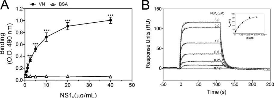
DENV NS1 directly binds VN.
To assess whether NS1 directly
interacts with VN, purified human VN was immobilized onto
microtiter plates, and serial dilutions of recombinant dimeric
NS1
e(
19
) were added. NS1
einteracted with VN in a
dose-depen-dent manner, whereas no binding was observed with BSA, used as
a negative control (
Fig. 2A
). This interaction was further
con-firmed by SPR analysis, in which VN was immobilized on a CM5
sensor chip dextran surface and the NS1
eprotein was injected at
different concentrations, ranging from 0.12
M to 3.0
M. A
concentration-dependent binding of NS1
eto immobilized VN
was observed (
Fig. 2B
). An evaluation of the global rates of
asso-ciation and dissoasso-ciation (
k
aand
k
d, respectively) revealed a
k
aof
4.23
⫻
10
4⫾
0.23
⫻
10
4M
⫺1s
⫺1and a
k
d
of 6.45
⫻
10
⫺2⫾
0.92
⫻
10
⫺2s
⫺1. The global dissociation equilibrium constant (
K
D
) was
1.52
⫾
0.17
M, assuming a simple 1:1 Langmuir binding mode.
Taken together, the ELISA and SPR data indicate that DENV2
NS1 directly binds VN.
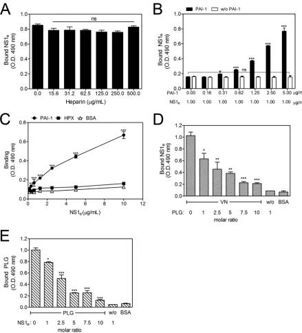
DENV NS1-VN interaction is increased by PAI-1 and
inhib-ited by plasminogen.
VN can bind several molecules, such as
hep-arin, PAI-1, HPX, and PLG, and these interactions occur at
dis-tinct sites in the VN molecule (
23–27
). Therefore, to identify the
FIG 1DENV2 NS1 interacts with vitronectin (VN) in yeast and mammalian cells. (A) Transformants carrying the bait (pGBKT7-NS1) and prey (pACT2-VN) plasmids were visualized by their growth on double (SD–Leu–Trp; column 1), triple (SD–His–Leu–Trp; column 2) and quadruple (SD–Ade–His–Leu–Trp; column 3) dropout media and by blue staining in the colony lift filter assay (column 4), indicating yeast growth andHIS3,ADE2, andlacZreporter gene activation, respectively. AH109 yeast cells cotransformed with the plasmids pGBKT7-53 (encoding murine p53 fused to the GAL4 DNA-binding domain) and pGADT7-T (encoding the simian virus 40 [SV40] large T antigen fused to the GAL4 activation domain) served as the positive control (C⫹). AH109 cotrans-formed with the plasmids pGBKT7-NS1 and pGADT7-AD served as the negative control (NC). (B) Supernatants from mock- or DENV2-infected HuH7.5.1 cells were immunoprecipitated with purified anti-NS1 polyclonal antibody. The supernatant (S), flowthrough (F), and eluted (E) fractions were analyzed by Western blotting with anti-VN and anti-NS1 polyclonal antibodies. Purified VN protein and DENVsupwere used as band mass controls (C). The presence of two bands,
of 75 and 65 kDa, corresponding to human VN was observed in the elution fraction of the DENV2 samples.
FIG 2DENV2 NS1 directly binds human vitronectin (VN). (A) Microtiter plates were coated with purified VN (100 nM), and increasing amounts of recombinant DENV2 NS1ewere added. Bound NS1ewas detected using an anti-NS1 polyclonal antibody. BSA was used as the negative control. Error bars
indicate standard deviations for three independent experiments, and asterisks indicate significant differences from the control based on two-way ANOVA and the Bonferroni posttest. ***,P⬍0.001. (B) Replicates (black solid lines) and 1:1 fitting (gray dashed lines) of the binding of recombinant DENV2 NS1eto
immobilized VN as analyzed by surface plasmon resonance analysis. Increasing concentrations of NS1e(0.12 to 3.0M) were injected onto a VN-coated CM5
sensor chip. The amount of NS1 associated with VN was measured in response units (RU). (Inset) The equilibrium response units (Req) obtained for binding of
NS1 to immobilized VN were plotted against the concentration of NS1e.
on November 7, 2019 by guest
http://jvi.asm.org/
[image:4.585.140.450.66.146.2] [image:4.585.65.520.492.655.2]putative binding sites of NS1 on the VN molecule, competitive
ELISAs were performed in which VN ligands were used as
com-petitors (
28
). VN-coated wells were incubated with fixed
concen-trations of NS1
eand various concentrations of heparin (15.6 to
500.0
g/ml) or PAI-1 (0.16 to 5.00
g/ml). The addition of
in-creasing amounts of heparin did not affect binding (
Fig. 3A
),
sug-gesting that the NS1-VN interaction does not occur through the
HBDs. However, when PAI-1 was coincubated with NS1
e, an
un-expected dose-dependent increase in the binding of NS1 to VN
was observed (
Fig. 3B
), thus indicating that PAI-1 facilitates
FIG 3The NS1-VN interaction is affected by PAI-1 and PLG but not by heparin. Microtiter plates were coated with VN (100 nM), and the effects of increasing concentrations of heparin (A) and PAI-1 (B) on the binding of NS1eto VN were analyzed. Bound NS1ewas detected using an anti-NS1 polyclonal antibody.
One-way ANOVA and the Bonferroni posttest were performed to calculate the significance of differences compared to the binding without heparin or PAI-1. (C) DENV NS1 directly interacts with PAI-1 but does not interact with HPX. Microtiter plates were coated with purified PAI-1 (100 nM) or HPX (100 nM), and increasing amounts of NS1ewere added. Bound NS1ewas detected using an anti-NS1 polyclonal antibody. BSA was used as the negative control. Two-way
ANOVA and the Bonferroni posttest were used to calculate the significance of differences. (D and E) NS1 and PLG compete for VN binding. VN was immobilized on microtiter plates, and a constant amount of NS1e(100 nM) together with increasing amounts of PLG (D) or a constant amount of PLG (100 nM) with
increasing amounts of NS1e(E) was added. Bound NS1eand PLG were detected using specific antibodies. One-way ANOVA and the Bonferroni posttest were
used to calculate the significance of differences compared to the binding without NS1 or PLG. Error bars indicate standard deviations for three independent experiments, and the asterisks indicate significant differences from the control. ns, not significant; *,P⬍0.05; **,P⬍0.01; ***,P⬍0.001.
on November 7, 2019 by guest
http://jvi.asm.org/
[image:5.585.70.509.64.548.2]VN-NS1 interactions. To evaluate whether NS1 directly interacts
with PAI-1, purified human PAI-1 was immobilized and
incu-bated with increasing concentrations of NS1
e. A dose-dependent
interaction was observed, thus confirming direct binding between
these molecules (
Fig. 3C
).
A canonical feature of the VN structure is the homology with
hemopexin-type repeats found in human HPX. Computationally
predicted models indicate that the VN central domain adopts a
full four-bladed

-propeller fold similar to that described for HPX
(
29
). Moreover, both HPX and VN hemopexin domains are
rec-ognized by surface proteins of
Streptococcus pyogenes
, and these
interactions are important for bacterial attachment (
30
). Based on
these findings, we assessed whether NS1 interacts directly with
HPX or, alternatively, if NS1 competes with HPX for binding to
VN. First, HPX-coated wells were incubated with increasing
con-centrations of NS1
e. No significant binding was observed (
Fig.
3C
). In a competitive binding assay in which VN-coated wells
were incubated with fixed concentrations of NS1
eand increasing
concentrations of HPX (1.8 to 30.0
g/ml), the addition of HPX
did not alter the NS1-VN interaction (data not shown). These two
assays indicate that NS1 does not bind HPX and does not interact
with VN via the hemopexin domains.
Because PLG binds VN, we hypothesized that PLG-binding
sites on VN may overlap the NS1-binding sites. A constant
amount of NS1
e(100 nM) with increasing amounts of PLG or a
constant amount of PLG (100 nM) with increasing amounts of
NS1
ewas added to immobilized VN. NS1 binding to VN was
affected in the presence of PLG and was reduced
⬃
80% at a
10-fold molar excess (
Fig. 3D
). Similarly, the presence of increasing
concentrations of NS1 affected the binding of PLG to immobilized
VN (
Fig. 3E
). Taken together, these results indicate that NS1 and
PLG have overlapping binding sites on VN.
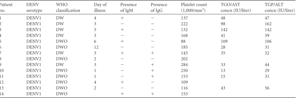
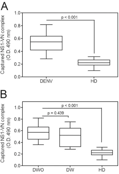
Identification of the NS1-VN complex in plasmas from
DENV-infected patients.
During DENV infection, the NS1
pro-tein is found at high concentrations in the sera of infected patients
(
31
,
32
). Because alterations in vascular homeostasis are the basis
of dengue pathophysiology and severity, we evaluated whether
NS1 interacts with circulating VN in the plasma in
DENV-in-fected patients. A capture ELISA probed with an anti-VN
mono-clonal antibody was performed using a plasma panel composed of
14 samples from DENV-infected patients (
Table 1
) and 40
sam-ples from healthy blood donors (HD). All DENV-positive samsam-ples
were from DENV1 infections and were NS1 positive according to
the Platelia Dengue NS1 Ag assay. The captured NS1-VN complex
was significantly increased in DENV-positive samples compared
to HD samples (
P
⬍
0.001) (
Fig. 4A
), clearly suggesting that this
interaction occurs during infection in the plasma. We then
evalu-ated whether this complex formation correlates with disease
se-verity. DENV-positive samples were divided according to the new
dengue classification guidelines (
2
), into samples from cases of
dengue without warning signs (DWO;
n
⫽
8) and those from cases
of dengue with warning signs (DW;
n
⫽
6). Although both groups
showed higher mean OD values than those for HD (
P
⬍
0.001 [for
each group]) (
Fig. 4B
), we did not observe a significant difference
in these values between DWO and DW samples (
P
⫽
0.439) (
Fig.
4B
), indicating that the presence of the NS1-VN complex during
infection does not correlate with disease progression.
Nonethe-less, we cannot exclude the possibility that the lack of a significant
difference between dengue patients with different severities of
dis-ease may have been due to the small sample size.
DENV NS1 binds to MAC components and inhibits its
for-mation.
To assess whether the NS1-VN interaction would
con-tribute to subverting the complement attack as described for
bac-terial pathogens (
28
,
33
), we evaluated whether this complex
blocks MAC formation by using purified MAC components. NS1
ewas preincubated with VN, mixed with purified C7, C8, and C9,
and then added to C5b6-treated sheep erythrocytes. In the
pres-ence of both NS1 and VN, erythrocyte lysis was significantly
in-hibited, whereas the NS1 buffer did not block the MAC cytolytic
activity (
Fig. 5A
). VN inhibited MAC formation, as expected (
17
).
Interestingly, recombinant NS1
ealso inhibited MAC activity, by
50%. However, erythrocyte lysis inhibition was more pronounced
in the presence of both NS1
eand VN (
P
⬍
0.05) (
Fig. 5A
),
sug-gesting that the NS1-VN interaction is important for avoiding
MAC activation.
To investigate whether NS1 glycosylation and oligomerization
have similar inhibitory effects on MAC formation, purified NS1
eexpressed in
E. coli
, which is in its dimeric conformation and is not
glycosylated, and purified NS1 expressed in
Drosophila
S2 cells
(NS1
dros), which is in its hexameric conformation and is
polyman-TABLE 1Characteristics of DENV-infected patientsa
Patient no.
DENV serotype
WHO classification
Day of illness
Presence of IgM
Presence of IgG
Platelet count (1,000/mm3)
TGO/AST concn (IU/liter)
TGP/ALT concn (IU/liter)
1 DENV1 DW 4 ⫹ ⫺ 137 48 47
2 DENV1 DW 3 ⫺ ⫺ 222 98 162
3 DENV1 DW 5 ⫹ ⫺ 132 142 142
4 DENV1 DW 3 ⫺ ⫺ 168 41 59
5 DENV1 DWO 6 ⫹ ⫺ 88 109 106
6 DENV1 DWO 12 ⫺ ⫺ 185 28 31
7 DENV1 DW 3 ⫹ ⫹ 143 35 32
8 DENV2 DWO 2 ⫺ ⫺ 202
9 DENV1 DW 3 ⫺ ⫹ 284 33 44
10 DENV1 DWO 1 ⫺ ⫹ 250 13 29
11 DENV1 DWO 1 ⫺ ⫹ 153 15 31
12 DENV1 DWO 4 ⫹ ⫺ 109
13 DENV1 DWO 2 ⫺ ⫺ 116 43 56
14 DENV1 DWO ⫹ ⫹ 153
aAll patients were positive for DENV NS1. DW, dengue with warning signs; DWO, dengue without warning signs; ALT, alanine aminotransferase; AST, aspartate aminotransferase; TGO, glutamicoxalacetic transaminase; TGP, glutamic-pyruvic transaminase.
on November 7, 2019 by guest
http://jvi.asm.org/
[image:6.585.42.547.77.245.2]nose glycosylated, were incubated with purified C7, C8, and C9
and then added to C5b6-treated sheep erythrocytes. Both proteins
significantly inhibited erythrocyte lysis in a dose-dependent
man-ner, whereas factor H and NS1 buffer, included as negative
con-trols, did not inhibit MAC-dependent lysis (
Fig. 5B
). Therefore, it
seems that NS1 glycosylation and oligomerization are not
re-quired for the protein to inhibit MAC activity.
We also evaluated whether NS1 interacts with purified MAC
components. Microtiter plates coated with NS1
ewere incubated
separately with purified C5, C6, C7, C8, and C9. NS1 strongly
bound to C5, C6, and C9, whereas a weaker interaction was
ob-served between NS1 and C7. Furthermore, no interaction was
detected with C8. C2 was included as a negative control because it
does not interact with NS1 (
Fig. 5C
). From these data, it is
plau-sible to conclude that NS1 may inhibit MAC formation through
direct interaction with MAC components.
The DENV2 NS1-VN complex inhibits C9 polymerization
in
vitro
.
Because DENV NS1 interacts with C9, we next investigated
the effects of NS1
eand DENV2 NS1
droson C9 polymerization.
Both NS1
e(
Fig. 6A
) and NS1
dros(
Fig. 6B
) inhibited C9
polymer-ization in a concentration-dependent manner, indicating that the
NS1-C9 complex affects MAC formation. We also evaluated
whether the NS1-VN complex affects C9 polymerization. A more
pronounced inhibition was observed in the presence of both NS1
eand VN than that with NS1
eor VN alone, suggesting that this
interaction enhances the inhibition of C9 polymerization (
Fig. 6C
and
D
). We also performed a competitive ELISA using a constant
amount of NS1
ein the presence of increasing amounts of VN. NS1
binding to C9 was not affected by VN even at a 10-fold molar
excess (
Fig. 6E
), thus indicating that NS1 and VN do not compete
for the same binding sites on C9. Taken together, our data strongly
suggest that NS1 and VN bind C9 simultaneously and interfere
with its polymerization.
Flavivirus NS1 binds C9 and inhibits C9 polymerization in
serum.
To address whether NS1 secreted by mammalian cells
(NS1
mam) is capable of inhibiting C9 polymerization, we used
recombinant NS1
mamproteins from DENV2, WNV, and ZIKV
expressed in HEK 293 cells. Initially, BHK-21 cells were
preincu-bated with C5b6, and DENV2, WNV, or ZIKV NS1
mamwas
pre-incubated with 5% NHS in a separate preparation. C5b6-treated
BHK-21 cells were then incubated with NS1
mamplus 5% NHS,
washed, and lysed. Samples were analyzed by Western blotting
with anti-C9 antibody. As the NS1
mamconcentration increased,
the band intensity corresponding to the C9 polymer decreased
(
Fig. 7A
) for all tested flavivirus proteins. Surprisingly, the C9
polymerization showed the largest inhibitory effect when cells
were incubated with the ZIKV NS1
mamprotein. When anti-NS1
was preincubated with NS1
mamat 50
g/ml, the inhibition of C9
polymerization was abolished, thus confirming a specific role of
NS1 in inhibition of C9 polymerization.
Because ZIKV NS1
maminhibited C9 polymerization more
strongly than the DENV2 and WNV NS1
mamproteins did, we
assessed binding of purified C9 to immobilized NS1 proteins. As
shown in
Fig. 7B
, the DENV2 and WNV NS1
mamproteins bound
C9 similarly, whereas ZIKV NS1
mambound C9 more avidly,
sug-gesting that the pronounced effect of ZIKV NS1 on C9
polymer-ization may be explained by its binding to C9. In addition,
prein-cubation of flavivirus NS1
mam(25
g/ml) with increasing
concentrations of VN was more effective at preventing C9
poly-merization in serum (
Fig. 7C
).
DISCUSSION
During the course of evolution, pathogens have developed
strate-gies to evade complement system activation in order to coexist
with the human host. Flavivirus NS1 has been described as an
immune evasion protein that can attenuate the activation of the
classical, lectin, and alternative pathways by interacting with
com-plement proteins and their regulators. It has been shown that
WNV NS1 recruits the complement regulator factor H as a
cofac-tor for C3b inactivation by faccofac-tor I (
11
). Additionally, flavivirus
NS1 binds C1s/pro-C1s and C4 in a complex to cleave C4 to C4b
in solution, thus reducing the deposition of C4b (
12
). NS1 also
recruits C4BP as a cofactor for factor I-mediated cleavage of C4b
in solution (
13
). In this study, we defined a novel mechanism by
which DENV NS1 inhibits MAC formation by interacting with
VN and terminal complement proteins.
VN is a multifunctional glycoprotein that plays an important
role in many biological processes, including pericellular
proteol-ysis, fibrinolproteol-ysis, and regulation of the terminal pathway of the
complement cascade. Several pathogens, including
Gram-nega-tive and -posiGram-nega-tive bacteria, possess proteins that recruit VN to help
them become serum resistant and adhere to host cells. By
recruit-FIG 4The NS1-VN interaction complex was captured in plasmas from DENV-infected patients. (A) Microtiter plates were coated with purified anti-NS1 polyclonal antibody (10g/ml), and NS1-positive plasma samples from DENV-infected patients and from healthy blood donors (HD) were added. Bound VN was detected using a specific monoclonal anti-VN antibody. (B) The data obtained from DENV-positive samples were separated into cases of dengue without warning signs (DWO) and dengue with warning signs (DW) and compared to the results for HD. The boxes indicate medians and inter-quartile ranges, and the whiskers indicate the 5th to 95th percentiles. To calculate the significance of differences between groups, Student’sttest was performed, and a significant difference was achieved when thePvalue was ⬍0.001.
on November 7, 2019 by guest
http://jvi.asm.org/
[image:7.585.67.261.65.347.2]ing VN to their membranes, they avoid lysis through inhibition of
C5b9 complex formation (
14
,
18
). Whereas the majority of the
described interactions are for bacterial proteins, there are a few
viral proteins that have been shown to interact with human VN,
including the HIV gp120/160 protein (
34
,
35
) and the hepatitis C
virus (HCV) F protein (
36
). This is the first study to report the
interaction of DENV NS1 with VN as a complement evasion
strategy.
Our findings indicate that both recombinant and native NS1
proteins from DENV-infected patients form complexes with VN.
Most pathogenic bacteria bind VN at either its N-terminal region
(
37
), its central domain (containing HPX-like domains) (
30
), or
the basic carboxy-terminal HBD-3 (
38
). Usually, bacterium-VN
binding does not interfere with the VN domain involved in the
inhibition of the C5b9 complex, thus allowing this complement
regulator to remain active in inhibiting MAC formation. Our data
suggest that the NS1-VN interaction does not involve the SMB
domain, HBDs, or HPX-like domains of the VN molecule.
How-ever, DENV NS1 competes with PLG for binding to VN,
suggest-ing that these molecules have overlappsuggest-ing bindsuggest-ing sites on VN.
The multifunctional nature of VN can be attributed to its
rec-ognition of various ligands, including PAI-1, which regulates PLG
activation by inhibiting tissue-plasminogen activator (tPA) and
urokinase-plasminogen activator (uPA) (
39
,
40
). VN regulates the
half-life of PAI-1, keeping its inhibitory form stable for a longer
period (
41
,
42
). Several studies have demonstrated increased
PAI-1 levels in severe cases of dengue. Interestingly, none of them
found a correlation between elevated levels of PAI-1 and disease
severity. However, both elevated PAI-1 levels and disease severity
did correlate with poor clinical outcome and platelet count (
43–
45
). Additionally, it has been demonstrated that the EIII domain
of the DENV2 E protein induces PAI-1 gene expression (
46
). Here
we demonstrate that NS1 directly binds PAI-1 and that this
asso-ciation seems to facilitate the NS1-VN interaction. Competition
FIG 5DENV2 NS1 binds MAC components and inhibits MAC formation. (A) NS1 inhibits erythrocyte lysis in the presence or absence of VN. NS1e(12.5g/ml)
and VN (20g/ml) were preincubated separately or together for 1 h at 37°C. The proteins were then incubated with C7, C8, and C9 and added to C5b6-coated sheep erythrocytes. Cell lysis was measured by determining the free hemoglobin absorbance at 540 nm. Lysis in the absence of inhibitor (MAC) was set to 100%. NS1 buffer was used as a negative control. (B) Both NS1 produced inE. coli(NS1e; nonglycosylated) and NS1 produced inDrosophilaS2 cells (NS1dros;
polymannose glycosylated) inhibited MAC formation in a dose-dependent manner. NS1eor NS1dros(6.25, 12.5, and 25g/ml) was preincubated with C7, C8,
and C9 and added to C5b6-coated sheep erythrocytes. After incubation, cell lysis was measured. Incubation with factor H (50g/ml) or NS1 buffer was included as a negative control. For panels A and B, error bars indicate standard deviations for three independent experiments, and the asterisks indicate the significance of differences from the control, determined using one-way ANOVA and the Bonferroni posttest. ns, not significant; *,P⬍0.05; ***,P⬍0.001. (C) NS1 binds the terminal complement components C5, C6, C7, and C9. Microtiter plates were coated with NS1e(10g/ml), and increasing amounts of C2, C5, C6, C7, C8,
and C9 (0 to 1,000 nM) were added. Binding was detected using specific polyclonal antibodies. C2 was used as an NS1-complement binding negative control. The error bars indicate standard deviations for three independent experiments, and the asterisks indicate the significance of differences from the control, determined using two-way ANOVA and the Bonferroni posttest. *,P⬍0.05; **,P⬍0.01; ***,P⬍0.001.
on November 7, 2019 by guest
http://jvi.asm.org/
assays suggest that NS1 and PAI-1 do not have overlapping
bind-ing sites on VN. Therefore, we speculate that these molecules may
form a ternary complex. However, the physiological significance
of NS1 binding simultaneously to PAI-1 and VN and the
conse-quences of such an interaction to the success of viral infectivity
remain to be elucidated.
According to our data, NS1 has a MAC-inhibitory activity that
can be attributed to its interaction with MAC components. NS1
binds the complement proteins C5, C6, C7, and C9 and may
thereby inhibit the assembly and membrane insertion of the
MAC. The functional consequences of the NS1-C9 interaction
were evaluated. NS1 by itself or complexed with VN inhibited C9
polymerization, thus preventing lytic pore formation. By
interact-ing with VN, NS1 enhanced MAC inhibition. This additive effect
may be attributed to simultaneous binding of NS1 and VN to C9.
We also demonstrated that glycosylation of NS1 is not a
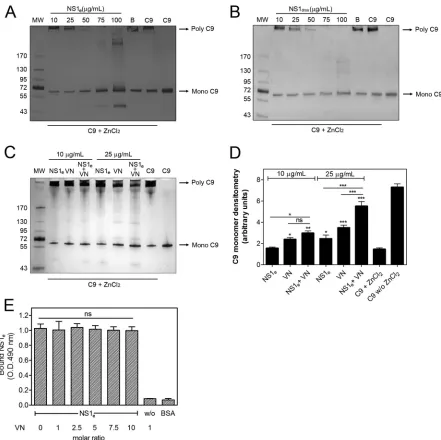
determi-FIG 6NS1 by itself or in association with vitronectin (VN) inhibits C9 polymerization. C9 was incubated with different amounts of NS1e(10 to 100g/ml) (A)
or NS1dros(10 to 100g/ml) (B) or with NS1 buffer (B; negative control) at 37°C for 40 min before the addition of 50M ZnCl2for 2 h at 37°C. (C) C9 was
incubated separately or with a mixture of NS1eand VN (10 and 25g/ml) at 37°C for 40 min before the addition of 50M ZnCl2for 2 h at 37°C. All samples were
subjected to SDS-PAGE in a gradient gel (4 to 20%) under nonreducing conditions, and C9 polymerization was visualized by silver staining. (D) The C9 monomer band intensities from panel C were measured by densitometry and reported in arbitrary units. The error bars indicate standard deviations for three independent experiments, and the asterisks indicate the significance of differences from the C9 band intensity in the absence of NS1 and VN (C9⫹ZnCl2),
determined using one-way ANOVA and the Bonferroni posttest. (E) NS1 and VN do not compete for C9 binding. C9 was immobilized on microtiter plates, and a constant amount of NS1e(100 nM) with increasing amounts of VN was added. Bound NS1ewas detected using an anti-NS1 polyclonal antibody. One-way
ANOVA and the Bonferroni posttest were used to calculate the significance of differences compared to the binding without NS1. Error bars indicate standard deviations for three independent experiments, and the asterisks indicate the significance of differences from the control. ns, not significant; *,P⬍0.05; **,P⬍ 0.01; ***,P⬍0.001. MW, molecular weight.
on November 7, 2019 by guest
http://jvi.asm.org/
[image:9.585.72.513.63.503.2]nant factor in inhibiting MAC formation because both
nonglyco-sylated and glycononglyco-sylated forms of NS1 hindered erythrocyte lysis
and C9 polymerization. We also compared the C9 polymerization
inhibition levels by DENV2, WNV, and ZIKV NS1 proteins
pro-duced in mammalian cells. Our data showed that these three
fla-vivirus proteins inhibited C9 polymerization in a way similar to
that for the NS1
eand NS1
drosproteins used in this study,
confirm-ing our results. It is worth mentionconfirm-ing that ZIKV NS1 bound and
inhibited C9 polymerization more strongly than the DENV2 and
WNV proteins did, suggesting a possible role of ZIKV NS1
com-plement evasion in assisting ZIKV infection in the brain (
47
,
48
).
This study provides novel insights into the mechanisms of
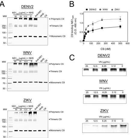
fla-FIG 7Flavivirus NS1 from mammalian cells binds and inhibits C9 polymerization in serum. (A) Flavivirus NS1 secreted by mammalian cells (NS1mam) inhibits
C9 polymerization. Initially, BHK-21 cells were preincubated with 5g/ml purified C5b6 for 2 h at 37°C in VBS. DENV2, WNV, or ZIKV NS1mamwas
preincubated with 5% NHS for 30 min at 37°C. Rabbit polyclonal anti-NS1 (50g/ml) was mixed with 50g/ml NS1mamto serve as a control. Cells were washed
and incubated with NS1mammixed with NHS for 50 min at 37°C. Thereafter, cells were washed and lysed. Samples were separated in a 10% polyacrylamide gel
under nonreducing conditions, and the C9 bands were analyzed by Western blotting using anti-C9. (B) Flavivirus NS1maminteracts with purified C9. C9 (0 to
500 nM) was incubated in DENV2, WNV, or ZIKV NS1mam-coated plates (10g/ml) for 2 h at 37°C. Wells were washed with PBS and then incubated with
polyclonal anti-C9 antibody, followed by incubation with peroxidase-conjugated anti-goat IgG antibody for 1 h at 37°C. The error bars indicate standard deviations for three independent experiments, and the asterisks indicate the significance of differences from the control, determined using two-way ANOVA and the Bonferroni posttest. ***,P⬍0.001. (C) Flavivirus NS1mamin complex with VN inhibited C9 polymerization. DENV2, WNV, or ZIKV NS1mam(25g/ml)
was preincubated with increasing concentrations of VN (0 to 25g/ml) for 30 min at 37°C, incubated with 5% NHS for 30 min at 37°C, and added to C5b6-treated BHK-21 cells for 50 min at 37°C. Thereafter, cells were washed three times with VBS and lysed with cell lysis buffer, and the C9 polymer was analyzed by Western blotting using anti-C9 antibody. Experiments were repeated three times.
on November 7, 2019 by guest
http://jvi.asm.org/
[image:10.585.78.513.65.523.2]vivirus manipulation of the host complement system. NS1 is a
pathogenic immune evasion protein that interacts with more than
one human complement molecule and controls multiple steps of
complement activation and function. In summary, NS1 by itself
or in association with VN is capable of inhibiting C9
polymeriza-tion and, consequently, MAC formapolymeriza-tion. These results suggest a
role for NS1 in dengue pathogenesis as a terminal pathway
inhib-itor of the complement system.
ACKNOWLEDGMENTS
We thank Cynthia Peterson and her student Letitia Olson at the Univer-sity of Tennessee for providing the PAI-1 protein.
This work was supported by Rio de Janeiro Research Foundation (FAPERJ) (grants 010.002879/2014, 010.001597/2014, 102.356/2013, 110.196/2013, and 112.671/2012) and from the Conselho Nacional de Desenvolvimento Científico e Tecnológico (CNPq) (grants 400213/ 2014-33 1 and 475104/2012-9).
FUNDING INFORMATION
This work, including the efforts of Ronaldo Mohana-Borges, was funded by International Centre for Genetic Engineering and Biotechnology (ICGEB) (CRP/BRA15-02). This work, including the efforts of Ronaldo Mohana-Borges, was funded by Fundação Carlos Chagas Filho de Amparo à Pesquisa do Estado do Rio de Janeiro (FAPERJ) (475104/2012-9).
The funders had no role in study design, data collection and interpreta-tion, or the decision to submit the work for publication.
REFERENCES
1.Bhatt S, Gething PW, Brady OJ, Messina JP, Farlow AW, Moyes CL, Drake JM, Brownstein JS, Hoen AG, Sankoh O, Myers MF, George DB, Jaenisch T, Wint GR, Simmons CP, Scott TW, Farrar JJ, Hay SI.2013. The global distribution and burden of dengue. Nature496:504 –507.http: //dx.doi.org/10.1038/nature12060.
2.WHO.2009. Dengue: guidelines for diagnosis, treatment, prevention and control, new ed. WHO, Geneva, Switzerland.
3.Lindenbach BD, Rice CM.2003. Molecular biology of flaviviruses. Adv Virus Res59:23– 61.http://dx.doi.org/10.1016/S0065-3527(03)59002-9. 4.Mackenzie JM, Jones MK, Young PR.1996. Immunolocalization of the
dengue virus nonstructural glycoprotein NS1 suggests a role in viral RNA replication. Virology220:232–240.http://dx.doi.org/10.1006/viro.1996 .0307.
5.Lindenbach BD, Rice CM.1997.trans-Complementation of yellow fever virus NS1 reveals a role in early RNA replication. J Virol71:9608 –9617. 6.Akey DL, Brown WC, Dutta S, Konwerski J, Jose J, Jurkiw TJ,
DelPro-posto J, Ogata CM, Skiniotis G, Kuhn RJ, Smith JL.2014. Flavivirus NS1 structures reveal surfaces for associations with membranes and the im-mune system. Science 343:881– 885. http://dx.doi.org/10.1126/science .1247749.
7.Gutsche I, Coulibaly F, Voss JE, Salmon J, d’Alayer J, Ermonval M, Larquet E, Charneau P, Krey T, Megret F, Guittet E, Rey FA, Flamand M.2011. Secreted dengue virus nonstructural protein NS1 is an atypical barrel-shaped high-density lipoprotein. Proc Natl Acad Sci U S A108: 8003– 8008.http://dx.doi.org/10.1073/pnas.1017338108.
8.Silva EM, Conde JN, Allonso D, Nogueira ML, Mohana-Borges R.2013. Mapping the interactions of dengue virus NS1 protein with human liver proteins using a yeast two-hybrid system: identification of C1q as an in-teracting partner. PLoS One8:e57514.http://dx.doi.org/10.1371/journal .pone.0057514.
9.Muller DA, Young PR.2013. The flavivirus NS1 protein: molecular and structural biology, immunology, role in pathogenesis and application as a diagnostic biomarker. Antiviral Res 98:192–208. http://dx.doi.org/10 .1016/j.antiviral.2013.03.008.
10. Modhiran N, Watterson D, Muller DA, Panetta AK, Sester DP, Liu L, Hume DA, Stacey KJ, Young PR.2015. Dengue virus NS1 protein acti-vates cells via Toll-like receptor 4 and disrupts endothelial cell monolayer integrity. Sci Transl Med 7:304ra142. http://dx.doi.org/10.1126 /scitranslmed.aaa3863.
11. Chung KM, Liszewski MK, Nybakken G, Davis AE, Townsend RR, Fremont DH, Atkinson JP, Diamond MS.2006. West Nile virus non-structural protein NS1 inhibits complement activation by binding the regulatory protein factor H. Proc Natl Acad Sci U S A103:19111–19116.
http://dx.doi.org/10.1073/pnas.0605668103.
12. Avirutnan P, Fuchs A, Hauhart RE, Somnuke P, Youn S, Diamond MS, Atkinson JP.2010. Antagonism of the complement component C4 by flavivirus nonstructural protein NS1. J Exp Med207:793– 806.http://dx .doi.org/10.1084/jem.20092545.
13. Avirutnan P, Hauhart RE, Somnuke P, Blom AM, Diamond MS, Atkinson JP.2011. Binding of flavivirus nonstructural protein NS1 to C4b binding protein modulates complement activation. J Immunol187:424 – 433.http://dx.doi.org/10.4049/jimmunol.1100750.
14. Singh B, Su YC, Riesbeck K.2010. Vitronectin in bacterial pathogenesis: a host protein used in complement escape and cellular invasion. Mol Mi-crobiol78:545–560.http://dx.doi.org/10.1111/j.1365-2958.2010.07373.x. 15. Milis L, Morris CA, Sheehan MC, Charlesworth JA, Pussell BA.1993. Vitronectin-mediated inhibition of complement: evidence for different binding sites for C5b-7 and C9. Clin Exp Immunol92:114 –119. 16. Podack ER, Preissner KT, Muller-Eberhard HJ.1984. Inhibition of C9
polymerization within the SC5b-9 complex of complement by S-protein. Acta Pathol Microbiol Immunol Scand284(Suppl):89 –96.
17. Sheehan M, Morris CA, Pussell BA, Charlesworth JA.1995. Comple-ment inhibition by human vitronectin involves non-heparin binding do-mains. Clin Exp Immunol101:136 –141.
18. da Silva LB, Miragaia LDS, Breda LC, Abe CM, Schmidt MC, Moro AM, Monaris D, Conde JN, Jozsi M, Isaac L, Abreu PA, Barbosa AS. 2015. Pathogenic Leptospira species acquire factor H and vitronectin via the surface protein LcpA. Infect Immun83:888 – 897.http://dx.doi.org/10 .1128/IAI.02844-14.
19. Allonso D, da Silva Rosa M, Coelho DR, da Costa SM, Nogueira RM, Bozza FA, Santos FB, de Barcelos Alves AM, Mohana-Borges R.2011. Polyclonal antibodies against properly folded dengue virus NS1 protein expressed in E. coli enable sensitive and early dengue diagnosis. J Virol Methods175:109 –116.http://dx.doi.org/10.1016/j.jviromet.2011.04.029. 20. Allonso D, Meneses MD, Fernandes CA, Ferreira DF, Mohana-Borges R.2014. Assessing positivity and circulating levels of NS1 in samples from a 2012 dengue outbreak in Rio de Janeiro, Brazil. PLoS One9:e113634.
http://dx.doi.org/10.1371/journal.pone.0113634.
21. Zhang Z, Yang J, Wei J, Yang Y, Chen X, Zhao X, Gu Y, Cui S, Zhu X. 2011. Trichinella spiralis paramyosin binds to C8 and C9 and protects the tissue-dwelling nematode from being attacked by host complement. PLoS Negl Trop Dis5:e1225.http://dx.doi.org/10.1371/journal.pntd.0001225. 22. Yasumitsu H, Seo N, Misugi E, Morita H, Miyazaki K, Umeda M.1993.
Vitronectin secretion by hepatic and non-hepatic human cancer cells. In vitro cellular & developmental biology. Animal29A:403– 407.
23. Kost C, Stuber W, Ehrlich HJ, Pannekoek H, Preissner KT. 1992. Mapping of binding sites for heparin, plasminogen activator inhibitor-1, and plasminogen to vitronectin’s heparin-binding region reveals a novel vitronectin-dependent feedback mechanism for the control of plasmin formation. J Biol Chem267:12098 –12105.
24. Lane DA, Flynn AM, Pejler G, Lindahl U, Choay J, Preissner K.1987. Structural requirements for the neutralization of heparin-like saccharides by complement S protein/vitronectin. J Biol Chem262:16343–16348. 25. Liang OD, Rosenblatt S, Chhatwal GS, Preissner KT.1997.
Identifica-tion of novel heparin-binding domains of vitronectin. FEBS Lett407:169 – 172.http://dx.doi.org/10.1016/S0014-5793(97)00330-X.
26. Schar CR, Jensen JK, Christensen A, Blouse GE, Andreasen PA, Peter-son CB.2008. Characterization of a site on PAI-1 that binds to vitronectin outside of the somatomedin B domain. J Biol Chem283:28487–28496.
http://dx.doi.org/10.1074/jbc.M804257200.
27. Preissner KT.1990. Specific binding of plasminogen to vitronectin. Evi-dence for a modulatory role of vitronectin on fibrin(ogen)-induced plas-min formation by tissue plasplas-minogen activator. Biochem Biophys Res Commun168:966 –971.
28. Su YC, Jalalvand F, Morgelin M, Blom AM, Singh B, Riesbeck K.2013. Haemophilus influenzae acquires vitronectin via the ubiquitous protein F to subvert host innate immunity. Mol Microbiol87:1245–1266.http://dx .doi.org/10.1111/mmi.12164.
29. Xu D, Baburaj K, Peterson CB, Xu Y. 2001. Model for the three-dimensional structure of vitronectin: predictions for the multi-domain protein from threading and docking. Proteins44:312–320.http://dx.doi .org/10.1002/prot.1096.
on November 7, 2019 by guest
http://jvi.asm.org/
30. Liang OD, Preissner KT, Chhatwal GS. 1997. The hemopexin-type repeats of human vitronectin are recognized by Streptococcus pyogenes. Biochem Biophys Res Commun234:445– 449.http://dx.doi.org/10.1006 /bbrc.1997.6663.
31. Alcon S, Talarmin A, Debruyne M, Falconar A, Deubel V, Flamand M. 2002. Enzyme-linked immunosorbent assay specific to dengue virus type 1 nonstructural protein NS1 reveals circulation of the antigen in the blood during the acute phase of disease in patients experiencing primary or secondary infections. J Clin Microbiol40:376 –381.http://dx.doi.org/10 .1128/JCM.40.02.376-381.2002.
32. Libraty DH, Young PR, Pickering D, Endy TP, Kalayanarooj S, Green S, Vaughn DW, Nisalak A, Ennis FA, Rothman AL.2002. High circu-lating levels of the dengue virus nonstructural protein NS1 early in dengue illness correlate with the development of dengue hemorrhagic fever. J Infect Dis186:1165–1168.http://dx.doi.org/10.1086/343813.
33. Hallstrom T, Blom AM, Zipfel PF, Riesbeck K. 2009. Nontypeable Haemophilus influenzae protein E binds vitronectin and is important for serum resistance. J Immunol183:2593–2601.http://dx.doi.org/10.4049 /jimmunol.0803226.
34. Su HR, Boackle RJ.1994. Heparin mediates binding of S-protein/ vitronectin to the envelope glycoprotein of the human immunodeficiency virus and CD4. Int Arch Allergy Immunol105:238 –244.http://dx.doi.org /10.1159/000236763.
35. Bozzini S, Falcone V, Conaldi PG, Visai L, Biancone L, Dolei A, Toniolo A, Speziale P.1998. Heparin-binding domain of human fibronectin binds HIV-1 gp120/160 and reduces virus infectivity. J Med Virol54:44 –53.
http://dx.doi.org/10.1002/(SICI)1096-9071(199801)54:1⬍44::AID -JMV7⬎3.0.CO;2-P.
36. Huang YP, Cheng J, Zhang SL, Wang L, Guo J, Liu Y, Yang Y, Zhang LY, Bai GQ, Gao XS, Ji D, Lin SM, Shao Q.2005. Screening of hepato-cyte proteins binding to F protein of hepatitis C virus by yeast two-hybrid system. World J Gastroenterol11:5659 –5665.http://dx.doi.org/10.3748 /wjg.v11.i36.5659.
37. Sa E Cunha C, Griffiths NJ, Virji M.2010. Neisseria meningitidis Opc invasin binds to the sulphated tyrosines of activated vitronectin to attach to and invade human brain endothelial cells. PLoS Pathog6:e1000911.
http://dx.doi.org/10.1371/journal.ppat.1000911.
38. Kohler S, Hallstrom T, Singh B, Riesbeck K, Sparta G, Zipfel PF, Hammerschmidt S.2015. Binding of vitronectin and factor H to Hic contributes to immune evasion of Streptococcus pneumoniae serotype 3. Thromb Haemost 113:125–142. http://dx.doi.org/10.1160/TH14-06 -0561.
39. Lawrence D, Strandberg L, Grundstrom T, Ny T.1989. Purification of
active human plasminogen activator inhibitor 1 from Escherichia coli. Comparison with natural and recombinant forms purified from eucary-otic cells. Eur J Biochem186:523–533.
40. Zhou A, Carrell RW, Huntington JA. 2001. The serpin inhibitory mechanism is critically dependent on the length of the reactive center loop. J Biol Chem 276:27541–27547. http://dx.doi.org/10.1074/jbc .M102594200.
41. Declerck PJ, De Mol M, Alessi MC, Baudner S, Paques EP, Preissner KT, Muller-Berghaus G, Collen D.1988. Purification and characteriza-tion of a plasminogen activator inhibitor 1 binding protein from human plasma. Identification as a multimeric form of S protein (vitronectin). J Biol Chem263:15454 –15461.
42. Hansen M, Busse MN, Andreasen PA.2001. Importance of the amino-acid composition of the shutter region of plasminogen activator inhibi-tor-1 for its transitions to latent and substrate forms. Eur J Biochem268: 6274 – 6283.http://dx.doi.org/10.1046/j.0014-2956.2001.02582.x. 43. Wills BA, Oragui EE, Stephens AC, Daramola OA, Dung NM, Loan HT,
Chau NV, Chambers M, Stepniewska K, Farrar JJ, Levin M. 2002. Coagulation abnormalities in dengue hemorrhagic fever: serial investiga-tions in 167 Vietnamese children with dengue shock syndrome. Clin Infect Dis35:277–285.http://dx.doi.org/10.1086/341410.
44. Mairuhu AT, Setiati TE, Koraka P, Hack CE, Leyte A, Faradz SM, ten Cate H, Brandjes DP, Osterhaus AD, Reitsma PH, van Gorp EC.2005. Increased PAI-1 plasma levels and risk of death from dengue: no associa-tion with the 4G/5G promoter polymorphism. Thromb J3:17.
45. Djamiatun K, Faradz SM, Setiati TE, Netea MG, van der Ven AJ, Dolmans WM.2011. Increase of plasminogen activator inhibitor-1 and decrease of transforming growth factor-b1 in children with dengue hae-morrhagic fever in Indonesia. J Trop Pediatr57:424 – 432.http://dx.doi .org/10.1093/tropej/fmq122.
46. Shyu HW, Lin YY, Chen LC, Wang YF, Yeh TM, Su SJ, Cheng WC, Chen CY, Lin KH, Chou MC.2010. The dengue virus envelope protein induced PAI-1 gene expression via MEK/ERK pathways. Thromb Hae-most104:1219 –1227.http://dx.doi.org/10.1160/TH10-05-0302. 47. Miner JJ, Cao B, Govero J, Smith AM, Fernandez E, Cabrera OH,
Garber C, Noll M, Klein RS, Noguchi KK, Mysorekar IU, Diamond MS. 2016. Zika virus infection during pregnancy in mice causes placental dam-age and fetal demise. Cell165:1081–1091.http://dx.doi.org/10.1016/j.cell .2016.05.008.
48. Veerhuis R, Nielsen HM, Tenner AJ.2011. Complement in the brain. Mol Immunol48:1592–1603.http://dx.doi.org/10.1016/j.molimm.2011 .04.003.
on November 7, 2019 by guest
http://jvi.asm.org/