Transcriptional Suppression of miR-181c by Hepatitis C Virus
Enhances Homeobox A1 Expression
Anupam Mukherjee,aShubham Shrivastava,aJoydip Bhanja Chowdhury,aRanjit Ray,bRatna B. Raya,b
Departments of Pathologyaand Internal Medicine,bSaint Louis University, St. Louis, Missouri, USA
ABSTRACT
Hepatitis C virus (HCV)-induced chronic liver disease is one of the leading causes of hepatocellular carcinoma (HCC). The mo-lecular events leading to HCC following chronic HCV infection remain poorly defined. MicroRNAs (miRNAs) have been impli-cated in the control of many biological processes, and their deregulation is associated with different viral infections. In this study, we observed that HCV infection of hepatocytes transcriptionally downregulates miR-181c expression by modulating CCAAT/enhancer binding protein(C/EBP-). Reduced expression of the pri-miR-181c transcript was noted following HCV infection.In silicoprediction suggests that homeobox A1 (HOXA1) is a direct target of miR-181c. HOXA1 is a member of the homeodomain-containing transcription factor family and possesses pivotal roles in normal growth, development, and differen-tiation of mammalian tissues. Our results demonstrated that HOXA1 expression is enhanced in HCV-infected hepatocytes. Ex-ogenous expression of the miR-181c mimic inhibits HOXA1 and its downstream molecules STAT3 and STAT5, which are in-volved in cell growth regulation. Interestingly, overexpression of miR-181c inhibited HCV replication by direct binding with E1 and NS5A sequences. Furthermore, accumulation of HCV genotype 2a RNA with miR-181c was observed in an RNA-induced silencing complex in Huh7.5 cells. Our results provide new mechanistic insights into the role of miR-181c in HCV-hepatocyte interactions, and miR-181c may act as a target for therapeutic intervention.
IMPORTANCE
Chronic HCV infection is one of the major causes of end-stage liver disease, including hepatocellular carcinoma. An understand-ing of the molecular mechanisms of HCV-mediated hepatocyte growth promotion is necessary for therapeutic intervention against HCC. In this study, we have provided evidence of mediated transcriptional downregulation of miR-181c. HCV-infected liver biopsy specimens also displayed lower expression levels of miR-181c. We have further demonstrated that inhibi-tion of miR-181c upregulates homeobox A1 (HOXA1), which is important for hepatocyte growth promoinhibi-tion. Exogenous expres-sion of miR-181c inhibited HCV replication by directly binding with HCV E1 and NS5A sequences. Taken together, our results provided new mechanistic insights for an understanding of the role of miR-181c in HCV-hepatocyte interactions and revealed miR-181c as a potential target for therapeutic intervention.
A
n estimated 170 million to 200 million people are chronically infected with hepatitis C virus (HCV) worldwide, and infec-tion often leads to severe liver disease, including advanced fibrosis, cirrhosis, and hepatocellular carcinoma (HCC) (1,2). The HCV genome is a single-stranded positive-sense RNA, and HCV be-longs to the familyFlaviviridaeand the genusHepacivirus. The 9.6-kb HCV genome encodes a large polyprotein, which is cessed by viral and cellular proteases to produce structural pro-teins (core, E1, and E2/p7) and nonstructural (NS) propro-teins (NS2, NS3, NS4A, NS4B, NS5A, and NS5B) (3).MicroRNAs (miRNAs) constitute a class of⬃18- to 22-nucle-otide-long noncoding RNAs and play a crucial role in the regula-tion of gene expression. The producregula-tion of miRNAs requires sev-eral processing steps. The first primary miRNAs are cleaved by Drosha to produce precursor miRNAs, which in turn are cleaved by Dicer to produce mature, single-stranded miRNAs (4,5). Once synthesized, mature miRNA associates with the RNA-induced si-lencing complex together with Argonaute/EIF2C. Target mRNA recognition is mediated through specific base-pairing interactions between the 5=end (“seed” region) of miRNA and sites within the coding and 3=untranslated regions (UTRs) of mRNAs, leading to mRNA destabilization (6). Deregulation of miRNA occurs fre-quently in a variety of diseases, including liver disease (7).
Several RNA viruses interact directly with cellular miRNAs in
modulating viral genome replication (8, 9). The liver-specific miRNA miR-122 is required for efficient HCV replication (9). miR-122 binds two target sites in the 5=UTR of the HCV genome and enhances intracellular viral RNA accumulation in hepatoma cells. Other miRNAs, such as miR-199a*, let-7b, miR-196, and miR-448, have also been reported to physically interact with HCV RNA and suppress viral replication (9). HCV infection can alter the miRNA expression profile of the host cell in facilitating repli-cation or escape from the immune system (10,11). We have pre-viously shown that miR-130a expression is upregulated in HCV-infected cells. Overexpression of miR-130a facilitates HCV replication by inhibiting the interferon (IFN)-induced transmem-brane protein IFITM1 (12).
In the present study, we observed that expression of miR-181c
Received17 March 2014Accepted26 April 2014 Published ahead of print30 April 2014 Editor:B. Williams
Address correspondence to Ratna B. Ray, rayrb@slu.edu. A.M. and S.S. contributed equally to this work.
Copyright © 2014, American Society for Microbiology. All Rights Reserved. doi:10.1128/JVI.00787-14
on November 7, 2019 by guest
http://jvi.asm.org/
is inhibited at the transcriptional level following HCV infection. HCV infection suppresses miR-181c expression by downregulat-ing the transcription factor CCAAT/enhancer binddownregulat-ing protein
(C/EBP-) and promotes homeobox A1 (HOXA1) expression,
which subsequently upregulates STAT3 and STAT5 expression. In addition, we observed that exogenous expression of miR-181c re-stricts HCV replication by binding to E1 and NS5A.
MATERIALS AND METHODS
Cells and viruses.Human hepatoma (Huh7.5) cells were maintained in Dulbecco’s modified Eagle’s medium (DMEM) supplemented with 10% fetal bovine serum (FBS), 100 U of penicillin G/ml, and 100g of strep-tomycin/ml. Huh7.5 cells harboring the HCV genotype 2a genome-length replicon with theRenillaluciferase reporter gene (Rep2a-Rluc cells, kindly provided by Hengli Tang) were cultured in DMEM containing 10% FBS with 1% antibiotics. All cells were maintained at 37°C in a 5% CO2
atmo-sphere.
HCV genotype 2a (clone JFH1) was grown in Huh7.5 cells as previ-ously described (13). Virus released into cell culture supernatant was fil-tered through a 0.45-m-pore-size cellulose acetate membrane and quan-titated in standard IU/ml. For infection, Huh7.5 cells were incubated with HCV genotype 2a (multiplicity of infection of 0.5) for 72 h.
RNA quantitation and reverse transcription-quantitative PCR (qRT-PCR).Total RNA was isolated by using TRIzol reagent (Invitro-gen). cDNA was synthesized by using miR-181c- or U6-specific primers with a TaqMan microRNA reverse transcription kit and a random hex-amer with Superscript III reverse transcriptase for HCV and 18S rRNA. Real-time PCR was performed for quantitation by using TaqMan univer-sal PCR master mix and a 6-carboxyfluorescein (FAM)-MGB probe for miR-181c (assay identification number 000482), pri-miR-181c (assay identification number Hs03303907_pri), and HCV (assay identification number AI6Q1GI). U6 (assay identification number 001973) or 18S rRNA (assay identification number Hs03928985_g1) was used as an en-dogenous control for miR-181c, pri-miR-181c, or HCV. The relative ex-pression levels of miR-181c and HCV were normalized to U6 or 18S rRNA by using the 2⫺⌬⌬CTformula (⌬⌬C
T⫽ ⌬CTof the sample⫺ ⌬CTof the
untreated control).
Patient samples.Liver biopsy specimens from HCV-infected adult patients for our study were approved by the Saint Louis University Insti-tutional Review Board, and written informed consent was obtained from all subjects.
Luciferase reporter assays.The full-length promoter (nucleotides
⫺1800 to⫹1; P0) or promoter fragments of the deletion mutant of
miR-181c (P1to P3) were PCR amplified from Huh7.5 genomic DNA by using
specific primers (Table 1). The fragments were digested with MluI and BglII and cloned into the pGL3-Basic luciferase vector (Promega). Huh7.5 cells were cotransfected with a luciferase reporter plasmid containing the miR-181c promoter (125 ng) and equal amounts of plasmid DNA
con-taining HCV core, E1E2p7, nonstructural proteins (NS2, NS3/4A, NS4B, NS5A, or NS5B), or the full-length genome (FL-HCV) by using Lipo-fectamine 2000 (Invitrogen). Luciferase activity was determined as previ-ously described (14).
The HOXA1 3=-UTR luciferase reporter construct was generated by cloning the PCR-amplified human HOXA1 mRNA 3=UTR (miR-181c target sites) into the MluI/HindIII site of the pMIR-Report miRNA ex-pression luciferase reporter plasmid (Ambion). The mutant 3=UTR of HOXA1 was used as a control in parallel. miR-181c binding-site-specific HCV E1 or NS5A gene segments were cloned into the pMIR-Report lu-ciferase reporter plasmid. Huh7.5 cells were cotransfected with the lucif-erase reporter plasmid and control miRNA or different doses of miR-181c mimic, and luciferase activity was determined. Primers used for HOXA1 3=-UTR, HCV E1, and NS5A cloning are listed inTable 1.
Rep2a-Rluc cells were transfected with either the miR-181c mimic or control miRNA, and HCV replication was determined by qRT-PCR or relativeRenillaluciferase activity (Promega) after 48 h of transfection.
RNA interference.Huh7.5 cells were cotransfected with 50 nM C/EBP- small interfering RNA (siRNA) (a pool of 3 target-specific siRNAs from Santa Cruz) or control siRNA and the promoter construct of miR-181c, and a lu-ciferase assay was performed at 48 h posttransfection.
Western blot analysis.Cell lysates were subjected to polyacrylamide gel electrophoresis and transferred onto a nitrocellulose membrane. The membranes were blocked with 5% nonfat dried milk and incubated with specific antibodies. The membrane was probed with antibodies to C/EBP-(Santa Cruz), HOXA1 (Sigma), STAT3, or STAT5 (Cell Signal-ing). Proteins were detected by using an enhanced chemiluminescence (ECL) Western blot substrate. The membrane was reprobed with actin as an internal control.
Ago2-RNA coimmunoprecipitation. The HCV RNA–miR-181c
complex was precipitated as described previously (15,16). Briefly, miR-181c-transfected Rep2a-Rluc cells were lysed with lysis buffer (150 mM KCl, 25 mM Tris-HCl [pH 7.4], 5 mM EDTA, 1% Triton X-100, 5 mM dithiothreitol [DTT], protease inhibitor mixture, and 100 U/ml RNase-OUT [Invitrogen]). Lysates were clarified, incubated with an anti-Ago2 monoclonal antibody (MAb) (11A9; Sigma) or isotype control IgG2a at 4°C overnight, and mixed with protein G-Sepharose (GE Healthcare) for 2 h. The beads were washed four times, and RNA was isolated by using the RNeasy minikit (Qiagen). HCV RNA was quantitated by qRT-PCR or one-step RT-PCR (Invitrogen) using specific primers.
RESULTS
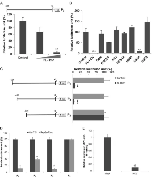
HCV infection downregulates miR-181c expression.The major targets and functions of a specific miRNA vary under different physiological or pathological conditions and in different cell types. Since HCV is primarily hepatotropic, and human hepato-cytes are the natural host of HCV, we initially performed an miRNA array (MAH-102A; Qiagen) using RNA from mock- or HCV-infected primary human hepatocytes from two different do-nors and observed a significant downregulation of miR-181c (⬃12-fold). We next validated the expression of miR-181c in pri-mary human hepatocytes infected with HCV and observed inhi-bition of miR-181c expression (Fig. 1A). The expression of miR-181c was significantly downregulated following HCV infection in Huh7.5 cells (Fig. 1B). A similar result was obtained when we examined the expression of miR-181c in Huh7.5 cells harboring the genome-length HCV replicon (Rep2a-Rluc cells) compared to parental Huh7.5 cells (Fig. 1C). These results indicated that HCV suppresses miR-181c expression in hepatocytes. RNA from HCV-infected liver biopsy specimens (12) also displayed reduced miR-181c expression levels compared to those of control liver biopsy specimens (not infected with HCV or hepatitis B virus [HBV]) (Fig. 1D). The intracellular HCV RNA expression level was in-TABLE 1Primer sequences used in this study
Primer Sequence
miR-181c-full promoter_For 5=-TTGACGCGTTCTGGACTAAC-3= miR-181c-F1 promoter_For 5=-TGTACGCGTGACAGGTATCT-3= miR-181c-F2 promoter_For 5=-CGTCACGCGTTGGCTTGCG-3= miR-181c-F3 promoter_For 5=-CGAACGCGTGTGCGATC-3= miR-181c promoter
F-F3_Rev
5=-AATAGATCTGTAATACGTTCTGG-3=
HOXA1-wt 3=UTR_For 5=-TGTGACGCGTTATTCAGAACCTAAAAG-3= HOXA1-mt 3=UTR_For 5=-AATGTATTACGCGTGTTATTCGTGAA-3= HOXA1 3=UTR_Rev 5=-CACAAAGCTTAACTGATAGAACTCAGT-3= miR-181c/HCV-E1_For 5=-TGCGACGCGTGCGAGAGAGTGGGGAATA-3= miR-181c/HCV-E1_Rev 5=-CCGCAAGCTTCACCCCGCCACAGAGGTCCC-3= miR-181c/HCV-NS5A_For 5=-CGAAACGCGTCACGAACTACAAGACCGCCA-3= miR-181c/HCV-NS5A_Rev 5=-ACCTAAGCTTGGAAAAACGGCTTTGGTGT-3=
on November 7, 2019 by guest
http://jvi.asm.org/
[image:2.585.41.287.77.209.2]versely related to the expression level of miR-181c in biopsy spec-imens (Fig. 1E). Together, our results suggested that HCV repli-cation inhibits miR-181c expression.
HCV transcriptionally downregulates miR-181c.Since HCV infection downregulates miR-181c expression, we examined how HCV modulates miR-181c promoter activity. For this, a miR-181c promoter sequence (nucleotides⫺1800 to ⫹1; P0) was cloned into pGL3 luciferase reporter plasmid DNA (Fig. 2A). Cells were transfected with the P0construct and plasmid DNA containing the
full-length HCV genome (FL-HCV) to examine promoter activ-ity. The introduction of FL-HCV downregulated miR-181c pro-moter activity in a dose-dependent manner (Fig. 2A). We next determined which HCV protein was responsible for miR-181c trans-suppression. Huh7.5 cells were cotransfected with plasmids expressing FL-HCV, core, E1E2p7, NS2, NS3/4A, NS4B, NS5A, or NS5B, encoding plasmid DNA with the P0construct. Our results demonstrated that the HCV NS5A protein significantly inhibited miR-181c promoter activity (Fig. 2B).
FIG 1HCV infection suppresses miR-181c expression. (A) Primary human hepatocytes were mock treated or HCV infected for 96 h. Total RNA from mock-or virus-infected cells was extracted, and the expression level of miR-181c was measured by qRT-PCR and nmock-ormalized to the expression level of U6. (B) Huh7.5 cells were mock treated or HCV infected (multiplicity of infection of 0.5) for 72 h. Total RNA from mock- or virus-infected cells was extracted, and the expression level of miR-181c was measured and normalized to the expression level of U6. (C) Total RNA was extracted from Huh7.5 cells as a control or Huh7.5 cells harboring the HCV genome-length replicon (Rep2a-Rluc), and the expression level of miR-181c was determined by qRT-PCR. Data are presented as the means and standard deviations from three independent experiments. (D) Total RNA was extracted from control (not infected with HCV or HBV) and HCV-infected liver biopsy specimens. miR-181c expression was analyzed by real-time PCR, using U6 RNA as an internal control. A comparison of miR-181c levels in control and HCV-infected (n⫽6) liver tissues is shown.ⴱ,P⬍0.05;ⴱⴱ,P⬍0.01. (E) Correlation plot for miR-181c expression versus HCV RNA levels in liver biopsy specimens. We observed a strong inverse correlation between miR-181c and intrahepatic HCV RNA levels (r⫽ ⫺0.54;P⫽0.06). Statistical analysis was done by using the Spearman correlation.
HCV-Mediated miR-181ctrans-Suppression
on November 7, 2019 by guest
http://jvi.asm.org/
[image:3.585.113.477.60.505.2]FIG 2HCV transcriptionally suppresses miR-181c promoter activity. (A) Schematic diagram of the miR-181c promoter region (nucleotides⫺1800 to⫹1; P0)
cloned into the luciferase reporter plasmid (P0). Huh7.5 cells were cotransfected with the miR-181c–luciferase reporter construct and FL-HCV plasmid DNAs
(200 or 400 ng). Relative luciferase activity was measured after 48 h of transfection. (B) Huh7.5 cells were cotransfected with the miR-181c–luciferase reporter construct, P0, and the plasmid DNA encoding HCV structural and nonstructural proteins. Relative luciferase activity was measured after 48 h of transfection. (C)
Deletion mutants of the miR-181c promoter construct (nucleotides⫺624 to⫹1 [P1],⫺432 to⫹1 [P2], and⫺349 to⫹1 [P3]) and FL-HCV plasmid DNA were
transfected into Huh7.5 cells. The effect of HCV on these mutants was examined by a luciferase reporter assay. (D) Huh7.5 cells harboring the HCV genome-length replicon (Rep2a-Rluc) were transfected with miR-181c promoter constructs (P0, P1, P2, or P3), and promoter activity was measured by a relative luciferase
assay after 48 h of transfection. Data are presented as the means and standard deviations from at least three independent experiments. (E) Pri-miR-181c levels measured by qRT-PCR in mock-treated or HCV-infected cells and normalized to 18S.ⴱⴱ,P⬍0.01;ⴱⴱⴱ,P⬍0.001.
on November 7, 2019 by guest
http://jvi.asm.org/
[image:4.585.51.538.63.627.2]To identify the specific region on the miR-181c promoter re-sponsible for HCV-mediated suppression, we constructed a num-ber of deletion mutants (P1to P3) of the miR-181c promoter in luciferase reporter plasmid DNA (Fig. 2C). The P1(nucleotides
⫺624 to⫹1) and P2(nucleotides⫺432 to⫹1) mutant fragments of the miR-181c promoter displayed an inhibitory effect in the presence of FL-HCV, whereas the P3(nucleotides⫺349 to⫹1) fragment did not exhibit an effect (Fig. 2C), suggesting that the HCV-mediated regulatory sequence is absent from this region. We also examined the expression of the P0, P1, P2, or P3construct of miR-181c in Rep2a-Rluc cells and observed an inhibition of miR-181c promoter activity from the P0, P1, or P2construct, but not from the P3construct, supporting the above-described obser-vations (Fig. 2D). Next, we examined the effect of HCV on pri-mary miRNA (pri-miRNA) processing of miR-181c. For this, total RNA was isolated from mock-treated or HCV-infected Huh7.5 cells. The pri-miR-181c level in mock-treated or HCV-infected cells was measured by qRT-PCR using specific primers (Invitro-gen) and normalized with 18S RNA. pri-miR-181c transcript was inhibited in Huh7.5 cells following HCV infection (Fig. 2E). Sim-ilar results were observed for Rep2a cells (data not shown). These results further emphasized that HCV transcriptionally downregu-lates miR-181c expression.
To search for thecis-regulatory elements present in the miR-181c promoter region (nucleotides⫺432 to⫺349), we performed in silicoanalysis with the Promo TransFac 8.3 and TFSEARCH algorithms (17). Several conserved regulatory elements were
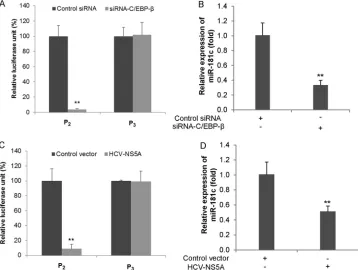
found in the designated consensus sequence of the miR-181c pro-moter, including binding sites for specificity protein 1 (SP-1) and CCAAT/enhancer binding protein(C/EBP-). We have previ-ously shown that HCV NS5A downregulates C3 promoter activity by modulation of C/EBP-(18). To analyze the role of C/EBP- in the regulation of HCV-mediated miR-181c expression, we knocked down C/EBP-using a specific siRNA and examined the activity of the P2or P3(as a negative control) construct of the miR-181c promoter. Silencing of C/EBP-reduced the activity of the miR-181c P2promoter, but not the P3construct, indicating that C/EBP-is an important player (Fig. 3A). To further verify the effect of C/EBP-on miR-181c transcriptional regulation, we silenced C/EBP-in Huh7.5 cells, and miR-181c transcript levels were examined by qRT-PCR. As anticipated, inhibition of miR-181c expression was observed in C/EBP-knockdown cells (Fig. 3B). Treatment of Huh7.5 cells with C/EBP-siRNA reduced target protein expression (data not shown). NS5A-mediated downregulation of C/EBP- was reported previously (18). We next examined the effect of a plasmid expressing HCV NS5A on the miR-181c P2construct. As expected, miR-181c promoter ac-tivity and miR-181c transcript levels were significantly decreased in the presence of NS5A with respect to the control vector (Fig. 3C andD). The P3construct was used as a negative control. Together, these results along with our previous findings suggested that HCV NS5A-mediated C/EBP- suppression is an important mecha-nism for miR-181c downregulation.
HOXA1 is a direct target of miR-181c.HCV infection also FIG 3Functional analysis ofcis-regulatory elements involved in the suppression of miR-181c by HCV NS5A. (A) Huh7.5 cells were cotransfected with an miR-181c–luciferase reporter construct (P2or P3) and C/EBP-or control siRNA. The activity of the miR-181c promoter was measured by a relative luciferase
assay after 48 h of transfection. (B) Huh7.5 cells were transfected with control or C/EBP-siRNA. RNA was extracted at 48 h posttransfection, and the expression level of miR-181c was determined by qRT-PCR. (C) miR-181c promoter (P2or P3) analysis by a relative luciferase assay was performed with Huh7.5 cells after
48 h of transfection with HCV NS5A or the control vector. Data are presented as the means and standard deviations from at least three independent experiments. (D) Huh7.5 cells were transfected with control or HCV NS5A plasmid DNA. RNA was extracted at 48 h posttransfection, and the expression level of miR-181c was determined by qRT-PCR.ⴱⴱ,P⬍0.01.
HCV-Mediated miR-181ctrans-Suppression
on November 7, 2019 by guest
http://jvi.asm.org/
[image:5.585.113.471.68.338.2]promotes cell growth and often leads to hepatocellular carcinoma (19). To identify the underlying mechanisms of HCV-mediated miR-181c inhibition, potential miR-181c target genes were searched by using the TargetScan and miRanda prediction meth-ods. We found a putative miR-181c target site in the HOXA1 3=
UTR. Homeobox-containing genes are a family of transcription factors that have crucial roles in cell growth and survival (20). Our in silicoanalysis suggested a single putative binding site of
miR-181c in the 3= UTR of HOXA1 with a high mirSVR score of
⫺1.2254 (Fig. 4A). The HOXA1 3=UTR with a potential binding site for miR-181c or a mutant fragment without the binding site was cloned into the pMIR-Report luciferase vector. The interac-tion between miR-181c and the 3=UTR of HOXA1 was examined by a luciferase reporter assay. The results showed that miR-181c represses the expression of the wild-type HOXA1 3=UTR in a dose-dependent manner, as evident from the decreased luciferase activity (Fig. 4B). The mutant 3=UTR of HOXA1 did not exhibit inhibition by miR-181c mimic (Fig. 4C). We further transfected the vector or the HOXA1 3=UTR into Rep2a cells and performed a HOXA1 3=UTR luciferase assay. As expected, luciferase activity
was not altered due to the lower expression level of miR-181c (data not shown). Next, we examined whether modulation of miR-181c has a role in cell growth. For this, Rep2a-Rluc cells were transfected with control miRNA or miR-181c, and cell viability was examined at different time points. Overexpression of miR-181c significantly inhibited Rep2a-Rluc cell proliferation (Fig. 4D).
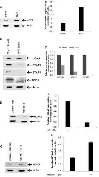
To determine the expression level of HOXA1 in HCV-infected hepatocytes, Huh7.5 cells were infected with HCV genotype 2a, and HOXA1 expression was analyzed by Western blotting using a specific antibody. Expression of HOXA1 was significantly upregu-lated in HCV-infected cells compared to mock-treated control cells (Fig. 5AandB). On the other hand, overexpression of the miR-181c mimic inhibited HOXA1 and HCV NS5A expression in HCV-infected Huh7.5 cells (Fig. 5C). Next, we determined whether downstream molecules of HOXA1 are altered by miR-181c. HOXA1 transcriptionally upregulates STAT3 and STAT5 expression in human mammary epithelial cells (20). Expressions of STAT3 and STAT5 were significantly inhibited in HCV-in-fected hepatocytes by the miR-181c mimic compared to control FIG 4miR-181c targets the HOXA1 3=untranslated region (UTR). (A)In silicoanalysis of the HOXA1 3=UTR revealing a single putative miR-181c binding site. The miR-181c target region of HOXA1 (GenBank accession numberNM_005522) is indicated. (B) Huh7.5 cells were cotransfected with the 3=UTR of HOXA1 cloned the into pMIR-Report luciferase vector and different doses of the miR-181c mimic (50, 100, or 200 nM). Relative luciferase activity was measured after 48 h of transfection. Control-miR, control miRNA. (C) Luciferase activity from the 3=UTR of the HOXA1 reporter was decreased after cotransfection of the HOXA1 3=UTR and the miR-181c mimic (50 nM) but not with the mutant 3=UTR of HOXA1. The results are shown as means and standard deviations from three independent experiments. (D) miR-181c inhibits cell proliferation. Cells harboring the HCV genome-length replicon (Rep2a-Rluc) were transfected with 50 nM control miRNA or the miR-181c mimic. Viable cells were counted by using Trypan blue at different time points. Data are presented as the means and standard deviations from three independent experiments.ⴱⴱ,P⬍0.01.
on November 7, 2019 by guest
http://jvi.asm.org/
[image:6.585.125.460.67.398.2]FIG 5HCV infection enhances expression of HOXA1, and exogenous expression of miR-181c inhibits HOXA1 and its downstream molecules STAT3 and STAT5. (A) Huh7.5 cells were mock treated or infected with HCV genotype 2a. Cell lysates were prepared after 72 h of postinfection, and HOXA1 expression was analyzed by Western blotting using a specific antibody. The blot was reprobed with an antibody to actin for normalization. (B) Fold changes of HOXA1 levels after normalization with actin from three independent experiments. (C) Huh7.5 cells were infected with HCV genotype 2a and then transfected with control miRNA (Control-miR) or the miR-181c mimic. Cell lysates were prepared after 48 h of miRNA transfection. Expression levels of HOXA1, STAT3, STAT5, or HCV NS5A were determined by Western blotting of HCV-infected and miR-181c-overexpressing cell lysates using specific antibodies. The blot was reprobed with an antibody to actin for comparison of protein loads. (D) Fold changes of all the proteins after normalization to actin from three independent experiments. (E) Introduction of the miR-181c mimic into Huh7.5 cells reduces HOXA1 expression levels. The blot was reprobed with an antibody to actin for comparison of protein loads. (F) Fold changes of HOXA1 after normalization with actin from two independent experiments. (G) Transfection of anti-miR-181c in Huh7.5 cells enhances HOXA1 expression. The blot was reprobed with an antibody to actin for comparison of protein loads. (H) Fold changes of HOXA1 after normalization with actin from two independent experiments.
on November 7, 2019 by guest
http://jvi.asm.org/
[image:7.585.124.455.34.626.2]miRNA (Fig. 5C). Relative expression levels of HOXA1, STAT3, and STAT5 were determined by densitometry scanning (Fig. 5D). We also examined the expression level of HOXA1 in Huh7.5 cells transfected with the miR-181c mimic, and inhibition of HOXA1 was noted compared to control miRNA (Fig. 5EandF). On the other hand, we observed a⬃2-fold upregulation of HOXA1 ex-pression, compared to control miRNA, when Huh7.5 cells were treated with anti-miR-181c (Fig. 5GandH). Furthermore, miR-181c and STAT3 expression levels were inversely correlated in liver biopsy specimens (Fig. 6A). On the other hand, HCV and STAT3 expression levels were positively correlated in liver biopsy specimens (Fig. 6B).
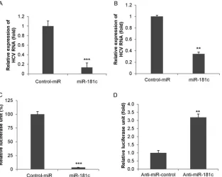
Exogenous expression of miR-181c suppresses HCV replica-tion.We next examined the effect of miR-181c on HCV replica-tion in Huh7.5 cells infected with HCV genotype 2a. Cells were transfected with the control or the miR-181c mimic after 2 days of infection, and RNA was isolated at 48 h posttransfection. HCV replication was significantly reduced in miR-181c-overexpressing cells compared to that in control-transfected cells (Fig. 7A). Rep2a-Rluc cells were also transfected with the control miRNA or the miR-181c mimic, and HCV replication was measured by qRT-PCR or a luciferase reporter assay after 48 h of transfection. A significant inhibition of HCV RNA expression was observed in miR-181c mimic-overexpressing Rep2a-Rluc cells compared to control cells (Fig. 7BandC). Knockdown of miR-181c using anti-miR-181c enhanced HCV replication, as evident from theRenilla luciferase activity (Fig. 7D). Together, these data suggested that FIG 6Correlation of STAT3, HCV RNA, and miR-181c expression levels in
liver biopsy specimens. Correlation plots are shown for miR-181c versus STAT3 levels (A) and STAT3 versus HCV RNA levels (B). Statistical analysis was done by using the Spearman correlation.
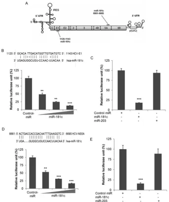
FIG 7Exogenous expression of the miR-181c mimic inhibits HCV replication. (A) Huh7.5 cells were infected with HCV genotype 2a (multiplicity of infection of 1) for 48 h and then transfected with either control miRNA (Control-miR) or the miR-181c mimic. Total RNA was extracted after 48 h of transfection, and HCV replication was measured by qRT-PCR. Cellular 18S RNA was used as an internal control for normalization. (B) Rep2a-Rluc cells were transfected with control miRNA or the miR-181c mimic, and HCV genomic RNA replication was measured by qRT-PCR after 48 h of transfection. (C) Rep2a-Rluc cells were transfected with control miRNA or the miR-181c mimic, and relative luciferase activity was measured by aRenillaluciferase assay after 48 h of transfection. Results are presented as the means and standard deviations from three independent experiments. (D) Rep2a-Rluc cells were transfected with control anti-miR or anti-miR-181c, and relative luciferase activity was measured. Results are presented as the means and standard deviations from three independent experiments.
ⴱⴱ,P⬍0.01;ⴱⴱⴱ,P⬍0.001.
on November 7, 2019 by guest
http://jvi.asm.org/
[image:8.585.60.266.67.331.2] [image:8.585.138.451.397.647.2]overexpression of miR-181c suppresses HCV replication in hepa-toma cells.
Since overexpression of miR-181c downregulates HCV repli-cation, we performedin silicoanalyses with ViTa algorithms (21) to identify whether miR-181c physically binds to the HCV ge-nome. Lower minimum free energy (MFE) values of the miRNAs and the target sites revealed energetically more probable hybrid-izations between miRNAs and target genes (21). Ourin silico anal-ysis suggested a putative binding site for miR-181c in the HCV E1 and NS5A genes, with lower MFE values of⫺20.2 and⫺18.2, respectively (Fig. 8A,B, andD). We next cloned the HCV E1 or HCV NS5A sequence with the potential binding site of miR-181c into the pMIR-Report luciferase vector. Cells were cotransfected
with the E1– or NS5A–pMIR-Report luciferase vector and the miR-181c mimic, and luciferase expression was measured. The results demonstrated dose-dependent inhibition of both E1 and NS5A expression upon miR-181c overexpression (Fig. 8BandD). To further verify the specificity of miR-181c binding to HCV E1 or NS5A sequences, we used an unrelated miRNA, miR-203. A sig-nificant change in luciferase activity was not observed when the unrelated miRNA miR-203 was cotransfected with the E1– or NS5A–pMIR-Report luciferase vector (Fig. 8CandE).
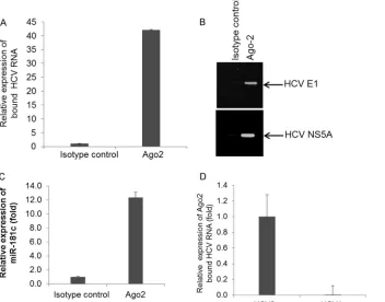
Finally, we examined the interaction between HCV RNA and miR-181c. For this, miR-181c-transfected Rep2a-Rluc cell lysates were immunoprecipitated with an Ago2-specific monoclonal an-tibody or an unrelated isotype control. RNA was isolated from the FIG 8miR-181c directly targets HCV E1 and NS5A. (A) Schematic representation of the HCV genome showing potential target sites for miR-181c. IRES, internal ribosome entry site. (B)In silicoanalysis of HCV E1 reveals a single putative miR-181c binding site. Huh7.5 cells were cotransfected with HCV E1 cloned into the pMIR-Report luciferase vector and different doses of the miR-181c mimic. Relative luciferase activity was measured after 48 h of transfection. Control-miR, control miRNA. (C) Huh7.5 cells were cotransfected with HCV E1 cloned into the pMIR-Report luciferase vector, control miRNA, the miR-181c mimic, or miR-203. Relative luciferase activity was measured after 48 h of transfection. (D)In silicoanalysis of the miR-181c target site in HCV NS5A. Huh7.5 cells were cotransfected with HCV NS5A cloned into the pMIR-Report luciferase vector and different doses of the miR-181c mimic. Relative luciferase activity was measured after 48 h of transfection. (E) Huh7.5 cells were cotransfected with HCV NS5A cloned into the pMIR-Report luciferase vector and control miRNA, the miR-181c mimic, or miR-203. Relative luciferase activity was measured after 48 h of transfection. Results are presented as the means and standard deviations from three independent experiments.ⴱⴱ,P⬍0.01;ⴱⴱⴱ,P⬍0.001.
HCV-Mediated miR-181ctrans-Suppression
on November 7, 2019 by guest
http://jvi.asm.org/
[image:9.585.121.464.65.474.2]immunoprecipitate by using an RNeasy kit and analyzed by qRT-PCR. The RNA level of the HCV replicon was approximately⬃ 40-fold higher in the Ago2 immunoprecipitate than in the control immunoprecipitate (Fig. 9AandB). Expression of miR-181c in the immunoprecipitate was also observed (Fig. 9C). We also ex-amined this specific interaction in Huh7.5 cell lysates transfected with control miRNA or miR-181c with HCV RNA and immuno-precipitated with the Ago2-specific monoclonal antibody. As ex-pected, only amplification of HCV RNA was observed for miR-181c-transfected cells (data not shown). To further investigate whether miR-181c targets other HCV genotypes, we took advan-tage of an HCV genotype 1a (clone H77) subgenomic replicon, where the miR-181c binding site of NS5A is mismatched between HCV genotype 2a and HCV genotype 1a. We did not observe HCV RNA accumulation in immunoprecipitates examined with the subgenomic replicon from HCV genotype 1a in Ago2 immu-noprecipitation (IP) experiments (Fig. 9D). Together, these re-sults suggested that miR-181c binds directly to the HCV genome and inhibits viral replication.
DISCUSSION
In this study, we have demonstrated that (i) HCV infection tran-scriptionally downregulates miR-181c, (ii) HCV-mediated down-regulation of miR-181c enhances HOXA1 and its downstream molecules involved in cell growth regulation, and (iii) exogenous expression of miR-181c exerts anti-HCV activity by binding to E1 and NS5A. To our knowledge, this is the first report showing
HCV-mediated transcriptional regulation of a cellular miRNA in hepatocytes.
miRNAs are responsible for regulating many cellular pro-cesses, and viruses modulate the host miRNA milieu in different ways to facilitate pathogenesis (5,8,9,22). In response to viral infection, signaling pathways in mammalian cells direct a variety of intracellular events that generate an immune response within the infected cells. Genes of the miR-181 family play a central role in malignant transformation, are downregulated in many tumors, and thus appear to function as tumor suppressor genes in chronic lymphocytic leukemia, breast cancer, and glioblastoma (23–25). We observed suppression of miR-181c by HCV in human hepa-tocytes, suggesting direct evidence for an association with viral infection. Downregulation of miR-181c in HCV-infected liver bi-opsy specimens, but not in liver bibi-opsy specimens from patients with unrelated liver disease, was also observed. Thus, inhibition of miR-181c appeared to be specific to HCV infection. miR-181c is in a cluster with miR-181d in an intron of the coding gene NANOS3, according to miRBase. When we performed an miRNA array using RNA from mock- or HCV-infected primary human hepatocytes from two different donors, significant downregula-tion of miR-181c was observed, but the alteradownregula-tion of miR-181d expression was not significant, which may be due to posttranscrip-tional regulation. Although miR-181c and NANOS3 are regulated by the same promoter, to our knowledge, there is no report of their coexpression. NANOS3 has been implicated as a repressor, FIG 9miR-181c directly binds to the HCV genotype 2a genome. (A) Rep2a-Rluc cell lysates were immunoprecipitated with Ago2-specific monoclonal antibody or an unrelated IgG2a isotype control. RNA was isolated from immunoprecipitates by using an RNeasy kit, and HCV RNA expression was analyzed by qRT-PCR. (B) HCV E1 or NS5A was amplified from immunoprecipitates by RT-PCR and analyzed by agarose gel electrophoresis. A representative sample of triplicates is shown. (C) Relative expression levels of miR-181c from Ago2 immunoprecipitates analyzed by qRT-PCR. (D) miR-181c-expressing HCV genotype 2a- or HCV genotype 1a-harboring Huh7.5 cell lysates were immunoprecipitated with Ago2-specific monoclonal antibody. RNA was isolated from immunoprecipitates, and
HCV RNA expression was analyzed by qRT-PCR.
on November 7, 2019 by guest
http://jvi.asm.org/
[image:10.585.125.477.69.345.2]and therefore, it will be important to study its function in the setting of HCV infection in the future. Our results further dem-onstrated that HCV transcriptionally inhibits miR-181c expres-sion through C/EBP-, a cellular transcription factor. C/EBP-is involved in several cellular functions, such as cell proliferation, apoptosis, and differentiation (26). Many viral proteins can in-duce multiple signaling pathways, which may cross talk with each other or converge on common downstream effectors. We have previously shown that C/EBP-is downregulated during HCV infection mediated by HCV NS5A (18). Here, we demonstrated that HCV NS5A inhibits miR-181c promoter activity by modulat-ing C/EBP-expression. Deletion of the C/EBP-binding site on an miR-181c promoter (P3fragment) or knockdown of C/EBP- restored HCV-mediated suppression of miR-181c promoter ac-tivity.
Our results further demonstrated that HOXA1 is a direct target of miR-181c. In vertebrates, the homeobox genes are found in clusters A, B, C, and D on four separate chromosomes (27,28). HOXA1 is a part of the A clusters on chromosome 7 and encodes a DNA binding transcription factor that regulates gene expres-sion, morphogenesis, cell proliferation, and differentiation (20, 29,30). HOXA1 expression is also known to potentiate oncogenic transformation in immortalized mammary epithelial cells by stimulating STAT3/5 (29). We have observed that expression lev-els of HOXA1 and its downstream signaling molecules STAT3 and STAT5 are significantly reduced in HCV-infected hepatocytes fol-lowing introduction of the miR-181c mimic. miR-181c also di-rectly targets the 3=untranslated region of HOXA1. Furthermore, we have shown that miR-181c and STAT3 expression levels are inversely correlated in liver biopsy specimens, whereas HCV and STAT3 expression levels are positively correlated in liver biopsy specimens. Moreover, STAT3 expression negatively regulates the type I IFN-mediated antiviral response (31). Therefore, it is plau-sible that HCV-mediated STAT3 activation limits the IFN-medi-ated antiviral response. Exogenous expression of miR-181c also inhibited the growth of Huh7.5 cells harboring the HCV genotype 2a replicon, suggesting that miR-181c-mediated HOXA1 modu-lation plays a role in cell proliferation and in HCV-mediated he-patocyte growth regulation.
Emerging evidence suggests that cellular miRNAs play a key role in the regulation of viral replication (8). miR-24 and miR-93 inhibit vesicular stomatitis virus replication by targeting genes encoding viral L and P proteins, respectively (32). miR-125-5p targets the S protein of HBV to inhibit replication (33). miR-122, miR-199a, and miR-210 also suppress HBV replication (34,35). On the other hand, miR-196, miR-351, miR-431, and miR-448 inhibit HCV replication in Huh7 cells by interacting with the HCV genome (36). Recently, miR-296-5p was shown to inhibit enterovirus 71 replication by binding to the VP-1 gene (37). Our study demonstrated that the introduction of miR-181c into HCV-infected hepatocytes or cells harboring the HCV genome-length replicon inhibits viral genome replication. We have furthermore shown that miR-181c has strong target sequences for the HCV envelope glycoprotein E1 and the nonstructural protein NS5A. The inhibitory effect of miR-181c on HCV replication appears to depend on complementarity between genotype and host se-quences.
In summary, we have demonstrated that HCV infection tran-scriptionally downregulates miR-181c expression by targeting C/EBP-, which in turn enhances the expression of the cell growth
regulator HOXA1. Furthermore, exogenous expression of miR-181c limits HCV replication. Inhibition of miR-miR-181c by HCV may suppress the antiviral activity of miR-181c. Taken together, our observations contribute to the understanding of the mechanistic role of miR-181c in HCV-host interactions, which may have po-tential for therapeutic strategies.
ACKNOWLEDGMENTS
We thank Adrian M. Di Bisceglie for providing coded liver biopsy speci-mens, Lynda Morrison for antibodies, Charles Rice for the H77 sub-genomic replicon, Hengli Tang for the Rep2a-Rluc cell line, and Robert Steele for technical assistance.
This work was supported by research grants DK081817 (R.B.R.) and DK080812 (R.R.) from the National Institutes of Health and the Doisy Research Fund from the Saint Louis University Pathology Department.
REFERENCES
1.Gravitz L.2011. Introduction: a smouldering public-health crisis. Nature 474:S2–S4.http://dx.doi.org/10.1038/474S2a.
2.Scheel TK, Rice CM.2013. Understanding the hepatitis C virus life cycle paves the way for highly effective therapies. Nat. Med.19:837– 849.http: //dx.doi.org/10.1038/nm.3248.
3.Bartenschlager R, Lohmann V, Penin F.2013. The molecular and structural basis of advanced antiviral therapy for hepatitis C virus infection. Nat. Rev. Microbiol.11:482– 496.http://dx.doi.org/10.1038/nrmicro3046.
4.Meister G, Tuschl T.2004. Mechanisms of gene silencing by double-stranded RNA. Nature 431:343–349. http://dx.doi.org/10.1038/nature 02873.
5.Bartel DP.2009. MicroRNAs: target recognition and regulatory func-tions. Cell136:215–233.http://dx.doi.org/10.1016/j.cell.2009.01.002. 6.Winter J, Jung S, Keller S, Gregory RI, Diederichs S.2009. Many roads
to maturity: microRNA biogenesis pathways and their regulation. Nat. Cell Biol.11:228 –234.http://dx.doi.org/10.1038/ncb0309-228. 7.Roberts AP, Lewis AP, Jopling CL.2011. The role of microRNAs in viral
infection. Prog. Mol. Biol. Transl. Sci.102:101–139.http://dx.doi.org/10 .1016/B978-0-12-415795-8.00002-7.
8.Cullen BR.2013. How do viruses avoid inhibition by endogenous cellular microRNAs? PLoS Pathog.9:e1003694.http://dx.doi.org/10.1371/journal .ppat.1003694.
9.Conrad KD, Niepmann M.2014. The role of microRNAs in hepatitis C virus RNA replication. Arch. Virol. 159:849 – 862.http://dx.doi.org/10 .1007/s00705-013-1883-4.
10. Shrivastava S, Mukherjee A, Ray RB.2013. Hepatitis C virus infection, microRNA and liver disease progression. World J. Hepatol.5:479 – 486. http://dx.doi.org/10.4254/wjh.v5.i9.479.
11. Cullen BR.2013. MicroRNAs as mediators of viral evasion of the immune system. Nat. Immunol.14:205–210.http://dx.doi.org/10.1038/ni.2537. 12. Bhanja Chowdhury J, Shrivastava S, Steele R, Di Bisceglie AM, Ray R,
Ray RB.2012. Hepatitis C virus infection modulates expression of inter-feron stimulatory gene IFITM1 by upregulating miR-130A. J. Virol.86: 10221–10225.http://dx.doi.org/10.1128/JVI.00882-12.
13. Kanda T, Basu A, Steele R, Wakita T, Ryerse JS, Ray R, Ray RB.2006. Generation of infectious hepatitis C virus in immortalized human hepa-tocytes. J. Virol.80:4633– 4639.http://dx.doi.org/10.1128/JVI.80.9.4633 -4639.2006.
14. Shrivastava S, Bhanja Chowdhury J, Steele R, Ray R, Ray RB.2012. Hepatitis C virus upregulates Beclin1 for induction of autophagy and ac-tivates mTOR signaling. J. Virol.86:8705– 8712.http://dx.doi.org/10.1128 /JVI.00616-12.
15. Shimakami T, Yamane D, Jangra RK, Kempf BJ, Spaniel C, Barton DJ, Lemon SM.2012. Stabilization of hepatitis C virus RNA by an Ago2-miR-122 complex. Proc. Natl. Acad. Sci. U. S. A.109:941–946.http://dx.doi.org /10.1073/pnas.1112263109.
16. Murakami Y, Aly HH, Tajima A, Inoue I, Shimotohno K.2009. Regu-lation of the hepatitis C virus genome replication by miR-199a. J. Hepatol. 50:453– 460.http://dx.doi.org/10.1016/j.jhep.2008.06.010.
17.Farré D, Roset R, Huerta M, Adsuara JE, Roselló L, Albà MM, Messeguer X.2003. Identification of patterns in biological sequences at the ALGGEN server: PROMO and MALGEN. Nucleic Acids Res.31:3651– 3653.http://dx.doi.org/10.1093/nar/gkg605.
HCV-Mediated miR-181ctrans-Suppression
on November 7, 2019 by guest
http://jvi.asm.org/
18. Mazumdar B, Kim H, Meyer K, Bose SK, Di Bisceglie AM, Ray RB, Ray R.2012. Hepatitis C virus proteins inhibit C3 complement production. J. Virol.86:2221–2228.http://dx.doi.org/10.1128/JVI.06577-11.
19. Banerjee A, Ray RB, Ray R.2010. Oncogenic potential of hepatitis C virus proteins. Viruses2:2108 –2133.http://dx.doi.org/10.3390/v2092108. 20. Mohankumar KM, Perry JK, Kannan N, Kohno K, Gluckman PD, Emerald BS, Lobie PE.2008. Transcriptional activation of signal trans-ducer and activator of transcription (STAT) 3 and STAT5B partially me-diate homeobox A1-stimulated oncogenic transformation of the immor-talized human mammary epithelial cell. Endocrinology149:2219 –2229. http://dx.doi.org/10.1210/en.2007-1320.
21. Hsu PW, Lin LZ, Hsu SD, Hsu JB, Huang HD.2007. ViTa: prediction of host microRNAs targets on viruses. Nucleic Acids Res.35:D381–D385. http://dx.doi.org/10.1093/nar/gkl1009.
22. Sarnow P, Jopling CL, Norman KL, Schütz S, Wehner KA. 2006. MicroRNAs: expression, avoidance and subversion by vertebrate viruses. Nat. Rev. Microbiol.4:651– 659.http://dx.doi.org/10.1038/nrmicro1473. 23. Pekarsky Y, Santanam U, Cimmino A, Palamarchuk A, Efanov A, Maximov V, Volinia S, Alder H, Liu CG, Rassenti L, Calin GA, Hagan JP, Kipps T, Croce CM.2006. Tcl1 expression in chronic lymphocytic leukemia is regulated by miR-29 and miR-181. Cancer Res.66:11590 – 11593.http://dx.doi.org/10.1158/0008-5472.CAN-06-3613.
24. Lowery AJ, Miller N, Devaney A, McNeill RE, Davoren PA, Lemetre C, Benes V, Schmidt S, Blake J, Ball G, Kerin MJ. 2009. MicroRNA signatures predict oestrogen receptor, progesterone receptor and HER2/ neu receptor status in breast cancer. Breast Cancer Res.11:R27.http://dx .doi.org/10.1186/bcr2257.
25. Conti A, Aguennouz M, La Torre D, Tomasello C, Cardali S, Angileri FF, Maio F, Cama A, Germanò A, Vita G, Tomasello F.2009. miR-21 and 221 upregulation and miR-181b downregulation in human grade II-IV astrocytic tumors. J. Neurooncol.93:325–332.http://dx.doi.org/10 .1007/s11060-009-9797-4.
26. Ramji DP, Foka P.2002. CCAAT/enhancer-binding proteins: structure, function and regulation. Biochem. J.365:561–575.http://www.ncbi.nlm .nih.gov/pmc/articles/PMC1222736/pdf/12006103.pdf.
27. Hassan NM, Hamada J, Murai T, Seino A, Takahashi Y, Tada M, Zhang X, Kashiwazaki H, Yamazaki Y, Inoue N, Moriuchi T.2006. Aberrant expression of HOX genes in oral dysplasia and squamous cell carcinoma tissues. Oncol. Res.16:217–224.
28. Grier DG, Thompson A, Kwasniewska A, McGonigle GJ, Halliday HL,
Lappin TR.2005. The pathophysiology of HOX genes and their role in cancer. J. Pathol.205:154 –171.http://dx.doi.org/10.1002/path.1710. 29. Mohankumar KM, Xu XQ, Zhu T, Kannan N, Miller LD, Liu ET,
Gluck-man PD, Sukumar S, Emerald BS, Lobie PE.2007. HOXA1-stimulated oncogenicity is mediated by selective upregulation of components of the p44/42 MAP kinase pathway in human mammary carcinoma cells. Oncogene 26:3998 – 4008.http://dx.doi.org/10.1038/sj.onc.1210180.
30. Wardwell-Ozgo J, Dogruluk T, Gifford A, Zhang Y, Heffernan TP, van Doorn R, Creighton CJ, Chin L, Scott KL.2014. HOXA1 drives mela-noma tumor growth and metastasis and elicits an invasion gene expres-sion signature that prognosticates clinical outcome. Oncogene33:1017– 1026.http://dx.doi.org/10.1038/onc.2013.30.
31. Wang WB, Lew SE, Lee CK.2011. STAT3 negatively regulates type I IFN-mediated antiviral response. J. Immunol.187:2578 –2585.http://dx .doi.org/10.4049/jimmunol.1004128.
32. Otsuka M, Jing Q, Georgel P, New L, Chen J, Mols J, Kang YJ, Jiang Z, Du X, Cook R, Das SC, Pattnaik AK, Beutler B, Han J.2007. Hyper-susceptibility to vesicular stomatitis virus infection in Dicer1-deficient mice is due to impaired miR24 and miR93 expression. Immunity27:123– 134.http://dx.doi.org/10.1016/j.immuni.2007.05.014.
33. Potenza N, Papa U, Mosca N, Zerbini F, Nobile V, Russo A.2011. Human microRNA hsa-miR-125a-5p interferes with expression of hepa-titis B virus surface antigen. Nucleic Acids Res.39:5157–5163.http://dx .doi.org/10.1093/nar/gkr067.
34. Chen Y, Shen A, Rider PJ, Yu Y, Wu K, Mu Y, Hao Q, Liu Y, Gong H, Zhu Y, Liu F, Wu J.2011. A liver-specific microRNA binds to a highly conserved RNA sequence of hepatitis B virus and negatively regulates viral gene expression and replication. FASEB J.25:4511– 4521.http://dx.doi .org/10.1096/fj.11-187781.
35. Zhang GL, Li YX, Zheng SQ, Liu M, Li X, Tang H.2010. Suppression of hepatitis B virus replication by 199a-3p and microRNA-210. Antiviral Res.88:169 –175.http://dx.doi.org/10.1016/j.antiviral.2010 .08.008.
36. Pedersen IM, Cheng G, Wieland S, Volinia S, Croce CM, Chisari FV, David M.2007. Interferon modulation of cellular microRNAs as an anti-viral mechanism. Nature449:919 –922.http://dx.doi.org/10.1038/nature 06205.
37. Zheng Z, Ke X, Wang M, He S, Li Q, Zheng C, Zhang Z, Liu Y, Wang H.2013. Human microRNA hsa-miR-296-5p suppresses enterovirus 71 replication by targeting the viral genome. J. Virol.87:5645–5656.http://dx .doi.org/10.1128/JVI.02655-12.