Role of WDHD1 in Human Papillomavirus-Mediated Oncogenesis
Identified by Transcriptional Profiling of E7-Expressing Cells
Yunying Zhou,aQishu Zhang,aGe Gao,bXiaoli Zhang,cYafei Liu,aShoudao Yuan,aXiaowei Wang,bJason J. Chena
The Cancer Research Center, Shandong University School of Medicine, Jinan, Shandong, Chinaa; Department of Radiation Oncology, Washington University School of Medicine, St. Louis, Missouri, USAb; The Qilu Hospital, Shandong University School of Medicine, Jinan, Shandong, Chinac
ABSTRACT
The E7 oncoprotein of the high-risk human papillomavirus (HPV) plays a major role in HPV-induced carcinogenesis. E7
abro-gates the G
1cell cycle checkpoint and induces genomic instability, but the mechanism is not fully understood. In this study, we
performed RNA sequencing (RNA-seq) to characterize the transcriptional profile of keratinocytes expressing HPV 16 (HPV-16)
E7. At the transcriptome level, 236 genes were differentially expressed between E7 and vector control cells. A subset of the
differ-entially expressed genes, most of them novel to E7-expressing cells, was further confirmed by real-time PCR. Of interest, the
ac-tivities of multiple transcription factors were altered in E7-expressing cells. Through bioinformatics analysis, pathways altered
in E7-expressing cells were investigated. The upregulated genes were enriched in cell cycle and DNA replication, as well as in the
DNA metabolic process, transcription, DNA damage, DNA repair, and nucleotide metabolism. Specifically, we focused our
stud-ies on the gene encoding WDHD1 (WD repeat and high mobility group [HMG]-box DNA-binding protein), one of the genes that
was upregulated in E7-expressing cells. WDHD1 is a component of the replisome that regulates DNA replication. Recent studies
suggest that WDHD1 may also function as a DNA replication initiation factor as well as a G
1checkpoint regulator. We found
that in E7-expressing cells, the steady-state level of WDHD1 protein was increased along with the half-life. Moreover,
downregu-lation of WDHD1 reduced E7-induced G
1checkpoint abrogation and rereplication, demonstrating a novel function for
WDHD1. These studies shed light on mechanisms by which HPV induces genomic instability and have therapeutic implications.
IMPORTANCE
The high-risk HPV types induce cervical cancer and encode an E7 oncoprotein that plays a major role in HPV-induced
carcino-genesis. However, the mechanism by which E7 induces carcinogenesis is not fully understood; specific anti-HPV agents are not
available. In this study, we performed RNA-seq to characterize transcriptional profiling of keratinocytes expressing HPV-16 E7
and identified more than 200 genes that were differentially expressed between E7 and vector control cells. Through
bioinformat-ics analysis, pathways altered in E7-expressing cells were identified. Significantly, the WDHD1 gene, one of the genes that is
up-regulated in E7-expressing cells, was found to play an important role in E7-induced G
1checkpoint abrogation and rereplication.
These studies shed light on mechanisms by which HPV induces genomic instability and have therapeutic implications.
H
uman papillomaviruses (HPVs) are small DNA viruses that
replicate in squamous epithelia. Specific types of HPV
(high-risk HPVs) are the causative agents for cervical and several other
cancers (
1
). The transforming properties of high-risk HPVs such
as HPV 16 (HPV-16) primarily depend on E7 as well as E6
onco-genes (
1
,
2
). HPV E6 and E7 proteins promote the degradation of
p53 and pRb, respectively (
3
,
4
). E7 from the high-risk HPV types
can abrogate cell cycle checkpoints and induces genomic
instabil-ity. Although several transcription profiling studies for E7 have
been conducted using DNA microarray analysis (
3
,
5–7
), the HPV
E7 activities downstream from, or independent of, pRb
responsi-ble for deregulation of cell cycle and induction of genomic
insta-bility are not fully understood.
Cell cycle progression is regulated by cyclins and by
cyclin-dependent kinases (Cdks) and their regulatory proteins at
sev-eral checkpoints (
8
). Once the checkpoint becomes abnormal,
genomic instability may occur (
8
). Genomic instability is a
hall-mark of cancer progression (
9
). Polyploidy is a type of genomic
instability where cells have more than two sets of chromosomes
and has been recognized as a causal factor for tumorigenesis (
10
).
Significantly, polyploidy can be detected in the early stage of
cer-vical carcinogenesis (
11
). Polyploidy can be formed via
rereplica-tion, a process of successive rounds of host DNA replication
with-out entering mitosis (
12
). Rereplication may lead to not only
polyploidy but also gene amplification, DNA fragmentation, DNA
breaks, and cellular DNA damage response (
13–15
). We recently
demonstrated that HPV-16 E7 induces rereplication and that the
cellular DNA replication initiation factor Cdt1 plays a role in this
process (
16
).
DNA replication is regulated by sequential and interactive
mechanisms to ensure that the genome is accurately replicated
only once per cell cycle. The process of replication initiation is
Received21 March 2016Accepted16 April 2016
Accepted manuscript posted online20 April 2016
CitationZhou Y, Zhang Q, Gao G, Zhang X, Liu Y, Yuan S, Wang X, Chen JJ. 2016. Role of WDHD1 in human papillomavirus-mediated oncogenesis identified by transcriptional profiling of E7-expressing cells. J Virol 90:6071–6084.
doi:10.1128/JVI.00513-16.
Editor:L. Banks, International Centre for Genetic Engineering and Biotechnology
Address correspondence to Xiaowei Wang, xwang@radonc.wustl.edu, or Jason J. Chen, jxchen@sdu.edu.cn.
Supplemental material for this article may be found athttp://dx.doi.org/10.1128 /JVI.00513-16.
Copyright © 2016, American Society for Microbiology. All Rights Reserved.
on November 7, 2019 by guest
http://jvi.asm.org/
divided into two steps, pre-replicative complex (pre-RC)
assem-bly and activation; the latter leads to generation of replication
forks. Pre-RC starts with the association of the origin recognition
complex (ORC), which then promotes the recruitment of two
proteins, Cdc6 and Cdt1, onto origins. This is followed by
recruit-ment of minichromosome maintenance 2-7 (MCM2-7) onto
chromatin as a result of concerted actions of Cdc6 and Cdt1 (
9
).
Prior to the S phase, origins are licensed by the binding of
com-ponents of the replicative DNA helicase MCMs in eukaryotes (
17
).
Afterward, licensing proteins are downregulated or inhibited such
that no more origins can be licensed and rereplication of DNA is
prevented. Cells employ a licensing checkpoint to monitor that
sufficient origins are licensed, inhibiting S-phase entry until that
state is established (
18
). The G
1arrest observed in cells that have
engaged in the licensing checkpoint is associated with low levels of
G
1Cdk-cyclin activity and pRb hypophosphorylation.
WDHD1 (WD repeat and high mobility group [HMG]-box
DNA-binding protein 1) contains multiple N-terminal WD40
do-mains and a C-terminal HMG box. WD40 dodo-mains are found in a
variety of eukaryotic proteins and may function as
adaptor/regu-latory modules in signal transduction, pre-mRNA processing, and
cytoskeleton assembly. HMG boxes are found in many eukaryotic
proteins involved in chromatin assembly, transcription, and
rep-lication. In addition to its established role in pre-RC activation
(
19–21
), WDHD1 is also involved in pre-RC assembly (
19
). The
WDHD1 gene is localized adjacent to replication foci, interacts
with human primase-DNA polymerase/MCM10, and is required
for DNA synthesis (
20
,
22
,
23
). A role for WDHD1 in G
1check-point control has recently been suggested (
23
). In addition,
deple-tion of WDHD-1 increases DNA damage, leading to the
accumu-lation of late S- and/or G
2-phase cells (
24
).
In this study, we performed high-throughput RNA sequencing
(RNA-seq) to characterize the transcriptional profile of
keratino-cytes expressing HPV-16 E7. In E7-expressing cells, the expression
levels of hundreds of genes were found to be differentially
regu-lated, the activity of multiple transcription factors was altered, and
multiple molecular pathways were changed. The WDHD1 gene is
among the genes upregulated in E7-expressing cells. Significantly,
downregulation of the WDHD1 gene reduced E7-induced G
1checkpoint abrogation and rereplication. These results should
help provide insights into the cellular pathways targeted during
tumor development caused by HPV.
MATERIALS AND METHODS
Cell culture.Primary human keratinocytes (PHKs) and spontaneously immortalized human foreskin keratinocytes (NIKS cells) were cultured on mitomycin C-treated J2-3T3 feeder cells with E medium composed of 3 parts Dulbecco’s modified Eagle medium (DMEM) and 1 part Ham’s F12 medium plus 5% fetal bovine serum (FBS). Cells of the human telo-merase reverse transcriptase-expressing human retinal pigment epithe-lium cell line RPE1 were maintained in a 1:1 dilution of DMEM-Ham’s F-12 medium plus 10% FBS. PHK and NIKS cells expressing HPV-16 E7 and RPE1 cells expressing HPV-16 E7 or HPV-6 E7 were established using a pBabe retroviral system as described previously (16). Populations of infected cells were pooled and expanded. PHKs, NIKS cells, and RPE1-derived cell lines were maintained in puromycin and used within 15 pas-sages.
RNA-seq.Total RNA from NIKS cells was used to construct cDNA libraries for RNA-seq. First, the rRNA was removed by the use of a Ribo-Minus kit (Life Technologies) combined with custom-designed DNA probes for rRNAs. The processed total RNA was then used to construct
RNA-seq libraries with a NEBNext mRNA library preparation kit (New England BioLabs). In this way, double-stranded cDNA was synthesized from rRNA-depleted total RNA. RNA was then end repaired, dA tailed, and ligated to standard Illumina adaptor oligonucleotides. Adaptor-li-gated cDNA libraries were amplified with Phusion PCR master mix. The cDNA libraries were then loaded into a HiSeq 2000 system (Illumina) for sequencing at the Washington University Genome Technology Access Center. The resulting raw sequence reads were first analyzed using a cus-tom bioinformatics pipeline to remove low-quality reads and then clus-tered before sequentially mapping to the human transcriptome and ge-nome with Bowtie was performed (25). In this way, about 90% of all sequence reads were mapped to known human sequences. Sequence reads mapping to the same transcript were combined and normalized on the basis of the length of the transcript and the number of total reads from each sample (reads per kilobase per million [RPKM]). Normalized read counts were compared across samples to identify changes in transcript abundance. Transcripts from the same gene locus in the human genome were combined for evaluation of expression changes at the gene level.
Bioinformatics analysis.The official gene designations for the genes that showed significant expression differences between E7 and vector cells were submitted to the Database for Annotation, Visualization and Inte-grated Discovery (DAVID, v6.7) Bioinformatics Resources.Homo sapiens were selected as the annotation species. Functional classification of differ-entially expressed genes was analyzed by gene ontology (GO) via the use of the Web-based Gene Set Analysis Toolkit. Pathway analysis was per-formed using the Kyoto Encyclopedia of Genes and Genomes (KEGG) PATHWAY database via Web Gestalt. To identify the statistical signifi-cance of the cluster results, we set aPvalue ofⱕ0.05 as the cluster filter. We then used the gene counts and terms to make the column chart sepa-rately for each category.
RT-PCR.Total RNA from RPE1, NIKS, and PHK control cells and the corresponding E7-expressing cells was isolated using an RNeasy kit (Qiagen) according to the manufacturer’s instruction. cDNA was synthe-sized with a Superscript VILO cDNA synthesis kit (Invitrogen). iTaq Uni-versal SYBR green Supermix (Bio-Rad) was used in a Bio-Rad CFX96 Touch Real-Time PCR detection system for quantitative real-time PCR (qRT-PCR). Data were analyzed using the threshold cycle (2⫺⌬⌬CT) method. The primer sequences are listed inTable 1.
ChIP assay.The chromatin immunoprecipitation (ChIP) assay was performed using a ChIP assay kit from Millipore, following the supplied protocol. Immunoprecipitations were performed using anti-MCM3 or control IgG antibodies. PCR was performed with the primers designed from the sequences of the human c-MYC gene as follows: sense, AAG CTGAATTGTGCAGTGCATC; antisense, CTCACCCAAAGGCATTTT AAG. It was shown that the MCM protein complex binds to the DNA replication initiation zone upstream of the c-MYC gene (26).
Flow cytometry.For cell cycle and polyploidy analysis, asynchronous cultured cells expressing HPV E7 or vector alone were treated with phos-phate-buffered saline (PBS) or bleomycin (Alexis Biochemicals) (4g/ml in PBS). At 24 h later, cells were fixed in 70% ethanol, treated with 50
g/ml RNase A plus 50g/ml propidium iodide, and analyzed by fluo-rescence-activated cell sorting (FACS). For the bromodeoxyuridine (BrdU) labeling experiment, BrdU (final concentration, 20M) was added to the medium 2 h before collection of cells. After fixation, cells were permeabilized with 2 N HCl– 0.5% Triton X-100, neutralized with 0.1 M sodium tetraborate, stained with monoclonal anti-BrdU (BD Bio-sciences) followed by treatment with anti-mouse IgG F(ab)2-fluorescein isothiocyanate (FITC) (Sigma), and counterstained with PBS–7-amino-actinomycin D (7-AAD)–RNase A. Flow cytometric analysis was per-formed on a BD FACSAria III sorter instrument equipped with BD FACSDiva 7.0 software (BD Biosciences, NJ, USA). FITC 490-nm fluo-rescence was acquired in logarithmic amplification in FL1, and 7-AAD 650-nm fluorescence was acquired in linear amplification in FL3. Cell cycle analysis was done using a Cytomics FC500 Flow Cytometry CXP 2.0 system.
on November 7, 2019 by guest
http://jvi.asm.org/
siRNAs and transfection.Cells were transfected with a final concen-tration of 20 nM small interfering RNA (siRNA) per target gene using Lipofectamine 2000 transfection reagent (Invitrogen) according to the manufacturer’s instructions. For gene knockdown analysis, cells were har-vested 48 h posttransfection and specific protein levels were analyzed by immunoblotting. For cell cycle analysis, 24 h after transfection, cells were treated with bleomycin (4g/ml) for an additional 24 h. For polyploidy analysis, cells were blocked in G1phase with thymidine for 16 h after transfection with 20 nM siRNA for 24 h, and then cells were treated with bleomycin (4g/ml) for an additional 48 h. The siRNA duplexes were as follows: for the small interfering WDHD1-1 (si-WDHD1-1) sense strand, 5=-GCAUGUACCCUAAGAAUAA-3=; for the si-WDHD1-2 sense strand, 5=-GCAAAGUUAUGGAAAGUAU-3=; for the si-MCM3 sense strand, 5=-GCATTGTCACTAAATGTTCTCTAGT-3=(27); and for the negative-control (NC) siRNA sense strand, 5=-UUCUCCGAA CGUGUCACGU-3=.
Immunoblotting.Total cellular protein was prepared in lysis buffer (10 mM Tris [pH 7.4], 1% SDS, 1.0 mM sodium orthovanadate). To obtain cytoplasmic and nuclear proteins, cells were extracted with a nu-clear extract kit (Active motif; 100946). The protein concentration was measured by the use of bicinchoninic acid (BCA) protein assay reagent (Pierce) and confirmed by Coomassie blue staining of membranes after blotting. Equal amounts of protein from each cell lysate were separated in an SDS polyacrylamide gel (PAGE) and transferred onto a nitrocellulose filter membrane (NC) membrane. Membranes were blotted with anti-bodies against WDHD1 (abcam; ab72436), MCM3 (abcam; ab4460), SP1 (Cell Signaling; catalog no. 9389), and tubulin (Sigma; T-4026). Protein bands were detected using an Odyssey infrared imaging system (Li-COR, Lincoln, NE) and quantified using ImageJ (NIH). WDHD1 half-life was measured following a cycloheximide (25g/ml) chase procedure and calculated using Half Life Calculator (www.calculator.net).
Statistical analysis.Data are presented as means and standard devia-tions (SDs). The Studentttest was used to evaluate the differences be-tween means.Pvalues of⬍0.05 were considered significant.
RESULTS
Gene expression profiling of HPV-16 E7-expressing cells.
To
identify genes differentially expressed between cells with and
with-out E7, we used NIKS cells (
28
). NIKS cells exhibit many
charac-teristics of early-passage human keratinocytes, the natural host
cells for HPV, including stratification, differentiation, and the
ability to sustain the HPV life cycle (
29–31
). The use of NIKS cells
for HPV oncogene studies can avoid comparisons being made
between senescing (vector) and proliferating (E7) cells, when the
experimental goal is to explore the biological activities of E7. In
addition, while PHKs do not proliferate efficiently, NIKS cells
grow relatively well in culture. For gene expression profiling
anal-ysis, the RNA-seq approach was used. From RNA-seq data, we
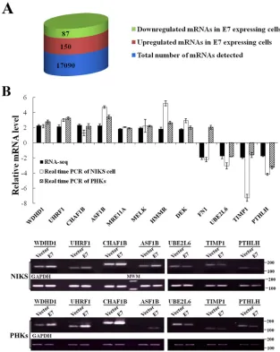
detected a total of 20,537 transcripts that included 17,090 mRNAs.
After normalization, 237 genes were identified that were
differen-tially expressed between E7-expressing and vector control NIKS
cells (
Fig. 1A
). Among these, 150 genes were upregulated
(be-tween 1.7-fold and 4.3-fold) and 87 genes were downregulated
(between 1.7-fold and 4.3-fold) in NIKS cells expressing E7 (
Fig.
1A
; see also Table S1 in the supplemental material).
To verify the RNA-seq results, we performed real-time PCR
assay for nearly a dozen genes selected on the basis of their
poten-tial E7-related biological functions. Among these genes, consistent
with RNA-seq analysis results, the WDHD1, UHRF1, CHAF1B,
ASF1B, MRE11, MELK, HMMR, and DEK genes were shown to
be upregulated whereas the FN1 (fibronectin), UBE2L6, and
TIMP1 genes were shown to be downregulated in E7-expressing
NIKS cells (
Fig. 1B
). For example, the WDHD1 (WD repeat and
HMG-box DNA-binding protein 1) gene was shown to be
up-regulated by more than 2-fold in E7-expressing NIKS cells by
RNA-seq and it was verified to be similarly upregulated by
real-time PCR; the UBE2L6 (ubiquitin/ISG15-conjugating enzyme E2
L6) gene was shown to be downregulated by 1.77-fold in
E7-ex-pressing NIKS cells by RNA-seq, and it was verified to be similarly
downregulated by real-time PCR. Although the fold changes as
demonstrated by real-time PCR for some genes such as those
en-coding CHAF1B and FN1 were not dramatic, the differences are
statistically significant. These are newly described differentially
expressed genes in E7-expressing cells, with the exception of the
DEK and FN1 genes. Thus, the RNA-seq results provided
ge-nome-wide gene expression profiling in E7-expressing cells that
can be verified by real-time PCR.
[image:3.585.41.548.76.256.2]Using real-time PCR, we also examined the expression of the
selected genes described above in primary human keratinocytes
(PHKs) expressing HPV-16 E7 or containing a vector. The
expres-sion profiles of the selected genes were similar to what was
ob-served in NIKS cells (
Fig. 1B
). However, the opposite results were
obtained for FN1 expression. While FN1 was downregulated in
E7-expressing NIKS cells, it was upregulated in E7-expressing
TABLE 1The primer sequencesGene Forward primer Reverse primer
WDHD1 GCTTCAGGTCGTCCTAGACAG CCTTTGGGATGTTACAAGTGGT
UHRF1 AGGTGGTCATGCTCAACTCAC ACAGTTGGCGTAGAGTTCCC
CHAF1B CGGAGCAGATCGCTTTTCAG CCCAGGTTACTCCTTGGACATAA
ASF1B TCCGGTTCGAGATCAGCTTC GTCGGCCTGAAAGACAAACA
MRE11A GGGGCAGATGCACTTTGTG GAAGCAAAACCGGACTAATGTCT
MELK TCTCCCAGTAGCATTCTGCTT TGATCCAGGGATGGTTCAATAGA
HMMR ATGATGGCTAAGCAAGAAGGC TTTCCCTTGAGACTCTTCGAGA
DEK AACTGCTTTACAACAGGCCAG ATGGTTTGCCAGAAGGCTTTG
FN1 CGGTGGCTGTCAGTCAAAG AAACCTCGGCTTCCTCCATAA
UBE2L6 TGGACGAGAACGGACAGATTT GGCTCCCTGATATTCGGTCTATT
TIMP1 AGAGTGTCTGCGGATACTTCC CCAACAGTGTAGGTCTTGGTG
PTHLH ATTTACGGCGACGATTCTTCC GCTTGGAGTTAGGGGACACC
c-MYC AAGCTGAATTGTGCAGGCATC CTCACCCAAAGGCATTTTAAG
HPV16E7 AGTGTGACTCTACGCTTCGGTTG CTGAGAACAGATGGGGCACAC
HPV6E7 GACGAAGTGGACGGACAAGA TCCGCCATCGTTGTTAGGTC
MCM3 GAAGACCAGGGAATTT AGGCAACCAGCTCCTACCAG
on November 7, 2019 by guest
http://jvi.asm.org/
PHKs. It was previously reported that FN1 was downregulated in
three immortal nontumorigenic cell lines that expressed the
high-risk HPV E7 protein (
32
). These results show that the expression
profiles for the selected genes are similar, with the exception of the
expression profiles of NIKS cells and PHKs expressing HPV E7.
Functional implications of differentially expressed genes in
HPV E7-expressing cells.
Some of the differentially expressed
genes in E7-expressing cells identified in this study have
previ-ously been shown to be regulated by HPV E7 at the transcription
level. These genes are summarized in
Table 2
. E7 is known to
abrogate cell cycle checkpoints. Consistent with this notion, a
group of cell cycle-related genes are differentially expressed in
E7-expressing cells. These include the CCNA2, Cdc25A, and
MYBL2 genes. Expression of the CCNA2 gene, which encodes
cyclin A2, is upregulated in E7-expressing cells (
51
). The tyrosine
phosphatase Cdc25A gene is an E2F1 target gene (
52
) and is
in-volved in the regulation of the G
1/S-phase transition (
53
). The
Cdc25A gene is upregulated in E7-expressing cells (
54
,
55
,
57
).
The MYBL2 (
B-Myb
) gene is an E2F-responsive gene and a
com-ponent of the DREAM complex that promotes expression of genes
during the G
2/M phase of the cell cycle (
48
).
B-Myb
has been
implicated in a positive regulatory role for Cdk1 expression (
49
,
56
). We and others have shown that E7 upregulates
B-Myb
expres-sion (
7
,
46
). Moreover, downregulation of
B-Myb
-induced G
1ar-rest in E7-expressing cells upon DNA damage has been described
previously (
59
).
Some other genes such as the APOBEC-3B, BARD1, DEK,
DHFR, MSH6, RAD51AP1, S100P, and S100A8 genes are
upregu-lated in E7-expressing cells and may play critical roles in cervical
carcinogenesis. Among these genes, the APOBEC-3B gene, a
member of the cytidine deaminase gene family, is activated by
HPV infection and increases genome instability (
46
). The nuclear
protein DEK has shown to be upregulated in E7-expressing cells as
well as cervical intraepithelial neoplasia (CIN) and cervical
can-cers. DEK was reported to be required for cellular immortalization
by HPV E7 (
47
,
48
). DEK upregulation may be a common event in
FIG 1Genes differentially expressed in HPV-16 E7-expressing cells. (A) Numbers of genes differentially expressed between NIKS cells expressing E7 and a vector control, as determined by RNA-seq, are presented as columns. Blue, total number of protein-coding mRNAs detected; red, upregulated genes in E7-expressing cells; green, downregulated genes in E7-expressing cells. (B) Real-time PCR assay for selected genes in keratinocytes. Upper panel, relative expression levels of mRNAs in E7-expressing NIKS cells or PHKs compared with vector, measured by real-time PCR and RNA-seq. Lower panels, images of DNA gel electrophoresis. MWM, molecular weight marker.on November 7, 2019 by guest
http://jvi.asm.org/
[image:4.585.134.447.66.459.2]human carcinogenesis and may reflect its senescence inhibitory
function (
47
). The RAD51AP1 gene (a DNA repair gene) is an
S-phase cell cycle checkpoint gene that is directly induced by E2F1
and becomes overexpressed when pRB is inactivated by E7 (
49
).
Bioinformatic analysis of genes differentially expressed in
E7-expressing cells.
We next performed bioinformatics analysis
on genes differentially expressed in E7-expressing cells. First we
used the Gene Ontology (GO) system, a gene function
classifica-tion system that provides a comprehensive descripclassifica-tion of gene
properties (
60
). Based on the GO annotation, the 150 upregulated
genes could be classified into multiple functional groups (
Fig. 2A
).
In the “biological processes” category, more than 50 genes
be-longed to the “cell cycle” functional group. Some additional cell
[image:5.585.40.551.77.235.2]cycle-related genes were in the “M phase,” “cell division,” and
“cell proliferation” functional groups. Taking the data together,
after eliminating overlapping genes, a total of 59 genes were shown
to be cell cycle related. These results are consistent with the known
cell cycle regulatory functions of HPV E7 (
61
). Interestingly, more
than 40 upregulated genes in E7-expressing cells were in the “DNA
metabolic process” (such as DNA repair and replication) group.
In addition, 33 upregulated genes belonged to the “DNA
replica-tion” functional group, which is consistent with the known
func-tion of E7 to establish an S-phase-like environment in otherwise
differentiating epithelial cells to support HPV replication (
62
).
There were also 41 genes and 28 genes, respectively, in the
“regu-lation of transcription” and “transcription” functional groups. It
TABLE 2Differentially expressed genes in E7-expressing cellsGene designation
Ratio of E7/vector by RNA-seq
Regulation categorya
(reference[s]) Product description
S100P 4.27 1(50,52) S100 calcium binding protein P DHFR 3.54 1(53) Dihydrofolate reductase CDC6 3.00 1(54) Cell division cycle 6 RAD51AP1 1.98 1(55) RAD51-associated protein 1
APOBEC3B 1.97 1(47) Apolipoprotein B mRNA editing enzyme catalytic polypeptide-like 3B
DEK 1.80 1(48,49) DEK oncogene
CDC25A 1.82 1(56) Cell division cycle 25A
MSH6 1.78 1(57) MutS homolog 6
CCNA2 1.78 1(57) Cyclin A2
BARD1 1.77 1(59) BRCA1-associated RING domain 1
MYBL2 1.79 1(7,46) V-myb myeloblastosis viral oncogene homolog (avian)-like 2 S100A8 1.70 1(58) S100 calcium binding protein A8
FN1 0.50 2(59) Fibronectin 1
aUpward-pointing arrows represent upregulation; downward-pointing arrows represent downregulation.
FIG 2GO categories of differentially expressed genes in NIKS cells expressing HPV-16 E7. (A) Biological process category. Ten groups (with more than 22 genes each) of upregulated genes (n⫽157) are listed. (B) Biological process category. Three groups (with more than 10 genes each) of downregulated genes (n⫽67) are listed.
on November 7, 2019 by guest
http://jvi.asm.org/
[image:5.585.111.474.437.694.2]is known that E7 is involved in transcriptional regulation, partly
through degradation of pRb and release of E2F.
E7 has been known to regulate the DNA damage response (
16
).
In fact, E7 also induces DNA damage (
63
,
64
). Consistently, a total
of 30 genes that were upregulated in E7-expressing cells belonged
to the “cellular response to stress,” “response to DNA damage
stimulus,” and “DNA repair” groups. In addition, a total of 30
genes belonged to the “chromosome organization” group and 24
genes belonged to the “organelle fission” group (the members of
which are employed to obtain a variety of organelles in cell
divi-sion). The significance of the latter two groups to E7 function
remains to be established.
Interestingly, genes in a group related to “regulation of
cata-bolic process” were downregulated in E7-expressing cells (
Fig.
2B
). Genes for “endoderm development” were also
downregu-lated in E7-expressing cells. However, the significance of these
findings is not known. Notably, antiapoptotic genes in the
“induc-tion of programmed cell death” func“induc-tional group are also
down-regulated in E7-expressing cells. Cells expressing HPV-16 E7 are
predisposed to undergo apoptosis (
65
,
66
), and the observation is
therefore consistent with this finding.
Next, we searched the differentially expressed genes using the
KEGG PATHWAY database, a collection of pathway maps
repre-senting knowledge on the molecular interaction and reaction
net-works. Nine biological pathways that were significantly
upregu-lated (6 or more genes with altered expression) in E7-expressing
NIKS cells are listed in
Table 3
. Consistent with results of GO
analysis and the known E7 functions, the “cell cycle” and “DNA
replication” pathways were significantly regulated in
E7-express-ing cells. As expected, the members of the “pathways in cancer”
group were changed. Up to 24% of upregulated genes were
asso-ciated with DNA repair (“mismatch repair,” “nucleotide excision
repair,” and “homologous recombination” groups), and 12% of
upregulated genes were associated with nucleotide metabolism
(“purine metabolism” and “pyrimidine metabolism” groups). In
addition, 6% of upregulated genes are in the “p53 signaling
path-way” group (data not shown).
Targets of specific transcription factors are differentially
ex-pressed in HPV E7-expressing cells.
We reasoned that, on the
basis of the results seen with differentially expressed genes, we
might be able to identify transcription factors whose activities are
differentially regulated in E7-expressing cells. For this, we
ana-lyzed 41 of the cancer-related transcription factor families
de-scribed in TRED (
https://cb.utdallas.edu/cgi-bin/TRED/tred.cgi
?process
⫽
searchTFGeneForm
) for their predicted target gene
expression characteristics. Significantly, targets of the
transcrip-tion family that includes E2F, MYB, MYC, NF-
B, p53, and SP are
significantly differentially expressed (with at least 10 target genes
that were up- or downregulated) in E7-expressing cells (
Fig. 3
).
E7 targets the degradation of pRb and release of E2F1. E7 may
also activate E2F2 transcription through interaction with HDACs
(
67
). In contrast, E7 can associate with and inactivate the
tran-scriptional repression activity of E2F6 (
68
). Consistent with these
observations, as many as 24 target genes of the E2F family are
upregulated in E7-expressing cells.
The MYB gene family consists of three members, namely,
A
-,
B
-, and
C-Myb
. Multiple lines of evidence suggest that
B-Myb
plays an important role in HPV-associated carcinogenesis (
7
,
42
,
69–71
). We recently showed that HPV E7 is able to activate the
B-Myb
gene (
42
,
44
,
45
).
[image:6.585.39.550.76.196.2]Up to 17 MYC targets were upregulated in E7-expressing cells.
This is consistent with the observation that HPV-18 E7 binds and
TABLE 3KEGG pathway analysis of differentially expressed genes in E7-expressing cellsPathway IDa KEGG pathway(s) Total no. of genes Highly expressed genes
04110 Cell cycle 20 E2F1, CDC6/7/25A/45, CDK1/2, RBL1, TTK, MCM27, CCNE2, CDKN2A, PCNA, MDM2, CCNA2
03030 DNA replication 15 POLE, POLA1, MCM2–7, RPA1, RFC5, POLD3, PRIM1, RPA2, RFC3, PCNA 05200 Cancer (several pathways) 9 E2F1, CCNE2, MSH6, CDKN2A, MSH2, MDM2, BRCA2, BIRC3, CDK2 03430 Mismatch repair 8 POLD3, RFC5, RPA1, MSH6, RPA2, RFC3, MSH2, PCNA
03420 Nucleotide excision repair 7 POLD3, RFC5, RPA1, RPA2, RFC3, POLE, PCNA 04115 p53 signaling 6 CCNE2, CDK1, CDKN2A, RRM2, MDM2, CDK2 03440 Homologous recombination 6 POLD3, RPA1, RPA2, MRE11A, BRCA2, RAD54B 00230 Purine metabolism 6 POLD3, PRIM1, RRM2, RRM1, POLE, POLA1 00240 Pyrimidine metabolism 6 POLD3, PRIM1, RRM2, RRM1, POLE, POLA1
aID, identifier.
FIG 3Differentially expressed transcription factor target gene families in HPV E7-expressing cells. Genes differentially expressed between NIKS-E7 and NIKS-vector cells are categorized as targets of transcription factors.yaxis, numbers of differentially expressed genes.xaxis, names of transcription factor gene families.
on November 7, 2019 by guest
http://jvi.asm.org/
[image:6.585.137.453.588.693.2]augments c-Myc transcriptional activity (
72
). We have also
dem-onstrated that activation of c-Myc contributes to bovine
papillo-mavirus type 1 E7-induced cell proliferation (
73
). Nonetheless,
the results cannot rule out the possibility that upregulation of
MYC targets is an indirect effect of E2F, as most (9 of 17) of the
upregulated MYC targets are also E2F targets.
Interestingly, most (10 of 14) of the targets of NF-
B family
were downregulated in E7-expressing cells. NF-
B plays a key role
in regulating the immune response to infection. Abnormal
regu-lation of NF-
B has been linked to cancer, viral infection, and
improper immune development. Inhibition of NF-
B may reflect
the function of HPV to downregulate the immune system.
Con-sistently, HPV-16 E7 inhibited NF-
B DNA binding activity (
74
)
and attenuated NF-
B activation (
75
,
76
). However, and in
con-trast, it was observed that E7, together with E6, upregulated
NF-
B-responsive genes (
77
). Regulation of NF-
B activity by E7 can
be complicated, depending on the cellular context.
Results from several studies indicate that in HPV
E7-express-ing cells, the activity of p53 is inhibited, although its steady-state
level is high (
78–81
). However, results of one study suggest that
p53 remains active in E7-expressing cells (
82
). The p53 family
targets include p53, p63, and p73. Some were not confirmed (data
not shown). Among the experimentally confirmed p53 targets, all
three genes that are positively regulated by p53 (the MDM2,
MSH2, and S100A9 genes) were upregulated in E7-expressing
cells whereas the one gene negatively regulated by p53, the
PTHLH gene (encoding parathyroid hormone-related protein),
was downregulated. These results favor the notion that p53
func-tions in E7-expressing cells.
The Sp family transcription factors are widely expressed in
hu-man tissues and involved in the regulation of multiple cellular
processes and responses to cellular microenvironment. We know
little about the functional interaction of HPV with Sp family
members other than SP1 binding to the HPV early promoter (
83
).
Interestingly, we found that most of the transcriptional targets of
Sp family members were downregulated in E7-expressing cells
(
Fig. 3
).
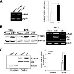
Expression and localization of WDHD1 in E7-expressing
cells.
Given the fact that cell cycle and DNA replication pathways
and their related genes have shown significant disregulation in
E7-expressing cells, we selected WDHD1, which is known to be
involved in both DNA replication and potentially G
1checkpoint
regulation, for further analysis. As transfection efficiencies in
ke-ratinocytes are not very high, we included RPE1 cells. RPE1 cells
expressing the wild-type E7 E7) and vector control
(RPE1-vector) have been used in our recent HPV-related functional
stud-ies (
16
,
84
,
85
). Similarly to what was observed in keratinocytes,
WDHD1 mRNA levels were increased (
⬃
2-fold) in E7-expressing
FIG 4Upregulation and nuclear localization of WDHD1 in E7-expressing cells. (A) WDHD1 mRNA levels in RPE1 cells determined by real-time PCR analysis. (B) WDHD1 protein levels in RPE1 and NIKS cells expressing E7 or vector examined by Western blotting (left panel). WDHD1 mRNA levels in RPE1 cells expressing HPV-6 E7 or⫺16 E7 (right panel) are indicated.gapdh, GAPDH (glyceraldehyde-3-phosphate dehydrogenase) gene. (C) Cellular localization of WDHD1 in E7-expressing cells. Right panel, quantification of WDHD1 protein expression in different cellular compartment. Error bars reflect the standard deviations of the means. Data from an experiment representative of 3 are shown. *,P⬍0.05.on November 7, 2019 by guest
http://jvi.asm.org/
[image:7.585.135.451.68.381.2]FIG 5WDHD1 plays a role at the G1/S transition in E7-expressing cells. (A) RPE1 cells expressing E7 or containing a vector were transfected with siRNAs targeting WDHD1. The steady-state levels of WDHD1 were measured by Western blotting. Tubulin was used as a loading control. Right panel, quantification of relative WDHD1 levels from 3 independent experiments. (B) RPE1 cells were incubated with cycloheximide (CHX) and harvested at the indicated times. The stability of WDHD1 was monitored by immunoblotting analyses (left panel). Data are summarized in the right panel. (C) After siRNA transfection, cells were treated with bleomycin, stained with propidium iodide (PI), and analyzed by flow cytometry. Results of 1 experiment representative of 3 are shown (upper panel). The percentages of cells with 2C and 4C DNA content are indicated. Data are summarized in the lower panel. (D) After siRNA transfection, cells were treated with bleomycin, stained with BrdU, and analyzed by flow cytometry. Data of 3 independent experiments are summarized in the lower panel. *,P⬍0.05; **,P⬍0.01. NC, negative control.
on November 7, 2019 by guest
http://jvi.asm.org/
[image:8.585.111.475.66.646.2]RPE1 cells (
Fig. 4A
). We then further examined the steady-state
level of WDHD1 protein. As shown in
Fig. 4B
, the levels of
WDHD1 protein were upregulated in both RPE1-E7 cells
(1.9-fold) and NIKS-E7 cells (2.2-(1.9-fold). As an initial step toward
un-derstanding the mechanism by which E7 regulates WDHD1, we
examined the steady-state level of WDHD1 protein in RPE1 cells
expressing HPV-6 E7, which does not degrade pRb (
86
). As shown
in
Fig. 4B
, the level of WDHD1 protein was comparable to that
seen with the vector control cells, suggesting that pRb degradation
is important for WDHD1 upregulation by HPV E7.
WDHD1 is usually localized in the nucleus adjacent to
replica-tion foci and is required for efficient DNA replicareplica-tion (
24
,
87
). We
examined cellular localization of WDHD1 in E7-expressing RPE1
cells. Our study demonstrated that WDHD1 was mainly located in
the nucleus, where it was present in levels that were about 2-fold
higher in E7-expressing cells than in control cells (
Fig. 4C
).
WDHD1 depletion induces G
1arrest.
Many HMG proteins
facilitate the assembly of nucleoprotein complexes to influence
cell cycle (
23–25
). DNA replication initiation factors may also be
involved in the licensing checkpoint to modulate S-phase entry
(
18
). As an HMG box-containing protein and a DNA replication
initiation factor, WDHD1 may play a role in cell cycle control in
E7-expressing cells. To test this possibility, we employed the RNA
interference (RNAi) approach by using two independent siRNAs.
The steady-state level of WDHD1 protein was downregulated (to
0.2-fold) after transfection with si-WDHD1-2 and (to 0.5-fold)
after transfection with si-WDHD1-1 in RPE1-E7 cells (
Fig. 5A
).
We also determined the protein stability of WDHD1 in
E7-ex-pressing cells. Two hours after cycloheximide treatment, the
steady-state level of WDHD1 in the vector-containing RPE1 cells
had dropped by more than 50%. In contrast,
⬃
75% of WDHD1
was maintained in E7-expressing cells (
Fig. 5B
). The protein
half-life of WDHD1 in E7-expressing cells was greatly increased
com-pared with that in the vector control cells (3.4 h versus 1.6 h).
Next, we examined changes of cell cycle profiles of E7-expressing
and vector-containing RPE1 cells after siRNA knockdown of
WDHD1. Consistent with what we have recently observed, upon
treatment using the DNA-damaging agent bleomycin, fewer cells
were arrested at the G
1phase in the nonsilencing siRNA
control-transfected E7-expressing cells than in the vector control cells (
Fig.
5C
), indicating abrogation of the G
1checkpoint in E7-expressing
cells. Notably, knockdown of WDHD1 with siRNAs led to an
in-crease in the G
1peak (from 26.9% to 67.5% for si-WDHD1-1 and
46.7% for si-WDHD1-2) in E7-expressing cells. To demonstrate
the role of WDHD1 in promoting S-phase entry of cells more
directly, we transfected siRNAs targeting WDHD1 into
E7-ex-pressing cells and measured BrdU incorporation upon bleomycin
treatment. Significantly, knockdown of WDHD1 by siRNAs led to
a significant reduction of BrdU incorporation (from 13.7% to
5.3% for si-WDHD1-1 and 5.7% for si-WDHD1-2) in RPE1-E7
cells (
Fig. 5D
). These results demonstrate an important role of
WDHD1 in the G
1cell cycle control and S-phase entry of
E7-expressing cells.
WDHD1 depletion reduces rereplication in E7-expressing
cells.
We have recently demonstrated that cells expressing
HPV-16 E7 undergo rereplication upon DNA damage and that
the DNA replication-initiating factor Cdt1 plays an important
role in this process (
16
). Since WDHD1 has been implicated as a
DNA replication-initiating factor (
22
), we examined its potential
role in E7-induced rereplication. As WDHD1 depletion may
block cells at the G
1/S phase, we synchronized cells with thymidine
and then released them into S phase and G
2phase (
Fig. 6A
). As
shown in
Fig. 6A
, after thymidine release for 2 h, 30% of the cells
synchronized in G
1bypassed S phase and entered G
2phase and
6% became polyploid. As time passed, 60% of cells entered G
2phase and 8% became polyploid by 6 h, suggesting that
rereplica-FIG 5continuedon November 7, 2019 by guest
http://jvi.asm.org/
[image:9.585.112.476.67.323.2]FIG 6WDHD1 depletion reduces rereplication and decreases MCM3 chromatin loading and expression. (A) RPE1-E7 cells were treated with thymidine for 16 h, harvested at different times after release from thymidine, and analyzed with flow cytometry. (B) RPE1-E7 cells were transfected with siRNAs targeting WDHD1 and treated with thymidine for 16 h followed by bleomycin treatment for 48 h after release for 2 h from thymidine. Cells were analyzed with flow cytometry. Cells with⬎4C DNA content were quantified (right panel). (C) RPE1 cells were subjected to ChIP assay using antibody against MCM3 (left panel). RPE1-E7 cells were transfected with siRNA targeting WDHD1 and cells were subjected to ChIP assay (right panel). (D) RPE1-E7 cells were transfected with siRNAs targeting WDHD1, and real-time PCR analysis was performed. Another set of cells (right panel) was analyzed for steady-state levels of MCM3 by Western blotting. (E) RPE1-E7 cells were transfected with siRNAs targeting MCM3. Cells with⬎4C DNA content were quantified (right panel). Data from an experiment representative of 3 are shown. *,P⬍0.05; **,P⬍0.01.
on November 7, 2019 by guest
http://jvi.asm.org/
[image:10.585.113.470.67.655.2]tion had occurred. We then used siRNAs to assess the role of
WDHD1 in E7-induced rereplication upon DNA damage.
Nota-bly, the percentage of polyploidy was significantly reduced in
E7-expressing cells after WDHD1 knockdown (
Fig. 6B
). Our recent
study demonstrated that, under those conditions, polyploidy in
E7-expressing cells was due to rereplication, where DNA
replica-tion occurs once again in the G
2phase of the cell cycle (
88
). These
results indicate that WDHD1 plays an important role in
E7-in-duced rereplication.
As an initial step toward understanding the mechanism by
which WDHD1 knockdown causes a reduction in rereplication,
we examined the loading of MCM3 onto chromatin by ChIP
as-say. It is known that DNA replication-initiating factors recruit
MCMs to the origin of replication (
89
). Consistent with this
no-tion, more MCM3 (
⬃
2.5-fold) bound to chromatin in
E7-ex-pressing cells than in vector control cells (
Fig. 6C
, left panel).
WDHD1 knockdown by siRNA significantly reduced MCM3
loading to the chromatin (
Fig. 6C
, right panel). Next, we
exam-ined the effect of knocking down WDHD1 on MCM3 expression.
As shown in
Fig. 6D
, both MCM3 mRNA levels (left panel) and
the steady-state levels of MCM3 went down after WDHD1
knock-down. In addition, WDHD1 knockdown significantly reduced
polyploidy (
Fig. 6E
). These results indicate that WDHD1
facili-tates rereplication in E7-expressing cells by modulating MCM3,
via either expression or loading. The extent to which the reduced
MCM3 chromatin levels seen after WDHD1 knockdown are due
to loading
per se
or to its expression remains to be determined.
DISCUSSION
Previous transcriptional profiles of E7 were mostly determined in
the presence of E6. Nonetheless, several gene expression profiling
studies have been conducted on HPV E7-expressing cells using
microarray analysis. The cells employed included mouse cells,
cer-vical cancer cell lines (C33A and CaSki), and PHKs (
3
,
5–7
,
90
).
However, there are major limitations with the DNA microarray
approach, such as the relatively high background noise resulting
from solution-based DNA probe hybridization as well as the
lim-ited scope of profiling analysis, which is confined by the DNA
probe set. RNA-seq is a more sensitive method for reliably
iden-tifying differentially expressed genes, especially low-abundance
genes, than traditional methods for transcriptome profiling such
as microarray analyses (
91
). Additionally, RNA-seq profiling is
more comprehensive than microarray analyses, as all expressed
transcripts in the cells are included in the analysis. Our study was
the first to profile the gene expression of HPV E7-expressing cells
using RNA-seq. With this new method, we have identified many
E7-induced gene expression changes that were not reported
pre-viously from microarray studies.
It was observed that although the steady-state level of p53 was
high in HPV E7-expressing cells, its activity was low (
78
,
80
,
92
). It
was therefore proposed that p53 activity is inhibited by HPV E7.
However, one study found the opposite phenomenon (
82
). Is p53
functioning in E7-expressing cells? This is an open question and
remains to be resolved. Using an unbiased approach, we showed
that among the experimentally confirmed p53 targets, all genes
positively regulated by p53 were upregulated in E7-expressing
cells while the one negatively regulated by p53 (the PTHLH gene)
(
93
) was downregulated. These results favor the notion that p53 is
functional in E7-expressing cells. Consistently, up to 6% of the
upregulated genes identified in this study have been shown to
participate in the p53 signaling pathway (data not shown).
While the established cellular function is pre-RC activation for
DNA synthesis, WDHD1 has also been implicated in a role in
pre-RC assembly, functioning as a DNA replication initiation
fac-tor (
19
). The known DNA replication initiation factors, Cdt1 and
Cdc6, have been shown to be required for rereplication (
9
,
10
,
14
,
34
,
44
,
63
). However, no such function has been reported
for WDHD1. The current study was the first to demonstrate
that WDHD1 plays a role in rereplication. The mechanism by
which WDHD1 contributes to rereplication is likely that of
load-ing MCMs. Consistent with this notion and with a previous
ob-servation, we have shown that WDHD1 knockdown reduced
chromatin loading of MCM3. Interestingly, WDHD1 knockdown
reduced both MCM3 expression and its chromatin loading. It is
therefore not clear whether the reduced chromatin loading of
MCM3 after WDHD1 knockdown is a result of reduced MCM3
expression or of loading to chromatin or of a combination of the
two. The detailed mechanism by which WDHD1 regulates MCM3
expression is also not known. Future studies will address this
ques-tion.
DNA replication initiation factors have been shown to play a
role in G
1checkpoint regulation. Consistently, we have shown
that knockdown of WDHD1 led to G
1arrest in E7-expressing
cells. It is believed that G
1arrest induced by knockdown of DNA
replication-initiating factors is due to their requirement for DNA
replication initiation. This is normally associated with reduced
cyclin D and cyclin E activities (
94
). Interestingly, our study
showed that cells still arrested in the G
1phase even with partial
knockdown of WDHD1 (
Fig. 5
). In this case, the steady-state level
of WDHD1 after knockdown in E7-expressing cells was similar to
that seen with the NC-transfected control cells, where normal
DNA replication is expected, suggesting that WDHD1 regulates
the G
1checkpoint in E7-expressing cells by a mechanism
inde-pendent of DNA replication initiation. Consistently, mimosine
can prevent WDHD1 binding to chromosomes and arrest the
cell cycle in the G
1phase by activating HIF1-
(
23
). How
WDHD1 precisely regulates the G
1checkpoint requires further
studies.
ACKNOWLEDGMENTS
We thank members of our laboratories for helpful discussions and Wesley Haynes for English editing.
We declare that we have no conflicts of interest.
FUNDING INFORMATION
This work, including the efforts of Xiaowei Wang, was funded by HHS | National Institutes of Health (NIH) (R01GM089784 and R21CA177902). This work, including the efforts of Jason J. Chen, was funded by National Natural Science Foundation of China (NSFC) (81471944).
REFERENCES
1.zur Hausen H.2002. Papillomaviruses and cancer: from basic studies to clinical application. Nat Rev Cancer2:342–350.http://dx.doi.org/10.1038 /nrc798.
2.Doorbar J.2006. Molecular biology of human papillomavirus infection and cervical cancer. Clin Sci (Lond)110:525–541.http://dx.doi.org/10 .1042/CS20050369.
3.Lee KA, Shim JH, Kho CW, Park SG, Park BC, Kim JW, Lim JS, Choe YK, Paik SG, Yoon DY.2004. Protein profiling and identification of modulators regulated by the E7 oncogene in the C33A cell line by
on November 7, 2019 by guest
http://jvi.asm.org/
teomics and genomics. Proteomics4:839 – 848.http://dx.doi.org/10.1002 /pmic.200300626.
4.Kuner R, Vogt M, Sultmann H, Buness A, Dymalla S, Bulkescher J, Fellmann M, Butz K, Poustka A, Hoppe-Seyler F.2007. Identification of cellular targets for the human papillomavirus E6 and E7 oncogenes by RNA interference and transcriptome analyses. J Mol Med (Berl)85:1253– 1262.http://dx.doi.org/10.1007/s00109-007-0230-1.
5. Cortés-Malagón EM, Bonilla-Delgado J, Díaz-Chávez J, Hidalgo-Miranda A, Romero-Cordoba S, Uren A, Celik H, McCormick M, Munguía-Moreno JA, Ibarra-Sierra E, Escobar-Herrera J, Lambert PF, Mendoza-Villanueva D, Bermudez-Cruz RM, Gariglio P.2013. Gene expression profile regulated by the HPV16 E7 oncoprotein and estradiol in cervical tissue. Virology447:155–165.http://dx.doi.org/10.1016/j.virol .2013.08.036.
6.Kim E, Kang J, Cho M, Lee S, Seo E, Choi H, Kim Y, Kim J, Kang KY, Kim KP, Han J, Sheen Y, Yum YN, Park SN, Yoon DY.2009. Profiling of transcripts and proteins modulated by the E7 oncogene in the lung tissue of E7-Tg mice by the omics approach. Mol Med Rep2:129 –137. 7.Pang CL, Toh SY, He P, Teissier S, Ben Khalifa Y, Xue Y, Thierry F.
2014. A functional interaction of E7 with B-Myb-MuvB complex pro-motes acute cooperative transcriptional activation of both S- and M-phase genes. (129 c). Oncogene33:4039 – 4049.http://dx.doi.org/10.1038/onc .2013.426.
8.Murray AW.2004. Recycling the cell cycle: cyclins revisited. Cell116:221– 234.http://dx.doi.org/10.1016/S0092-8674(03)01080-8.
9.Bochman ML, Schwacha A.2009. The Mcm complex: unwinding the mechanism of a replicative helicase. Microbiol Mol Biol Rev73:652– 683. http://dx.doi.org/10.1128/MMBR.00019-09.
10. Fujiwara T, Bandi M, Nitta M, Ivanova EV, Bronson RT, Pellman D.
2005. Cytokinesis failure generating tetraploids promotes tumorigenesis in p53-null cells. Nature437:1043–1047.http://dx.doi.org/10.1038 /nature04217.
11. Olaharski AJ, Sotelo R, Solorza-Luna G, Gonsebatt ME, Guzman P, Mohar A, Eastmond DA.2006. Tetraploidy and chromosomal instability are early events during cervical carcinogenesis. Carcinogenesis27:337– 343.http://dx.doi.org/10.1093/carcin/bgi218.
12. Porter AC.2008. Preventing DNA over-replication: a Cdk perspective. Cell Div3:3.http://dx.doi.org/10.1186/1747-1028-3-3.
13. Davidson IF, Li A, Blow JJ. 2006. Deregulated replication licensing causes DNA fragmentation consistent with head-to-tail fork collision. Mol Cell24:433– 443.http://dx.doi.org/10.1016/j.molcel.2006.09.010. 14. Vaziri C, Saxena S, Jeon Y, Lee C, Murata K, Machida Y, Wagle N,
Hwang DS, Dutta A.2003. A p53-dependent checkpoint pathway pre-vents rereplication. Mol Cell 11:997–1008. http://dx.doi.org/10.1016 /S1097-2765(03)00099-6.
15. Mariani BD, Schimke RT.1984. Gene amplification in a single cell cycle in Chinese hamster ovary cells. J Biol Chem259:1901–1910.
16. Fan X, Liu Y, Heilman SA, Chen JJ.2013. Human papillomavirus E7 induces rereplication in response to DNA damage. J Virol87:1200 –1210. http://dx.doi.org/10.1128/JVI.02038-12.
17. Parker MJ, Gillespie LD, Gillespie WJ.2001. Hip protectors for prevent-ing hip fractures in the elderly. Cochrane Database Syst Rev 2001:
CD001255.http://dx.doi.org/10.1002/14651858.
18. Shreeram S, Sparks A, Lane DP, Blow JJ.2002. Cell type-specific re-sponses of human cells to inhibition of replication licensing. Oncogene
21:6624 – 6632.http://dx.doi.org/10.1038/sj.onc.1205910.
19. Li Y, Xiao H, de Renty C, Jaramillo-Lambert A, Han Z, DePamphilis ML, Brown KJ, Zhu W.2012. The involvement of acidic nucleoplasmic DNA-binding protein (And-1) in the regulation of prereplicative complex (pre-RC) assembly in human cells. J Biol Chem287:42469 – 42479.http: //dx.doi.org/10.1074/jbc.M112.404277.
20. Zhu W, Ukomadu C, Jha S, Senga T, Dhar SK, Wohlschlegel JA, Nutt LK, Kornbluth S, Dutta A.2007. Mcm10 and And-1/CTF4 recruit DNA polymerase alpha to chromatin for initiation of DNA replication. Genes Dev21:2288 –2299.http://dx.doi.org/10.1101/gad.1585607.
21. Sato N, Koinuma J, Fujita M, Hosokawa M, Ito T, Tsuchiya E, Kondo S, Nakamura Y, Daigo Y.2010. Activation of WD repeat and high-mobility group box DNA binding protein 1 in pulmonary and esophageal carcinogenesis. Clin Cancer Res16:226 –239.http://dx.doi.org/10.1158 /1078-0432.CCR-09-1405.
22. Esposito F, Tornincasa M, Federico A, Chiappetta G, Pierantoni GM, Fusco A.2014. Retraction. High-mobility group A1 protein inhibits
p53-mediated intrinsic apoptosis by interacting with Bcl-2 at mitochondria. Cell Death Dis5:e1206.
23. Park SY, Im JS, Park SR, Kim SE, Wang HJ, Lee JK.2012. Mimosine arrests the cell cycle prior to the onset of DNA replication by preventing the binding of human Ctf4/And-1 to chromatin via Hif-1alpha activation in HeLa cells. Cell Cycle11:761–766.http://dx.doi.org/10.4161/cc.11.4 .19209.
24. Yoshizawa-Sugata N, Masai H.2009. Roles of human AND-1 in chro-mosome transactions in S phase. J Biol Chem284:20718 –20728.http://dx .doi.org/10.1074/jbc.M806711200.
25. Langmead B, Trapnell C, Pop M, Salzberg SL. 2009. Ultrafast and memory-efficient alignment of short DNA sequences to the human ge-nome. Genome Biol10:R25.http://dx.doi.org/10.1186/gb-2009-10-3-r25. 26. Kinoshita Y, Johnson EM.2004. Site-specific loading of an MCM protein complex in a DNA replication initiation zone upstream of the c-MYC gene in the HeLa cell cycle. J Biol Chem279:35879 –35889.http://dx.doi.org/10 .1074/jbc.M401640200.
27. Li J, Deng M, Wei Q, Liu T, Tong X, Ye X.2011. Phosphorylation of MCM3 protein by cyclin E/cyclin-dependent kinase 2 (Cdk2) regulates its function in cell cycle. J Biol Chem286:39776 –39785.http://dx.doi.org/10 .1074/jbc.M111.226464.
28. Lambert PF, Ozbun MA, Collins A, Holmgren S, Lee D, Nakahara T.
2005. Using an immortalized cell line to study the HPV life cycle in orga-notypic “raft” cultures. Methods Mol Med119:141–155.
29. Zehbe I, Lichtig H, Westerback A, Lambert PF, Tommasino M, Sher-man L.2011. Rare human papillomavirus 16 E6 variants reveal significant oncogenic potential. Mol Cancer10:77.http://dx.doi.org/10.1186/1476 -4598-10-77.
30. Genther SM, Sterling S, Duensing S, Munger K, Sattler C, Lambert PF.
2003. Quantitative role of the human papillomavirus type 16 E5 gene during the productive stage of the viral life cycle. J Virol77:2832–2842. http://dx.doi.org/10.1128/JVI.77.5.2832-2842.2003.
31. Zehbe I, Richard C, DeCarlo CA, Shai A, Lambert PF, Lichtig H, Tommasino M, Sherman L.2009. Human papillomavirus 16 E6 variants differ in their dysregulation of human keratinocyte differentiation and apoptosis. Virology383:69 –77.http://dx.doi.org/10.1016/j.virol.2008.09 .036.
32. Mannhardt B, Weinzimer SA, Wagner M, Fiedler M, Cohen P, Jansen-Durr P, Zwerschke W.2000. Human papillomavirus type 16 E7 onco-protein binds and inactivates growth-inhibitory insulin-like growth factor binding protein 3. Mol Cell Biol20:6483– 6495.http://dx.doi.org/10.1128 /MCB.20.17.6483-6495.2000.
33. Mansour M, Touka M, Hasan U, Bellopede A, Smet A, Accardi R, Gabet AS, Sylla BS, Tommasino M.2007. E7 properties of mucosal human papillomavirus types 26, 53 and 66 correlate with their intermediate risk for cervical cancer development. Virology367:1–9.http://dx.doi.org/10 .1016/j.virol.2007.05.005.
34. Zhang W, Chen H, Chen Y, Liu J, Wang X, Yu X, Chen JJ, Zhao W.
2015. Cancerous inhibitor of protein phosphatase 2A contributes to hu-man papillomavirus oncoprotein E7-induced cell proliferation via E2F1. Oncotarget6:5253–5262.http://dx.doi.org/10.18632/oncotarget.2867. 35. Blomberg I, Hoffmann I.1999. Ectopic expression of Cdc25A accelerates
the G(1)/S transition and leads to premature activation of cyclin E- and cyclin A-dependent kinases. Mol Cell Biol19:6183– 6194.http://dx.doi .org/10.1128/MCB.19.9.6183.
36. Wu L, Goodwin EC, Naeger LK, Vigo E, Galaktionov K, Helin K, DiMaio D.2000. E2F-Rb complexes assemble and inhibit cdc25A tran-scription in cervical carcinoma cells following repression of human pap-illomavirus oncogene expression. Mol Cell Biol20:7059 –7067.http://dx .doi.org/10.1128/MCB.20.19.7059-7067.2000.
37. Shen T, Huang S.2012. The role of Cdc25A in the regulation of cell proliferation and apoptosis. Anticancer Agents Med Chem12:631– 639. http://dx.doi.org/10.2174/187152012800617678.
38. Moon MS, Lee CJ, Um SJ, Park JS, Yang JM, Hwang ES.2001. Effect of BPV1 E2-mediated inhibition of E6/E7 expression in HPV16-positive cer-vical carcinoma cells. Gynecol Oncol80:168 –175.http://dx.doi.org/10 .1006/gyno.2000.6053.
39. Sadasivam S, DeCaprio JA.2013. The DREAM complex: master coordi-nator of cell cycle-dependent gene expression. Nat Rev Cancer13:585– 595.http://dx.doi.org/10.1038/nrc3556.
40. Sala A, Calabretta B.1992. Regulation of BALB/c 3T3 fibroblast prolif-eration by B-myb is accompanied by selective activation of cdc2 and cyclin
on November 7, 2019 by guest
http://jvi.asm.org/
D1 expression. Proc Natl Acad Sci U S A89:10415–10419.http://dx.doi .org/10.1073/pnas.89.21.10415.
41. Zhu W, Giangrande PH, Nevins JR.2004. E2Fs link the control of G1/S and G2/M transcription. EMBO J 23:4615– 4626. http://dx.doi.org/10 .1038/sj.emboj.7600459.
42. Fan X, Chen JJ.2014. Role of Cdk1 in DNA damage-induced G1 check-point abrogation by the human papillomavirus E7 oncogene. Cell Cycle
13:3249 –3259.http://dx.doi.org/10.4161/15384101.2014.953879. 43. Johung K, Goodwin EC, DiMaio D.2007. Human papillomavirus E7
repression in cervical carcinoma cells initiates a transcriptional cascade driven by the retinoblastoma family, resulting in senescence. J Virol81:
2102–2116.http://dx.doi.org/10.1128/JVI.02348-06.
44. Armstrong DJ, Roman A.1997. The relative ability of human papilloma-virus type 6 and human papillomapapilloma-virus type 16 E7 proteins to transacti-vate E2F-responsive elements is promoter- and cell-dependent. Virology
239:238 –246.http://dx.doi.org/10.1006/viro.1997.8885.
45. Lam EW, Morris JD, Davies R, Crook T, Watson RJ, Vousden KH.
1994. HPV16 E7 oncoprotein deregulates B-myb expression: correlation with targeting of p107/E2F complexes. EMBO J13:871– 878.
46. Ohba K, Ichiyama K, Yajima M, Gemma N, Nikaido M, Wu Q, Chong P, Mori S, Yamamoto R, Wong JE, Yamamoto N.2014. In vivo and in vitro studies suggest a possible involvement of HPV infection in the early stage of breast carcinogenesis via APOBEC3B induction. PLoS One
9:e97787.http://dx.doi.org/10.1371/journal.pone.0097787.
47. Wise-Draper TM, Allen HV, Thobe MN, Jones EE, Habash KB, Munger K, Wells SI.2005. The human DEK proto-oncogene is a senescence in-hibitor and an upregulated target of high-risk human papillomavirus E7. J Virol79:14309 –14317.http://dx.doi.org/10.1128/JVI.79.22.14309-14317 .2005.
48. Adams AK, Hallenbeck GE, Casper KA, Patil YJ, Wilson KM, Kimple RJ, Lambert PF, Witte DP, Xiao W, Gillison ML, Wikenheiser-Brokamp KA, Wise-Draper TM, Wells SI.2015. DEK promotes HPV-positive and -negative head and neck cancer cell proliferation. Oncogene
34:868 – 877.http://dx.doi.org/10.1038/onc.2014.15.
49. Zhou X, Han S, Wang S, Chen X, Dong J, Shi X, Xia Y, Wang X, Hu Z, Shen H.2009. Polymorphisms in HPV E6/E7 protein interacted genes and risk of cervical cancer in Chinese women: a case-control analysis. Gynecol Oncol114:327–331.http://dx.doi.org/10.1016/j.ygyno.2009.05 .011.
50. Jakubickova L, Barathova M, Pastorekova S, Pastorek J, Gibadulinova A.2005. Expression of S100P gene in cervical carcinoma cells is indepen-dent of E7 human papillomavirus oncogene. Acta Virol49:133–137.http ://www.elis.sk/index.php?page⫽shop.product_details&flypage⫽flypage .tpl&product_id⫽99&category_id⫽4&option⫽com_virtuemart&vmcchk
⫽1&Itemid⫽1.
51. Hellung Schonning B, Bevort M, Mikkelsen S, Andresen M, Thomsen P, Leffers H, Norrild B. 2000. Human papillomavirus type 16 E7-regulated genes: regulation of S100P and ADP/ATP carrier protein genes identified by differential-display technology. J Gen Virol81:1009 –1015. http://dx.doi.org/10.1099/0022-1317-81-4-1009.
52. Aisner DL, Nguyen TT, Paskulin DD, Le AT, Haney J, Schulte N, Chionh F, Hardingham J, Mariadason J, Tebbutt N, Doebele RC, Weickhardt AJ, Varella-Garcia M.2014. ROS1 and ALK fusions in colo-rectal cancer, with evidence of intratumoral heterogeneity for molecular drivers. Mol Cancer Res 12:111–118. http://dx.doi.org/10.1158/1541 -7786.
53. Yeager TR, Reznikoff CA.1998. Methotrexate resistance in human uro-epithelial cells with p53 alterations. J Urol159:581–585.http://dx.doi.org /10.1016/S0022-5347(01)63988-0.
54. Martin CM, Astbury K, McEvoy L, O’Toole S, Sheils O, O’Leary JJ.
2009. Gene expression profiling in cervical cancer: identification of novel markers for disease diagnosis and therapy. Methods Mol Biol511:333– 359.http://dx.doi.org/10.1007/978-1-59745-447-6_15.
55. Martinez I, Wang J, Hobson KF, Ferris RL, Khan SA.2007. Identifica-tion of differentially expressed genes in HPV-positive and HPV-negative oropharyngeal squamous cell carcinomas. Eur J Cancer43:415– 432.http: //dx.doi.org/10.1016/j.ejca.2006.09.001.
56. Nguyen DX, Westbrook TF, McCance DJ.2002. Human papillomavirus type 16 E7 maintains elevated levels of the cdc25A tyrosine phosphatase during deregulation of cell cycle arrest. J Virol76:619 – 632.http://dx.doi .org/10.1128/JVI.76.2.619-632.2002.
57. Myklebust MP, Bruland O, Fluge O, Skarstein A, Balteskard L, Dahl O.
2011. MicroRNA-15b is induced with E2F-controlled genes in
HPV-related cancer. Br J Cancer105:1719 –1725.http://dx.doi.org/10.1038/bjc .2011.457.
58. Li JZ, Pan HY, Zheng JW, Zhou XJ, Zhang P, Chen WT, Zhang ZY.
2008. Benzo (a) pyrene induced tumorigenesity of human immortalized oral epithelial cells: transcription profiling. Chin Med J (Engl)121:1882– 1890.
59. Rey O, Lee S, Park NH.2000. Human papillomavirus type 16 E7 onco-protein represses transcription of human fibronectin. J Virol74:4912– 4918.http://dx.doi.org/10.1128/JVI.74.10.4912-4918.2000.
60. Zhang X, Chen H, Wang X, Zhao W, Chen JJ.2014. Expression and transcriptional profiling of the LKB1 tumor suppressor in cervical cancer cells. Gynecol Oncol 134:372–378. http://dx.doi.org/10.1016/j.ygyno .2014.04.050.
61. Moody CA, Laimins LA.2010. Human papillomavirus oncoproteins: pathways to transformation. Nat Rev Cancer10:550 –560.http://dx.doi .org/10.1038/nrc2886.
62. Banerjee NS, Genovese NJ, Noya F, Chien WM, Broker TR, Chow LT.
2006. Conditionally activated E7 proteins of high-risk and low-risk man papillomaviruses induce S phase in postmitotic, differentiated hu-man keratinocytes. J Virol80:6517– 6524.http://dx.doi.org/10.1128/JVI .02499-05.
63. Park JW, Shin MK, Lambert PF.2014. High incidence of female repro-ductive tract cancers in FA-deficient HPV16-transgenic mice correlates with E7’s induction of DNA damage response, an activity mediated by E7’s inactivation of pocket proteins. Oncogene33:3383–3391.http://dx.doi .org/10.1038/onc.2013.327.
64. Park JW, Shin MK, Pitot HC, Lambert PF.2013. High incidence of HPV-associated head and neck cancers in FA deficient mice is associated with E7’s induction of DNA damage through its inactivation of pocket proteins. PLoS One 8:e75056. http://dx.doi.org/10.1371/journal.pone .0075056.
65. Howes KA, Ransom N, Papermaster DS, Lasudry JG, Albert DM, Windle JJ.1994. Apoptosis or retinoblastoma: alternative fates of photo-receptors expressing the HPV-16 E7 gene in the presence or absence of p53. Genes Dev8:1300 –1310.http://dx.doi.org/10.1101/gad.8.11.1300. 66. Pan H, Griep AE.1995. Temporally distinct patterns of p53-dependent
and p53-independent apoptosis during mouse lens development. Genes Dev9:2157–2169.http://dx.doi.org/10.1101/gad.9.17.2157.
67. Longworth MS, Wilson R, Laimins LA. 2005. HPV31 E7 facilitates replication by activating E2F2 transcription through its interaction with HDACs. EMBO J24:1821–1830. http://dx.doi.org/10.1038/sj.emboj .7600651.
68. McLaughlin-Drubin ME, Huh KW, Munger K.2008. Human papillo-mavirus type 16 E7 oncoprotein associates with E2F6. J Virol82:8695– 8705.http://dx.doi.org/10.1128/JVI.00579-08.
69. Chen Y, Miller C, Mosher R, Zhao X, Deeds J, Morrissey M, Bryant B, Yang D, Meyer R, Cronin F, Gostout BS, Smith-McCune K, Schlegel R.
2003. Identification of cervical cancer markers by cDNA and tissue mi-croarrays. Cancer Res63:1927–1935.
70. Santin AD, Zhan F, Bignotti E, Siegel ER, Cane S, Bellone S, Palmieri M, Anfossi S, Thomas M, Burnett A, Kay HH, Roman JJ, O’Brien TJ, Tian E, Cannon MJ, Shaughnessy J, Jr, Pecorelli S.2005. Gene expres-sion profiles of primary HPV16- and HPV18-infected early stage cervical cancers and normal cervical epithelium: identification of novel candidate molecular markers for cervical cancer diagnosis and therapy. Virology
331:269 –291.http://dx.doi.org/10.1016/j.virol.2004.09.045.
71. Rampias T, Sasaki C, Weinberger P, Psyrri A.2009. E6 and e7 gene silencing and transformed phenotype of human papillomavirus 16-positive oropharyngeal cancer cells. J Natl Cancer Inst101:412– 423.http: //dx.doi.org/10.1093/jnci/djp017.
72. Wang YW, Chang HS, Lin CH, Yu WC.2007. HPV-18 E7 conjugates to c-Myc and mediates its transcriptional activity. Int J Biochem Cell Biol
39:402– 412.http://dx.doi.org/10.1016/j.biocel.2006.09.006.
73. Fan X, Liu Y, Chen JJ.2003. Activation of c-Myc contributes to bovine papillomavirus type 1 E7-induced cell proliferation. J Biol Chem278:
43163– 43168.http://dx.doi.org/10.1074/jbc.M306008200.
74. Perea SE, Massimi P, Banks L.2000. Human papillomavirus type 16 E7 impairs the activation of the interferon regulatory factor-1. Int J Mol Med
5:661– 666.
75. Spitkovsky D, Hehner SP, Hofmann TG, Moller A, Schmitz ML.2002. The human papillomavirus oncoprotein E7 attenuates NF-kappa B acti-vation by targeting the Ikappa B kinase complex. J Biol Chem277:25576 – 25582.http://dx.doi.org/10.1074/jbc.M201884200.
on November 7, 2019 by guest
http://jvi.asm.org/
76. Vandermark ER, Deluca KA, Gardner CR, Marker DF, Schreiner CN, Strickland DA, Wilton KM, Mondal S, Woodworth CD.2012. Human papillomavirus type 16 E6 and E 7 proteins alter NF-kB in cultured cervi-cal epithelial cells and inhibition of NF-kB promotes cell growth and immortalization. Virology 425:53– 60. http://dx.doi.org/10.1016/j.virol .2011.12.023.
77. Nees M, Geoghegan JM, Hyman T, Frank S, Miller L, Woodworth CD.
2001. Papillomavirus type 16 oncogenes downregulate expression of in-terferon-responsive genes and upregulate proliferation-associated and NF-kappaB-responsive genes in cervical keratinocytes. J Virol75:4283– 4296.http://dx.doi.org/10.1128/JVI.75.9.4283-4296.2001.
78. Shaikh F, Sanehi P, Rawal R. 2012. Molecular screening of com-pounds to the predicted protein-protein interaction site of Rb1-E7 with p53-E6 in HPV. Bioinformation8:607– 612.http://dx.doi.org/10 .6026/97320630008607.
79. Nair P, Somasundaram K, Krishna S.2003. Activated Notch1 inhibits p53-induced apoptosis and sustains transformation by human papilloma-virus type 16 E6 and E7 oncogenes through a PI3K-PKB/Akt-dependent pathway. J Virol77:7106 –7112.http://dx.doi.org/10.1128/JVI.77.12.7106 -7112.2003.
80. Massimi P, Banks L.1997. Repression of p53 transcriptional activity by the HPV E7 proteins. Virology227:255–259.http://dx.doi.org/10.1006 /viro.1996.8315.
81. Wang Y, Okan I, Pokrovskaja K, Wiman KG. 1996. Abrogation of p53-induced G1arrest by the HPV 16 E7 protein does not inhibit p53-induced apoptosis. Oncogene12:2731–2735.
82. Eichten A, Westfall M, Pietenpol JA, Munger K.2002. Stabilization and functional impairment of the tumor suppressor p53 by the human papil-lomavirus type 16 E7 oncoprotein. Virology295:74 – 85.http://dx.doi.org /10.1006/viro.2002.1375.
83. Dong XP, Pfister H.1999. Overlapping YY1- and aberrant SP1-binding sites proximal to the early promoter of hu