Replicate Vigorously and Cause CD4
ⴙ
T Cell Depletion in Humanized
BLT Mice
Maria Salgado,a,bMichael D. Swanson,cChristopher W. Pohlmeyer,aRobert W. Buckheit III,aJin Wu,dNancie M. Archin,c Thomas M. Williams,dDavid M. Margolis,cRobert F. Siliciano,a,eJ. Victor Garcia,cJoel N. Blanksona
Department of Medicine, Center for AIDS Research, Johns Hopkins University School of Medicine, Baltimore, Maryland, USAa; AIDS Research Institute IrsiCaixa, Institut
d’Investigació en Ciències de la Salut Germans Trias i Pujol, Universitat Autònoma de Barcelona, Badalona, Spainb; Division of Infectious Diseases, Department of Internal
Medicine, Center for AIDS Research, University of North Carolina at Chapel Hill, Chapel Hill, North Carolina, USAc; Department of Pathology, University of New Mexico
School of Medicine, Albuquerque, New Mexico, USAd; Howard Hughes Medical Institute, Johns Hopkins University School ofMedicine, Baltimore, Maryland, USAe
ABSTRACT
Elite controllers or suppressors (ES) are HIV-1-infected patients who maintain undetectable viral loads without antiretroviral
therapy. The mechanism of control remains unclear, but the HLA-B
ⴱ
57 allele is overrepresented in cohorts of these patients.
However, many HLA-B
ⴱ
57 patients develop progressive disease, and some studies have suggested that infection with defective
viruses may be the cause of the lack of high levels of virus replication and disease progression in ES. We therefore performed a
comprehensive comparative
in vivo
and
in vitro
characterization of viruses isolated from well-defined ES. For this purpose, we
first performed full-genome sequence analysis and
in vitro
fitness assays on replication-competent isolates from HLA-B
ⴱ
57 ES
and HLA-B
ⴱ
57 chronic progressors (CPs). Under our experimental conditions, we found that isolates from ES and CPs can
repli-cate
in vitro. However, since inherently these assays involve the use of unnaturally
in vitro-activated cells, we also investigated
the replication competence and pathogenic potential of these HIV isolates
in vivo
using humanized BLT mice. The results from
these analyses demonstrate that virus isolates from ES are fully replication competent
in vivo
and can induce peripheral and
sys-temic CD4 T cell depletion. These results provide the first direct
in vivo
evidence that viral fitness does not likely determine
clini-cal outcome in HLA-B
ⴱ
57 patients and that elite suppressors can control replication-competent, fully pathogenic viruses. A
bet-ter understanding of the immunological bases of viral suppression in ES will serve to inform novel approaches to preventive and
therapeutic HIV vaccine design.
IMPORTANCE
Elite suppressors are HIV-1-infected patients who have undetectable levels of viremia despite not being on antiviral drugs. One
of the most fundamental questions about this phenomenon involves the mechanism of control. To address this question, we
iso-lated virus from elite suppressors and from HIV-1-infected patients who have the usual progressive disease course. We
com-pared how well the isolates from the two groups of patients replicated in culture and in humanized mice. Our results suggest that
elite suppressors are capable of controlling HIV-1 due to the possession of unique host factors rather than infection with
defec-tive virus.
U
nderstanding the mechanisms involved in the natural control
of human immunodeficiency virus type 1 (HIV-1)
replica-tion may lead to the design of an effective HIV-1 vaccine. Patients
known as elite controllers or suppressors (ES), who maintain viral
loads below the limit of detection of clinical assays without
anti-retroviral therapy (ART), represent fewer than 1% of all HIV-1
infected patients (
1–3
). Previous reports have suggested that some
ES and long-term nonprogressors, who maintain stable CD4
counts for prolonged periods, are infected with attenuated or
de-fective virus (
4–12
). However, in other studies,
replication-com-petent HIV-1 isolates were cultured from some ES (
13–15
), and
full-genome sequence analysis of these replication-competent
iso-lates did not reveal any large deletions or signature mutations
(
13
). It has been very challenging to isolate virus from ES, and
full-length genotypic analyses have been performed on
replica-tion-competent isolates obtained from fewer than 10 ES (
11
,
13
,
16–18
) and just 3 HLA-B
ⴱ
57-positive ES (
13
,
16
,
18
).
Further-more, studies comparing the growth kinetics of
replication-com-petent virus from ES to those of multiple isolates from patients
with progressive disease have not been performed. Host factors
clearly contribute to elite suppression of viral replication. The
HLA-B
ⴱ
57 allele is overrepresented in cohorts of ES (
19–24
) and
has been associated with HIV control in large genome-wide
asso-ciation studies (
25
,
26
). HIV-1 epitopes that are presented by
HLA-B
ⴱ
57 proteins are conserved and immunodominant, and
robust HIV-1-specific T cell responses have been documented in
HLA-B
ⴱ
57 ES (
19
,
20
,
27–30
). However, many HLA-B
ⴱ
57
pa-Received16 November 2013Accepted27 December 2013 Published ahead of print3 January 2014
Editor:G. Silvestri
Address correspondence to J. Victor Garcia, [email protected], or Joel N. Blankson, [email protected].
M.S. and M.D.S. contributed equally to this article.
Supplemental material for this article may be found athttp://dx.doi.org/10.1128 /JVI.03380-13.
Copyright © 2014, American Society for Microbiology. All Rights Reserved.
doi:10.1128/JVI.03380-13
on November 7, 2019 by guest
http://jvi.asm.org/
tients are viremic and develop progressive disease (
19
). To
deter-mine the
in vivo
contribution of viral fitness to the clinical
out-come in HLA-B
ⴱ
57 patients, we (i) isolated
replication-competent virus from CD4
⫹T cells from 18 HLA-B
ⴱ
57 patients,
(ii) performed full-genome sequence analysis, (iii) demonstrated
their
in vitro
replication competence, and (iv) evaluated their
abil-ity to establish a
de novo
infection, deplete CD4
⫹T cells, and
establish latency
in vivo
using BLT humanized mice. Our results
strongly suggest that some ES are indeed capable of controlling
replication of fully pathogenic HIV-1 isolates.
MATERIALS AND METHODS
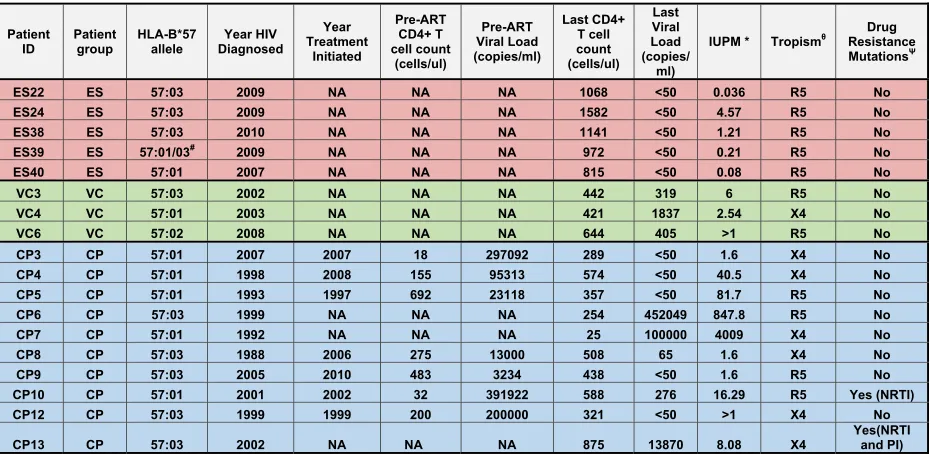
Patient population. We studied 24 HIV-1-seropositive individuals. Eleven were elite suppressors (ES), who maintained viral loads of⬍50 copies/ml without antiretroviral therapy; 3 were viremic controllers (VCs), who had viral loads of greater than 50 copies/ml but less than 2,000 copies/ml; and 10 were chronic progressors (CPs), who had pre-ART viral loads of⬎2,000 copies/ml. Replication-competent virus was obtained from 5 of the 11 ES.Table 1lists the clinical characteristics of the patients used for the study. The protocol was approved by the Institutional Review Board of Johns Hopkins University School of Medicine. Informed con-sent was obtained before phlebotomy. The study was approved by the Johns Hopkins University Institutional Review Board. All study subjects were older than 21 years of age, and informed written consent was ob-tained from all subjects prior to enrollment into the study.
Virus isolation and sequence analysis.Isolation of replication-com-petent virus from bulk CD4⫹T cells was performed as previously de-scribed (13). Briefly, total CD4⫹T cells were isolated by negative selection using the Miltenyi CD4⫹T cell isolation kit II. The CD4⫹T cells were cultured with irradiated donor cells and phytohemagglutinin (PHA) at a final concentration of 0.5g/ml in the presence of interleukin-2 (IL-2) and T cell growth factors. After 2 days, PHA was removed and PHA-activated donor blasts were added to the culture. HIV p24 Gag antigen was measured at days 14 and 21 (Perkin-Elmer). Replication-competent
iso-lates were obtained from 10 CPs, 3 VCs, and 5 ES (Table 1). We were not able to isolate replication-competent virus from 6 other ES. Isolates from one ES (ES 38) have been previously described (18). RNA was isolated from positive-well supernatants. Full-genome sequence analysis of viral isolates was performed as previously described (13).nefclones from the replication-competent virus were compared to proviralnefclones ampli-fied by PCR. Phylogenetic analysis suggested that the replication-compe-tent isolates were representative of the virus archived in the lareplication-compe-tent reser-voir of these patients (data not shown). Resistance mutation predictions were performed using Geno2Pheno database (http://www.geno2pheno .org/).
Sequence analysis of virus amplified from humanized mice.Mouse spleen and thymus samples were collected, and proviral DNA was ex-tracted using the QIAamp DNA Blood Minikit (Qiagen). Nested PCR was then performed to amplify proviral Gag and Nef, and sequence analysis was performed as previously described (13).
Generation of humanized BLT mice.BLT mice were generated as previously described (31,32). Briefly, 6- to 8-week-old NOD.Cg-Prkdc-scid Il2rgtm1Wjl/SzJ (NSG) mice (Jackson Laboratory) were irradiated with 200 cGy and implanted with fetal thymus and liver tissue underneath the kidney capsule. CD34⫹cells isolated from autologous fetal liver were used to transplant between 2⫻105and 3⫻105cells per mouse. Recon-stitution of BLT mice with human immune cells was monitored in periph-eral blood by flow cytometry every 3 to 4 weeks as previously described (31). Mice were maintained by the Division of Laboratory Animal Medi-cine under specific-pathogen-free conditions at the University of North Carolina at Chapel Hill in accordance with protocols approved by the Institutional Animal Care and Use Committee.
[image:2.585.62.527.80.307.2]Flow cytometry analysis.Mononuclear cells (MNCs) from BLT mice were isolated from the bone marrow, spleen, lymph nodes, lung, liver, and thymic organoid tissues as previously described (32). Live cells were iden-tified based on their characteristic side scatter versus forward scatter. Sub-sequently, live human MNCs were identified with mouse anti-human CD45⫹(clone HI30; BD Pharmingen) to determine the percentage of human reconstitution. Lymphocytes were gated through human CD45⫹ TABLE 1Clinical dataa
Patient ID
Patient group
HLA-B*57 allele
Year HIV Diagnosed
Year Treatment
Initiated
Pre-ART CD4+ T cell count
(cells/ul)
Pre-ART Viral Load (copies/ml)
Last CD4+ T cell count (cells/ul)
Last Viral Load (copies/
ml)
IUPM * Tropismθ
Drug Resistance MutationsΨ
ES22 ES 57:03 2009 NA NA NA 1068 <50 0.036 R5 No
ES24 ES 57:03 2009 NA NA NA 1582 <50 4.57 R5 No
ES38 ES 57:03 2010 NA NA NA 1141 <50 1.21 R5 No
ES39 ES 57:01/03# 2009 NA NA NA 972 <50 0.21 R5 No
ES40 ES 57:01 2007 NA NA NA 815 <50 0.08 R5 No
VC3 VC 57:03 2002 NA NA NA 442 319 6 R5 No
VC4 VC 57:01 2003 NA NA NA 421 1837 2.54 X4 No
VC6 VC 57:02 2008 NA NA NA 644 405 >1 R5 No
CP3 CP 57:01 2007 2007 18 297092 289 <50 1.6 X4 No
CP4 CP 57:01 1998 2008 155 95313 574 <50 40.5 X4 No
CP5 CP 57:01 1993 1997 692 23118 357 <50 81.7 R5 No
CP6 CP 57:03 1999 NA NA NA 254 452049 847.8 R5 No
CP7 CP 57:01 1992 NA NA NA 25 100000 4009 X4 No
CP8 CP 57:03 1988 2006 275 13000 508 65 1.6 X4 No
CP9 CP 57:03 2005 2010 483 3234 438 <50 1.6 R5 No
CP10 CP 57:01 2001 2002 32 391922 588 276 16.29 R5 Yes (NRTI)
CP12 CP 57:03 1999 1999 200 200000 321 <50 >1 X4 No
CP13 CP 57:03 2002 NA NA NA 875 13870 8.08 X4
Yes(NRTI and PI)
a#, could not distinguish between HLA-Bⴱ57:01 and HLA-Bⴱ57:02;ⴱ, infectious units per million (IUPM) was determined by limiting-dilution culture analysis using purified CD4⫹T cells;, tropism of isolates as determined by culture analysis in MT-2 cells;⌿, drug resistance mutations were predicted using the Geno2Pheno database.
Elite Suppressor Viruses Replicate in Humanized Mice
on November 7, 2019 by guest
http://jvi.asm.org/
cells and CD3 (clone HIT3a; BD Pharmingen) cells for T cell subsets. T cells (CD45⫹CD3⫹gate) were further analyzed for CD4 (RPA-T4; BD Pharmingen) and CD8 (clone SK1; BD) subsets. For the identification of resting CD4⫹T cells, analysis was performed as previously described (32). All flow cytometry data were collected and analyzed using BD FACSDiva software.
Exposure of BLT mice to HIV.Humanized BLT mice were adminis-tered 200l of HIV at a concentration of approximately 325 ng/ml p24. All mice were exposed via tail vein injection. Mice were bled to determine the presence of viral RNA in the plasma beginning at 1 week postexposure. Analysis of HIV infection.RNA from the plasma was isolated using the RNeasy Minikit (Qiagen). Levels of viral RNA were quantified with a one-step real-time reverse transcriptase PCR (RT-PCR) assay using the following primers and probe: 5=-CATGTTTTCAGCATTATCAGAAGG A-3=, 5=-TGCTTGATGTCCCCCCACT-3=, and 5=-6-carboxyfluorescein (FAM)-CCACCCCACAAGATTTAAACACCATGCTAA-Q (nonfluores-cent quencher)-3=(Applied Biosystems).
Establishment and assessment of HIV latency in BLT mice.HIV ex-posure was performed as described above. At 2 weeks postexex-posure, an-tiviral therapy consisting of tenofovir disoproxil fumarate, emtricitabine, and raltegravir was administered intraperitoneally (i.p.) as previously de-scribed (32). On day 20 of therapy, the dose of tenofovir disoproxil fuma-rate was lowered to 102 mg/kg body weight and maraviroc was added at 61.5 mg/kg body weight. After a period of suppression of viral replication was achieved, lymph nodes, spleen, liver, lung, and bone marrow were harvested from the animal, and cells were isolated from these tissues and from peripheral blood as described above. The cells from all tissues were combined, and the resting human CD4⫹T cells were isolated via negative selection (Stemcell Technologies, Vancouver, Canada) (32). The enriched resting cells were cultured in the presence of 15 nM efavirenz and 1M raltegravir for 1 day as to limit the potential contribution of nonintegrated HIV DNA to the outgrowth assay results. The resting cells were then stimulated and cocultured with feeder cells as previously described (33). A maximum-likelihood method was used to calculate the frequency of rest-ing cell infection (33). The results are expressed as infectious units per million resting CD4⫹T cells (IUPM).
Viral tropism assay.Positive supernatants from each patient were used to infect MT-2 cells (obtained from the NIH AIDS Research and Reference Program) as previously described (34). Tropism was deter-mined by the degree of replication in these cells as deterdeter-mined by the p24 assay (Perkin-Elmer).
Viral fitness assay.Viral fitness was analyzed as described previously (13). Peripheral blood mononuclear cells (PBMCs) from a healthy donor were activated for 2 days with IL-2 and PHA. CD4⫹T cells were isolated (by magnetically activated cell sorting [MACS] with a CD4⫹T cell isola-tion kit II) and infected by spinoculaisola-tion (1,200⫻gfor 2 h) with equal quantities (200 ng/ml) of p24 from primary patient isolates or with Ba-L or IIIB laboratory HIV-1 strains as controls. Supernatant samples were taken over the course of 7 days. Viral replication was quantified using p24 enzylinked immunosorbent assay (ELISA) (Perkin-Elmer). The me-dian p24 concentrations for each point were determined for each group of patients and were compared using the Mann-Whitney nonparametric test.
CD4 and HLA downregulation.CD4⫹T cells from healthy HLA-A2⫹ donors were obtained and infected as described above. On days 3, 5, and 7, the cells were stained with allophycocyanin (APC)-Cy7-conjugated anti-CD4 antibody, APC-conjugated anti-CD3 antibody, and phycoerythrin (PE)-conjugated HLA-A2 antibody (Becton Dickinson) and then fixed and permeabilized with Cytofix/Cytoperm solution (Becton Dickinson). Intracellular staining for Gag was then performed with phycoerythrin-conjugated KC57 antibody (Beckman Coulter) as previously described (35,36). The HLA-A2 downregulation ratio was defined as the mean fluorescence intensity (MFI) of HLA-A2 on cells that were positive for intracellular Gag divided by the MFI of HLA-A2 on CD4⫹T cells that were Gag negative. The CD4 downregulation ratio represents the fraction
of all CD4⫹T cells that were Gag positive and CD4 low. The Mann-Whitney nonparametric test was used to compare CD4 and HLA down-regulation for each group of patients.
HLA typing. Genomic DNA was isolated from peripheral blood mononuclear cells using the QIAamp DNA Blood Minikit (Qiagen). The HLA-B locus was amplified, followed by bidirectional sequencing of ex-ons 2, 3, and 4 with AlleleSEQR HLA-B (Abbott). Sequences were then obtained on 3130 XL (Applied Biosystems) and assembled with Assign software (Conexio Genomics, Australia).
For HLA typing of the human tissue used to reconstitute the human-ized BLT mice, the thymus/liver organoid was harvested at necropsy and cells were extracted. DNA was purified using the QIAamp DNA Blood Minikit (Qiagen).
Nucleotide sequence accession numbers.The sequences determined in this study have been submitted to GenBank (accession numbers KF384798toKF384908).
RESULTS
We studied virus isolated from 18 HLA-B
ⴱ
57 patients; 5 of these
were ES, 10 were chronic progressors (CPs), and 3 were patients
known as viremic controllers (VCs), who maintained plasma virus
levels of between 50 and 2,000 copies of HIV-1 RNA/ml (
Table 1
).
As shown in
Fig. 1
, all the isolates from the 3 patient groups
rep-licated vigorously
in vitro
in IL-2/PHA-activated CD8-depleted
CD4
⫹T cells, and the median growth curves from the 3 patient
groups were not significantly different. Full HIV-1 genome
se-quencing performed for all 18 isolates revealed no large deletions
in any of the genes, and drug resistance mutations, which could
potentially affect viral fitness, were present in only 2 CPs (
Table 1
).
However, viral isolates from CPs were more likely to be
CXCR4-tropic than virus isolated from ES, consistent with higher levels of
ongoing viral replication in CPs (
Table 1
).
Escape mutations in HLA-B
ⴱ
57-restricted Gag epitopes have
been well characterized and have been shown to have a fitness cost
in vivo
(
37–39
) and
in vitro
(
40–42
). We compared the frequency
of mutations in HLA-B
ⴱ
57 epitopes in the different groups of
patients and found that isolates from CPs and VCs were more
likely to contain escape mutations in Gag, Nef, and integrase than
isolates from ES (
Fig. 1
and
2
; see Table S1 in the supplemental
material). In contrast, there was no significant difference in the
frequency of escape mutants in HLA-B
ⴱ
57-restricted epitopes in
other viral genes (
Fig. 2
). Multiple studies have highlighted the
importance of Nef in viral pathogenesis
in vivo
(
43
,
44
). Nef plays
a key role in CD4 (
45
) and HLA-A and HLA-B molecule (
46
)
downregulation. Sequence analysis demonstrated that all isolates
had intact
nef
genes. We thus assessed these parameters in all 18
isolates. As shown in
Fig. 3
, there was no significant difference in
the downregulation of CD4 or HLA-A2 by isolates from the
dif-ferent patient groups. Taken together, our findings suggested that
isolates from HLA-B
ⴱ
57 ES are fully replication competent, with
functional
nef
genes and fewer cytotoxic T lymphocyte (CTL)
es-cape mutations in Gag and Nef than seen in CPs.
Having established the
in vitro
replication competence of these
viruses, we proceeded to perform an
in vivo
analysis of their
rep-lication competence and their ability to induce CD4
⫹T cell
de-pletion and to establish latency. To address these important issues,
we used BLT humanized mice (
47
,
48
). BLT mice are generated by
transplantation of immunodeficient mice previously implanted
with human fetal thymic and liver tissue with autologous CD34
⫹hematopoietic stem cells (
31
). The humanized BLT mice used for
the experiments in this study were derived from 5 different tissue
on November 7, 2019 by guest
http://jvi.asm.org/
samples. Prior to infection, the presence of human cells in
periph-eral blood was confirmed by flow cytometry. The periphperiph-eral blood
of the BLT mice used for experiments in this study had on average
68.5% (
⫾
6.6%) human (CD45
⫹) cells, of which 47.9%
(
⫾
12.45%) expressed human CD3. Of the CD3
⫹human T cells,
83.6% (
⫾
2.9%) also expressed human CD4. Once the presence of
human cells was confirmed in all the animals, they were
inocu-lated via tail vein injection with 4 isolates from 3 ES (including the
(1/1)
KAF9 HW9 YT9
85 120 ...|....| ....|....|...
CONSENSUS_B Nef KGALDLSHF HTQGYFPDWQNYT
ES22 (1/1) .A...I... .............
ES24 (12/12) .A....... .............
ES38 (1/1) .A....... N............
ES39 (1/1) SA.M..... ....F.....C..
ES40 .A.V..... ....F........
VC3 (4/5) ......... ............. VC3 (1/5) ......... .............
VC4 (3/4) ......... ............. VC4 (1/4) .....F... .............
VC6 (1/1) Q..F..... .............
CP3 (4/7) .A.V..... N........H... CP3 (3/7) ...V..... N........H...
CP4 (5/6) ......... N............ CP4 (1/6) ......... N............
CP5 (10/10) .A....... N............
CP6 (4/5) NA.R...F. N............ CP6 (1/5) NA.R...F. N............
CP7 (3/5) ...V..... N............ CP7 (2/5) ...V.F... N............
CP8 (5/5) ...F..... N...F........
CP9 (2/2) ......... .............
CP10 (3/4) ...V..... N...F....H... CP10 (1/4) ...V.F... N...F....H...
CP12 (1/1) .....F... N............
CP13 (1/2) ...I.I... ............. CP13 (1/2) ...V.I... ............. ISW9 KF11 TW10
150 165 240 ....|....| ...|....|.. |....|....
CONSENSUS_B Gag AISPRTLNAW KAFSPEVIPMF TSTLQEQIGW
ES22 (1/1) .......... ........... ........D.
ES24 (13/13) .......... ........... ..........
ES38 (1/1) .L........ ........... ........A.
ES39 (1/1) .......... ........... .....D..A.
ES40 (1/1) .L........ ........... ..........
VC3 (3/3) PL........ ........... ..N.....A.
VC4 (4/6) .L........ ........... ..N.....A.
VC4 (2/6) .......... ..N.....A.
VC6 (1/1) .L........ ........... ....R...D.
CP3 (4/5) .L........ ........... ..N....VA.
CP3 (1/5) .L........ ........... ..N.....A.
CP4 (6/6) .L........ ........... ..N.....A.
CP5 (1/3) P......... ........... ..N....... CP5 (1/3) PL........ ........... ..N....... CP5 (1/3) .L........ ........... ..N.......
CP6 (4/4) PL........ .N......... ..N.....A.
CP7 (1/1) P.T....... ........... ..N.....R.
CP8 (1/2) P......... ........... ..N.......
CP8 (1/2) ...I....... ..N.......
CP9 (2/2) PL........ ........... ..N.....N.
CP10 (2/2) .L........ ........... ..N.......
CP12 (1/1) PL........ .N......... ..N.....T.
CP13 (1/2) PR........ ........... ..N.....A.
CP13 (1/2) R...TS.... ........... ..N.....A.
0 1 2 3 4 5 6 7
1 10 100 1000
ES VC CP IIIB Ba-L
Days post-Infection
p2
4
(
ng/
m
L)
0 1 2 3 4 5 6 7
1 10 100 1000
Days post-Infection
p2
4
(
n
g/
m
L
)
C
D
B
A
ES24 ES22
ES38 ES40 ES39
VC4 VC3
VC6 CP3 CP4 CP5
CP8 CP9 CP10 CP12 CP13 CP6 CP7
III-B Ba-L
FIG 1(A) Growth kinetics of 18 HIV-1 isolates cultured from CD4⫹T cells from HLA-Bⴱ57⫹patients. The patients were ES (red), VCs (green), or CPs (blue). Two laboratory strains (black) were included for comparison. The isolates were all cultured in activated CD4⫹T cells from the same HIV-1 negative donor. (B) Median growth curve for each group of patients. (C and D) Sequence variation within the three HLA-Bⴱ57-03-restricted Gag (C) and Nef (D) epitopes for each isolate. The red and blue boxes denote two distinct but overlapping Nef epitopes, HW9 (116 to 124) and YT9 (120 to 128), respectively. Comparisons of the degree of sequence variation were made using the Mann-Whitney test.
Elite Suppressor Viruses Replicate in Humanized Mice
on November 7, 2019 by guest
http://jvi.asm.org/
[image:4.585.49.490.67.613.2]previously described CCR5-tropic isolate ES8-43 [
13
]) and 2
iso-lates from 2 CPs. Infection was monitored longitudinally using
plasma viral load analysis and by determining the levels of CD4
⫹T
cells in peripheral blood. As shown in
Fig. 4
, mice infected with all
ES and CP viral isolates developed persistent viremia, with
steady-state viral loads ranging from 10
3to 10
6copies/ml. A significant
decline (
⬎
20%) in peripheral CD4
⫹T cells was observed over the
time course of the experiment in the majority of infected mice.
Mice infected with the CXCR4-tropic CP4-2B isolate had the most
dramatic decline in CD4
⫹T cells, consistent with data from prior
studies in this model with R5 (
68
) and X4 (
44
) laboratory isolates.
Interestingly, significant differences in viral loads were seen in 2
isolates cultured from the same ES (ES38-5 and ES38-9) that could
not be attributed to sequence differences or to different donor
tissue used for the generation of the humanized mice (see Table S2
in the supplemental material). These results demonstrate the
in
vivo
replication capacity of these viruses and their intrinsic ability
to induce CD4
⫹T cell depletion.
Having observed a decrease in the levels of peripheral blood
CD4
⫹T cells, we investigated the effect of each of these viruses on
the levels of CD4
⫹T cells in different tissues. For this purpose,
tissues from each infected BLT mouse were collected and used to
prepare single-cell suspensions for flow cytometry analysis. As
shown in
Fig. 5
, there was a reduction in the levels of CD4
⫹T cells
A
RT
B
Integrase
C
Vpr
D
Vif
E
Rev
F
Envelope
IVW9 IAW9
250 380 .|....|.. |....|...
CONSENSUS_B RT IVLPEKDSW IATESIVIW
ES22 ......... .........
ES24 ......... .T.......
ES38 .M..D.E.. .T.......
ES39 V....Q... .S.......
ES40 V....QE.. .T.......
VC3 ......... ....G.I..
VC4 .E..D.... .........
VC6 .E...R... .........
CP3 ......ED. .........
CP4 .E..D.... .........
CP5 .E....... .........
CP6 ......... .........
CP7 .E....E.. .........
CP8 ......... .........
CP9 .E....E.. .T....I..
CP10 .K....... .........
CP12 ......... .........
CP13 ......... .S.......
SW10 KF9
130 180 ..|....|.. ..|....|.
CONSENSUS_B Int STTVKAACWW KTAVQMAVF
ES22 .A........ .........
ES24 .......... .........
ES38 .......... .........
ES39 .......... .........
ES40 .S........ R........
VC3 .N........ .........
VC4 ..M....... .........
VC6 .A........ .........
CP3 .N........ .........
CP4 .NA....... ........L
CP5 CN........ .........
CP6 .AA....... .........
CP7 .NA....... .........
CP8 .N........ .........
CP9 ..A....... .........
CP10 .AAL...... .........
CP12 ..A....... .........
CP13 ..A....... .........
AW9
30 |....|...
CONSENSUS_B Vpr AVRHFPRIW
ES22 .......V.
ES24 .......L.
ES38 .........
ES39 .......P.
ES40 .......V.
VC3 .......T.
VC4 ..K....P.
VC6 .......A.
CP3 .......P.
CP4 .........
CP5 .......P.
CP6 .......V.
CP7 ..K.Y..P.
CP8 .......V.
CP9 .......P.
CP10 .........
CP12 .......P.
CP13 .......L.
KY10
20 .|....|...
CONSENSUS-B Rev KTVRLIKFLY
ES22 .......TI.
ES24 ..........
ES39 Q...F.....
ES38 .A..I.....
ES40 .......L..
VC3 ....F.....
VC4 QA..I..A..
VC6 RII.I..IF.
CP3 E..KV.....
CP4 .A........
CP5 .A.....V..
CP6 LA..A..Y..
CP7 .A........
CP8 ......RR..
CP9 .......Y..
CP10 ....F.....
CP12 .A.....S..
CP13 .IA.I.....
IF9 LW9
35 85 ....|.... ....|....
CONSENSUS-B Vif ISRKAKGWF LGQGVSIEW
ES22 V..G.Q..L .........
ES24 ..K...... .........
ES40 .T...Q... .........
ES39 T..R..... .........
ES38 V....R... .........
VC3 S.K...R.I .........
VC4 V.G.V.... .........
VC6 V.K..MR.A .........
CP3 ..KE..... .........
CP4 ...R.Q... .........
CP5 ....T.R.. .........
CP6 ......... .........
CP7 N.K...... .........
CP8 R.G..RK.. .........
CP9 ..K...E.S .........
CP10 R.G.T...G .........
CP12 ......... .........
CP13 ..K.....V .........
KW11
60 .|....|....
CONSENSUS-B Env KAYDTEVHNVW
ES22 ...........
ES24 .V.........
ES38 .G.KK.A..I.
ES39 ...........
ES40 ......M....
VC3 ...N.......
VC4 ..FE.......
VC6 .S.........
CP3 RG....A....
CP4 RG.EK.A....
CP5 .S.EA.A....
CP6 ...N..K....
CP7 ...EK......
CP8 R..EK......
CP9 ...K..A....
CP10 ..HH.......
CP12 .S.N..K....
CP13 ...EK......
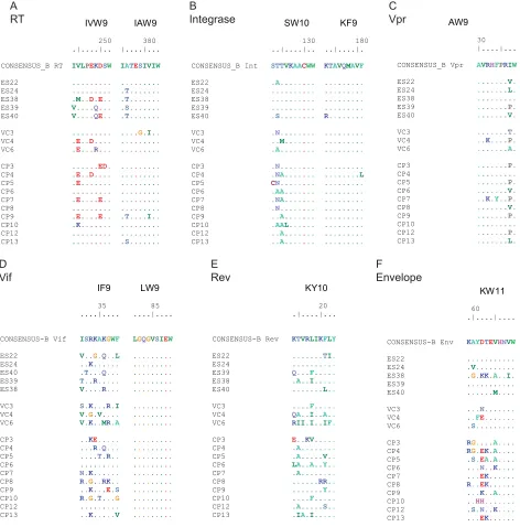
FIG 2Variations within HLA-Bⴱ57-restricted epitopes. Sequence analysis of epitopes in reverse transcriptase (A), integrase (B), Vpr (C), Vif (D), Rev (E), and Env (F) is shown.
on November 7, 2019 by guest
http://jvi.asm.org/
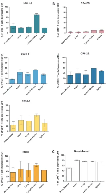
[image:5.585.45.517.68.546.2]obtained from the tissues from infected animals that was similar to
that observed in peripheral blood. Specifically, the mice infected
with the CXCR4-tropic virus had the largest reduction in systemic
CD4
⫹T cell levels (
Fig. 5B
). The rest of the mice infected with the
ES viruses and the mice infected with the CP6 virus showed
inter-mediate levels of CD4
⫹T cell depletion in all tissues analyzed.
These results demonstrate that ES viruses are capable of
replicat-ing systemically and depletreplicat-ing CD4
⫹T cells in tissues in a manner
that reflects peripheral blood levels.
Having established that these viruses are capable of robust
rep-lication
in vivo
and systemic depletion of CD4
⫹T cells, we
pro-ceeded to investigate whether viruses from ES patients can
estab-lish latent infection
in vivo. For this purpose we used an animal
infected with the ES38-9 virus. We chose this virus because it
demonstrated a typical profile of partial systemic CD4
⫹T cell
depletion (
Fig. 5
). After confirming sustained HIV infection in
peripheral blood at two different time points, ART consisting of
raltegravir, tenofovir, and emtricitabine was initiated (
Fig. 6
).
Upon therapy initiation, a dramatic drop in viral load was noted.
The viral load remained suppressed for the duration of treatment.
At 5 weeks after therapy initiation, lymphoid tissue was harvested
and a mononuclear cell suspension from each tissue prepared.
Resting cells were then isolated via negative antibody selection
using magnetic beads. The resting state of the CD4
⫹T cells was
confirmed by the lack of expression of HLA-DR and CD25 (
Fig.
6C
). HIV expression was induced by maximum stimulation of the
cells via addition of medium containing PHA, IL-2, and allogeneic
irradiated PBMCs. Induced HIV was then further propagated by
the addition of allogeneic CD8 T cell-depleted, PHA-activated
PBMCs. Under these conditions, HIV induction from latency was
evident in 6/6 cultures containing 5
⫻
10
5resting cells and in 3/8
cultures containing 10
5resting cells. No outgrowth was observed
in 8 wells containing 2.5
⫻
10
4resting cells. Based on these data,
the frequency of resting cell infection was determined, using a
maximum-likelihood method, to be 5.2 infectious units per
mil-lion resting CD4
⫹T cells (IUPM). These results are similar to
what was observed previously in this system with the reference
HIV-1 isolate JR-CSF and demonstrate the susceptibility of the ES
virus to ART and its ability to establish latency
in vivo
(
32
).
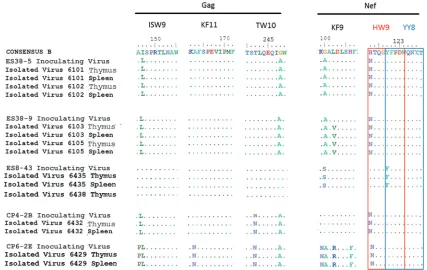
Escape mutations in HLA-B
ⴱ
57 epitopes have been associated
with diminished viral fitness, and reversion to the wild-type
se-quence has been observed after the virus is transmitted to
HLA-B
ⴱ
5701-negative recipients (
37
,
39
). Isolates ES38-5, ES38-9, and
CP4-2B all contained multiple escape mutations and were
inocu-lated into humanized mice that were negative for the B
ⴱ
57 allele
(see Table S2 in the supplemental material). No reversion of these
escape mutations was seen in bulk sequence of virus amplified
from thymus and spleen, even after 4 months of infection in the
case of mice infected with ES38 isolates (
Fig. 7
), consistent with a
recent study that showed that no reversion of Gag escape
muta-tions occurred until after more than a year after transmission of
HIV-1 to HLA-B
ⴱ
57-negative donors (
39
). These data suggest
that the virus can replicate efficiently
in vivo
and induce CD4
⫹T
cell depletion even when potentially attenuating escape mutations
are present.
DISCUSSION
In the vast majority of cases, when untreated, HIV infection
re-sults in progressive loss of CD4
⫹lymphocytes, resulting in
immu-nodeficiency, susceptibility to rare opportunistic infections and
cancers, and ultimately death. The most notable exemptions are
individuals who can naturally and completely control HIV
infec-tion. These rare individuals are designated elite suppressors. The
mechanisms by which ES control viral replication and avoid
dis-ease progression are still not fully understood. Studies have shown
that some macaques are capable of controlling pathogenic simian
immunodeficiency virus (SIV) isolates (
49
,
50
), but studies in
hu-man ES have yielded conflicting results. While some studies have
suggested that some ES are infected with attenuated or defective
virus, others have shown that some ES are infected with
replica-tion-competent virus. We have documented the transmission of
replication-competent HIV-1 isolates from CPs to ES (
16
,
18
),
and studies have shown persistent viremia in ES (
51–53
),
evolu-tion of plasma virus over time (
54–56
), and a decrease in the
frequency of latently infected CD4
⫹T cells in ES treated with
highly active ART (HAART) (
57
). However, other studies
com-paring individual viral proteins from ES and CPs have reported
3 5 7
0.5 1.0 1.5
VC CP III-B Ba-L ES
Day post-Infection
C
D
4
dow
n
re
gul
a
ti
on r
a
ti
o
3 5 7
0.5 1.0 1.5
VC CP III-B Ba-L ES
Day post-Infection
H
LA
-A
2
dow
n
re
gul
a
ti
on r
a
ti
o
A
B
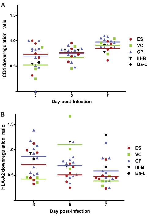
FIG 3Downregulation of CD4 and HLA-A2 in primary CD4⫹T cells infected with HIV-1 isolates. The isolates were laboratory isolates (black) or HIV-1 isolates cultured from ES (red), VCs (green), or CPs (blue). Each symbol represents an individual isolate. The HLA-A2 downregulation ratio (A) is the MFI of HLA-A2 on cells that were positive for intracellular Gag divided by the MFI of HLA-A2 on CD4⫹T cells that were Gag negative. The CD4 downregu-lation ratio (B) represents the fraction of all CD4⫹T cells that were Gag pos-itive and CD4 low. The horizontal lines represent the median level of down-regulation for each group of patients. The Mann-Whitney test was used to compare the degrees of CD4 and HLA-A2 downregulation in the different patient populations.
Elite Suppressor Viruses Replicate in Humanized Mice
on November 7, 2019 by guest
http://jvi.asm.org/
[image:6.585.43.283.65.417.2]reduced fitness of ES Gag (
10
,
58
), Env (
59
), reverse transcriptase
(
60
), and Nef (
61
) proteins. It seems unlikely that the majority of
ES are infected with isolates that have four or more different
at-tenuated genes, and a major caveat is that these studies have
uni-formly analyzed plasma isolates. There is strong evidence that ES
plasma isolates have accumulated escape mutations which may
have a negative effect on fitness (
62–64
). These attenuating
muta-tions are largely absent from proviral clones and
replication-com-petent isolates cultured from the latent reservoir of ES (
62–64
).
Since isolates from the reservoir are more likely to be
representa-tive of the transmitted virus, it is important to study the fitness of
virus from this compartment rather than virus that has
subse-quently evolved to evade the immune response. This is illustrated
by a prior study where we demonstrated a significant reduction in
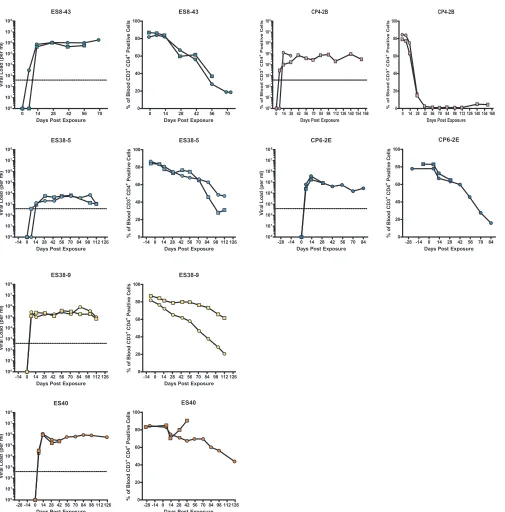
FIG 4In vivoreplication and pathogenesis of HIV isolates from ES and CPs. BLT humanized mice were exposed to HIV-1 isolates that were cultured from either ES or CPs. Each isolate was used to infect two mice. The mice were bled periodically to obtain plasma for viral load analysis via real-time RT-PCR and blood mononuclear cells for flow cytometric analysis. For each isolate, the left panel shows viral load analysis (the dotted line represents the limit of detection for the viral load assay), and the right panel shows the percentage of peripheral blood CD4⫹T cells. Different symbols represent different mice. One mouse infected with isolate CP4-2B and one mouse infected with isolate ES40 died shortly after day 28 and day 42 of infection, respectively.on November 7, 2019 by guest
http://jvi.asm.org/
[image:7.585.45.550.59.571.2]ES8-43 % o f C D 3
+T cells Expressing CD4
Bone Marro w Liv er Lun g Lym ph No des Sple en 0 20 40 60 80 100 CP4-2B
% of C
D
3
+T cells Expressing
C D 4 Bone Marro w
Liver Lun g Lymph Node s Spleen 0 20 40 60 80 100 ES38-5
% of C
D
3
+T cells Expressing
C D 4 Bone Marro w
Liver Lun g Lymph Node s Spleen 0 20 40 60 80 100 CP6-2E % o f C D 3
+T cells Expressing CD4
Bone Marro w Live r Lun g Lym ph No des Sple en 0 20 40 60 80 100 ES38-9
% of C
D
3
+T cells Expressing CD4
Bone Marro w Liv er Lun g Lym ph No des Sple en 0 20 40 60 80 100 Non-infected % o f C D 3
+T cells Expressing CD4
Bone Marro w Live r Lun g Lym ph No des Sple en 0 20 40 60 80 100
A
B
C
ES40 % o f CD3 +T cel ls E xpr essi n g C D 4 Bone Marro w Live r LungLymph Nodes Spleen 0 20 40 60 80 100
FIG 5HIV isolates derived from elite suppressors are pathogenicin vivoand result in systemic CD4⫹T cell depletion. Mononuclear cells were isolated from various tissues at necropsy. The percentage of T cells that express CD4 was calculated for each tissue. The bars represent the average for two animals. Results are from BLT mice infected with HLAⴱB57 elite suppressor-derived isolates (A), HLAⴱB57 chronic progressor-derived isolates (B), or noninfected BLT mice (C).
on November 7, 2019 by guest
http://jvi.asm.org/
[image:8.585.122.482.29.683.2]fitness of a replication-competent isolate containing escape
mu-tations in HLA-B
ⴱ
57-restricted epitopes in Gag compared to an
isolate without escape mutations in these epitopes obtained from
the same ES (
65
). In this study, we show that the growth kinetics of
replication-competent virus isolated from CD4
⫹T cells of
HLA-B
ⴱ
57-positive ES and CPs are comparable. We also demonstrate
that the isolates have a similar ability to downregulate HLA and
CD4 proteins
in vitro.
Having established the
in vitro
fitness of the viruses obtained
from the ES, we evaluated their replication capacity and ability to
induce CD4
⫹T cell depletion
in vivo. This analysis is particularly
important because the
in vivo
substrate for replication represents a
rich milieu of different components, all interacting in multiple
ways that cannot be recapitulated
ex vivo
with cultured cells that
represent only a single substrate entity in an artificial activation
state. Furthermore, prior studies have shown that SIV isolates that
appeared to be fully replication competent
in vitro
were
attenu-ated when they were inoculattenu-ated into nonhuman primates (
43
,
66
).
A
-21 -14 -7 0 7 14 21 28 35 42 49 0
20 40 60 80 100
103
104
105
106
107
Days Post Exposure
%
of
B
lood C
D
3
+
C
D
4
P
o
s
it
ive C
e
ll
s
Vi
ra
l Load
(p
e
r m
l)
ART
B
p24 Status of Wells: Resting CD4+ T cells
Cultured Per Well:
5 x 10⁵
1 x 10⁵
2.5 x 10⁴
Calulated IUPM: 5.12
D
+
HIV Exposure Via Tail Vein
Begin Antiretroviral Therapy
Sacrifice BLT Mice and Isolate Cells from
Multiple Tissues
Magnetic Negative Selection for Resting
Human CD4+ T cells
Culture Resting CD4+ T cells for
36 Hours Without Stimulation in the Presence of Efavirenz and
Raltegravir
Harvest Culture Supernatant and Test for
Evidence of HIV Replication
% 8 . 2 %
2 . 2 1
77.4% 7.6%
Pre-column
HLA-DR
CD25
% 0 %
3 . 0
% 1 . 0 %
5 . 9 9
Post-column
HLA-DR
CD25
C
Stimulate resting CD4 T cells with IL2, PHA and allogeneic
irradiated PBMC
Co-culture stimulated resting CD4 T cells with CD8-depleted
PHA blasts
+
+
FIG 6Establishment of a latent viral reservoir by an ES-derived HIV isolate. (A) Experimental overview for theex vivodetermination of latency. (B) Longitudinal analysis of plasma viral loads (orange circles) and the levels of CD4⫹T cells in the blood (blue circles) of a BLT mouse exposed to the ES-derived isolate ES38-9. The shaded area indicates the duration that antiretroviral therapy was administered. The dashed line indicates the limit of quantitation of HIV RNA. (C) Enrichment of resting CD4⫹T cells from the suppressed, ES38-9-infected BLT mouse. Mononuclear cells from all tissues were pooled and subjected to magnetic negative selection of CD4⫹resting T cells. Samples were analyzed by flow cytometry for the expression of CD25 and HLA-DR before (left) and after (right) negative selection. (D) Results of a viral outgrowth assay to quantify the latent reservoir. Shaded squares indicate that the well was positive for p24 in the supernatant as determined by ELISA, while nonshaded squares represent wells that were negative for p24 in the supernatant.
on November 7, 2019 by guest
http://jvi.asm.org/
We therefore determined the replication capacity of a subset of
these isolates in humanized mice. We chose BLT humanized mice
because they represent the most advanced and complete system to
investigate HIV replication
in vivo. Specifically, BLT mice are fully
reconstituted with all the types of human cells involved in HIV
replication, including T cells, macrophages, and dendritic cells.
BLT mice have been validated for the study of HIV transmission,
pathogenesis, and HIV persistence (
32
,
68
), and our recent studies
have shown significant differences in the level of viremia
in vivo
when these mice are infected with attenuated versus wild-type
viral isolates (
44
,
67
). The presence of robust replication of all the
isolates studied in BLT mice and their ability to induce CD4
⫹T
cell depletion presented here represent the first evidence to date
that isolates from ES are capable of establishing a pathogenic
in-fection
in vivo. Our data also show that viral fitness is not likely to
determine whether an HLA-B
ⴱ
57-infected patient becomes an ES
or a CP. The fact that viruses from ES patients replicate efficiently
in vivo
allowed us to show that replication of ES viruses
in vivo
is
efficiently suppressed by ART. The ability to suppress the
replica-tion of ES viruses by ART made it possible to then demonstrate
that these viruses can persist
in vivo
and establish a latent
infec-tion.
Our study has some limitations. We were not able to isolate
virus from all ES, which is consistent with our prior work that
showed that these patients have a very low frequency of latently
infected CD4
⫹T cells. However, we cannot rule out the
possi-bility that some HLA-B
ⴱ
57 ES are not infected with
replica-tion-competent virus. Furthermore, the
in vitro
evaluation of
virus fitness is dependent on the use of primary cells activated
in vitro
in an artificial manner. This can mask important effects
such as the role of Nef in HIV replication. However, the
dem-onstration that isolates from HLA-B
ⴱ
57 ES have intact
nef
genes, can replicate efficiently
in vitro, and are capable of
effec-tive downregulation of CD4 and HLA-A2 strongly suggests that
Nef is functional in these viruses and not likely to contribute to
the ES phenotype observed. Another limitation is the fact that
the
in vivo
evaluation of these viruses was performed in a
mouse model where human cells replace the endogenous
im-mune system. However, it should be noted that this type of
experiment cannot be performed in humans. Also, because of
the limited species tropism of HIV, these experiments cannot
be performed in nonhuman primates either. Therefore, BLT
humanized mice represent a viable and useful alternative to
perform these types of investigations.
In summary, this is the first study to show that these ES
isolates replicate effectively
in vivo
and eventually cause CD4
⫹T cell depletion. This study also shows that ES isolates are
pathogenic and capable of causing immunosuppression
in vivo.
Therefore, our results imply that infection with attenuated or
defective viruses is not likely to be the cause of elite suppression
in all patients. The finding that control of fully pathogenic
HIV-1 is possible has major implications for the design of
HIV-1 vaccines. In addition, in future experiments it will be
important to determine whether this ES phenotype can be
re-capitulated
in vivo
by creating humanized mice with
hemato-poietic stem cells derived from these patients and challenging
them with autologous and heterologous viruses.
FIG 7Limited variation occurs within three HLA-Bⴱ57-03-restricted Gag and Nef epitopes during HIV infection of BLT Mice. HIV-1 isolates were amplified from the thymuses and spleens of BLT humanized mice at week 16 (isolates ES38-5 and ES38-9), week 8 (isolate ES8-43), and week 4 (isolates CP4-2B and CP6-2E), and Gag and Nef epitopes were analyzed. The red and blue boxes denote two distinct but overlapping Nef epitopes, HW9 (116 to 124) and YT9 (120 to 128), respectively.
Elite Suppressor Viruses Replicate in Humanized Mice
on November 7, 2019 by guest
http://jvi.asm.org/
[image:10.585.78.505.66.336.2]ACKNOWLEDGMENTS
This work was supported by the HHMI (R.F.S.), by NIH grants 5R01DA030156 (D.M.M.), AI096113, AI073146, and AI096138 (J.V.G.), AI080328 (J.N.B.), and AI100775 (M.D.S), and by the ‘‘Sara Borrell’’ grant from the Spanish Health Institute (M.S.).
REFERENCES
1.Deeks SG, Walker BD.2007. Human immunodeficiency virus control-lers: mechanisms of durable virus control in the absence of antiretroviral therapy. Immunity 27:406 – 416. http://dx.doi.org/10.1016/j.immuni .2007.08.010.
2. Migueles SA, Connors M. 2010. Long-term nonprogressive disease among untreated HIV-infected individuals: clinical implications of un-derstanding immune control of HIV. JAMA304:194 –201.http://dx.doi .org/10.1001/jama.2010.925.
3.Blankson JN.2010. Control of HIV-1 replication in elite suppressors. Discov. Med.9:261–266.
4.Deacon NJ, Tsykin A, Solomon A, Smith K, Ludford-Menting M, Hooker DJ, McPhee DA, Greenway AL, Ellett A, Chatfield C, Lawson VA, Crowe S, Maerz A, Sonza S, Learmont J, Sullivan JS, Cunningham A, Dwyer D, Dowton D, Mills J.1995. Genomic structure of an attenu-ated quasi species of HIV-1 from a blood transfusion donor and recipi-ents. Science 270:988 –991. http://dx.doi.org/10.1126/science.270.5238 .988.
5.Calugi G, Montella F, Favalli C, Benedetto A.2006. The entire genome of a nef-deleted strain of human immunodeficency virus type 1 recovered 20 years after primary infection: large pool of env-deleted proviruses. J. Virol.80:11892–11896.http://dx.doi.org/10.1128/JVI.00932-06. 6.Mariani R, Kirchhoff F, Greenough TC, Sullivan JL, Desrosiers RC,
Skowronski J.1996. High frequency of defective nef alleles in a long-term survivor with nonprogressive human immunodeficiency virus type 1 in-fection. J. Virol.70:7752–7764.
7.Kirchhoff F, Greenough TC, Brettler DB, Sullivan JL, Desrosiers RC. 1995. Absence of intact nef sequences in a long-term survivor with non-progressive HIV-1 infection. N. Engl. J. Med.332:228 –232.http://dx.doi .org/10.1056/NEJM199501263320405.
8.Iversen AK, Shpaer EG, Rodrigo AG, Hirsch MS, Walker BD, Sheppard HW, Merigan TC, Mullins JI.1995. Persistence of attenuated rev genes in a human immunodeficiency virus type 1-infected asymptomatic individ-ual. J. Virol.69:5743–5753.
9.Huang Y, Zhang L, Ho DD. 1998. Characterization of gag and pol sequences from long-term survivors of human immunodeficiency virus type 1 infection. Virology 240:36 – 49. http://dx.doi.org/10.1006/viro .1997.8913.
10. Miura T, Brumme ZL, Brockman MA, Rosato P, Sela J, Brumme CJ, Pereyra F, Kaufmann DE, Trocha A, Block BL, Daar ES, Connick E, Jessen H, Kelleher AD, Rosenberg E, Markowitz M, Schafer K, Vaida F, Iwamoto A, Little S, Walker BD.2010 Impaired replication capacity of acute/early viruses in persons who become HIV controllers. J. Virol.84: 7581–7591.http://dx.doi.org/10.1128/JVI.00286-10.
11. Alexander L, Weiskopf E, Greenough TC, Gaddis NC, Auerbach MR, Malim MH, O’Brien SJ, Walker BD, Sullivan JL, Desrosiers RC.2000. Unusual polymorphisms in human immunodeficiency virus type 1 asso-ciated with nonprogressive infection. J. Virol.74:4361– 4376.http://dx .doi.org/10.1128/JVI.74.9.4361-4376.2000.
12. Alexander L, Aquino-DeJesus MJ, Chan M, Andiman WA.2002. Inhi-bition of human immunodeficiency virus type 1 (HIV-1) replication by a two-amino-acid insertion in HIV-1 vif from a nonprogressing mother and child. J. Virol.76:10533–10539.http://dx.doi.org/10.1128/JVI.76.20 .10533-10539.2002.
13. Blankson JN, Bailey JR, Thayil S, Yang HC, Lassen K, Lai J, Gandhi SK, Siliciano JD, Williams TM, Siliciano RF.2007. Isolation and character-ization of replication-competent human immunodeficiency virus type 1 from a subset of elite suppressors. J. Virol.81:2508 –2518.http://dx.doi .org/10.1128/JVI.02165-06.
14. Lamine A, Caumont-Sarcos A, Chaix ML, Saez-Cirion A, Rouzioux C, et al.2007. Replication-competent HIV strains infect HIV controllers despite undetectable viremia (ANRS EP36 study). AIDS21:1043–1045. http://dx.doi.org/10.1097/QAD.0b013e3280d5a7ac.
15. Julg B, Pereyra F, Buzón MJ, Piechocka-Trocha A, Clark MJ, Baker BM, Lian J, Miura T, Martinez-Picado J, Addo MM, Walker BD. 2010. Infrequent recovery of HIV from but robust exogenous infection of
acti-vated CD4(⫹) T cells in HIV elite controllers. Clin. Infect. Dis.51:233– 238.http://dx.doi.org/10.1086/653677.
16. Bailey JR, O’Connell K, Yang HC, Han Y, Xu J, Jilek B, Williams TM, Ray SC, Siliciano RF, Blankson JN.2008. Transmission of human im-munodeficiency virus type 1 from a patient who developed AIDS to an elite suppressor. J. Virol. 82:7395–7410.http://dx.doi.org/10.1128/JVI .00800-08.
17. Gaillard S, Dinoso JB, Marsh JA, DeZern AE, O’Connell KA, Spivak AM, Alwood K, Durand CM, Ambinder RF, Blankson JN.2011. Sus-tained elite suppression of replication competent HIV-1 in a patient treated with rituximab based chemotherapy. J. Clin. Virol.51:195–198. http://dx.doi.org/10.1016/j.jcv.2011.04.002.
18. Buckheit RW, III, Allen TG, Alme A, Salgado M, O’Connell KA, Huculak S, Falade-Nwulia O, Williams TM, Gallant JE, Siliciano RF, Blankson JN.2012. Host factors dictate control of viral replication in two HIV-1 controller/chronic progressor transmission pairs. Nat. Commun. 3:716.http://dx.doi.org/10.1038/ncomms1697.
19. Migueles SA, Sabbaghian MS, Shupert WL, Bettinotti MP, Marincola FM, Martino L, Hallahan CW, Selig SM, Schwartz D, Sullivan J, Connors M.2000. HLA Bⴱ5701 is highly associated with restriction of virus replication in a subgroup of HIV-infected long term nonprogressors. Proc. Natl. Acad. Sci. U. S. A.97:2709 –2714.http://dx.doi.org/10.1073 /pnas.050567397.
20. Migueles SA, Osborne CM, Royce C, Compton AA, Joshi RP, Weeks KA, Rood JE, Berkley AM, Sacha JB, Cogliano-Shutta NA, Lloyd M, Roby G, Kwan R, McLaughlin M, Stallings S, Rehm C, O’Shea MA, Mican J, Packard BZ, Komoriya A, Palmer S, Wiegand AP, Maldarelli F, Coffin JM, Mellors JW, Hallahan CW, Follman DA, Connors M. 2008. Lytic granule loading of CD8⫹T cells is required for HIV-infected cell elimination associated with immune control. Immunity29:1009 – 1021.http://dx.doi.org/10.1016/j.immuni.2008.10.010.
21. Pereyra F, Addo MM, Kaufmann DE, Liu Y, Miura T, Rathod A, Baker B, Trocha A, Rosenberg R, Mackey E, Ueda P, Lu Z, Cohen D, Wrin T, Petropoulos CJ, Rosenberg ES, Walker BD.2008. Genetic and immu-nologic heterogeneity among persons who control HIV infection in the absence of therapy. J. Infect. Dis.197:563–571.http://dx.doi.org/10.1086 /526786.
22. Han Y, Lai J, Barditch-Crovo P, Gallant JE, Williams TM, Siliciano RF, Blankson JN.2008. The role of protective HCP5 and HLA-C associated polymorphisms in the control of HIV-1 replication in a subset of elite suppressors. AIDS 22:541–544. http://dx.doi.org/10.1097/QAD.0b013e 3282f470e4.
23. Lambotte O, Boufassa F, Madec Y, Nguyen A, Goujard C, Meyer L, Rouzioux C, Venet A, Delfraissy JF, SEROCO-HEMOCO Study Group. 2005. HIV controllers: a homogeneous group of HIV-1-infected patients with spontaneous control of viral replication. Clin. Infect. Dis.41:1053– 1056.http://dx.doi.org/10.1086/433188.
24. Emu B, Sinclair E, Hatano H, Ferre A, Shacklett B, Martin JN, McCune JM, Deeks SG.2008. HLA class I-restricted T cell responses may contrib-ute to the control of HIV infection, but such responses are not always necessary for long-term virus control. J. Virol.82:5398 –5407.http://dx .doi.org/10.1128/JVI.02176-07.
25. Fellay J, Shianna KV, Ge D, Colombo S, Ledergerber B, Weale M, Zhang K, Gumbs C, Castagna A, Cossarizza A, Cozzi-Lepri A, De Luca A, Easterbrook P, Francioli P, Mallal S, Martinez-Picado J, Miro JM, Obel N, Smith JP, Wyniger J, Descombes P, Antonarakis SE, Letvin NL, McMichael AJ, Haynes BF, Telenti A, Goldstein DB.2007. A whole-genome association study of major determinants for host control of HIV-1. Science317:944 –947.http://dx.doi.org/10.1126/science.1143767. 26. International HIV Controllers Study, Pereyra F, Jia X, McLaren PJ, Telenti A, de Bakker PI, Walker BD, Ripke S, Brumme CJ, Pulit SL, Car-rington M, Kadie CM, Carlson JM, Heckerman D, Graham RR, Plenge RM, Deeks SG, Gianniny L, Crawford G, Sullivan J, Gonzalez E, Davies L, Camargo A, Moore JM, Beattie N, Gupta S, Crenshaw A, Burtt NP, Guiducci C, Gupta N, Gao X, Qi Y, Yuki Y, Piechocka-Trocha A, Cutrell E, Rosenberg R, Moss KL, Lemay P, O’Leary J, Schaefer T, Verma P, Toth I, Block B, Baker B, Rothchild A, Lian J, Proudfoot J, Alvino DM, Vine S, Addo MM, Allen TM, Altfeld M, Henn MR, Le Gall S, Streeck H, Haas DW, Kuritzkes DR, Robbins GK, Shafer RW, Gulick RM, et al. 2010. The major genetic determinants of HIV-1 control affect HLA class I peptide presentation. Science330:1551–1557.http://dx.doi.org/10.1126/science .1195271.
27. Migueles SA, Laborico AC, Shupert WL, Sabbaghian MS, Rabin R,
on November 7, 2019 by guest
http://jvi.asm.org/
Hallahan CW, Van Baarle D, Kostense S, Miedema F, McLaughlin M, Ehler L, Metcalf J, Liu S, Connors M.2002. HIV-specific CD8⫹T cell proliferation is coupled to perforin expression and is maintained in nonprogressors. Nat. Immunol.3:1061–1068.http://dx.doi.org/10.1038 /ni845.
28. Betts MR, Nason MC, West SM, De Rosa SC, Migueles SA, Abraham J, Lederman MM, Benito JM, Goepfert PA, Connors M, Roederer M, Koup RA.2006. HIV nonprogressors preferentially maintain highly func-tional HIV-specific CD8⫹T cells. Blood107:4781– 4789.http://dx.doi .org/10.1182/blood-2005-12-4818.
29. Sáez-Cirión A, Lacabaratz C, Lambotte O, Versmisse P, Urrutia A, Boufassa F, Barré-Sinoussi F, Delfraissy JF, Sinet M, Pancino G, Venet A, Agence Nationale de Recherches sur le Sida EP36 HIV Controllers Study Group.2007. HIV controllers exhibit potent CD8 T cell capacity to suppress HIV infection ex vivo and peculiar cytotoxic T lymphocyte acti-vation phenotype. Proc. Natl. Acad. Sci. U. S. A.104:6776 – 6781.http://dx .doi.org/10.1073/pnas.0611244104.
30. Hersperger AR, Pereyra F, Nason M, Demers K, Sheth P, Shin LY, Kovacs CM, Rodriguez B, Sieg SF, Teixeira-Johnson L, Gudonis D, Goepfert PA, Lederman MM, Frank I, Makedonas G, Kaul R, Walker BD, Betts MR.2010. Perforin expression directly ex vivo by HIV-specific CD8 T-cells is a correlate of HIV elite control. PLoS Pathog.6:e1000917. http://dx.doi.org/10.1371/journal.ppat.1000917.
31. Melkus MW, Estes JD, Padgett-Thomas A, Gatlin J, Denton PW, Othieno FA, Wege AK, Haase AT, Garcia JV.2006. Humanized mice mount specific adaptive and innate immune responses to EBV and TSST-1. Nat. Med.12:1316 –1322.http://dx.doi.org/10.1038/nm1431. 32. Denton PW, Olesen R, Choudhary SK, Archin NM, Wahl A, Swanson
MD, Chateau M, Nochi T, Krisko JF, Spagnuolo RA, Margolis DM, Garcia JV.2012. Generation of HIV latency in humanized BLT mice. J. Virol.86:630 – 634.http://dx.doi.org/10.1128/JVI.06120-11.
33. Archin NM, Eron JJ, Palmer S, Hartmann-Duff A, Martinson JA, Wiegand A, Bandarenko N, Schmitz JL, Bosch RJ, Landay AL, Coffin JM, Margolis DM.2008. Valproic acid without intensified antiviral ther-apy has limited impact on persistent HIV infection of resting CD4⫹T cells. AIDS 22:1131–1135. http://dx.doi.org/10.1097/QAD.0b013e3282 fd6df4.
34. Salgado M, Rabi SA, O’Connell KA, Buckheit RW, III, Bailey JR, Chaudhry AA, Breaud AR, Marzinke MA, Clarke W, Margolick JB, Siliciano RF, Blankson JN. 2011. Prolonged control of replication-competent dual-tropic human immunodeficiency virus-1 following ces-sation of highly active antiretroviral therapy. Retrovirology8:97.http://dx .doi.org/10.1186/1742-4690-8-97.
35. Nou E, Zhou Y, Nou DD, Blankson JN.2009. Effective downregulation of HLA-Aⴱ2 and HLA-Bⴱ57 by primary human immunodeficiency virus type 1 isolates cultured from elite suppressors. J. Virol.83:6941– 6946. http://dx.doi.org/10.1128/JVI.00306-09.
36. Sampah ME, Ceccato CM, Blankson JN.2011. HIV type 1-mediated downregulation of HLA-Bⴱ57/Bⴱ5801 proteins on elite suppressor CD4⫹ T cells. AIDS Res. Hum. Retroviruses27:183–186.http://dx.doi.org/10 .1089/aid.2010.0144.
37. Leslie AJ, Pfafferott KJ, Chetty P, Draenert R, Addo MM, Feeney M, Tang Y, Holmes EC, Allen T, Prado JG, Altfeld M, Brander C, Dixon C, Ramduth D, Jeena P, Thomas SA, St John A, Roach TA, Kupfer B, Luzzi G, Edwards A, Taylor G, Lyall H, Tudor-Williams G, Novelli V, Mar-tinez-Picado J, Kiepiela P, Walker BD, Goulder PJ.2004. HIV evolution: CTL escape mutation and reversion after transmission. Nat. Med.10:282– 289.http://dx.doi.org/10.1038/nm992.
38. Goepfert PA, Lumm W, Farmer P, Matthews P, Prendergast A, Carlson JM, Derdeyn CA, Tang J, Kaslow RA, Bansal A, Yusim K, Heckerman D, Mulenga J, Allen S, Goulder PJ, Hunter E.2008. Transmission of HIV-1 gag immune escape mutations is associated with reduced viral load in linked recipients. J. Exp. Med. 12;.205:1009 –1017.http://dx.doi.org/10 .1084/jem.20072457.
39. Crawford H, Lumm W, Leslie A, Schaefer M, Boeras D, Prado JG, Tang J, Farmer P, Ndung’u T, Lakhi S, Gilmour J, Goepfert P, Walker BD, Kaslow R, Mulenga J, Allen S, Goulder PJ, Hunter E.2009. Evolution of HLA-Bⴱ5703 HIV-1 escape mutations in HLA-Bⴱ5703-positive individ-uals and their transmission recipients. J. Exp. Med.206:909 –921. 40. Martinez-Picado J, Prado JG, Fry EE, Pfafferott K, Leslie A, Chetty S,
Thobakgale C, Honeyborne I, Crawford H, Matthews P, Pillay T, Rousseau C, Mullins JI, Brander C, Walker BD, Stuart DI, Kiepiela P, Goulder P.2006. Fitness cost of escape mutations in p24 gag in association
with control of human immunodeficiency virus type 1. J. Virol.80:3617– 3623.http://dx.doi.org/10.1128/JVI.80.7.3617-3623.2006.
41. Brockman MA, Schneidewind A, Lahaie M, Schmidt A, Miura T, Desouza I, Ryvkin F, Derdeyn CA, Allen S, Hunter E, Mulenga J, Goepfert PA, Walker BD, Allen TM.2007. Escape and compensation from early HLA-B57-mediated CTL pressure on HIV-1 gag alters capsid interactions with cyclophilin A. J. Virol.81:12608 –12618.http://dx.doi .org/10.1128/JVI.01369-07.
42. Boutwell CL, Rowley CF, Essex M. 2009. Reduced viral replication capacity of human immunodeficiency virus type 1 subtype C caused by cytotoxic-T-lymphocyte escape mutations in HLA-B57 epitopes of capsid protein. J. Virol.83:2460 –2468.http://dx.doi.org/10.1128/JVI.01970-08. 43. Kestler HW, III, Ringler DJ, Mori K, Panicali DL, Sehgal PK, Daniel MD, Desrosiers RC.1991. Importance of the nef gene for maintenance of high virus loads and for development of AIDS. Cell65:651– 662.http://dx .doi.org/10.1016/0092-8674(91)90097-I.
44. Zou W, Denton PW, Watkins RL, Krisko JF, Nochi T, Foster JL, Garcia JV.2012 Nef functions in BLT mice to enhance HIV-1 replication and deplete CD4⫹CD8⫹thymocytes. Retrovirology9:44.http://dx.doi.org /10.1186/1742-4690-9-44.
45. Garcia JV, Miller AD.1991. Serine phosphorylation-independent down-regulation of cell-surface CD4 by nef. Nature350:508 –511.http://dx.doi .org/10.1038/350508a0.
46. Schwartz O, Marechal V, Le Gall S, Lemonnier F, Heard JM.1996. Endocytosis of major histocompatibility complex class I molecules is in-duced by the HIV-1 nef protein. Nat. Med.2:338 –342.http://dx.doi.org /10.1038/nm0396-338.
47. Denton PW, Garcia JV.2011. Humanized mouse models of HIV infec-tion. AIDS Rev.13:135–148.
48. Marsden MD, Kovochich M, Suree N, Shimizu S, Mehta R, Cortado R, Bristol G, An DS, Zack JA.2012. HIV latency in the humanized BLT mouse. J. Virol.86:339 –347.http://dx.doi.org/10.1128/JVI.06366-11. 49. Mudd PA, Watkins DI.2011. Understanding animal models of elite
control: windows on effective immune responses against immunodefi-ciency viruses. Curr. Opin. HIV AIDS6:197–201.http://dx.doi.org/10 .1097/COH.0b013e3283453e16.
50. Pandrea I, Gaufin T, Gautam R, Kristoff J, Mandell D, Montefiori D, Keele BF, Ribeiro RM, Veazey RS, Apetrei C.2011. Functional cure of SIVagm infection in rhesus macaques results in complete recovery of CD4⫹T cells and is reverted by CD8⫹cell depletion. PLoS Pathog. 7:e1002170.http://dx.doi.org/10.1371/journal.ppat.1002170.
51. Dinoso JB, Kim SY, Siliciano RF, Blankson JN.2008. A comparison of viral loads between HIV-1-infected elite suppressors and individuals who receive suppressive highly active antiretroviral therapy. Clin. Infect. Dis. 47:102–104.http://dx.doi