This is a repository copy of A systematic review and meta-analysis of the risk of increasing
adiposity on Barrett's Esophagus.
White Rose Research Online URL for this paper:
http://eprints.whiterose.ac.uk/79617/
Version: Accepted Version
Article:
Cook, MB, Greenwood, DC, Hardie, LJ et al. (2 more authors) (2008) A systematic review
and meta-analysis of the risk of increasing adiposity on Barrett's Esophagus. American
Journal of Gastroenterology, 103 (2). 292 - 300. ISSN 0002-9270
https://doi.org/10.1111/j.1572-0241.2007.01621.x
eprints@whiterose.ac.uk https://eprints.whiterose.ac.uk/
Reuse
Unless indicated otherwise, fulltext items are protected by copyright with all rights reserved. The copyright exception in section 29 of the Copyright, Designs and Patents Act 1988 allows the making of a single copy solely for the purpose of non-commercial research or private study within the limits of fair dealing. The publisher or other rights-holder may allow further reproduction and re-use of this version - refer to the White Rose Research Online record for this item. Where records identify the publisher as the copyright holder, users can verify any specific terms of use on the publisher’s website.
Takedown
If you consider content in White Rose Research Online to be in breach of UK law, please notify us by
Title: A Systematic Review and Meta-Analysis of the Risk of Increasing Adiposity on Barrett’s Esophagus.
Authors:
M B Cook, D C Greenwood, L J Hardie, C P Wild, D Forman
Centre for Epidemiology and Biostatistics, Leeds Institute for Genetics Health and Therapeutics, Faculty of Medicine and Health, University of Leeds, Leeds, LS2 9JT, England, United Kingdom
Corresponding author: Professor David Forman
Centre for Epidemiology and Biostatistics
Leeds Institute of Genetics, Health and Therapeutics University of Leeds
Arthington House Cookridge Hospital Leeds, LS16 6QB, UK Tel: (0113) 392 4164 Fax: (0113) 392 4178 Email: d.forman@leeds.ac.uk
Abstract
Objectives
Esophageal adenocarcinoma and its precursor lesion, Barrett’s esophagus, are
increasing in incidence in Western populations. Gastro-esophageal reflux disease and
high body mass index are known risk factors but it is unclear whether body mass
index mediates its risk on Barrett’s esophagus independently. This systematic review
and meta-analysis investigated whether increasing body mass index is associated with
Barrett’s esophagus compared to general population and gastro-esophageal reflux
disease controls.
Methods
Search strategies were conducted in MEDLINE (US National Library of Medicine,
Bethesda, Maryland) (1966–2005) and EMBASE (Reed Elsevier PLC, Amsterdam,
Netherlands) (1980–2005). Studies to be included were required to present ‘current’
body mass index data for consecutively recruited Barrett’s esophagus patients and
appropriate comparison arms with a minimum number of 30 subjects in each.
Results
The literature search produced 5,501 hits from which 295 papers were extracted.
Only 10 studies met the criteria for inclusion. STATA was used to conduct random
effects meta-analyses. Nine studies comparing the body mass index of Barrett’s
esophagus and gastro-esophageal reflux disease groups produced a pooled odds ratio
of 0.99 per kg/m2 (95% CI: 0.97, 1.01; I2=52%), whilst the pooled estimate of three
studies comparing Barrett’s esophagus with general population controls was 1.02 per
kg/m2 (95% CI: 1.01, 1.04; I2=0%).
Increasing adiposity is only an indirect risk factor for Barrett’s esophagus through the
precursor lesion of gastro-esophageal reflux disease. Hence body mass index status
has no predictive value with respect to gastro-esophageal reflux disease patients and
Abbreviations
BMI Body Mass Index
GERD Gastro-Esophageal Reflux Disease
GERD-HUK GERD-histology unknown
Introduction
Barrett’s esophagus is a metaplastic lesion usually confined to the lower region of the
esophagus which substantially increases the risk of developing esophageal
adenocarcinoma. Estimates of risk of progression to malignancy are approximately
0.5–1% per annum (1, 2). The strongest associated risk factor for this precancerous
condition is gastro-esophageal reflux disease (GERD) (3). Frequent exposure to
caustic refluxate erodes the regular squamous epithelium which may subsequently be
replaced with the goblet cell-containing metaplasia termed Barrett’s esophagus (4).
The increasing incidence of esophageal adenocarcinoma in Caucasian populations is
well documented (5). Recent evidence also suggests that the incidence of Barrett’s
esophagus is following a similar pattern in these populations (6, 7). In addition, a
progressive imbalance in the sex ratio throughout the progression from reflux disease,
Barrett’s esophagus and on to esophageal adenocarcinoma has been confirmed (8). Of
relevance to these observations is the obesity pandemic (9). In England the prevalence
of obesity has tripled in twenty years and continues to rise (10). Excess adiposity is a
known risk factor for much morbidity, including several cancers (11). The prevalence
of obesity has increased at similar rates in parts of Europe and the United States (12,
13).
Recent meta-analyses published statistically significant pooled risk estimates for
overweight and obese groups for the development of GERD and esophageal
adenocarcinoma (14, 15). Previous studies have not been able to investigate the risk of
failure for any to meet the eligibility criterion of having a general population control
group.
The present study aimed to investigate the effect of BMI on risk of Barrett’s
esophagus by comparison with GERD controls as well as general population controls.
Increasing BMI is already known to be a risk factor for GERD (14), which is itself a
risk factor for Barrett’s esophagus (3). It is unknown whether the increased risk for
Barrett’s esophagus associated with BMI is mediated by GERD directly or whether
there is an elevated risk regardless of reflux. This study aimed to investigate these
questions by conducting meta-analyses of the BMI of Barrett’s esophagus patients
Methods
Highly sensitive search strategies were designed and executed in MEDLINE (US
National Library of Medicine, Bethesda, Maryland) (1966–2006), EMBASE (Reed
Elsevier PLC, Amsterdam, Netherlands) (1980–2006) and MEDLINE in Process (US
National Library of Medicine, Bethesda, Maryland) on 20th January 2006 (copies are
available on request).
Studies to be included could be of any design but were required to present categorical
or mean BMI data for a Barrett’s esophagus population and a comparison arm. The
comparison arm was required to be general population, GERD with esophagitis
(GERD-ESO) or GERD with histology unknown (GERD-HUK). GERD-ALL is used
to refer to both of these groups combined. For studies presenting with more than one
GERD comparison group GERD-ESO was used in preference over GERD-HUK to
provide a more homogenous disease group. A minimum study population of 30 was
required in each arm. Barrett’s esophagus could be diagnosed by endoscopy or by
histology. Short segment Barrett’s esophagus (less than 3cm in length) was not
excluded from the analysis as long as the study had not excluded long-segment
Barrett’s esophagus patients (more than or equal to 3cm). BMI was required to be
‘current’; that is, measured at study entry. Recruitment of patients had to be
consecutive and have no methodological bias, which may lead to misrepresentation of
BMI for the respective disease groupsopen to all; studies from institutes with an
inherent selection bias were not included (e.g. Veteran’s Affairs Hospitals) nor were
studies with specific age criteria or restrictions on the maximum number of GERD
also not included as it is likely that many such patients will have been referred due to
GERD symptomatology and inclusion of such studies may risk masking any true
association between BMI and Barrett’s esophagus if GERD is considered an
intermediary in the causal pathway. Also a minimum study population of 30 was
required in each arm. Duplicate citations were deleted using the reference
management software EndNote (16). Selected references had their citations checked
for any articles which may have been missed or which were absent in the databases
utilized. Where required, authors were contacted with requests for additional
information.
BMI data were extracted from each study and analyzed with STATA 8.2 (17) and
linear trend meta-analytic statistical methodology previously described (18). Briefly,
BMI data was stratified using the cut-points 24.9 and 29.9 kg/m2. Assuming a normal
distribution, the mean of each BMI tertile was estimated for Barrett’s esophagus and
comparison arms combined. A logistic regression was then undertaken of patient
group on BMI categorical means using frequency weights. This produced an odds
ratio and standard error for each study, estimates of risk which are per 1kg/m2
increase in BMI. Thus, the assumption is made that any relationship between BMI and
risk of developing Barrett’s esophagus is linear.
These risk estimates were pooled using random effects (DerSimonian-Laird)
meta-analyses using I2 as the chosen measure of heterogeneity (19). An I2 value of 0%
indicates no observed heterogeneity, and larger values show increasing heterogeneity.
Random effects meta-regressions were used to investigate possible effect modifiers
population size, method of BMI data collection and year of patient recruitment.
Funnel plots were produced and Egger’s test (21) was conducted to inspect potential
small study bias. and aA sensitivity analysis was also conducted whereby each study
Results
There were 5,501 hits from which 295 studies and 121 reviews were extracted.
Citations in reviews were checked for any studies which may have been missed. Of
the 295 studies extracted, 17 studies were identified as investigating the variable of
body weight (M Gough, University of Sheffield, UK, personal communication, 2005)
(22-37). Sixteen authors were contacted for further information and 10 provided it.
Eight of these 10 replies enabled additional unpublished data to be incorporated in the
meta-analyses (M Gough, personal communication) (24, 25, 31, 32, 34, 37, 38).
Authors from seven of the studies initially identified either failed to reply, replied but
could not provide the BMI data for various reasons (e.g. lost due to a computer virus,
no longer had access) or replied and sent the data only for it to be inadequate fordata
for inclusion (e.g. Barrett’s esophagus group included esophageal adenocarcinoma
cases). This, left leaving a total of 10 studies available for meta-analyses, as shown in
Table 1 ((M Gough, personal communication) (24-26, 30-32, 34, 36, 37). All of these
studies either explicitly stated or are assumed, from the recruitment dates and
respective regional practice guidelines, to have diagnosed Barrett’s esophagus
histologically (Table 1).
For the Barrett’s esophagus and GERD-ALL groups a random effects meta-analysis
produced an odds ratio of 0.99 per kg/m2 (95% CI: 0.97, 1.01) with an I2 of 52%
(Figure 1). The odds ratio for the GERD-ESO comparison arm was 0.99 per kg/m2
(95% CI: 0.96, 1.01; I2=62%) whilst the estimate for those studies with a GERD-HUK
Data stratified by sex enabled sex-specific meta-analyses of Barrett’s esophagus and
GERD-ALL groups to be undertaken (M Gough, personal communication) (24, 25,
31, 32, 34, 38). The male sex random effects meta-analysis included these seven
studies and provided a pooled odds ratio of 0.99 per kg/m2 (95% CI: 0.96, 1.03) with
an overall heterogeneity of I2=50%. The pooled estimate for the GERD-ESO
comparison subgroup was 0.97 per kg/m2 (95% CI: 0.92, 1.03; I2=70%) whilst the
equivalent for the GERD-HUK comparison was 1.02 per kg/m2 (95% CI: 0.98, 1.07;
I2=0%). The random effects meta-analysis for females also provided no statistically
significant point estimates with an odds ratio of 0.98 per kg/m2 (95% CI: 0.94, 1.02;
I2=66%) for the overall analysis, 0.97 per kg/m2 (95% CI: 0.92, 1.03; I2=70.3%) for
the GERD-ESO comparison arm and 0.99 per kg/m2 (95% CI: 0.92, 1.07; I2=60.4) for
the GERD-HUK comparison group.
In addition to the statistically non-significant result of the Barrett’s esophagus and
GERD-ALL groups using the logistic regression methodology, random effects
meta-analyses of odds ratios calculated by analyzing BMI as a categorical variable in the
six relevant datasetsderived by cross tabulations of the six categorical datasets were
also null (overweight (BMI=25) OR=0.97 per kg/m2; 95% CI: 0.78, 1.20) (obese
(BMI=30) OR=1.06 per kg/m2; 95% CI: 0.91, 1.24).
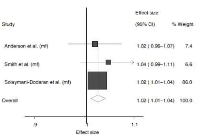
In the Barrett’s esophagus and general population control comparison, shown in
Figure 2, the random effects meta-analysis gave a pooled odds ratio of 1.02 per kg/m2
(95% CI: 1.01, 1.04; I2=0%; p=0.002). When stratified by sex there was no difference
between both the male (OR=1.02; 95% CI: 0.98, 1.08; p=0.32) and female (OR=1.03;
non-significant. A sensitivity analysis was conducted due to the relative study sizes. When
the largest study was omitted the point estimate increased slightly but became
statistically non-significant (OR=1.03 per kg/m2; 95% CI: 0.99, 1.07). Additional
sub-group analyses were not undertaken for the Barrett’s esophagus and general
population control analysis due to the inclusion of only three studies.
To investigate possible small study bias in the Barrett’s esophagus and GERD-ALL
analysis a funnel plot of the log odds ratio against the inverse of the standard error of
the log odds ratio was produced and did not appear to show any bias (data not shown)
and this was confirmed by a statistically non-significant Egger’s test (p=0.08)..
A random effects sensitivity analysis was conducted whereby each study was
excluded in turn to give an indication of how much influence each individual study
had on the pooled estimate. No single study significantly altered the pooled estimate
for the Barrett’s esophagus and GERD-ALL analysis (data not shown).
The I2 value for the Barrett’s esophagus and GERD-ALL meta-analysis was 52%
while the subgroup values for comparison to the ESO group and the
GERD-HUK group were 62% and 35% respectively. The full dataset was utilized for
investigation of heterogeneity by meta-regression. When study population size was
dichotomized by the median and entered into meta-regression it proved not to be
statistically significant source of heterogeneity (p=0.202). The method of BMI data
collection (clinical measurement or self-reported) could not be investigated due to
only one dataset confirming clinical measurement as its method (34). The remaining
meta-regression on the mid-point of patient recruitment year could also not be
undertaken as this was unknown for four of the nine studies.
In the Barrett’s esophagus and GERD-ALL analysis one dataset, from Ireland,
provided an estimate which was considerably lower than all others (24). When this
dataset was temporarily excluded from the meta-analysis the I2 for the GERD-ESO
comparison group was reduced to 36%; this was also reflected in the GERD-ALL I2
value which decreased from 52% to 33%. This difference was highlighted in
subsequent meta-regressions of geographical location; a comparison of the three US
and the three UK studies was not statistically significant (p=0.9) whilst there were
statistical significant differences between the three US studies and the Irish study
Discussion
The systematic review and meta-analysis presented provides evidence that increasing
BMI does not present an increased risk for Barrett’s esophagus above what would be
expected from GERD alone. Point estimates calculated for the Barrett’s esophagus
and GERD-ALL meta-analysis, and those detailed for the subgroup and sex specific
analyses, were not statistically significant.
There was a ‘moderate’ amount of heterogeneity with an I2
value of 52% for the
Barrett’s esophagus and GERD-ALL analysis (19). An investigation of study size by
meta-regression provided no evidence to support this as a source of heterogeneity.
Method of data analysis and year of patient recruitment variables could not be
investigated due to lack of data whilst it is not obvious why the only Irish study (24)
included provided a protective odds ratio for males with increasing BMI and thus
contributed significantly to the heterogeneity statistic.
The Barrett’s esophagus and general population control comparison gave an odds
ratio of 1.12 per five unit increase of BMI. This analysis was heavily dominated by
the Solaymani-Dodaran study (see Figure 2) (25). When excluded in a sensitivity
analysis, the point estimate increased slightly but was no longer statistically
significant.
A previous meta-analysis of esophagitis and esophageal adenocarcinoma is suggestive
to a hypothesis that increasing adiposity is a risk factor for the development of
concluding that it is an increased risk of GERD, caused by increasing BMI, which
underlies this association; once GERD occurs it would seem that there is no additional
effect of BMI on progression to Barrett’s esophagus. BMI is, therefore, of no value in
predicting which GERD patients may be at risk of developing Barrett’s esophagus and
consequently such information is of no value in making decisions about which GERD
patients would benefit from endoscopic screening or surveillance.
This indirect mechanism of association could, potentially, be explanatory of observed
increases in esophageal adenocarcinoma risk in higher BMIs categories. (14, 15)
Thus, it is proposed that increasing adiposity is only a direct risk factor for GERD and
that this association acts as an intermediary in the etiology of Barrett’s esophagus and,
possibly, esophageal adenocarcinoma. There is a lack of data on BMI comparisons
between patients with this cancer and GERD or Barrett’s control groups. One cohort
study has investigated the risk of BMI for developing low grade dysplasia from BO
(39). The reported OR was 1.01 per kg/m2 (95% CI: 0.92, 1.11; p=0.862) indicating
no association. Two other studies have adjusted for GERD in their investigations of
esophageal adenocarcinoma and BMI compared to population controls (40, 41). Both
found evidence for BMI acting as an independent risk factor for esophageal
adenocarcinoma and this suggests that increasing adiposity has additional effects on
cancer risk other than propagating GERD. Both of these studies measured GERD on
symptom questionnaires and responses from such are known to have a relatively low
diagnostic sensitivity (42), thus further investigation into this association and its
The mechanistic explanation of why increasing adiposity should increase the
likelihood of GERD remains enigmatic. Several hypotheses have been forwarded to
explain the association including a decrease in pressure of the lower esophageal
sphincter (43), hiatus hernia (43, 44), altered refluxate composition (45), high fat diet
(46-48), estrogens (49), Helicobacter pylori (50, 51) and visceral fat (43, 52). It
appears unlikely that the risk is mediated wholly through any one of these; the route
of association is likely to be complex and multi-factorial.
The meta-analysis undertaken has several limitations. Firstly, the accuracy of BMI
measurement and its reliability as a measure of adiposity are known to be imperfect,
although this measure it still more applicable to epidemiological studies then other
methodologies such as computed tomography, which is expensive and impractical for
large populations.
Second, the timing of the BMI measurement was denoted as ‘current’ in all studies.
This is not ideal as it is not truly representative of the situation earlier in the disease
process. It is, however, the only time-point for which BMI is consistently recorded for
Barrett’s esophagus patients in a research field which already exhibits a paucity of
adiposity data. Given that Barrett’s esophagus is a pre-cancerous condition, it is likely
that this measurement is less susceptible to reporting bias than in the case of studies of
cancer.
Thirdly, we assume that all BO patients also have GERD. Although for two of the
included studies this is true (31, 36), three other studies included in this meta-analysis
It is conceivable that the remaining 20% may not have GERD. Conversely BO is
known to cause de-sensitization of the esophagus (53) and, coupled with the fact that
GERD is the most potent and consistent risk factor for BO (3, 54), it is therefore
likely that the majority of BO patients have GERD and this provides confidence to
our conclusions.
A final weakness of this meta-analysis is that all odds ratios are unadjusted.
Exposures including smoking, alcohol, diet and medication may be hypothesized as
effect modifiers but data on these variables have rarely been published on Barrett’s
esophagus patients, hence their omission from the analysis. In addition there was no
study which provided unadjusted and adjusted odds ratios which may have allowed
artificial adjustment of study point estimates (55).
This meta-analysis indicates that increasing adiposity presents no additional risk for
Barrett’s esophagus above that which it presents by increasing risk of GERD alone.
The large body of evidence of the associated risk between obesity and GERD, when
compared to general population controls, is conclusive. In summary, the causality of
the association between obesity and esophageal adenocarcinoma requires further
investigation, although it may be postulated that the effect is indirect via the known
associations with GERD.
Acknowledgements
The authors would like to thank the following for their kind provision of data which
enabled this meta-analysis to be undertaken:
Christine P.J. Caygill David C. Whiteman Gaile L. Moe
Guilherme M.R. Campos Liam J. Murray
Lesley A. Anderson Martin D. Gough
Masoud Solaymani-Dodaran Nicholas J. Shaheen
Shanmugarajah Rajendra Steven R. DeMeester
1) What is current knowledge
Increasing body mass index is a risk factor for gastro-esophageal reflux disease
Increasing body mass index is a risk factor for esophageal adenocarcinoma
Gastro-esophageal reflux disease is the main risk factor for Barrett’s esophagus
2) What is new here
In gastro-esophageal reflux disease patients increasing body mass index does not alter the risk of Barrett’s esophagus
Increasing body mass index is an indirect risk factor for Barrett’s
esophagus mediated through gastro-esophageal reflux
Formatted: Bullets and Numbering
1. Jankowski JA, Provenzale D, Moayyedi P. Esophageal adenocarcinoma
arising from Barrett's metaplasia has regional variations in the west.
Gastroenterology 2002;122:588-90.
2. Shaheen NJ, Crosby MA, Bozymski EM, et al. Is there publication bias in the
reporting of cancer risk in Barrett's esophagus? Gastroenterology
2000;119:333-8.
3. Conio M, Filiberti R, Blanchi S, et al. Risk factors for Barrett's esophagus: a
case-control study. Int J Cancer 2002;97:225-9.
4. Wild CP, Hardie LJ. Reflux, Barrett's oesophagus and adenocarcinoma:
burning questions. Nat Rev Cancer 2003;3:676-84.
5. Vizcaino AP, Moreno V, Lambert R, et al. Time trends incidence of both
major histologic types of esophageal carcinomas in selected countries,
1973-1995. Int J Cancer 2002;99:860-8.
6. van Soest EM, Dieleman JP, Siersema PD, et al. Increasing incidence of
Barrett's oesophagus in the general population. Gut 2005;54:1062-6.
7. Irani S, Parkman H, Krevsky B, et al. A decade (1991-2000) of increasing
incidence of endoscopic and histologic Barrett's esophagus (BE) at a single
academic medical center. Am J Gastroenterol 2003;98:S16.
8. Cook MB, Wild CP, Forman D. A systematic review and meta-analysis of the
sex ratio for Barrett's esophagus, erosive reflux disease, and nonerosive reflux
disease. Am J Epidemiol 2005;162:1050-61.
10. The National Audit Office, The Audit Commission, The Healthcare
Commission, Tackling Child Obesity - First Steps. London. Her Majesty's
Stationary Office, 2006.
11. International Agency for Research on Cancer, Handbooks of cancer
prevention. Weight control and physical activity., Lyon: International Agency
for Research on Cancer, 2002.
12. Seidell JC. Prevalence and time trends of obesity in Europe. J Endocrinol
Invest 2002;25:816-22.
13. Flegal KM. Epidemiologic aspects of overweight and obesity in the United
States. Physiol Behav 2005;86:599-602.
14. Hampel H, Abraham NS, El-Serag HB. Meta-analysis: obesity and the risk for
gastroesophageal reflux disease and its complications. Ann Intern Med
2005;143:199-211.
15. Kubo A, Corley DA. Body mass index and adenocarcinomas of the esophagus
or gastric cardia: a systematic review and meta-analysis. Cancer Epidemiol
Biomarkers Prev 2006;15:872-8.
16. The EndNote Team. 2003. EndNote 7. Berkeley, CA: Thomson ISI
ResearchSoft.
17. StataCorp. 2004. Stata Statistical Software: Release 8.2. College Station,
18. Chene G, Thompson SG. Methods for summarizing the risk associations of
quantitative variables in epidemiologic studies in a consistent form. Am J
Epidemiol 1996;144:610-21.
19. Higgins JP, Thompson SG, Deeks JJ, et al. Measuring inconsistency in
meta-analyses. Br Med J 2003;327:557-60.
20. Sharp SJ. Meta-analysis regression. Stata Technical Bulletin 1998;16-22.
21. Egger M, Davey Smith G, Schneider M, et al. Bias in meta-analysis detected
by a simple, graphical test. Br Med J 1997;315:629-34.
22. Banki F, Demeester SR, Mason RJ, et al. Barrett's esophagus in females: a
comparative analysis of risk factors in females and males. Am J Gastroenterol
2005;100:560-7.
23. Reed PI, Caygill CPJ, Watson A, et al. The United Kingdom Barrett's
Oesophagus Registry (UKBOR) - The first six years. Gastroenterol Pol
2003;10:299-304.
24. Anderson LA, Murphy SJ, Johnston BT, et al. Obesity and smoking in patients
with Barrett's esophagus, esophageal adenocarcinoma and esophageal
adenocarcinoma: Results from the Finbar study. Gastroenterology
2005;128:A49.
25. Solaymani-Dodaran M, Logan RF, West J, et al. Risk of oesophageal cancer in
26. Gudlaugsdottir S, Verschuren W, Dees J, et al. Hypertension is frequently
present in patients with reflux esophagitis or Barrett's esophagus but not in
those with non-ulcer dyspepsia. Eur J Intern Med 2002;13:369-75.
27. Rajendra S, Kutty K, Karim N. Ethnic differences in the prevalence of
endoscopic esophagitis and Barrett's esophagus: the long and short of it all.
Dig Dis Sci 2004;49:237-42.
28. Chak A, Lee T, Kinnard MF, et al. Familial aggregation of Barrett's
oesophagus, oesophageal adenocarcinoma, and oesophagogastric junctional
adenocarcinoma in Caucasian adults. Gut 2002;51:323-8.
29. Vaughan TL, Kristal AR, Blount PL, et al. Nonsteroidal anti-inflammatory
drug use, body mass index, and anthropometry in relation to genetic and flow
cytometric abnormalities in Barrett's esophagus. Cancer Epidemiol
Biomarkers Prev 2002;11:745-52.
30. Cameron AJ. Barrett's esophagus: prevalence and size of hiatal hernia. Am J
Gastroenterol 1999;94:2054-9.
31. Campos GM, DeMeester SR, Peters JH, et al. Predictive factors of Barrett
esophagus: multivariate analysis of 502 patients with gastroesophageal reflux
disease. Arch Surg 2001;136:1267-73.
32. Caygill CP, Johnston DA, Lopez M, et al. Lifestyle factors and Barrett's
33. Moe GL, Kristal AR, Levine DS, et al. Waist-to-hip ratio, weight gain, and
dietary and serum selenium are associated with DNA content flow cytometry
in Barrett's esophagus. Nutr Cancer 2000;36:7-13.
34. Shaheen N. Body fat distribution predicts the presence of Barrett's esophagus:
a case-control study. Gastroenterology 2005;128:A231.
35. Casson AG, Veugelers PJ, Fitzgerald AL, et al. Dietary and lifestyle risk
factors in the etiology of gastroesophageal reflux disease, Barrett esophagus
and esophageal adenocarcinoma. The Canadian Journal of Gastroenterology
2005;19:3.
36. Kulig M, Nocon M, Vieth M, et al. Risk factors of gastroesophageal reflux
disease: methodology and first epidemiological results of the ProGERD study.
J Clin Epidemiol 2004;57:580-9.
37. Smith KJ, O'Brien SM, Smithers BM, et al. Interactions among smoking,
obesity, and symptoms of acid reflux in Barrett's esophagus. Cancer Epidemiol
Biomarkers Prev 2005;14:2481-6.
38. Cameron AJ. Barrett's esophagus: prevalence and size of hiatal hernia. Am J
Gastroenterol 1999;94:2054-9.
39. Oberg S, Wenner J, Johansson J, et al. Barrett esophagus: risk factors for
progression to dysplasia and adenocarcinoma. Ann Surg 2005;242:49-54.
40. Chow WH, Blot WJ, Vaughan TL, et al. Body mass index and risk of
adenocarcinomas of the esophagus and gastric cardia. J Natl Cancer Inst
41. Lagergren J, Bergstrom R, Nyren O. Association between body mass and
adenocarcinoma of the esophagus and gastric cardia. Ann Intern Med
1999;130:883-90.
42. Klauser AG, Schindlbeck NE, Muller-Lissner SA. Symptoms in
gastro-oesophageal reflux disease. Lancet 1990;335:205-8.
43. Pandolfino JE, El-Serag HB, Zhang Q, et al. Obesity: a challenge to
esophagogastric junction integrity. Gastroenterology 2006;130:639-49.
44. Wilson LJ, Ma W, Hirschowitz BI. Association of obesity with hiatal hernia
and esophagitis. Am J Gastroenterol 1999;94:2840-4.
45. Wisen O, Rossner S, Johansson C. Impaired pancreatico-biliary response to
vagal stimulation and to cholecystokinin in human obesity. Metabolism
1988;37:436-41.
46. Nebel OT, Castell DO. Lower esophageal sphincter pressure changes after
food ingestion. Gastroenterology 1972;63:778-83.
47. Holloway RH, Kocyan P, Dent J. Provocation of transient lower esophageal
sphincter relaxations by meals in patients with symptomatic gastroesophageal
reflux. Dig Dis Sci 1991;36:1034-9.
48. Becker DJ, Sinclair J, Castell DO, et al. A comparison of high and low fat
meals on postprandial esophageal acid exposure. Am J Gastroenterol
1989;84:782-6.
49. Nilsson M, Lagergren J. The relation between body mass and
50. Loffeld RJ. Helicobacter pylori, obesity and gastro-oesophageal reflux disease.
Is there a relation? A personal view. Neth J Med 2005;63:344-7.
51. Suzuki M, Suzuki H, Masaoka T, et al. Helicobacter pylori eradication
treatment modulates epithelial cell proliferation and tissue content of
hepatocyte growth factor in the gastric mucosa. Aliment Pharmacol Ther
2004;20:158-64.
52. El-Serag HB, Kvapil P, Hacken-Bitar J, et al. Abdominal obesity and the risk
of Barrett's esophagus. Am J Gastroenterol 2005;100:2151-6.
53. Byrne PJ, Mulligan ED, O'Riordan J, et al. Impaired visceral sensitivity to acid
reflux in patients with Barrett's esophagus. The role of esophageal motility.
Dis Esophagus 2003;16:199-203.
54. Eisen GM, Sandler RS, Murray S, et al. The relationship between
gastroesophageal reflux disease and its complications with Barrett's
esophagus. Am J Gastroenterol 1997;92:27-31.
55. Greenland S. Quantitative methods in the review of epidemiologic literature.
Table 1. Studies Included in the Meta-Analyses
Authors Year
of Publication
Country Patients (N) Comparison
Arm (N)
Comparison
Group(s)
Data format Barrett’s
Esophagus
Definition
Cameron 1999 USA 61 103
GERD-HUK
Categorical SIM
Campos et al.
(31)
2001 USA 189 313 GERD-HUK Categorical SIM
Caygill et al.†
(32)
2002 U.K. 101 101 GERD-ESO Means SIM
Gudlaugsdottir
et al. (26)
2002 The
Netherlands
34 31 GERD-ESO Means ≥3 cm CLE
or SIM
(24) and population
Kulig et al.
(36)
2004 Germany,
Austria, and
Switzerland
702 2660 GERD-ESO Categorical CLE or SIM
Solaymani-Dodaran et al.
(25)
2004 U.K. 1269 4935 and 9320 GERD-ESO
and population
Categorical SIM‡
Gough§ 2005 U.K. 150 151 GERD-ESO Categorical SIM
Shaheen (34) 2005 USA 169 302 GERD-HUK Means SIM
Smith et al.
(37)
2005 Australia 115 259 Population Categorical SIM
As well as the GERD patients, this comparison group may also include some patients presenting for endoscopy with no GERD symptoms.
†The data from one study (31) provided only mean weights, by sex, for the Barrett’s esophagus and reflux esophagitis groups, as height had not
‡This study cannot verify method of diagnosis, but is assumed to be representative of SIM; in consideration of the dates of the study and the
current U.K. practice guidelines, the majority of such patients are assumed to have undergone histologic diagnosis.
§M. Gough, The University of Sheffield, United Kingdom, personal communication, 2005.
CLE = columnar-lined epithelium; GERD-HUK = gastroesophageal reflux disease histology unknown; GERD-ESO = gastroesophageal reflux
disease with esophagitis;
Figure 1. Forest plot of random effects meta-analysis of the risk of BMI on Barrett’s
esophagus compared to the GERD comparison arms.
Each study’s odds ratio (OR) is represented by the corresponding black square with
the arms representing 95% confidence intervals. The pooled estimate subtotals are
designated by the diamonds, which follow each subgroup; these are 0.99 per kg/m2
(95% CI 0.96–1.01) and 1.00 per kg/m2 (95% CI 0.96–1.04), respectively, while the
last diamond is the overall pooled estimate, which is 0.99 (95% CI 0.97–1.01).
Figure 2. Forest plot of random effects meta-analysis of the risk of BMI on Barrett’s
esophagus compared to general population controls.
Each study’s odds ratio (OR) is represented by the corresponding black square with
the arms representing 95% CI. The pooled estimate is designated by the diamond and
APPENDIX
Search Strategy 1
1. exp obesity/
2. obes$.tw.
3. exp body mass index/
4. (body adj2 mass adj2 index$).tw.
5. bmi.tw.
6. exp body weight/
7. overweight$.tw.
8. (body adj5 fat).tw.
9. over-weight.tw.
10. exp adipose tissue/
11. adipose tissue.tw.
12. physical$ inactiv$.tw.
13. exp energy intake/
14. exp caloric intake/
15. energ$ intake.tw.
16. calor$ intake.tw.
17. exp energy expenditure/
18. exp energy metabolism/
19. energy balance.tw.
20. energy expend$.tw.
22. exp anthropometry/
23. anthropom$.tw.
24. (quetelet$ adj index$).tw.
25. exp body composition/
26. body weight.tw.
27. body size.tw.
28. fat$ distribut$.tw.
29. (waist adj3 hip adj3 ratio).tw.
30. (waist adj3 circumference$).tw.
31. (hip adj3 circumference$).tw.
32. or/1-31
33. exp gastroesophageal reflux/
34. (gastroesophageal adj2 reflux).tw.
35. (gastro?esophageal adj2 reflux).tw.
36. (gastro-esophageal adj2 reflux).tw.
37. (gastro-oesophageal adj2 reflux).tw.
38. gord.tw.
39. gerd.tw.
40. erd.tw.
41. enrd.tw.
42. erosive reflux.tw.
43. non-erosive reflux.tw.
44. non erosive reflux.tw.
45. endoscopy-negative reflux.tw.
47. negative-endoscopy reflux.tw.
48. negative endoscopy reflux.tw.
49. exp esophagitis/
50. esophagitis.tw.
51. oesophagitis.tw.
52. (reflux$ adj3 disease$).tw.
53. heartburn.tw.
54. indigestion.tw.
55. or/33-54
56. exp barrett esophagus/
57. (barret$ adj3 esophagus).tw.
58. (barret$ adj3 oesophagus).tw.
59. (metaplas$ adj5 epitheli$).tw.
60. (columnar adj5 line$).tw.
61. (columnar adj5 metaplas$).tw.
62. (intest$ adj5 metaplas$).tw.
63. brachyesophag$.tw.
64. brachyoesophag$.tw.
65. brachy-esophag$.tw.
66. brachy-oesophag$.tw.
67. endobrachy$.tw.
68. or/56-67
69. exp esophageal neoplasms/
70. (esophag$ adj250 neoplas$).tw.
72. (esophag$ adj250 cancer$).tw.
73. (esophag$ adj250 carcin$).tw.
74. (oesophag$ adj250 carcin$).tw.
75. (esophag$ adj250 tumo$).tw.
76. (oesophag$ adj250 tumo$).tw.
77. (esophag$ adj250 metasta$).tw.
78. (oesophag$ adj250 metasta$).tw.
79. (esophag$ adj250 malig$).tw.
80. (oesophag$ adj250 malig$).tw.
81. (adenocarcinoma$ adj250 esophag$).tw.
82. (adenocarcinoma$ adj250 oesophag$).tw.
83. or/69-82
84. 32 and 55
85. 32 and 68
86. 32 and 83
87. 84 or 85 or 86
Search Strategy 2
1. exp gastroesophageal reflux/
2. (gastroesophageal adj2 reflux).tw.
3. (gastro?esophageal adj2 reflux).tw.
4. (gastro-esophageal adj2 reflux).tw.
298 Cook et al.
5. (gastro-oesophageal adj2 reflux).tw.
6. gord.tw.
7. gerd.tw.
8. erd.tw.
9. enrd.tw.
10. erosive reflux.tw.
11. non-erosive reflux.tw.
12. non erosive reflux.tw.
13. endoscopy-negative reflux.tw.
14. endoscopy negative reflux.tw.
15. negative-endoscopy reflux.tw.
16. negative endoscopy reflux.tw.
17. exp esophagitis/
18. esophagitis.tw.
19. oesophagitis.tw.
20. (reflux$ adj3 disease$).tw.
21. heartburn.tw.
23. or/1-22
24. exp barrett esophagus/
25. (barret$ adj3 esophagus).tw.
26. (barret$ adj3 oesophagus).tw.
27. (metaplas$ adj5 epitheli$).tw.
28. (columnar adj5 line$).tw.
29. (columnar adj5 metaplas$).tw.
30. (intest$ adj5 metaplas$).tw.
31. brachyesophag$.tw.
32. brachyoesophag$.tw.
33. brachy-esophag$.tw.
34. brachy-oesophag$.tw.
35. endobrachy$.tw.
36. or/24-35
37. exp esophageal neoplasms/
38. (esophag$ adj250 neoplas$).tw.
39. (oesophag$ adj250 neoplas$).tw.
40. (esophag$ adj250 cancer$).tw.
41. (esophag$ adj250 carcin$).tw.
42. (oesophag$ adj250 carcin$).tw.
43. (esophag$ adj250 tumo$).tw.
44. (oesophag$ adj250 tumo$).tw.
45. (esophag$ adj250 metasta$).tw.
46. (oesophag$ adj250 metasta$).tw.
48. (oesophag$ adj250 malig$).tw.
49. (adenocarcinoma$ adj250 esophag$).tw.
50. (adenocarcinoma$ adj250 oesophag$).tw.
51. or/37-50
52. (risk$ adj2 factor$).ti.
53. exp Risk factors/
54. 52 or 53
55. 23 and 54
56. 36 and 54
57. 51 and 54
58. or/55-57