THE MOLECULAR AND CELLULAR DIFFERENCES BETWEEN
TENDONS AND LIGAMENTS
Thesis submitted in accordance with the requirements of the University of
Liverpool for the degree of Doctor in Philosophy
By
Yalda Ashraf Kharaz
ii
ABSTRACT
Tendons and ligaments play key roles in the musculoskeletal system in both man and animals. Both tissues can often be injured as result of contact based accidents or ageing and disease, causing discomfort, pain and increased susceptibility to degenerative joint disease. To date, tendon and ligament biology is relatively under-studied in healthy, non-diseased tissues. This information is essential to understand the pathology of these tissues and vital for future development of tendon and ligament tissue-engineered structures.
This thesis aims to investigate the molecular and cellular differences between tendons and ligaments around the canine stifle joint. The biochemical composition, structural, and morphological characteristics were identified between the different regions of the intra- articular cranial cruciate ligament (CCL) and extra-articular medial collateral ligament (MCL), and the positional long digital extensor tendon (LDET) and energy storing superficial digital flexor tendons (SDFT). Differences in proteome composition were also assessed between CCL and LDET. Cells isolated from canine CCL and LDET were cultured in a 3D in vitro fibrin culture model and measured for differences in structural, biochemical and proteome composition.
iii
TABLE OF CONTENTS
ABSTRACT ... ii
TABLE OF CONTENTS ... iii
INDEX TO FIGURES ... v
INDEX TO TABLES ... viii
LIST OF ABBREVIATIONS ... ix
ACKNOWLEDGEMENT ... xi
CHAPTER 1: GENERAL INTRODUCTION ... 1
1.1 CONNECTIVE TISSUE ... 2
1.2 TENDON ... 6
1.3 LIGAMENT ... 7
1.4 STRUCTURE OF TENDON AND LIGAMENT ... 8
1.5 TENDON AND LIGAMENT COMPOSITION ... 10
1.6 LIGAMENTS AND TENDONS OF THE KNEE JOINT ... 24
1.7 TENDON AND LIGAMENT INJURY AND DISEASE ... 28
1.8 HEALING OF TENDINOUS AND LIGAMENTOUS TISSUE ... 31
1.9 CURRENT METHOD OF TENDON AND LIGAMENT REPAIR AND TISSUE ENGINEERING ……….34
1.10 SUMMARY OF TENDON AND LIGAMENT COMPARISON STUDIES ... 36
1.11 HYPOTHESIS AND AIMS ... 37
CHAPTER 2: GENERAL MATERIAL AND METHODS ... 38
2.1 TISSUE COLLECTION ... 39
2.2 TISSUE PREPARATION ... 42
2.3 DEVELOPMENT OF TISSUE ENGINEERED LIGAMENT AND TENDON CONSTRUCTS .. 43
2.4 BIOCHEMICAL ANALYSIS ... 45
2.5 HISTOLOGY STAINING AND SCORING... 50
2.6 TISSUE IMMUNOSTAINING ... 56
2.7 TRANSMISSION ELECTRON MICROSCOPY ... 59
2.8 PROTEOMIC ANALYSIS ... 60
2.9 GENERAL STATISTICAL ANALYSIS ... 64
CHAPTER 3: A BIOCHEMICAL COMPARISON OF THE EXTRACELLULAR MATRIX COMPOSITION OF TENDONS AND LIGAMENTS AROUND THE CANINE STIFLE JOINT ... 65
3.1 INTRODUCTION ... 66
3.2 HYPOTHESIS & AIM ... 68
3.3 EXPERIMENTAL PROCEDURE ... 69
3.4 RESULTS ... 72
3.5 DISCUSSION ... 78
iv
CHAPTER 4: THE MORPHOLOGICAL AND STRUCTURAL DIFFERENCES AND EXTRACELLULAR MACROMOLECULES DISTRIBUTION BETWEEN TENDONS AND LIGAMENTS AROUND THE
CANINE STIFLE JOINT ... 83
4.1 INTRODUCTION ... 84
4.2 HYPOTHESIS AND AIMS ... 85
4.3 EXPERIMENTAL PROCEDURES ... 87
4.4 RESULTS ... 89
4.5 DISCUSSION ... 109
4.6 CONCLUSION ... 120
CHAPTER 5: A COMPARISON OF THE EXTRACELLULAR MATRIX COMPOSITION OF NATIVE TENDON/LIGAMENT AND 3D TENDON/LIGAMENT CONSTRUCTS ... 121
5.1 INTRODUCTION ... 122
5.2 HYPOTHESIS & AIMS ... 123
5.3 EXPERIMENTAL PROCEDURES ... 124
5.4 RESULTS ... 127
5.5 DISCUSSION ... 138
5.6 CONCLUSION ... 142
CHAPTER 6: PROTEOMIC COMPARISON OF TENDON, LIGAMENT AND 3D TENDON/LIGAMENT CONSTRUCTS ... 143
6.1 INTRODUCTION ... 144
6.2 HYPHOTHESIS & AIMS ... 147
6.3 EXPERIMENTAL PROCEDURE ... 148
6.4 RESULTS ... 151
6.5 DISCUSSION ... 173
CHAPTER 7: GENERAL DISCUSSION AND FUTURE DIRECTION ... 180
7.1 GENERAL DISCUSSION ... 181
7.2 CONCLUSION ... 188
7.3 FUTURE WORK ... 189
CHAPTER 8: SUPPLEMENTARY DATA ... 190
v
INDEX TO FIGURES
Figure 1.1. Classification of connective tissues ... 3
Figure 1.2. Hierarchical structure of tendon and ligament ... 9
Figure 1.3 Intracellular and extracellular synthesis and processing of the collagen triple helix ... 15
Figure 1.4. Proteoglycans present in tendon and ligaments ... 23
Figure 1.5. Anatomy of the human knee joint ... 25
Figure 1.6 A typical tensile stress-strain curve for tendon and ligament ... 29
Figure 1.7. Typical tendon and ligament healing process. ... 32
Figure 2.1. Anatomy of canine stifle or knee joint ... 40
Figure 2.2. Typical standard curve for dsDNA quantification ... 46
Figure 2.3.Typical standard curve for sGAG quantification.. ... 47
Figure 2.4. Typical standard curve for hydroxproline quantification. ... 48
Figure 2.5. Typical standard curve for elastin quantification. ... 49
Figure 2.6. Typical standard curve for PIERCETM protein assay. ... 61
Figure 3.1. Water content of CCL, MCL, LDET and SDFT ... 72
Figure 3.2. DNA content (µg DNA/ mg dry weight) of the proximal, middle and distal regions of CCL, MCL, LDET and SDFT. ... 73
Figure 3.3. Total collagen content (% /mg dry weight) of proximal, middle and distal regions of CCL, MCL, LDET and SDFT. ... 74
Figure 3.4. Elastin content (% /mg dry weight) of the proximal middle, distal regions of CCL, MCL, LDET and SDFT ... 75
Figure 3.5. sGAG content (µg sGAG/ mg dry weight) of the proximal, middle and distal regions of CCL, MCL, LDET and SDFT. ... 76
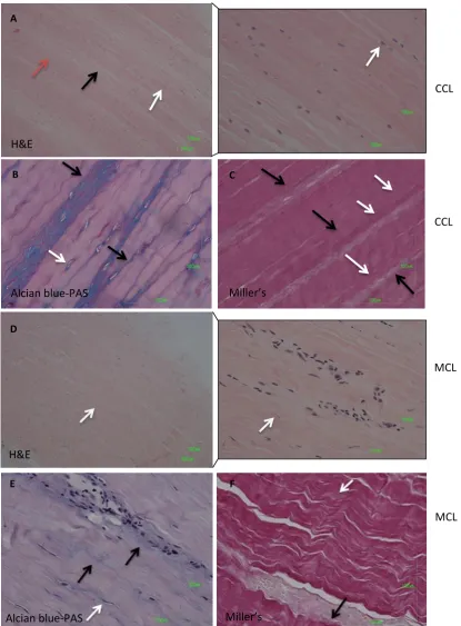
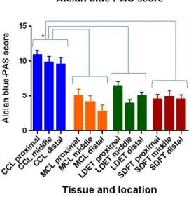
Figure 4.1. Representative images of histological staining of the mid-region of CCL and MCL.. ... 91
Figure 4.2. Representative images of histological staining of the mid-region of LDET and SDFT.. ... 92
Figure 4.3. Histology scoring results of ECM organisation, cell shape, cell distribution, cell alignment, vascularisation and inflammation ... 93
vi
Figure 4.5. Miller’s stain score for CCL, MCL, LDET and SDFT ... 95
Figure 4.6. Representative immunostaining pictures of negative controls. ... 99
Figure 4.7. Immunostaining of collagen I and III in CCL and LDET ... 100
Figure 4.8. Immunostaining of collagen type VI in CCL and LDET ... 101
Figure 4.9. Immunostaining of aggrecan and versican in CCL and LDET. ... 103
Figure 4.10. Immunostaining of decorin and biglycan in CCL and LDET. ... 104
Figure 4.11. Immunostaining of keratocan and asporin in CCL an LDET. ... 105
Figure 4.12. Immunostaining of fibrillin 1 and fibrillin 2 in CCL and LDET ... 107
Figure 4.13. Immunostaining of elastic fibres with fibrillin 1 and 2 in CCL and LDET. ... 108
Figure 5.1. Canine CCL and LDET cells embedded in a 3D fibrin gel ... 128
Figure 5.2. Representative histology staining pictures of 3D LDET construct.. ... 130
Figure 5.3. Representative histology staining pictures of 3D CCL construct.. ... 131
Figure 5.4. Representative pictures of transmission electron analysis of 3D CCL construct.132 Figure 5.5. Representative transmission electron pictures of 3D LDET constructs ... 133
Figure 5.6. DNA content (µg/ mg dry weight) of native tissues (CCL and LDET) and 3D constructs (CCL and LDET) ... 134
Figure 5.7. Collagen content/ mg % dry weight of native tissues (CCL and LDET) and 3D constructs (CCL and LDET).. ... 135
Figure 5.8. sGAG content (µg/ mg dry weight) of native tissue (CCL and LDET) and 3D construct (CCL and LDET) ... 136
Figure 5.9. Elastin content (% mg dry weight) of native tissue and 3D constructs (CCL and LDET) ... 137
Figure 6.1. Outline diagram of mass spectrometer adapted from Matthiesen and Jensen (2008) ... 145
Figure 6.2. Venn diagram of native CCL/LDET and 3D tissues with total number of protein identified each tissue as well as common proteins between tissues. ... 154
Figure 6.3. Overview of native ligament and tendon proteomes which were subdivided based on matrisomal protein and on function with the associated subcellular location of protein in both tissues ... 156
Figure 6.4. String analysis of native canine CCL.. ... 157
vii Figure 6.6. Overview of the proteomes of 3D ligament and 3D tendon constructs hich were
subdivided based on matrisomal protein and on function with the associated
subcellular location of protein in both tissues ... 159 Figure 6.7. String analysis of 3D ligament construct. ... 160 Figure 6.8. String analysis of 3D tendon construct ... 161 Figure 6.9. Principal component analysis between native and tissue engineered tendon and
ligament constructs ... 164 Figure 6.10. Volcano plots for native ligament vs native tendon, 3D ligament vs 3D tendon,
native ligament tendon vs 3D ligament, and native tendon vs 3D tendon ... 165 Figure 6.11. Different abundant proteins between native tissues and 3D constructs. ... 168 Figure 6.12. Expression plots of 3D ligament versus 3D tendon construct of selected proteins produced by Progenesis™ LC-MS ... 170 Figure 6.13. Post-translational modification of collagen alpha-1(I) and collagen alpha-2 (I)
between native tendon and ligament. ... 171 Figure 6.14. Post-translational modification of collagen alpha-1(III) between native tendon
viii
INDEX TO TABLES
Table 1.1. Collagen types, classification and distribution ... 12
Table 2.1. Tissue collection and associated experimental testing purposes ... 41
Table 2.2. H&E scoring sheet ... 53
Table 2.3. Miller’s scoring sheet ... 54
Table 2.4. Alcian-blue-PAS scoring sheet ... 55
Table 2.5. Primary and secondary antibodies used in tissue immunostaining for detection ECM macromolecules. ... 58
Table 3.1. Water content and tissue concentration of DNA, collagen, elastin and sGAG of CCL, MCL, LDET and SDFT at proximal, middle and distal regions. ... 77
Table 4.1. H&E scoring Kendall’s coefficient of concordance results. ... 96
Table 4.2. Kendall’s coefficients of concordance for Alcian blue-PAS and Miller’s histology score. ... 97
Table 6.1. Overview of PEAKS® database search results for native CCL and LDET tissue and 3D CCL and LDET construct. ... 154
Table 6.2.Matrisomal proteins in common between tendon and ligament native tissues and 3D constructs. ... 155
Table 6.3. Identified protein differences between native ligament and tendon with statistical significance. ... 166
ix
LIST OF ABBREVIATIONS
AB-PAS Alcian blue- periodic acid Schiff
ACL Anterior cruciate ligament
ANOVA Analysis of variance
BSA Bovine serum albumin
CCL Cranial cruciate ligament
Da Dalton
DAB 3,3'-Diaminobenzidine
dH20 Distilled water
DMBA Dimethylaminobenzaldehyde
DMEM Dulbecco’s Modified Eagles Medium
DNA Deoxyribonucleic acid
DMMB Dimethyl-methylene blue
DTT Dithiothreitol
ECM Extracellular matrix
EDTA Ethylenediaminetetraacetic acid
FBS Fetal bovine serum
FDR False discovery rate
Glycosaminoglycan GAG
GnHCl Guanidine hydrochloride
GO Gene ontology
H & E Hematoxylin and Eosin
HCL Hydrochloride
HPLC High-performance liquid chromatography
HRP Horse-radish peroxidase
x
kN Kilonewtons
LDET Long digital extensor tendon
M Molar
mM Millimolar
MCL Medical collateral ligament
mg Miligram,
ml Mililitre
MS Mass spectrometry
Nm Nanomolar
SDS-PAGE Sodium dodecyl sulphate polyacrylamide gel electrophoresis
SLRPs Small leucine rich proteoglycans
sGAG Sulphated glycosaminoglycans
SEM Standard error of the mean
SDFT Superficial digital flexor tendon
TBS Tris-buffered saline
TEM Transmission electron microscopy
TFA Trifluoroacetic acid
TGF Transforming growth factor
OD Optical density
PBS Phosphate buffer saline
PCA Principle component analysis
PG Proteoglycan
xi
ACKNOWLEDGEMENT
I would like to take this opportunity to acknowledge and express my gratitude to everyone in the Comparative Musculoskeletal Science Research Group, Department of Musculoskeletal Biology who has supported me throughout my PhD.
First and foremost I would like to express my sincerest gratitude to my primary supervisor, Dr Eithne Comerford, for her profound expertise, continued guidance, invaluable support and for giving me opportunity to advance my research career. I would also like to thank my secondary supervisors, Dr Elizabeth Canty-Laird and Simon Tew for their help, profound knowledge and assistance.
I would like to acknowledge Medical Research Council and University of Liverpool for the generous funding. I would also like to acknowledge the University of Liverpool Technology Directorate for co-funding the proteomic study
I am also very grateful to Deborah Simpson at Proteomic Facility Centre and Marion Pope for processing my samples for proteomic analysis and transmission electron microscopy, respectively. I also want to express my gratitude to Mandy Peffers for helping and assisting me with the proteomic data analysis.
I must also thank my laboratory colleagues Ben, Sumaya, Alan, Rhianon, Kate, Louise, Eleri and Othman for their positive energy and encouragement throughout my PhD.
1
1
CHAPTER 1
2
1.1 CONNECTIVE TISSUE
There are four basic types of tissues in the human or animal body, namely epithelial, muscle, nervous and connective tissue. Of these, connective tissues are one of the most abundant, being extensively distributed and encompassing a variety tissues with differing functional properties (Van de Graaff et al. 2010, Tortora and Derrickson 2013).
In general, the key functions of connective tissues are to bind, support and strengthen other body tissues, protect and insulate internal organs, and serve as major transport with the body (Van de Graaff et al. 2010, Tortora and Derrickson 2013). Connective tissues consist of cells and an extracellular matrix (ECM) of fibres, fluid and ground substance (Ross and Pawlina 2006, Tortora and Derrickson 2013). The role and function of the various connective tissues is reflected in the types of cells, fibres and the character of ground substance in the ECM (Ross and Pawlina 2006). The ground substance is a unstructured material that fills the space between cells and fibres of connective tissue and is primarly composed of water and proteoglycans and cell adhesion proteins (Marieb and Hoehn 2007).
3 Figure 1.1. Classification of connective tissues. Connective tissues are categorised into subgroups based on differences in cellular and ECM composition, organisation and functional properties adapted from Ross and Pawlina (2006) and Aughey and Frye (2001)
Embryonic connective tissue
mesenchyme
mucous connective tissues
Connective tissues proper
Loose connective tissue
Dense connective tissue
Irregular
Regular
Specialised connective tissue
Adipose
Bone
Cartilage
Blood
Hemapoietic
Lymphatic
Embryo
Umbilical cord
Dermis
Lamina propria
Tendon
Fasciae
[image:14.595.77.508.77.587.2]4
Embryonic connective tissue 1.1.1
Almost all connective tissues develop from the mesoderm, which is the middle embryonic germ layer that forms through proliferation and migration of the mesodermal and specific neural crest cells into primitive connective tissue referred to as mesenchyme (Ross and Pawlina 2006, Marieb and Hoehn 2007). The mesenchyme is established in the early embryo and leads to the formation of several connective tissues such as cartilage, tendon or bone (Lorda-Diez et al. 2014). It also develops into other tissues e.g. muscle, vascular and urogenital systems (Ross and Pawlina 2006). Mesenchymal tissue consist of unspecialised cells, that have a homogenous and spindle shaped appearance, and ECM with a sparse arrangement of fine reticular fibres and ground substance (Ross and Pawlina 2006, Tortora and Derrickson 2013).
Another subtype of embryonic connective tissue is mucous connective tissue, which is located in the umbilical cord. The mucous connective tissue cells are widely scattered and appear much like fibroblasts and are embedded in a jelly-like extracellular matrix, known as Wharton’s jelly (Aughey and Frye 2001, Ross and Pawlina 2006, Tortora and Derrickson 2013).
Connective tissue proper 1.1.2
Within this category connective tissues is further categorised into loose connective tissue and dense connective tissue.
Loose connective tissue 1.1.2.1
5 between skin and musculature and between muscle fibres. It is found underneath epithelial tissue and is also a component of lamina propria (Krstić 1985).
Dense connective tissue 1.1.2.2
Dense connective tissues have substantially less cells, but contain more fibres that are thicker and more tightly packed together compared to loose connective tissue. This subtype can further be divided into dense irregular and regular subcategories (Aughey and Frye 2001, Tortora and Derrickson 2013).
Dense Irregular Connective Tissue: This type of tissue contains sparse cells and little ground substance, but a high proportion of collagen fibres that are usually irregularly arranged. The fibres are arranged in bundles and run in more than one plane. This irregular arrangement allows tissues of this type to resist extensive stretching and extension (Ross and Pawlina 2006, Marieb and Hoehn 2007). Examples of dense irregular connective tissue include the fascia, dermis of skin and the fibrous covering that surround some organs such as the heart and kidney (Marieb and Hoehn 2007, Tortora and Derrickson 2013).
Dense Regular Connective Tissue: This type of tissue provides strong attachment between various structures, which are able to withstand tension along long axis of fibres (Tortora and Derrickson 2013). Unlike in dense irregular connective tissue, collagen fibres are arranged in parallel array and are tightly packed to provide maximum strength. Ligaments and tendons are examples of dense regular connective tissue, where ligament joins bone to bone and tendon joins muscle to bone (Aughey and Frye 2001, Ross and Pawlina 2006). The structure of tendon and ligaments are being examined in this thesis and will be discussed in further detail later on in this Chapter.
Specialised connective tissue 1.1.3
6 ECM differ in quantity, type and organisation (Ross and Pawlina 2006). For instance one feature that distinguishes bone from other connective tissues is the addition of collagen fibres and mineralisation of the bone matrix, giving it substantial strength and rigidity (Weatherholt et al. 2012). Conversely blood is a connective tissue that consists of different types of cells and a liquid ECM, allowing it to travel and transport nutrients, wastes, enzymes, plasma proteins and hormones (Marieb and Hoehn 2007, Tortora and Derrickson 2013). Another example of specialised connective tissue is cartilage, which contains specialised cells, chondrocytes, that maintain the ECM. Unlike other connective tissues cartilage does not contain nerves or blood vessels in its ECM. Cartilage resists both tension and compression, as it is composed of large amounts of glycosaminoglycans, firmly bound collagen fibres and has a high water content (Marieb and Hoehn 2007).
1.2 TENDON
Tendon function and gross structure 1.2.1
Tendons are dense connective tissues and are dominated by regularly arranged collagen fibres. They serve primarily to transfer the pull of muscles to bone (Benjamin and Ralphs 1998). They also play a fundamental role in locomotion, transferring the forces generated by our muscles to the skeleton, and thus facilitating movement (Screen 2009). In addition, they stabilise the joint, and act as shock absorber to limit muscle damage (Clegg et al. 2007). Some tendons have an additional energy storing function which when stretched under load they then recoil. This lessens the energetic cost of locomotion, as a reduced muscular effort is required to return the limb to the starting position (Birch et al. 2013). Examples of energy storing tendons are the human Achilles tendon and equine superficial digital flexor tendon (SDFT) (Thorpe et al. 2012, Birch et al. 2013). However, tendons such as the human anterior tibialis tendon and the equine common digital extensor tendon (CDET) act purely to position the limb and are relatively inextensible to allow efficient transfer of force from muscle to bone and precise placement of the limb (Thorpe et al. 2012).
7 to the bone; some tendon can be rounded or cord-like, straplike bands appear like, or flattened ribbons (Benjamin and Ralphs 1997, Kannus 2000, Benjamin et al. 2008). The point of unification of tendon to the muscle is referred to as the myotendinous junction (MTJ) and the point of unification of tendon with bone as the osteotendinous junction (OTJ). The connection of the proximal tendon of a muscle to bone is called the muscle origin, and the distal tendon connection is known as an insertion (Kannus 2000).The MTJ is important for force transmission of contracted muscle to tendon (Benjamin et al. 2002, Kostrominova et al. 2009). At this region collagen fibres of tendon are inserted into deep recesses formed by myocyte processes, which allows the tension that has been generated by contractile proteins of muscle fibres to be transferred to the collagen fibrils (Sharma and Maffulli 2005). The OTJ or enthesis is the interface between tendon and bone and is classified in to four zones: fibrous tissue, fibrocartilage, mineralised fibrocartilage and bone (Benjamin et al. 1986, Benjamin et al. 1995, Doschak and Zernicke 2005). The presence of fibrocartilage in tendon is an adaptation to resist compression and/or shear forces (Benjamin and Ralphs 1998).
1.3 LIGAMENT
Ligament function and gross structure 1.3.1
Ligament is another dense regular connective, that joins bone to bone (Ross and Pawlina 2006). Similar to tendons, ligaments vary in size, form, orientation and location (Frank 2004). Grossly, ligament appears white, firm, homogenous and fibrous (Frank et al. 1985). Ligament can be categorised into at least two major subgroups; those found in the musculoskeletal system (usually crossing joints) and those connecting other soft tissues, for instance the suspensory ligament in the abdomen. They are named based on the point of bony attachment, shape, function, their relation to joints or surfaces and their relationship to each other (Frank et al. 1985).
8 set limits. Another function of articular ligament role in joint is in proprioception, which provides feedback relating position space of the joint and contributes to the coordinated movement of the limbs (Frank 2004, Birch et al. 2013).
Ligament contains different regions similar to what has been previously described in tendon. For instance the anterior cruciate ligament has a proximal origin from the femur and a distal insertion on the tibia (Zantop et al. 2006). The direct insertion or enthesis of ligament to bone also consists of four gradual transition zones as mentioned earlier (Doschak and Zernicke 2005), while indirect insertion of the ligament passes along the surface of the bone rather than bony or gradient transition zone. The cruciate ligaments of the knee joint are examples of ligaments with direct entheses at both the femural and tibial insertions, whilst medial collateral ligament (MCL) has an indirect enthesis at the tibial insertion (Woo et al. 2006).
The formation of fibrocartilaginous matrix in both tendon and ligament is thought to occur at sites that are under compression. Cells in both tendon and ligament are capable of detecting changes in mechanical load and co-ordinate their response to alter the composition of ECM (Benjamin and Ralphs 1998)
1.4 STRUCTURE OF TENDON AND LIGAMENT
Hierarchical structure 1.4.1
9 The orientation of collagen fibrils tends to be in the direction of applied force (Kastelic et al. 1978) relative to the axis of tendon or ligament. In tendon forces are applied in a uniaxial direction, resulting in parallel alignment of collagen fibrils. However, in ligament collagen fibrils are not as uniformly orientated as forces are applied in more than one direction (Amis 1998, Rumian et al. 2007).
Figure 1.2. Hierarchical structure of tendon and ligament. Image from Kastelic et al. (1978) and Thorpe et al. (2012).
Crimping
Interbundle cells
Tenocytes/Ligamentocytes cells
Interfascicular cells cells
Endotendon/Endoligament Epitenon/Endoligament
1.5 nm 10 nm 10-350
nm
1-20 µm
nm
50-300 µm
nm
15 mm
nm
Tropocollagen Microfibrils Fibrils Fibre Fascicle Whole tendon/ligament
10
1.5 TENDON AND LIGAMENT COMPOSITION
Cells 1.5.1
Both tendon and ligament cells reside between collagen fibres within the fascicle and at the interfascicular regions (Lo et al. 2002, Clegg et al. 2007) (Figure 1.2). In both tendon and ligament the major cell type is the fibroblast which is responsible for the synthesis and assembly of ECM molecules (Tozer and Duprez 2005). Fibroblasts are referred to as tenoblasts/tenocytes in tendon and ligamentoblast/ligamentocytes in ligament, and comprises about 90-95% of the cellular element of both tissues (Kannus 2000, Hoffmann and Gross 2007). In tendon and ligament different morphological appearances of fibroblasts have been reported (Murray and Spector 1999, Kannus 2000, Clegg et al. 2007, Smith et al. 2012) Other populations of cells are also present but in lower number (5-10%)(Riley 2005, Hoffmann and Gross 2007) and include chondrocyte-like cells (fibrochondrocytes) at the bone origin and insertion sites, synovial cells, and vascular cells (Kannus 2000, Riley 2005, Hoffmann and Gross 2007). The existence of stem cells has also been documented in both tendon (Bi et al. 2007) and ligament tissue (Zhang et al. 2011).
11
Extracellular matrix 1.5.2
The ECM is the non-cellular structural network that is secreted by cells, and provides not only essential physical scaffolding for the cellular constituents but also initiates essential biochemical and biomechanical cues that are required for tissue morphogenesis, differentiation and homeostasis (Benjamin and Ralphs 1997, Kjaer 2004, Frantz et al. 2010). The general ECM composition of tendon and ligament consists of water and collagens, proteoglycans, elastin and glycoproteins, which are further described below (Benjamin and Ralphs 1997, Frank 2004, Thorpe et al. 2013).
Water 1.5.2.1
Tendon and ligament are composed of two–thirds water (Frank 2004, Kjaer 2004). A significant amount of this is associated with proteoglycans (PGs). The water and proteoglycans are thought to have spacing and lubricating roles necessary for the gliding of the fibres in the ECM of ligament and tendon (Amiel et al. 1995, Kjaer 2004).
Collagen 1.5.2.2
Collagen is the most abundant protein in the human body and makes up to 30% of its total protein content (Liu et al. 1995). Collagens are important for a wide range of functions including tissue scaffolding, maintaining the tissue structure, cell adhesion, chemotaxis, cell migration and the regulation of tissue remodelling during growth, morphogenesis and wound healing (Myllyharju and Kivirikko 2004, Kadler et al. 2007). Tendon and ligament contain approximately 70-80% dry weight collagen, of which ~85-95% is type I collagen (Frank 2004, Kjaer 2004, Riley 2005) depending on the type of tendon or ligament.
12 Collagen type Classification Distribution
I Fibril –forming Fibrils in tendon, bone, skin, cornea and blood vessels walls
II Fibril-forming Fibrils in cartilage
III Fibril-forming Forms heterotypic fibrils with type I collagen, especially in
embryonic skin and hollow organs
IV Network-forming Network in basement membrane
V Fibril-forming Forms heterotypic fibrils with type I, especially in embryonic skin
and in cornea
VI Beaded
filament-forming
Ubiquitous distribution, especially in muscle
VII Anchoring fibrils Fibrils in skin at the dermal/epidermal junction
VIII Network-
forming
Descement’s membrane
IX FACIT Associated with type II collagen fibril, especially in cartilage
X Network-forming Hypertrophic cartilage
XI Fibril-forming Heterotypic fibrils with type II
XII FACIT Associated with type I fibrils
XII Transmembrane Neuromuscular junction, skin
XIV FACIT Associated with type I fibrils
XV Endostatin Specialised basement membranes, close structural homology to
XVIII
XVI FACIT Specialised components of fibrillin 1 microfibrils and collagen
fibrils
XVII Transmembrane Transmembrane component of hemidesmosomes, which attach epidermis to basement membrane skin
XVIII Endostatin Associated with basement membranes
XIX FACIT Infrequent, localised to basement membrane zones; contributes
to muscle physiology and differentiation
XX FACIT Ubiquitous, most dominant in corneal epithelium
XXI FACIT Wide spread expression pattern
XXII FACIT Localised in specific tissue junctions such as myotendinous junction, cartilage and synovial fluid, hair follicle dermis XXIII Transmembrane Limited tissue distribution
XXIV Fibril-forming Expressed in tissues containing type I collagen
XXV Transmembrane Precursor protein for CLAC (collagenous Alzheimer amyloid
plaque component) XXVI Beaded filament
forming
Expressed in testis and ovary of adult tissues XXVII Fibril-forming Widespread expression especially in cartilage Ectodysplasin Transmembrane Ectoderm
Gliomedlin Transmembrane Myelinating Schwan cells
13 Collagen Structure- Collagen molecules consist of three polypeptide chains called α chains, in which each chain comprises a repeating Gly-X-Y triplet, where X and Y can be any residue but are usually proline and hydroxyproline respectively (van der Rest and Garrone 1991, Myllyharju and Kivirikko 2004). This triplet motif results in a left-handed helix that intertwines with two other helices to form a right hand triple-helical structure (van der Rest and Garrone 1991).
Collagen fibrils are the principal source of tensile strength in mammalian tissue. Fibrils can be identified by a characteristic banding pattern with 67nm axial periodicity. Fibrils can be up to millimetres in length and range in diameter from 12 nm to approximately 500nm, depending on the tissue and stage of development (Canty and Kadler 2005, Kadler et al. 2007). The three polypeptide α1 or α2 chains combine together to form a heterotrimer (two or three different chains) or a homotrimer (three identical chains) (Riley 2005). Fibrillar collagen type I is a heterotrimeric collagen, as it consists of two α1 chains and one α2 chain (Kadler et al. 2007). Heterotrimeric type I collagen is the main constituent of tendon and ligament and contributes to the high tensile strength of both tissues. Collagen type III is another fibrillar collagen present in both ligaments and tendon (Frank 2004, Kjaer 2004, Riley 2005). Collagen type III is believed to regulate the size of collagen type I fibrils and has been demonstrated to be important for normal fibrillogenesis (Kadler et al. 2007). Type III collagen has been found to be predominately localised at endotendon (Duance et al. 1977, Sodersten et al. 2013). Other types of collagen are also present but are present in much lower amounts. These include the fibrillar collagens type II and V, basement membrane collagen type IV and beaded filament forming collagens VI, XII and XIV (Frank 2004, Riley 2005).
14 a) Cleavage of the signal peptides
b) Hydroxylation of certain proline and lysine residues:
Hydroxylation to 4-hydroxyproline, 3-hydroxyproline and hydroxylysine is due to action of the three hydroxylases; prolyl-4-hydroxylase (P4H), proline 3-hydroxylase (P3H) and lysine hydroxylase.
c) Glycosylation of some of the hydroxylysine residues to galactosylhydroxylysine and glucosylgalactosylhydroxylysine
d) Glycosylation of certain aspargine residues in the C or both N and C propeptides e) Association of three C propeptides directed by specific recognition sequences f) Creation of intramolecular and intermolecular disulfide bonds
15 Figure 1.3 Intracellular and extracellular synthesis and processing of the collagen triple helix. Image adapted
from Myllyharju and Kivirikko (2004).
Synthesis and modification of procollagen chains
Chain association and nucleation
Triple helix formation and secretion
ROUGH ENDOPLASMIC RETICULUM
INTRACELLULAR MATRIX LATE TRANSPORT VESICLES AND EXTRACELLULAR MATRIX
Cleavage of N- and C-propetides
Assemble and crosslinking of fibrils
16 Collagen Crosslinks- After extracellular fibril formation, covalent crosslinks are formed between collagen molecules (Kielty and Grant 1993). The type of cross-link can vary according to type of tissue, but is generally divided into three types; immature, mature and non-enzymatic (Knott and Bailey 1998).
Tendon and ligament contain both of the immature hydroxylysine aldehyde derived cross-links and the lysine aldehyde derived cross-cross-links. Ligament mainly contains hydroxylysine aldehyde derived cross-links; whilst tendon has a higher content of lysine aldehyde derived cross-links (Amiel et al. 1984). Ligament also poses significant amount of type III collagen and a higher level of lysyl hydroxylation. This phenomenon might be due to the differences in turnover rate between these two tissues (Amiel et al. 1984).
Elastic Fibers 1.5.2.3
Although collagen fibrils are the principal source of tensile strength in ligament and tendon, (Canty and Kadler 2005), other non-collagenous components may contribute to the overall mechanical function of the tendon and ligament complex (Benjamin and Ralphs 1997, Frank 2004, Kjaer 2004, Thorpe et al. 2013)
Elastic fibres are major ECM assemblies that provide elasticity and resilience to many vertebrate tissues including arteries, lung, ligament, tendon, skin and elastin cartilage (Mithieux and Weiss 2005). Elastic fibres have at least three critical functions: 1) important mechanical properties in tissue elastic recoil and resilience (Butler et al. 1978, Eriksen et al. 2001), 2) regulation of cell functions such cell migration, and differentiation (Ito et al. 1997, Wendel et al. 2000) 3) regulation of the activity of the growth factor TGFβ family (Charbonneau et al. 2004, Feng and Derynck 2005). The arrangement, structure and organisation of elastic fibres are reflected in the function of the tissue. For instance, in arteries, elastic fibres are organised in concentric rings, while in the lung elastic fibres form fine branched network throughout the organ. In ligaments and tendons fibres are oriented longitudinally, parallel to collagen fibrils (Mithieux and Weiss 2005).
17 scaffold where secreted elastin molecules are deposited on the surface (Kielty 2006). The absence of fibrillin microfibrils during elastogenesis results in elastin sheets rather than formation of elastic fibres (Ross and Pawlina 2006).
Molecular Composition Of Elastic Fibres- Elastin is the most abundant component of elastin fibres and is extremely durable with little turnover in healthy tissues (Mithieux and Weiss 2005, Kielty 2006). Like collagen, elastin is rich in proline and glycine, but is not glycosylated and contains some hydroxylysine but no hydroxyproline (Alberts et al. 2002). Elastin is secreted as a 65-70 kDa protein, whose length depends on alternate splicing and is formed through lysine mediated crosslinking. Tropoelastin has a multidomain structure with repeating hydrophobic and lysine-rich crosslinking domains, each encoded by separate exons. Crosslinked elastin is formed through the action of the enzyme lysyl oxidase (LOX) and other members of this family (Mithieux and Weiss 2005, Kielty 2006). Proteoglycans, including biglycan have been detected within the elastin core (Baccarani-Contri et al. 1990). Moreover, it has been demonstrated that the elastin assembly may be influenced by the presence of sulphated proteoglycans (Kozel et al. 2004).
18 binding protein (LTBP)-1, which also co-localise in certain tissues such as nuchal ligament and dermis (Gibson et al. 1998, Kielty 2006).
Elastic fibres are thought to account for about 1-2% of the dry weight of tendon (Kannus 2000) and ligament (Frank 2004), however this can vary between tendon and ligament type. For instance nuchal ligament has been found to contain 7.8% elastin (Uitto 1979), while the ACL was found to contain 5% elastin (Dodds and Arnoczky 1994). The distribution of elastic fibres in bovine tendon (Grant et al. 2013) and canine cruciate ligament complex (Smith et al. 2011) has been described, with elastic fibres being sparse and microfibrils abundant in both tissue types. In bovine tendon both fibrillins 1 and 2 were found to co-localise with elastin (Grant et al. 2013), while in ligament the majority of fibrillin 2 fibres did not stain for elastin (Smith et al. 2011). This different localisation of fibrillin 2 between ligament and tendon may demonstrate a fundamental difference between these two tissues.
Proteoglycans 1.5.2.4
19 (about 70% water) within tissues (Franchi et al. 2010). GAGs are distinguished according to their sugar, the type of linkage between sugars, and the number and location of sulphate groups (Alberts et al. 2002). These are:
1. Chondroitin sulphate (CS) and dermatan sulphate (DS) 2. Heparan sulfate (HS)
3. Keratan sulphate (KS) 4. Hyaluronan
Chondroitin sulphate plays a role in articular cartilage and bone metabolism by controlling cartilaginous matrix integrity and bone mineralisation. It contains disaccharide repeats of glucuronic acid and N-acetylgalactosamine (Bali et al. 2001). Dermatan sulphate proteoglycans are widely distributed in the ECM of skin, sclera, tendon and a wide variety of connective tissues. Its chains are a variant of chondroitin sulphate and are also O-linked to a protein core via a serine xylose-galactose-galactose structure (Rosenberg et al. 1986). Keratan sulphate is primarily found in cornea and skeletal tissues and brain. It is the only GAG without uronic acid residues. The hexosamine residue is commonly sulphated at its 6-position, but sulphation may also occur at the 6-position of the galactose residues (Yoon and Halper 2005). Heparan sulphate is present on the cell surface of all human and animal cells. It has a similar tetrasaccharides linkage as CS, and an analogous consensus sequence for attachment to serine residues (Yoon and Halper 2005). Hyaluronan serves as a lubricant and is believed to have a role in resisting compressive forces in tissues and joints as well as being an important component of joint fluid (Alberts et al. 2002). Within the GAGs hyaluronan is unique, as it does not contain a sulphate chain. In addition unlike the other GAGs, hyaluronan is not covalently attached to the proteoglycan protein core and binds indirectly to proteoglycans (Ross and Pawlina 2006).
20 (ACL and PCL) have a higher GAG content than tendons (LDET, SDFT, and PT) (Rumian et al. 2007).
Proteoglycans are usually divided into two classes of small and large proteoglycans (Halper 2014). Their overall structure with their associated GAG chain is shown in Figure 1.4.
Small Leucine Rich Proteoglycans (SLRPs)- These proteoglycans are tissue organisers by orientating and ordering collagen fibrils during ontogeny, wound healing, tissue repair and interact with a number of surface receptors and growth factors (Iozzo et al. 2011). The central part of SLRPs contains 10 leucine-rich repeats region forming a parallel beta sheet which comprises a site for collagen binding (Halper 2014). The SLRPs are dividided into five classes (Halper 2014), however members of the first two classes are only described in this Chapter.
In tendon, SLRPs act to modulate formation and diameter of collagen fibrils during fibrillogenesis (Parkinson et al. 2011). The four principal SLRPs in ligament and tendon are decorin, biglycan, fibromodulin and lumican (Ilic et al. 2005, Yoon and Halper 2005). Decorin is a class I member SLRP with either single dermatan or chondroitin sulphate at its N-terminal region. At the leucine rich repeats region it contains binding sites for collagen type I, II, III and VI (Halper 2014). Decorin also binds to other proteins such as transforming growth factor β (TGFβ) (Hildebrand et al. 1994) and epidermal growth factor (EGF) (Santra et al. 2002). Apart from collagen fibrillogenesis, decorin hinders TGFβ activity (Hildebrand et al. 1994), normalises cell proliferation, stimulates the immune response (Yoon and Halper 2005) and can substitute for absent biglycan (Ameye and Young 2002).
Biglycan is another member of class I SLRPs, has similar homology to decorin and also plays important roles in collagen fibrillogenesis (Halper 2014). It has either two chondroitin or dermatan sulphate chains (Yoon and Halper 2005, Halper 2014) and can interact with collagen type I (Schonherr et al. 1995) and VI (Wiberg et al. 2002). Together, biglycan and aggrecan have been found to be increased at the fibrocartilaginous regions of tendon (Rees et al. 2009).
21 the high affinity site located in the C-terminal part of the protein (Kalamajski and Oldberg 2010). Asporin also plays a role in biomineralisation as it has the ability to bind to calcium in the polyaspartic N-terminal domain of the protein (Kalamajski et al. 2009).
Fibromodulin and lumican are members of class II SLRPs that consist of four keratan chains that are bound to the core protein. Both SLRPS have a binding sites at the leucine rich region for collagen I and II, though these are different to the decorin binding site (Yoon and Halper 2005, Halper 2014). Together decorin, biglycan, fibromodulin and lumican SLRPs play an important roles in the organisation of collagen fibrils, as mice deficent in these SLRPs demonstrated irregular collagens fibril diameters and reduced tensile strenghth of tendon (Danielson et al. 1997, Svensson et al. 1999, Ameye and Young 2002, Chakravarti 2002). Keratocan is another member of class II SLRPS. Keratocan and lumican together are the major keratan sulfate containing SLRPs that are primarly found in the cornea (Carlson et al. 2005). In the cornea the keratocan has been found to highly glycolysated, while in the tendon it is poorly sulphated. This SLRP is also likely to play an important role in regulating the collagenous matrix of tendon (Rees et al. 2009).
Large Aggregating Proteoglycans- Proteoglycans that indirectly bind to hyaluranon form giant macromolecules and are there referred to large aggregating proteoglycans (Ross and Pawlina 2006). Aggrecan is one of large aggregating that contains numerous chondroitin and keratan sulphate GAGs and three globular domains (G1,G2,G3) (Hardingham and Fosang 1992). The CS and KS GAG chains are attached to specific sites between the G2 and G3 domain, whilst the G1 domain interacts with hyaluronan (Hardingham and Fosang 1992, Riley 2005). Aggrecan is most abundant in cartilage. A major function of aggrecan is its capacity to retain water in this tissue due to the presence of many negatively charged GAG chains, which creates a strongly hydrophilic environment (Kiani et al. 2002, Heinegard 2009).
22 binding domain (Wight 2002). Versican has been found in bovine ligaments (Campbell et al. 1996) and in tendon, where it forms an integral part of a pericellular matrix that organises the tendon cells in linear arrays between collagen fascicles (Ritty et al. 2003). Versican has been found to interact with other ECM proteins and plays a important role in ECM assembly. Versican interacts occurs with proteins such as hyaluronan, tenascin R, fibrillin-1, fibrillin-2 and elastic fibers (Wight 2002). It has been noted that Versican interacts with the elastic fibre associated protein fibrillin-1 and co-localises with microfibrils (Isogai, Aspberg et al. 2002).
23
[image:34.595.92.460.71.642.2]
Figure 1.4. Proteoglycans present in tendon and ligament. The structure of each proteoglycan with its associated GAGs is represented. This image is modified fromRiley (2005).
GAGα GAGβ
G3 G1
KS CS1 CS2
G3 G2 G1 Decorin Biglycan Fibromodulin C terminus NH2 terminus Lumican
24
1.6 LIGAMENTS AND TENDONS OF THE KNEE JOINT
Functional anatomy 1.6.1
The knee joint (tibiofemoral joint) is the largest and most complex joint of the body (Tortora and Derrickson 2013). The knee joint is a synovial joint formed between the three bones, tibia, femur and patella. It is a hinge joint allowing for wide range of flexion and extension. The articulations consist of the patellofemoral and tibiofemoral joint. The joint capsule within the knee is thin and attaches on the margin of the femur and tibia and encloses the joint (Gosling et al. 2008). The patellar tendon, medial collateral ligament (MCL) and lateral collateral ligament (LCL), oblique popliteal and arcuate popliteal ligaments are extracapsular. The patellar tendon is located below the patella. It inserts at the top of the tibia and spreads over the top of the patella where it connects to the quadriceps tendon (Figure 1.5). The MCL and LCL extend from the medial and lateral condyles of the femur to the medial condyle of the tibia and lateral side of the head of the fibula and provide stability of the inner and outer part of the knee, respectively (Tortora and Derrickson 2013).
Knee intracapsular ligaments include the anterior cruciate ligament (ACL) and the posterior cruciate ligament (PCL). The cruciate ligaments (CLs) are the primary stabilisers of the kee joint which are attached to the tibia, located within the joint capsule and are surrounded by a layer of synovium. The ACL and PCL cross each other and are in intimate contact, forming the CL complex (Arnoczky and Marshall 1977, Woo et al. 2006, Smith et al. 2011). The PCL is twice the strength of ACL, and infrequently injured (Kannus et al. 1991). The increased strength of the PCL is thought to be with the large cross-sectional area of the PCL, and the large broad femoral attachment (Amis et al. 2006).
25 ranges of motion. The ACL is differentiated into two distinct fibre bundles, namely the anteromedial (AM) and posterolateral (PL) bundle (Zantop et al. 2006, Petersen and Zantop 2007). The AM bundle is thought to be important as a restraint to anterior–posterior translation of the knee, while the PL bundle is thought to be an important restraint to rotational moments about the knee (Woo et al. 2006).
Figure 1.5. Anatomy of the human knee joint. Image adapted from Calmbach and Hutchens (2003). The patellar tendon has been cut to allow observation of cruciate ligaments.
Translational animal models including mouse, rabbit, goat and sheep, pig, dog and horse are used to study the human knee structures. From this group, the dog model is thought to be the closest to a gold-standard animal model for knee osteoarthritis currently available (Gregory et al. 2012). The canine stifle (knee) joint is remarkably similar to the human knee.
Quadriceps
tendon Patellar
26 Apart from size, the macroscopic and microscopic anatomical structures are very similar (Cook et al. 2010). As in the human knee joint, the canine stifle joint consists of medial and lateral femorotibial compartments and a patellofemoral compartment. The canine stifle joint also contains similar structures such as the ACL, PCL, menisci, meniscal ligament, fat pad and patellar tendon to the human knee joint (Cook et al. 2010). The major anatomical difference is that the canine stifle joint consists of sesamoid bones in the popliteus and lateral and medial heads of the gastrocnemius muscles and a long digital extensor tendon (LDET) that is intra-articulary located. In the canine stifle joint the ACL is referred as the cranial cruciate ligament (CCL) and is comparable to the human ACL (Arnoczky 1983).
In this thesis, the canine stifle joint was the model chosen to examine knee joint tendon and ligaments including, the intra-articular CCL, LDET and extra- articular MCL and SDFT are studied. The CCL originates at lateral femoral condyle runs diagonally across and is inserted at the tibial eminence. The MCL runs its course from femur to tibia by fusing with the join capsule and medial meniscus. (Evans et al. 1979). The LDET originates from the extensor fossa on the lateral aspect of the femoral condyle and runs lateral to the stifle joint and becomes the muscle located along the lateral side of the tibia. The SDF muscle originates from the lateral supracondylar tuberosity of the femur, and continues distally into SDF tendon. The SDFT twists across the medial surface of the gastrocnemius muscle, travels distally over the tuber calcanei and attaches on each side. It then runs further distally before bifurcating at the tarsus and each of these tendons bifurcates to insert on the middle phalanges of digits II, III, IV and V. (Evans et al. 1979).
Blood supply 1.6.2
27 In this chapter we will first describe the general blood supply in tendon, ligament and the focus on ACL, MCL blood supply.
General Tendon Blood Supply- The vascularisation of tendon and the presence of vessels is important for the normal function of tendon cells and the ability of tendons to repair. The vessels generally run longitudinally, parallel to the fascicle and within the endotenon (O’Brien 2005). The blood supply in tendons could occur from three main sources 1) the musculotendinous junction; 2) the osteotendinous juction; 3) the extrinsic system through the paratenon or the synovial sheath (Carr and Norris 1989, Fenwick et al. 2002, Sharma and Maffulli 2005). At the musculotendinous junction the blood supply is from the superficial vessels in surrounding tissues, in which perimysial vessels continue between the fascicles of the tendon (Carr and Norris 1989, O’Brien 2005). Blood vessels originating from the muscle are not likely to extend past the proximal third of tendons (Sharma and Maffulli 2005). In the middle portion of tendon the main blood supply is via the paratenon, in which small blood vessels run transversely towards the tendon, and branch several times before running parallel to the long axis of tendon. The vessels enter tendon along the endotenon and the arterioles run longitudinally flanked by two venules (O’Brien 2005). The blood supply from the osteotendinous region is limited, as vascular supply is at the lower one third of the and there is no direct communication between the vessels because of the avascular fibrocartilaginous layer between the tendon and bone (Benjamin et al. 1986, Carr and Norris 1989, O'Brien 1992, O’Brien 2005).
28 The distribution of blood supply in the ACL is not homogenous, as the mid part of the cruciate ligaments is less vascularised than the proximal and distal parts. The PCL may have a more substantial blood supply as they appear to have more epiligamentous vessels (Arnoczky et al. 1979). In the ACL avascular zones are located within the fibrocartilaginous regions at the femoral and tibial insertion site and at the anterior part where the ligament faces the anterior rim of the intercondylar fossa (Petersen and Tillmann 1999, Petersen and Tillmann 2002).
The ACL may gain nutrition from the synovial fluid, as even though the ACL has been considered extra-articular due to the enveloping synovial epiligament, free passage of macromolecules from intra-articular synovial fluid to the substance of the ACL has been demonstrated (Kobayashi et al. 2006).
The MCL is supplied from the inferior medial geniculate artery, which travels longitudinally, transversely within and next to the substance of the ligament (Wallace and Amiel 1991). The MCL is more vascularised than ACL, as it has a relatively vascularised epiligament, with vessels penetrating the mid substance region (Bray et al. 2003).
In the canine the LDET and SDFT blood supply is not well described. However in the horse the blood supply of SDFT is described to be afforded proximally and distally through connection with arterial supply of the SDFT muscle and by vessels carried through the digital sheath. The tendon is least well vascularised within its middle third and is dependent on the paratendinous covering with this region, which supplied by many branches arising from the medial palmar artery (Kraus‐Hansen et al. 1992).
1.7 TENDON AND LIGAMENT INJURY AND DISEASE
Fatigue and stress 1.7.1
29 the collagen fibres (crimp) straightening out. The strain of the tissue at the end of this region has been reported to be between 1.5% and 4% (Butler et al. 1978). In the elastic region (Region II, Figure 1.6) the tendon or ligament shows a relatively linear response to stress, as the collagen fibres take up force, become more parallel and lose their wavy or crimped appearance. The slope of the curve in this region is often referred to as the elastic modulus or Young’s modulus of elasticity. Beyond the elastic limits of the tissue (Region III, Figure 1.6), fibre failures occur strain limit the strain of this region (linear region). Higher levels of strain beyond the linear region result in tensile failure and shear failure occurring between the fibres progressing to complete rupture (Region IV, Figure 1.6) (Butler et al. 1978, Goh et al. 2003, Doschak and Zernicke 2005).
Figure 1.6 A typical tensile stress-strain curve for tendon and ligament. Image from Butler and others. (Butler et al. 1978).
30 tissues has been shown between tendons such as, the human Achilles tendon and the anterior tibialis, with the Achilles tendon possessing ultimate tensile strength (force to failure) of 4 kN and anterior tibialis 1.5 kN. Differences in mechanical properties have also been shown in material strength (ultimate stress) and stiffness between tendon and ligament, such as the equine suspensory ligament (SL), SDFT and CDET, where the SL was found to contain lower ultimate stress and elastic modulus (Birch et al. 2013). Similar findings have also been reported between human Achilles tendon, patellar tendon, meniscofemoral ligament and PCL, with both ligaments containing a lower elastic modulus and tensile strength than tendons (An et al. 2004, Harner et al. Martin et al. 1998). These diverse mechanical properties of tendons and ligaments are as result of a distinct matrix composition and structural alignment of these tissues and contribute to the understanding of how these two connective tissues function.
Tendon and ligament injuries 1.7.2
31 Riley 2004). Histological observation of tendinopathies shows different features such as collagen fibre disorganisation, cell rounding, cell density change, increased glycosaminoglycans deposition, lipid accumulation and calcification (Astrom and Rausing 1995, Movin et al. 1997, Khan and Maffulli 1998, Riley et al. 2001, Riley 2005).
The ACL is one of the most frequently injured ligaments (Woo et al. 2000), with ~37 tears per 100,000 people (Gianotti et al. 2009). The ACL together with the MCL accounts for 95% of all multi-ligament injuries in the knee joint (Funakoshi et al. 2007), resulting in significant joint instability causing pain and immobility in the affected individual (Woo et al. 1999). Injury also leads to significant functional impairment in athletes, and is associated with induction of degenerative joint disease such as osteoarthritis (OA) (Daniel et al. 1994, Maffulli et al. 2003). ACL injury caused by trauma or contact sport only accounts for about 30 percent of ACL injuries. The remaining 70 percent of ACL tears are from non-contact injuries (Cimino et al. 2010). Risk factors such as age (Hasegawa et al. 2012), gender (Harmon and Ireland 2000, Toth and Cordasco 2000), bodyweight (Uhorchak et al. 2003) and genetics (Posthumus et al. 2009) are involved in the pathogenesis of non-contact ligament ruptures. Histological examination of degenerative ACL has been shown to be associated with an increase in cell number, disorientation of collagen fibres, inflammation and GAG accumulation and chondroid metaplasia (Hasegawa et al. 2012, Hasegawa et al. 2013). Chondroid metaplasia is characterised by cells becoming more rounded with chondroid transformation that are being arranged in chains (Vasseur et al. 1985, Narama et al. 1996, Comerford et al. 2006).
1.8 HEALING OF TENDINOUS AND LIGAMENTOUS TISSUE
Tendon and ligament have slower healing rates than other connective tissues, most likely due to the hypocellular and hypovascular nature of these tissues (Tozer and Duprez 2005). The process of ligament and tendon healing is complex, with the responses of these specific tissues being dramatically different and ranging from spontaneous healing to little or no healing (Woo, Hildebrand et al. 1999; Jung, Fisher et al. 2009).
32 rupture (Murray 2009). Conventional ligament and tendon healing occurs in three phases: inflammation, proliferation, and matrix remodeling (Figure 1.7) (Woo et al. 1999, Sharma and Maffulli 2005, James et al. 2008).
Figure 1.7. Typical tendon and ligament healing process. Image adapted from Lin et al. (2004).
33 collagen ratio, water, GAGs and DNA concentration (Lin et al. 2004, James et al. 2008). The repaired tendon tissue never achieves the mechanical properties it had prior to injury and the biochemical and ultrastructural characteristics remain abnormal even at 12 months post injury (Miyashita et al. 1997, Yang et al. 2013). Histologic studies of healing tissue have shown higher cell numbers and a less parallel collagen fibre organisation (Proctor et al. 1997, Provenzano et al. 2001). These altered structural properties may contribute to degenerative changes, inferior function, and an increased risk of re-rupture (Proctor et al. 1997, Rokito et al. 1999, Lin et al. 2004). Matrix metalloproteinases (MMPs) are important regulators of ECM network remodelling and are also altered during tendon and ligament healing (Foos et al. 2001, Ireland et al. 2001, Riley et al. 2002, Jones et al. 2006).
The extra- articular MCL tends to heal successfully, whilst the intra-articular ACL has poor healing capacity (Murray 2009). This could be due to several factors such as inadequate blood supply of the ACL (Arnoczky et al. 1979, Vasseur et al. 1985, Bray et al. 1996), presence of synovial fluid (Kobayashi et al. 2006), deficiencies in stimulation or intrinsic deficiencies of cell migration or proliferation (Nagineni et al. 1992, Geiger et al. 1994, Amiel et al. 1995, Spindler et al. 1996). Murray et al. (2000) illustrated that following rupture repair of the human ACL undergoes four histological phases, inflammation, epiligamentous regeneration, proliferation, and remodelling, however the ligament ends retract by forming layers of synovial tissue over the ruptured surface, which may impede ligament repair. Studies on ACL and MCL healing have demonstrated differences in terms of cellular properties and proliferation (Yoshida and Fujii 1999, Chun et al. 2003), stem cells properties (Zhang et al. 2011), MMP expression (Zhang et al. 2009) and lysyl oxidase expression (Xie et al. 2013).
34
1.9 CURRENT METHOD OF TENDON AND LIGAMENT REPAIR AND TISSUE ENGINEERING
The current methods of tendon and ligament repair depend on the grade of injury. In tendon, injuries with mild tissue defects have been found to respond to a gradual return to exercise (Soroceanu et al. 2012, Moshiri and Oryan 2013). About 80 percent of patients with mild tendon injury recover within 12 months with conservative therapy, however surgery may remain an option if the conservative therapy doesn’t work (Moshiri and Oryan 2013). Large tendon and ligament injuries require surgical treatment and/or surgical reconstruction (Ronel et al. 2004, Maffulli et al. 2012, Moshiri and Oryan 2013). Surgical reconstruction involves the use autografts or allografts as the current biological substitutes in large tendon and ligament injuries (Rodrigues et al. 2013), however both grafts have associated limitations. For example ACL replacement with allogeneic tissue which is from donor recipients, is associated with an increased risk of infection, disease transmission and graft rejection as well as limited tissue availability (Strickland et al. 2003, Robertson et al. 2006). Whilst ACL autogenous graft sources for ACL injury are from the patient’s own central third patellar or hamstring tendon, which can avoid complications associated with the use of allografts. Limitations with autografts include donor site morbidity, anterior knee pain, patella fracture, and residual hamstring weakness (Kartus et al. 2001, Tashiro et al. 2003).
Tissue engineering of tendons and ligaments offers an attractive approach to treat tendon and ligament rupture and could have the potential to provide an alternative graft source that avoids the donor site morbidity and is readily available (Rodrigues et al. 2013). A variety of methods are being currently investigated for enhancing repair and regeneration of tendon such as cell therapy, growth factors and gene therapy (Sharma and Maffulli 2005, Moshiri and Oryan 2013, Hirzinger et al. 2014), however this thesis will only focus on currently described scaffold based tendon and ligament tissue engineering strategies
35 degradation products not provoking inflammation or toxicity when implanted in vivo (Freed et al. 1994).
36
1.10 SUMMARY OF TENDON AND LIGAMENT COMPARISON STUDIES
Studies have demonstrated that while tendons and ligaments are composed of similar proteins, they contain different proportions of ECM macromolecules. This has been demonstrated in both rabbit (Amiel et al. 1984) and ovine (Rumian et al. 2007) tendons and ligaments. While the above studies used biochemical analysis, a recent proteomic study between human patellar tendon and ACL demonstrated differential protein expression of several ECM proteins (Little et al. 2014). These findings might relate to different functions between these tissue types. However function can also be related to the dissimilar structure of tendon and ligament tissue types. Structural differences between tendon and ligament have been demonstrated to some extent by Amiel et al. (1984), Rumian et al. (2007) and Zhu et al. (2012). Amiel et al. (1984) showed a different cell morphology between rabbit intra- and extra-articular ligaments and tendons, while Rumian et al. (2007) demonstrated different collagen fibril diameters between ovine tendons and ligaments, with ligaments (ACL, MCL and LCL) containing a greater proportion of smaller diameter fibrils. Zhu et al. (2012) compared structural differences between human ACL and hamstring tendon and found a more complex arrangement of collagen fibres and a different proteoglycan and distribution in ACL. Neither of the above mentioned studies fully examined the distinct morphological and structural characteristics and the arrangement of ECM components between tendons and ligaments at the different regions.
37
1.11 HYPOTHESIS AND AIMS
This study will address the hypothesis that canine tendons and ligaments around the stifle joint will be different in terms of their cellular and ECM composition. This difference will be reflected in a distinct cellular morphology, structural arrangement, localisation of ECM molecules and proteome characteristics between tendons and ligaments. It is further hypothesised that canine tendon and ligament cells retain similar morphological, ECM composition and proteomic characteristics to the original tendon and ligament tissue when cultured in 3D fibrin culture.
To answer our hypothesis the aims and objectives of this study are to:
1. Determine measurable differences in cellular and ECM macromolecular composition between canine tendons and ligaments with regard to the location and function of the tissue
2. Identify structural and morphological tissue characteristics using a semi-objective histological scoring system and identify the distribution and localisation of the ECM components in both tissue types.
3. Determine whether canine tendon/ligament cells are able to recapitulate the formation of tendinous or ligamentous tissue using 3D cultures and characterise whether 3D tendon and ligament constructs retain biochemical and morphological characteristic of the original tissue.
38
2
CHAPTER 2
39
2.1 TISSUE COLLECTION
41
Sample ID
Tissues obtained
Experimental testing protocol Left stifle Right stifle
A B C D E CCL, MCL, LDET and SDFT CCL, MCL, LDET and SDFT CCL, MCL, LDET and SDFT CCL, MCL, LDET and SDFT CCL, MCL, LDET and SDFT CCL, MCL, LDET and SDFT
CCL, MCL, LDET and SDFT
CCL, MCL, LDET and SDFT
CCL, MCL, LDET and SDFT
CCL, MCL, LDET and SDFT
Both left and right knee tissues were used biochemical and histological analysis
Samples A, B and C were used for tissue immunohistochemistry/ immunofluorescence F G H I J
CCL and LDET
CCL and LDET
CCL and LDET
CCL and LDET
CCL and LDET
CCL and LDET
CCL and LDET
CCL and LDET
CCL and LDET
CCL and LDET
Left stifle joint samples were used for cell isolation and creation tendon and ligament constructs for biochemical analysis, histology and mass spectrometry
Right stifle joint tissues were only used for mass spectrometry
TEM analysis was also performed with constructs created from isolated cells from samples F,G and H of left stifle joint