R E S E A R C H
Open Access
A next-generation microarray further
reveals stage-enriched gene expression
pattern in the blood fluke
Schistosoma
japonicum
Pengfei Cai
1,2*†, Shuai Liu
1†, Xianyu Piao
1, Nan Hou
1, Hong You
2, Donald P. McManus
2*and Qijun Chen
1,3*Abstract
Background:Schistosomiasis is caused by infection with blood flukes of the genusSchistosoma, and ranks, in terms of disability-adjusted life years (DALYs), as the third most important neglected tropical disease. Schistosomes have several discrete life stages involving dramatic morphological changes during their development, which require subtle gene expression modulations to complete the complex life-cycle.
Results:In the current study, we employed a second generation schistosome DNA chip printed with the most comprehensive probe array for studying theSchistosoma japonicumtranscriptome, to explore stage-associated gene expression in different developmental phases ofS. japonicum. A total of 328, 95, 268 and 532 mRNA transcripts were enriched in cercariae, hepatic schistosomula, adult worms and eggs, respectively. In general, genes associated with transcriptional regulation, cell signalling and motor activity were readily expressed in cercariae; the expression of genes involved in neuronal activities, apoptosis and renewal was modestly upregulated in hepatic schistosomula; transcripts involved in egg production, nutrition metabolism and glycosylation were enriched in adult worms; while genes involved in cell division, microtubule-associated mobility, and host-parasite interplay were relatively highly expressed in eggs.
Conclusions:The study further highlights the expressional features of stage-associated genes in schistosomes with high accuracy. The results provide a better perspective of the biological characteristics among different developmental stages, which may open new avenues for identification of novel vaccine candidates and the development of novel control interventions against schistosomiasis.
Keywords:Schistosoma japonicum, Microarray, Gene profiling, Stage-enriched expression, Developmental biology
Background
Schistosomiasis, a debilitating and chronic disease caused by infection with blood flukes (digenetic trematodes) of the genus Schistosoma, remains one of the most signifi-cant parasitic diseases worldwide, afflicting more than 230 million people, with about 800 million exposed to the risk of the infection [1, 2]. Schistosomiasis caused about 3.31
million DALYs in 2010, exceeded only by intestinal nema-tode infections and leishmaniasis, in the list of global neglected tropical diseases [3]. Schistosoma mansoni, S. haematobiumandS. japonicumare the three main species of clinical relevance. Currently, there are no practical anti-schistosome vaccines available. The repeated use of a single effective drug, praziquantel, is required for schistosomiasis treatment, while a variety of morbidity management strategies have been adopted for control of the disease [4, 5].
The schistosome life-cycle involves an aquatic snail as an intermediate host and a mammal as definitive host [6]. Schistosome cercariae are shed from infected snails under a light stimulus and are released into water * Correspondence:[email protected];
[email protected];[email protected] †Equal contributors
1MOH Key Laboratory of Systems Biology of Pathogens, Institute of Pathogen
Biology, Chinese Academy of Medical Sciences & Peking Union Medical College, Beijing, People’s Republic of China
2Molecular Parasitology Laboratory, QIMR Berghofer Medical Research
Institute, Queensland, Australia
Full list of author information is available at the end of the article
resources. The free-swimming cercariae infect a mam-malian host by skin contact. After skin penetration, the larvae lose their tails and transform into schistosomula. Once entering into capillaries or lymphatic vessels, they are carried to the heart and lungs within 3–5 days de-pending on the species. The lung-stage schistosomula continue migration to the hepatic portal system at about 14-days post-infection, where the juveniles pair up and become sexually mature. Then the schistosomes in cop-ula migrate to the mesenteric veins (S. mansoni and S. japonicum) or the pelvic venous plexus (S. haemato-bium), where the female worms lay eggs intravascularly, with varied patency periods among the species. Some eggs are lodged in tissues causing disease whereas others enter the intestine or bladder and are excreted from the host. The mature eggs hatch under favourable conditions to release miracidia which penetrate a snail host and de-velop asexually into mother and then daughter sporocysts, within which cercariae are produced, which are then re-leased from the snail and continue the life-cycle.
The availability of schistosome transcriptome [7, 8] and genome sequences [9–11] for the three major Schis-tosoma spp., provides an invaluable resource to profile gene expression across different developmental stages and between the sexes. In this respect, high-throughput technologies, such as microarrays [12–18], serial analysis of gene expression (SAGE) [19–21], digital gene expres-sion (DGE) [22], and, more recently, RNAseq [23, 24] have been employed in the analysis of gene profiling in schistosomes. These pioneering investigations have pro-vided unique information on developmental-enriched, gender-biased, tissue-specific, strain-specific and host-associated gene expression features within schistosomes [12, 14, 25–28], revealing critical insight on the biology of these parasites. With respect to using microarray plat-forms, the interpretation of microarray experiment de-pends on the quality of genetic information contained in the collection of DNA templates employed for probe de-sign. The first-generation of schistosome cDNA chips were printed based on EST transcripts, so that the data obtained from these chip experiments resulted in a poor interpretation due to the problems in annotating these ESTs [12–14]. We considered it essential to generate a second generation DNA microarray with a well-curated probe design, based on both transcriptomic and genomic sequences, in order to increase our understanding of schistosome biology.
We have constructed a second generation schistosome DNA chip printed with the most comprehensive cover-age of probes, designed based on S. japonicum and S. mansoni genomic and transcriptomic sequences for transcriptomic studies [29–31]. Here, we have identified stage-enriched transcripts in cercariae, hepatic schistoso-mula, adult worms and eggs using this next-generation
DNA microarray. This study presents a comprehensive view of the expression features of stage-enriched genes for four developmental phases ofS. japonicum, and provides novel insights on schistosome developmental biology.
Methods
Parasite materials
Schistosoma japonicum-infected snails (Oncomelania hupensis) were purchased from Hunan Institute of Para-sitic Diseases, Yueyang, China. Cercariae were shed from these snails under light stimulation and were collected. Hepatic schistosomula at 14 days post-infection (p.i.) were perfused from S. japonicum-infected New Zealand rabbitsviathe vascular system. Mixed adult worms were perfused from S. japonicum-infected rabbits at 6 weeks p.i. Schistosome eggs were purified from liver tissues of infected rabbits (6 weeks p.i.) by enzyme digestion [32]. All parasite samples (except eggs) were soaked in RNA-later (Ambion, CA, USA), and stored at -80 °C until total RNA extraction. Total RNA from eggs was isolated immediately after purification.
Total RNA isolation
Total RNA samples were isolated fromS. japonicum cer-cariae, hepatic schistosomula, adult worms and eggs using RNeasy Mini kits (QIAGEN, GmbH, Hilden, Germany) according to the manufacturer’s instructions. Potential contaminating genomic DNA was removed from RNA samples using a Turbo DNA-free kit (Ambion, CA, USA). The quantity of RNA in each sample was assessed by a NanoDropND-1000 spectrophotometer (NanoDrop Tech-nologies, Wilmington, DE). The integrity of total RNA in each sample was checked by denaturing agarose gel elec-trophoresis (Additional file 1: Figure S1).
Microarray construction and hybridization and subsequent data analysis
three biological replicates for all samples by CapitalBio, Beijing, China. Procedures for array hybridization, wash-ing, scannwash-ing, and data acquisition were performed ac-cording to the NimbleGen Arrays User’s Guide. The arrays were scanned using a MS200 scanner (NimbleGen Systems) at 2-μm resolution, and NimbleScan software (NimbleGen) was employed to extract fluorescent inten-sity raw data from the scanned images. Normalized gene expression data were generated using the Robust Multi-chip Average (RMA) algorithm [35, 36]. Outlier probes were identified and their contribution was reduced at the reported gene expression level [36]. The expression value of a gene is a weighted average of all forward or reverse probe sets when both background correction and quantile normalization are performed.
Bioinformatics analysis on stage-enriched mRNA and EST transcripts
mRNA and EST transcripts highly enriched in cercariae, hepatic schistosomula, adult worms and eggs ofS. japoni-cum were retrieved from the NCBI database (http:// www.ncbi.nlm.nih.gov/sites/batchentrez) based on fold-change (FC = the mean intensity/the median of the mean intensity values of the four developmental stages) values. (FC≥2 for both forward and reverse probe sets, and three biological replicates were used for each stage). Student’st -test was used to determine differentially expressed genes between one particular stage and any of the other three stages [28, 30] (P< 0.05). Heat maps were constructed based on the transformed log2FC values (forward probe
sets) using HemI 1.0 software [37]. Blast2GO was used to annotate the four gene sets functionally [38]. A compre-hensive re-annotation was performed against these gene sets using the BLASTx algorithm, with the annotation of S. mansoni, S. haematobium and Clonorchis sinensis homologues as a reference. For possible improved annota-tion, potential conserved protein domains were searched against genes annotated as hypothetical protein or un-known in the NCBI CDD database (v3.14) [39].
Quantitative real-time PCR
A total of 20 stage-enriched genes were selected for valid-ation using qRT-PCR as described [29]. One microgram total RNA samples were reverse transcribed into first-strand cDNA using a SuperScript III Reverse Transcript-ase Kit (Invitrogen, Carlsbad, CA, USA) with oligo (dT) 15 primer. The cDNA products were diluted 20-fold with nuclease-free water before undertaking the qPCR. Each 25 μl PCR reaction contained 12.5 μl of 2 × Brilliant II SYBR Green QPCR Master Mix (Agilent, USA), 1 μl cDNA, 1 μl of the forward and reverse primer pair (Additional file 2: Table S1), and 10.5μl of sterile water. PCR cycling conditions were as follows: 95 °C for 10 min, followed by 40 cycles of 30 s denaturation at
95 °C and 1 min annealing and extension at 60 °C. A dissociation step (95 °C for 15 s, 60 °C for 1 min, 95 °C for 15 s and 60 °C for 15 s) was performed to confirm the amplification specificity for each gene. 26S proteasome non-ATPase regulatory subunit 4 (PSMD4) [29, 40] was employed as a house-keeping gene in the assays. PCR reactions were performed in technical trip-licates on the 7300 Real-Time PCR system (Applied Biosystems). The relative expression level of each gene was analysed using SDS 1.4 software (Applied Biosys-tems). Correlations between the microarray and qPCR results for 20 stage-enriched genes were analysed with the Spearman’s rho.
Results and discussion
Global view of stage-enriched mRNA transcripts inS. japonicum
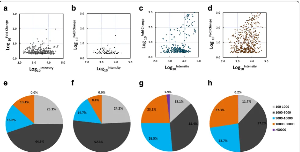
By employing a microarray with the most comprehensive probe coverage design to date, signal intensities from 3571, 1014, 1728 and 3381 sequences were found to be enriched (FC of mean of intensity value to the median of the mean of intensity values of the four stages≥2) in cer-cariae, hepatic schistosomula, adult worms and eggs, re-spectively. Based on the initial screening, we further retrieved a total of 1768 potential mRNA transcripts and 470 expressed sequence tags (ESTs) associated with developmental stages from the NCBI database (Additional file 3: Table S2). The gene collection was further filtered by requiring FC values from both forward and reverse probe sets≥2. This filtration finally retained 328, 95, 268 and 532 mRNA transcripts highly enriched in cercariae, hepatic schistosomula, adult worms and eggs, respectively (Additional files 4, 5, 6 and 7: Tables S3–S6), which contrasted with 128, 31, 83 and 84 ESTs, respectively, highly enriched in these four stages (Additional files 8, 9, 10 and 11: Tables S7–S10). However, the percentage of genes that were annotated as hypothetical protein or un-known (23.57% in the mRNA data in contrast to 69.01% in the EST data), highlights the utility of the second gener-ationS. japonicumDNA chip in profiling gene expression in this parasite.
adult worms and eggs, respectively showed an average intensity value > 10,000 (Fig. 2e-h).
Comparing the results with previous transcriptome data
A complete and accurate comparison of the results ob-tained in the current study with data from previous re-ports is hindered due to the following reasons. Firstly, the annotation of stage-enriched genes was not ideal in previous reports due to the fact that EST sequences were used for probe design coupled with less sequence hom-ology information from other trematode species being available. Secondly, the annotation for the same gene may not have been unique. Thirdly, the screening cri-teria for stage-enriched genes may have varied among different studies. Nevertheless, we compared our data
with these from previous Schistosoma transcriptome data [7, 13, 14, 23, 28] by manual checking. Globally, about 4.57, 10.07 and 12.97% genes enriched in cercariae, adult worms and eggs, respectively, were re-ported in previous studies (Additional files 4, 5 and 7: Tables S3, S4, S6). With respect to hepatic schistosomula (14 days p.i.), to our knowledge the only other relevant investigation on this particular stage was carried out on S. mansoni by Fitzpatrick et al. [28], but no enriched gene clustering was evident in that study. This was prob-ably due to the fact a large number (15) of distinct stages were analysed [28], and this has made comparison with our data for hepatic schistosomula difficult.
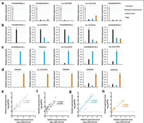
qPCR validation of the expression pattern of stage-enriched genes
A subset of 20 representative stage-enriched genes was se-lected for qRT-PCR validation (Figs 3 and 4a-d). Most genes were associated with important biological functions in each of the parasite forms. The expression of these genes at the four developmental stages validated by qRT-PCR analysis significantly correlated with the results obtained by microarray: for cercariae-enriched genes selected, r(30)= 0.8959, P< 0.0001 (Fig. 4e); for hepatic
schistosomula-enriched genes selected, r(30)= 0.7375, P< 0.0001 (Fig. 4f ); for adult-enriched genes selected, r(20)= 0.9082, P< 0.0001 (Fig. 4g); for egg-enriched
genes selected,r(21)= 0.8983,P< 0.0001 (Fig. 4h).
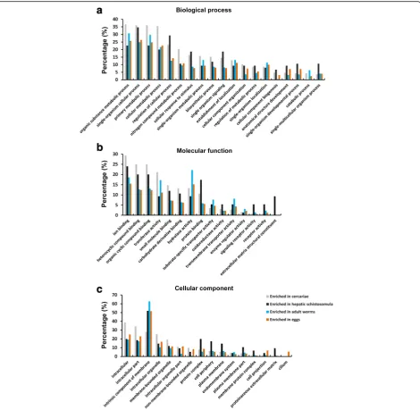
Putative functions predicted by GO analysis
We analysed the potential biological functions of the stage-enriched genes in S. japonicum using GO classifi-cation [41] (Fig. 5, Additional files 13, 14, 15 and 16: Tables S11–S14). Of the biological process categories, the most highly enriched GO terms were organic sub-stance metabolic process, single-organism cellular process, primary metabolic process and cellular metabolic process for cercariae, adult worms and eggs; the first three of these GO terms and regulation of cellular process were the most highly enriched GO terms for hepatic schistosomula. The percentages of genes involved in regulation of cellular process, cellular response to stimulus, and single organism signaling were higher in cercariae and schistosomula than those in adults and eggs. Of the molecular function cat-egories, the percentages of genes involved in ion, hetero-cyclic compound and organic hetero-cyclic compound, small molecule and carbohydrate derivative binding were higher in cercariae and schistosomula than in adults and eggs. A higher percentage of genes related to protein binding, sig-naling receptor activity and receptor activity were ob-served in schistosomula, while the GO term extracellular matrix structural constituent was only evident for this stage. In addition, a higher percentage of genes involved in hydrolase activity were assigned to adult worms. In the
[image:4.595.56.292.87.472.2]cellular component categories, gene products localised to intracellular, intracellular part and intracellular organelle were more abundant in cercariae, while gene products localised to intrinsic component of membrane were more enriched in the other three stages. Further, genes with GO terms of protein complex, cell periphery, plasma membrane, plasma membrane part and proteinaceous extracellular matrix were relatively enriched in hepatic schistosomula. In addition, the GO term cilium was present only in the egg stage.
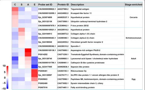
The top 25 genes enriched inS. japonicumcercariae, hepatic schistosomula, adult worms and eggs
The top 25 highly stage-associated genes in cercariae, hep-atic schistosomula, adult worms and eggs were analysed (Table 1). Collectively, the upregulated expression of these genes in cercariae indicates that signal transduction (ribo-somal protein S6 kinase beta-2 [42]), vesicular trafficking (calcium-dependent secretion activator [43] and small GTPase Rab-protein 11 [44]) and energy metabolism (AMP deaminase [45] and 5′-AMP-activated protein kin-ase [46]) and transcriptional regulation (krueppel-like factor 11, homeobox protein SMOX-1, and retinoid X re-ceptor RXR-2) are active processes in this stage.
The over-expression of the top 25 genes in hepatic schistosomula appears to reflect a diversity of physio-logical activities, including transcriptional (homeobox
protein engrailed-like SMOX-2 [47, 48], serum and glucocorticoid-regulated kinase 1 (SGK1) [49] and nu-clear receptor subfamily 4 group A [50, 51]) and neur-onal (protocadherin FAT4 [52], Aromatic-L-amino-acid decarboxylase [53] and delphilin [54]) activities, to-gether with tegumental integrity (annexin A3 [55, 56]), skeletal morphogenesis (protocadherin FAT4 [57]) and endosome-to-Golgi retrieval (vacuolar protein sorting-associated protein 29 [58]).
In mixed adult worms, genes encoding a number of trematode eggshell synthesis (TES) domain-containing proteins, DDR48 stress proteins, an asparagine-rich anti-gen Pfa35-2, two distinct tyrosinase homologues, cadherin, female-specific protein 800 and a prostatic spermine-binding protein are listed in the top 25 enriched mRNA transcripts (Table 1). Most of these genes are female-biased expressed genes [59] with potential molecular func-tions in egg production [60].
In the egg stage, genes encoding a glutenin high mo-lecular weight subunit DX5, egg protein CP1531, two histidine-rich glycoproteins, three ribonucleases, two tet-raspanins, three venom allergen-like (VAL) proteins and cell wall integrity and stress response component 1 are present in the top 25 upregulated mRNA transcripts (Table 1). Notably, it has been shown that T2 ribonuclease omega-1 in soluble egg antigen is a major Th2 polarizing component, which is capable of regulating inflammasome
[image:5.595.58.540.87.332.2]activity [61]. It has been shown previously that VAL-5 is mainly present in the egg, miracidium and sporocyst de-velopmental stages [62].
Genes enriched in cercariae
Interestingly, a group of genes encoding transcription factors, i.e. homeobox protein SMOX-1 (AY915497), bhlhzip transcription factor max/bigmax (FN314500), pre-B-cell leukemia transcription factor 2 (AY809282), transcription factor 25 (AY808969), 20 (AY813668), BTF3 (EZ000130), TFIID subunit 3 (AY812404) and 7 (FN317813), IIIB subunit (AY812330), LIM/homeobox protein (AY915618) and transcriptional repressor NF-X1(AY813973) were actively transcribed in cercariae (Additional file 4: Table S3), indicating gene transcrip-tion may not be as silent as previously suggested in this stage. It has been shown that the highest ratio of miR-NAs, the critical post-transcriptional regulators, in the total small RNA population was observed in cercariae compared with other different developmental stages of S. japonicum [32, 63], leading us to hypothesise that a specific group of genes may be actively transcribed in this aquatic stage. In addition, miRNAs may inhibit the translation of a subset of these transcripts, forming a
repertoire of genes that make schistosomula ready to adapt to subsequent intra-mammalian life. Further, there is epigenetic control of gene expression in S. mansoni cercariae [64]. Overall, these observations in-dicate that active transcriptional regulation occurs at different layers in cercariae to subtly control gene ex-pression in this stage.
We also observed that an extensive gene panel in-volved in cellular signalling transduction, i.e. F-box protein 25/32 (EZ000162), dual specificity mitogen-activated protein kinase 2 (AY815572), Serine/threo-nine kinase NLK (FN317434), Rho GTPase-activating protein 39 (FN317833), GDP/GTP exchange factor Sec2p domain containing protein (FN317362), Rho-associated protein kinase 1 (FN330915), mitogen-activated protein kinase 3 (EZ000180), Ran binding protein 9-related protein (AY812647), GTP-binding protein 2 (FN317377), NF-kappa-B inhibitor-interacting Ras-like protein 1 (AY812481), son of sevenless (AY915633), MAP kinase (AY594257), C-Jun-amino-terminal kinase-interacting pro-tein 4 (AY808598), and regulator of G-propro-tein signaling 7 (AY810841), were over-expressed in cercariae (Additional file 4: Table S3). These results support recent finding that three signaling pathways, extracellular signal-regulated
[image:6.595.58.539.89.389.2]kinase (ERK), p38 mitogen-activated protein kinase (MAPK), and protein kinase C (PKC), are modulated in cercariae in response to light and temperature cues as well as the skin fatty acid linoleic acid (LA) and are important in host penetration mechanisms [65].
In line with, and expanding on, previous transcriptional studies on schistosomes [13, 14, 66], genes encoding an array of cytoskeleton motor proteins, including dynein light intermediate chain 1, cytosolic (AY809199), tropo-nins (FN317001 and AY809606), tensin-1 (AY809674), vil-lin (AY808977), myosin light chain kinase, actin-related protein 5 (FN326677), dynamin (AY809889), catenin beta (AY814842), coronin (AY814365), dynein light chain
Tctex-type 1 (AY811669) and alpha-actinin (FN326862) (Additional file 4: Table S3) were more highly expressed in cercariae than the other stages evaluated. Transcripts en-coding LIM or PDZ domain-containing proteins, which contribute to cytoskeletal organisation, such as LIM/ homeobox protein (AY915618), actin binding LIM protein 1 (AY813306), four and a half LIM domains protein 2 (FN317368), and PDZ and LIM domain protein 7 (FN317962) (Additional file 4: Table S3), were also enriched in cercariae. Proteomic studies also revealed that cytoskeleton-related proteins are abundant in schistosome cercariae [67]. Together, these data indicate modulated signalling and motor activities and rigid transcriptional
[image:7.595.58.539.88.498.2]regulation are the most important biological events in cer-cariae, which enable them to seek, invade and adapt to a suitable definitive host.
Genes enriched in hepatic schistosomula
On invading a mammalian host, schistosomes have evolved several mechanisms to adapt to, and survive in, the hostile host environment; in particular, they develop a unique syncytial tegument, as well as mechanisms of antigenic mimicry [33], immune modulation [68] and
evasion [69, 70]. In this study, we found extracellular matrix constituents, that are located in the tegumental protein assemblage, were enriched in hepatic schistoso-mula. These collagen components included, for example, collagen alpha-1(V) chain (AY810683, AY811988, and AY815998), alpha-1(IV) chain (AY809845), alpha-1(XXIV) chain (AY814344), alpha-2(I) chain (AY810097, FN313634) and alpha-2(V) chain (AY813923) (Additional file 5: Table S4). This observation raises the possibility that collagen components may form a protective barrier on
[image:8.595.66.537.87.546.2]Table 1The top 25 genes enriched inS. japonicumcercariae, hepatic schistosomula, mixed adult worms and eggs
NCBI Nucleotide NCBI Protein Annotation FC
Enriched in cercariae
AY811679.1 AAX27568.2 Tegumental antigen 94.004
AY812964.1 AAW24696.1 Lysophosphatidic acid phosphatase type 6 89.015
AY808793.1 AAX24682.2 Krueppel-like factor 11 20.463
AY814888.1 AAP06195.1 Hypothetical protein 20.323
AY915869.1 AAX31090.1 UPF0506 domain containing protein 15.144
AY811006.1 AAX26895.2 Putative sodium-dependent transporter 14.884
FN319257.1 CAX74986.1 Ribosomal protein S6 kinase beta-2 13.668
AY813254.1 CAX83692.1 Gag-Pol polyprotein 11.090
AY812158.1 AAX28047.2 Calcium-dependent secretion activator 10.898
FN327240.1 CAX82964.1 UPF0364 protein 10.005
FN319112.1 CAX74840.1 Anti-inflammatory protein 16 9.750
AY809199.1 AAX25088.2 Dynein light intermediate chain 1 cytosolic 9.060
AY815066.1 AAW26798.1 Calpain 8.200
FN314407.1 CAX70140.1 Rab-protein 11 8.118
AY813232.1 AAW24964.1 DM9 domain-containing protein 7.327
AY915497.1 AAX30718.2 Homeobox protein SMOX-1 7.320
AY813605 AAW25337.1 Hypothetical protein 7.234
FN319705.1 CAX75429.1 THO complex subunit 1 6.827
AY813585.1 AAW25317.1 Hypothetical protein 6.756
AY811834.1 AAX27723.2 AMP deaminase 6.524
AY813088.1 AAW24820.1 Hypothetical protein 6.357
FN314484 CAX70217.1 Hypothetical protein 6.196
AY811464.1 ABA40369.1 5′-AMP-activated protein kinase subunit gamma-1 6.165
EU046089.1 AAW25910.1 Cercarial stage-specific protein Sj20H8 6.075
AY808884.1 AF129816_1 Retinoid X receptor RXR-2 6.011
Enriched in hepatic schistosomula
AY809629.1 AAX25518.2 Hypothetical protein 33.897
AY810683 AAX26572.2 Putative collagen alpha-1(V) chain precursor 9.200
AY815366.1 AAW27592.1 Alpha-ketoglutarate-dependent dioxygenase alkB 6 6.931
AY813429.1 AAW25161.1 Hypothetical protein 5.290
AY810949.1 AAX26838.2 Homeobox protein engrailed-like SMOX-2 5.057
EZ000055.1 ACE06835.1 Vacuolar protein sorting-associated protein 29 5.021
AY810397.1 AAX26286.2 Protocadherin Fat 4 4.839
AY811075.1 AAX26964.2 Hypothetical protein 4.831
AY815532.1 AAW27264.1 Hypothetical protein 4.727
AY814356 AAW26088.1 RhoGAP domain containing protein 4.610
AY811025.1 AAX26914.2 Serine/threonine-protein kinase Sgk1 4.342
AY809477.1 AAX25366.2 SAM and SH3 domain-containing protein 1 4.248
FN314446.1 CAX70179.1 Annexin A3 (Annexin III) 4.037
AY814048.1 AAW25780.1 Basic proline-rich protein-like isoform 3.967
AY808501.1 AAR28090.2 Nuclear receptor subfamily 4 group A 3.956
AY809584.1 AAX25473.2 Hypothetical protein 3.894
Table 1The top 25 genes enriched inS. japonicumcercariae, hepatic schistosomula, mixed adult worms and eggs(Continued)
AY813648.1 AAW25380.1 Hypothetical protein 3.439
AY915540.1 ABA40872.1 Leishmanolysin-like peptidase 3.419
AY812557.1 AAX28446.2 Aromatic-L-amino-acid decarboxylase 3.335
AY808377.1 AAX24266.2 Regulator of G-protein signaling 3 3.250
FN313634.1 CAX69368.1 Collagen alpha-2(I) chain 3.244
AY813683.1 AAW25415.1 Delphilin 3.240
AY812144.1 AAX28033.2 Hypothetical protein 3.212
AY813563 AAW25295.1 Hypothetical protein 3.203
Enriched in mixed adult worms
FN314868.1 CAX70600.1 Asparagine-rich antigen Pfa35-2 1651.245
EZ000096 ACE06876.1 Putative eggshell protein precursor 934.084
FN314999 CAX70731.1 TES domain containing protein 704.455
AY813556.1 AAW25288.1 Hypothetical protein 692.180
AY814029 AAW25761.2 Stress protein DDR48 (DNA damage-responsive protein 48) 678.514
FN313935.1 CAX69669.1 Stress protein DDR48 (DNA damage-responsive protein 48) 665.581
FN317103 CAX72834.1 Stress protein DDR48 (DNA damage-responsive protein 48) 645.627
FN313912 CAX69646.1 TES domain containing protein 604.574
FN313715.1 CAX69449.1 TES domain containing protein 561.444
AY812810.1 AAW24542.1 Histidine-rich glycoprotein precursor 526.698
FN315504.1 CAX71236.1 TES domain containing protein 517.929
AY815518 AAW27250.1 TES domain containing protein 489.519
FN314997 CAX70729.1 TES domain containing protein 422.784
AY813405 AAW25137.1 TES domain containing protein 407.588
AY815264.1 AAW26996.1 Tyrosinase 1 346.094
AY812315.1 AAX28204.2 Hypothetical protein 330.410
FN330801 CAX83018.1 Stress protein DDR48 (DNA damage-responsive protein 48) 235.455
AY814142.1 AAW25874.1 Putative FAM75 family member 224.325
AY812904 AAW24636.1 Tyrosinase 2 209.523
FN315510.1 CAX71242.1 Hypothetical protein 164.941
AY814814 AAW26546.1 Cadherin 145.264
AY815418 AAW27150.1 Female-specific protein 800 135.097
FN316955 CAX72686.1 Prostatic spermine-binding protein precursor 132.448
AY222885 AAP05897.1 Stress protein DDR48 (DNA damage-responsive protein 48) 127.238
FN314903.1 CAX70635.1 Hypothetical protein 107.908
Enriched in eggs
FN317800 CAX73529.1 Glutenin high molecular weight subunit DX5 1794.846
FN319280 CAX75008.1 Tetraspanin 22 1769.270
FN322023.1 CAX77751.1 Histidine-rich glycoprotein 1656.913
FN324495.1 CAX80219.1 Hypothetical protein 1549.720
FN326817 CAX82541.1 Histidine-rich glycoprotein 1523.735
FN317759.1 CAX73488.1 Similar to venom allergen-like (VAL) 25 protein 1062.695
FN324480.1 CAX80126.1 Hypothetical protein 938.553
FN321785 CAX77509.1 Ribonuclease T2 850.487
FN321171.1 CAX76897.1 Hypothetical protein 831.194
the worm surface, which may help the schistosomula evade host attack.
Schistosomula undertake a lengthy migration in the mammalian host to the portal venous system, where they mature into adult worms and pair. This migration is closely associated with locomotion activity controlled by the neuronal system. The data presented here show that neuronal activities may be particularly active in hep-atic schistosomula, which could be linked to the fact that responses to environmental cues from the host and the subsequent control of mobility are required to guarantee that they reach their destination [22]. A cohort of genes involved in neuronal activities in this stage includes netrin receptor unc5B (AY915275), nephrin (AY809045), caskin 2 (AY812623), spondin-1 (AY812421), as well as the previously described genes protocadherin FAT4, aromatic-L-amino-acid decarboxylase and delphilin. Although the precise functions of these genes in schisto-somes remain unknown, there is evidence from other studies that at least three are involved in axon guidance. In mammals, it has been shown that the unc5B receptor, interacting with netrin-1, activates the downstream sig-nal transduction pathway that mediates axon guidance [71]. A caskin ortholog in Drosophila is a cytoplasmic adaptor protein, which has been shown to mediate Lar signal transduction motor axon guidance [72]. Similarly, spondin-1 is an extracellular matrix protein, and previ-ous research showed that its C. elegans ortholog func-tions in axon guidance and fasciculation in motoneurons [73]. Also, the expression of nephrin homologues has been observed in the central nervous system of mam-mals, and nephrin may potentially interact with glutam-ate receptors [74, 75].
In multicellular organisms, apoptosis is a highly con-trolled cellular process of programmed cell death which plays a key role in maintaining cell populations during an organism’s life-cycle. The apoptosis pathway has been sug-gested as a potential intervention target in schistosomes [76]. The activities of two central proteolytic enzymes in-volved in the apoptosis process, caspase-3 and -7, were shown to peak in S. japonicum schistosomula (14 days p.i.) [77]. The upregulated expression of caspase 7 (AY813428) in hepatic schistosomula was confirmed in this study (Additional file 5: Table S4). It is of note that a cohort of planarian neoblast-like cells with self-renewal function has been identified in S. mansoni, with a po-tential role in renewal of the tegument [78]. In this re-spect, fibroblast growth factor receptor 2, a crucial gene for the maintenance of neoblast-like cell popula-tion in schistosomes [79], was enriched in hepatic schistosomula (Additional file 5: Table S4), emphasising the requirement for rapid tegumental renewal during this period of fast-growth.
Genes enriched in adult worms
[image:11.595.57.540.102.313.2]One of the major biological roles of adult worms is to pro-duce a large number of eggs, a key process in the schisto-some life-cycle. As earlier mentioned, within the top 25 adult-enriched genes, most are associated with egg pro-duction. However, two pre-requisites for egg production are mating and nutrient acquisition. In fulfilment of the former process, the gene encoding gynecophoral canal protein has been shown upregulated in adults, with a dra-matic bias towards male worms [59]. In regards to nutri-ent uptake, and consistnutri-ent with a previous study [18], over-expression of a number of ‘blood processing’
Table 1The top 25 genes enriched inS. japonicumcercariae, hepatic schistosomula, mixed adult worms and eggs(Continued)
FN319117.1 CAX74843.1 CIA30 domain containing protein 665.147
FN317754 CAX73483.1 Tetraspanin 663.055
FN322724.1 CAX78439.1 Peptidase inhibitor 16 651.579
FN319142 CAX74870.1 Hypothetical protein 628.202
FN320551 CAX76277.1 Egg protein CP1531 592.491
FN326664 CAX82388.1 Hypothetical protein 577.505
AY816014.1 AAW27746.1 Ribonuclease S-4 534.668
FN321764.1 CAX77484.1 Cell wall integrity and stress response component 1 488.342
FN326758 CAX82480.1 Hypothetical protein 484.608
FN317167 CAX72898.1 Hypothetical protein 481.352
FN319216.1 CAX74944.1 Hypothetical protein 453.890
FN320451 CAX76177.1 GLIPR1-like protein 1/venom allergen-like protein 5 422.820
FN317231 CAX72962.1 GLIPR1-like protein 1/venom allergen-like protein 5 417.438
FN326877 CAX82601.1 Hypothetical protein 416.455
proteases in adult worms was also revealed here. For instance, cathepsin family members, i.e. cathepsin C (FN315267), cathepsin D2-like (AY812817), cathepsin B-like (AY814095), cathepsin L (FN313884) and cathepsin L-like isoforms (AY222874, FN314782, and FN314778), and aminopeptidase N (FN317672) were readily identi-fied as adult worm-enriched genes (Additional file 6: Table S5). In addition, saposin B domain-containing proteins (FN314931, FN315898 and FN314355), which have been proposed as being involved in nutrient acqui-sition by disrupting the membrane of red blood cells to release haemoglobin [80], were highly expressed in adult worms.
In schistosomes, glycosylation is a complex process which plays a crucial role in their biology, particularly in terms of immune modulation [81]. A subset of tran-scripts involved glycosylation in was enriched in adult worms of S. japonicum. These genes included beta-1,4-galactosyltransferase 4 (AY813412), glycosyltrans-ferase 1 domain-containing protein 1 (FN319898), GDP-fucose protein O-fucosyltransferase 2 (AY810860), beta-1,3-galactosyltransferase 5 (AY814132), glycoprotein-N-acetylgalactosamine 3-beta-galactosyltransferase 1 (AY809881), glycoprotein 3-alpha-L-fucosyltransferase A (FN317387), alpha-1,3-mannosyl-glycoprotein 2-beta-N-acetylglucosaminyltransferase (AY812621), and alpha-L-fucosidase-like protein (FN317475) (Additional file 6: Table S5). However, given the inherent complexity of glycosylation and that multiple glycosyltransferases re-sponsible for similar molecular functions are present in the Schistosoma genomes [81, 82], it is difficult to con-clude that the global level of glycosylation or the expres-sion of specific glycans is higher in adults than in the other stages examined here.
Genes enriched in eggs
Globally, genes associated with the egg stage are involved in a diversity of biological functions, which may be the re-sult of using samples for analysis that comprise a mixture of immature and mature eggs. In addition to anticipated genes encoding egg proteins, immunogenic miracidial an-tigens and major egg anan-tigens, a number of genes involved in the cell cycle and proliferation, including meiosis expressed protein 1 (FN317540), meiosis-specific nuclear structural protein 1 (AY810474), mitogen-activated protein kinase 15 (FN317209), putative chromosome seg-regation protein SMC (AY812773), different isoforms of leishmanolysin-like peptidase (AY811259, FN317512, AY810562 and FN319863) and probably protein VHS3 (FN330961), placenta-specific gene 8 protein (FN317134), placental protein 25 homolog (FN317187) and centroso-mal protein of 162 kDa (AY810094), were upregulated in eggs (Additional file 7: Table S6). These transcripts may
be enriched in immature eggs, hinting that active cell div-ision is essential for embryonic development.
Further, a group of transcripts encoding tubulin and microtubule-associated motor proteins, i.e. tubulin alpha (FN317215), tubulin beta (FN320386), tubulin beta-2C chain (FN320061), cytoplasmic dynein light chain 1 (FN317588) and 2 (AY914882), dynein light chain 1, axo-nemal (FN317727), inner dynein arm light chain, axoaxo-nemal (FN317915), outer dynein arm protein 1 (AY813443), dy-nein heavy chain 5, axonemal (AY810177), as well as the ciliary and flagellar microtubule components, i.e. tektins (AY814061, AY914954, FN317819 and FN314465), dynein intermediate chain 3 (AY810742) and outer dense fibre protein 3-B (FN318315) were over-expressed in eggs (Additional file 7: Table S6). These transcriptional dif-ferences may reflect the fact that a miracidium is enclosed in the eggshell of the mature egg, and once the egg is released into the external environment and contacts freshwater, a high level of movement is re-quired for the larva to hatch and escape from the eggs [83], and to seek the snail intermediate host in order to establish an infection.
Though the miracidium is enclosed by an eggshell, an active parasite-host interplay takes place via pores in the egg [83]. On one hand, nutrients (e.g. iron, amino acid and lipid) are acquired by eggs from the host, a process supported by the upregulation of genes in-volved in transport and exchange activities, such as putative sodium-dependent transporter (FN318875), so-dium/hydrogen exchanger (AY815720), sodium/calcium exchanger (FN318247), large neutral amino acids trans-porter small subunit 2 (FN327074), Y + L amino acid transporter 2 (FN313722), high-affinity choline trans-porter 1 (FN317430), iron channels (i.e. voltage-gated hydrogen channel (FN318209), two pore calcium channel protein 2 (FN326741), and TWiK family of potassium channels protein (AY813707), and lipid metabolism (i.e. fatty acid-binding protein (FN318753) (Additional file 7: Table S6). On the other hand, it has been shown that major egg products fromS. mansonisuch as ribonuclease omega-1, kappa 5 (FN329842) and IPSE/alpha-1 are released into host tissues and modulate host immune re-sponses [84–87]. In this study,S. japonicum homologues of ribonuclease omega-1 (FN330952) and kappa 5 (FN321248) were also enriched in the egg stage, although as yet, no homologue of IPSE/alpha-1 has been identified in this schistosome species.
Conclusions
hepatic schistosomula, adult parasites and eggs. Overall, this study adds new insights on the developmental biol-ogy ofS. japonicumwhich further the discovery of novel intervention targets against this persistent parasite and the disease it causes.
Additional files
Additional file 1: Figure S1.Denaturing agarose gel electrophoresis of RNA samples isolated from different developmental stages (1, cercariae; 2, hepatic schistosomula; 3, adult worms; 4, eggs); one of three biological replicates for each stage are presented. (TIF 82 kb)
Additional file 2: Table S1.List of primer pairs used for qPCR validation. (XLSX 10 kb)
Additional file 3: Table S2.Retrieval ofS. japonicumstage-enriched genes from the NCBI database based on the DNA chip results. (XLSX 9 kb)
Additional file 4: Table S3.Information on mRNA transcripts enriched in cercariae (forward probe). (XLSX 188 kb)
Additional file 5: Table S4.Information on mRNA transcripts enriched in hepatic schistosomula (forward probe). (XLSX 64 kb)
Additional file 6: Table S5.Information on mRNA transcripts enriched in mixed adult worms (forward probe). (XLSX 150 kb)
Additional file 7: Table S6.Information on mRNA transcripts enriched in eggs (forward probe). (XLSX 287 kb)
Additional file 8: Table S7.Information on EST transcripts enriched in cercariae (forward probe). (XLSX 81 kb)
Additional file 9: Table S8.Information on EST transcripts enriched in hepatic schistosomula (forward probe). (XLSX 25 kb)
Additional file 10: Table S9.Information on EST transcripts enriched in mixed adult worms (forward probe). (XLSX 51 kb)
Additional file 11: Table S10.Information on EST transcripts enriched in eggs (forward probe). (XLSX 51 kb)
Additional file 12: Figure S2.Heatmap for EST transcripts enriched in cercariae, hepatic schistosomula, adult worms and eggs. The heatmap was created by HemI 1.0 based on the transformed data of log2FC value. The data are based on the mean of weighted signal intensity value of forward probe sets (three biological replicates). (TIF 110 kb)
Additional file 13: Table S11.Detailed GO annotation for mRNA transcripts enriched in cercariae. (XLSX 26 kb)
Additional file 14: Table S12.Detailed GO annotation for mRNA transcripts enriched in hepatic schistosomula. (XLSX 18 kb)
Additional file 15: Table S13.Detailed GO annotation for mRNA transcripts enriched in mixed adult worms. (XLSX 22 kb)
Additional file 16: Table S14.Detailed GO annotation for mRNA transcripts enriched in eggs. (XLSX 24 kb)
Abbreviations
CDS:Coding DNA sequences; DALYs: Disability adjusted life years; DGE: Digital gene expression; ERK: Extracellular signal-regulated kinase; ESTs: Expressed sequence tags; FC: Fold-changes; LAPs: Hydrolysis of lysophosphatidic acids; MAPK: Mitogen-activated protein kinase; PKC: Protein kinase C; SAGE: Serial analysis of gene expression; TES: Trematode eggshell synthesis;
UTRs: Untranslated regions; VAL: Venom allergen-like
Acknowledgements
We thank the Chinese National Genome Center at Shanghai for makingS. japonicumgenome publicly available.
Funding
This study was supported by the National Natural Science Foundation of China (Grant No. 81270026), the National S & T Major Program (Grant No. 2012ZX10004-220), the Special Fund for Health Research in the Public Interest (Grant No. 201202019), and the Program for Changjiang Scholars and
Innovative Research Team in University (IRT13007). DPM is a NHMRC Senior Principal Research Fellow and Senior Scientist at QIMR Berghofer Medical Research Institute. The funders had no role in study design, data collection and analysis, decision to publish, or preparation of the manuscript.
Availability of data and materials
Raw data and the normalized data have been deposited at the public domain Gene Expression Omnibus under the accession number for the platform GPL18617 and series GSE57143.
Authors’contributions
PC and QC conceived the project and designed the strategy. PC, SL, XP and NH carried out the experiments. PC, SL, DPM and QC analysed the data. PC, HY, DPM and QC wrote the manuscript. All authors read and approved the final manuscript.
Competing interests
The authors declare that they have no competing interests.
Consent for publication Not applicable.
Ethics approval and consent to participate
All procedures performed on animals within this study were conducted following animal husbandry guidelines of the Chinese Academy of Medical Sciences and with permission from the Experimental Animal Committee (Institute of Pathogen Biology, CAMS) with Ethical Clearance Number IPB-2011-6.
Author details
1MOH Key Laboratory of Systems Biology of Pathogens, Institute of Pathogen
Biology, Chinese Academy of Medical Sciences & Peking Union Medical College, Beijing, People’s Republic of China.2Molecular Parasitology Laboratory, QIMR Berghofer Medical Research Institute, Queensland, Australia.
3Key Laboratory of Zoonosis, Shenyang Agriculture University, Shenyang,
People’s Republic of China.
Received: 5 November 2016 Accepted: 21 December 2016
References
1. Steinmann P, Keiser J, Bos R, Tanner M, Utzinger J. Schistosomiasis and water resources development: systematic review, meta-analysis, and estimates of people at risk. Lancet Infect Dis. 2006;6:411–25.
2. Weerakoon KG, Gobert GN, Cai P, McManus DP. Advances in the diagnosis of human schistosomiasis. Clin Microbiol Rev. 2015;28:939–67.
3. Hotez PJ, Alvarado M, Basanez MG, Bolliger I, Bourne R, Boussinesq M, et al. The global burden of disease study 2010: interpretation and implications for the neglected tropical diseases. PLoS Negl Trop Dis. 2014;8:e2865. 4. Mutapi F, Rujeni N, Bourke C, Mitchell K, Appleby L, Nausch N, et al.
Schistosoma haematobiumtreatment in 1-5 year old children: safety and efficacy of the antihelminthic drug praziquantel. PLoS Negl Trop Dis. 2011;5:e1143.
5. Chen MG. Assessment of morbidity due toSchistosoma japonicuminfection in China. Infect Dis Poverty. 2014;3:6.
6. Cai P, Gobert GN, You H, McManus DP. The Tao survivorship of schistosomes: implications for schistosomiasis control. Int J Parasitol. 2016;46:453–63. 7. Hu W, Yan Q, Shen DK, Liu F, Zhu ZD, Song HD, et al. Evolutionary and
biomedical implications of aSchistosoma japonicumcomplementary DNA resource. Nat Genet. 2003;35:139–47.
8. Verjovski-Almeida S, DeMarco R, Martins EA, Guimaraes PE, Ojopi EP, Paquola AC, et al. Transcriptome analysis of the acoelomate human parasite Schistosoma mansoni. Nat Genet. 2003;35:148–57.
9. Berriman M, Haas BJ, LoVerde PT, Wilson RA, Dillon GP, Cerqueira GC, et al. The genome of the blood flukeSchistosoma mansoni. Nature. 2009;460: 352–8.
10. The Schistosoma japonicum Genome Sequencing and Functional Analysis Consortium. TheSchistosoma japonicumgenome reveals features of host-parasite interplay. Nature. 2009;460:345–51.
12. Waisberg M, Lobo FP, Cerqueira GC, Passos LK, Carvalho OS, Franco GR, et al. Microarray analysis of gene expression induced by sexual contact in Schistosoma mansoni. BMC Genomics. 2007;8:181.
13. Jolly ER, Chin CS, Miller S, Bahgat MM, Lim KC, DeRisi J, et al. Gene expression patterns during adaptation of a helminth parasite to different environmental niches. Genome Biol. 2007;8:R65.
14. Gobert GN, Moertel L, Brindley PJ, McManus DP. Developmental gene expression profiles of the human pathogenSchistosoma japonicum. BMC Genomics. 2009;10:128.
15. Chai M, McManus DP, McInnes R, Moertel L, Tran M, Loukas A, et al. Transcriptome profiling of lung schistosomula, in vitro cultured schistosomula and adultSchistosoma japonicum. Cell Mol Life Sci. 2006;63:919–29. 16. Moertel L, McManus DP, Piva TJ, Young L, McInnes RL, Gobert GN.
Oligonucleotide microarray analysis of strain- and gender-associated gene expression in the human blood fluke,Schistosoma japonicum. Mol Cell Probes. 2006;20:280–9.
17. Fitzpatrick JM, Johansen MV, Johnston DA, Dunne DW, Hoffmann KF. Gender-associated gene expression in two related strains ofSchistosoma japonicum. Mol Biochem Parasitol. 2004;136:191–209.
18. Parker-Manuel SJ, Ivens AC, Dillon GP, Wilson RA. Gene expression patterns in larvalSchistosoma mansoniassociated with infection of the mammalian host. PLoS Negl Trop Dis. 2011;5:e1274.
19. Ojopi EP, Oliveira PS, Nunes DN, Paquola A, DeMarco R, Gregorio SP, et al. A quantitative view of the transcriptome ofSchistosoma mansoni adult-worms using SAGE. BMC Genomics. 2007;8:186.
20. Williams DL, Sayed AA, Bernier J, Birkeland SR, Cipriano MJ, Papa AR, et al. ProfilingSchistosoma mansonidevelopment using serial analysis of gene expression (SAGE). Exp Parasitol. 2007;117:246–58.
21. Cogswell AA, Kommer VP, Williams DL. Transcriptional analysis of a unique set of genes involved inSchistosoma mansonifemale reproductive biology. PLoS Negl Trop Dis. 2012;6:e1907.
22. Piao X, Cai P, Liu S, Hou N, Hao L, Yang F, et al. Global expression analysis revealed novel gender-specific gene expression features in the blood fluke parasiteSchistosoma japonicum. PLoS One. 2011;6:e18267.
23. Anderson L, Amaral MS, Beckedorff F, Silva LF, Dazzani B, Oliveira KC, et al. Schistosoma mansoniegg, adult male and female comparative gene expression analysis and identification of novel genes by RNA-Seq. PLoS Negl Trop Dis. 2015;9:e0004334.
24. Picard MA, Boissier J, Roquis D, Grunau C, Allienne JF, Duval D, et al. Sex-biased transcriptome ofSchistosoma mansoni: host-parasite interaction, genetic determinants and epigenetic regulators are associated with sexual differentiation. PLoS Negl Trop Dis. 2016;10:e0004930. 25. Jones MK, Higgins T, Stenzel DJ, Gobert GN. Towards tissue specific
transcriptomics and expression pattern analysis in schistosomes using laser microdissection microscopy. Exp Parasitol. 2007;117:259–66.
26. Gobert GN, McManus DP, Nawaratna S, Moertel L, Mulvenna J, Jones MK. Tissue specific profiling of females ofSchistosoma japonicumby integrated laser microdissection microscopy and microarray analysis. PLoS Negl Trop Dis. 2009;3:e469.
27. Nawaratna SS, McManus DP, Moertel L, Gobert GN, Jones MK. Gene Atlasing of digestive and reproductive tissues inSchistosoma mansoni. PLoS Negl Trop Dis. 2011;5:e1043.
28. Fitzpatrick JM, Peak E, Perally S, Chalmers IW, Barrett J, Yoshino TP, et al. Anti-schistosomal intervention targets identified by life-cycle transcriptomic analyses. PLoS Negl Trop Dis. 2009;3:e543.
29. Liu S, Cai P, Hou N, Piao X, Wang H, Hung T, et al. Genome-wide identification and characterization of a panel of house-keeping genes in Schistosoma japonicum. Mol Biochem Parasitol. 2012;182:75–82. 30. Liu S, Cai P, Piao X, Hou N, Zhou X, Wu C, et al. Expression profile of the
Schistosoma japonicumdegradome reveals differential protease expression patterns and potential anti-schistosomal intervention targets. PLoS Comput Biol. 2014;10:e1003856.
31. Liu S, Zhou X, Piao X, Wu C, Hou N, Chen Q. Comparative analysis of transcriptional profiles of adultSchistosoma japonicumfrom different laboratory animals and the natural host, water buffalo. PLoS Negl Trop Dis. 2015;9:e0003993.
32. Cai P, Piao X, Hao L, Liu S, Hou N, Wang H, et al. A deep analysis of the small non-coding RNA population inSchistosoma japonicumeggs. PLoS One. 2013;8:e64003. 33. Wu C, Hou N, Piao X, Liu S, Cai P, Xiao Y, et al. Non-immune immunoglobulins
shieldSchistosoma japonicumfrom host immunorecognition. Sci Rep. 2015;5:13434.
34. Li DD, Zhao LX, Mylonakis E, Hu GH, Zou Y, Huang TK, et al. In vitro and in vivo activities of pterostilbene againstCandida albicansbiofilms. Antimicrob Agents Chemother. 2014;58:2344–55.
35. Irizarry RA, Hobbs B, Collin F, Beazer-Barclay YD, Antonellis KJ, Scherf U, et al. Exploration, normalization, and summaries of high density oligonucleotide array probe level data. Biostatistics. 2003;4:249–64.
36. Irizarry RA, Bolstad BM, Collin F, Cope LM, Hobbs B, Speed TP. Summaries of Affymetrix GeneChip probe level data. Nucleic Acids Res. 2003;31:e15. 37. Deng W, Wang Y, Liu Z, Cheng H, Xue Y. HemI: a toolkit for illustrating
heatmaps. PLoS One. 2014;9:e111988.
38. Gotz S, Garcia-Gomez JM, Terol J, Williams TD, Nagaraj SH, Nueda MJ, et al. High-throughput functional annotation and data mining with the Blast2GO suite. Nucleic Acids Res. 2008;36:3420–35.
39. Marchler-Bauer A, Derbyshire MK, Gonzales NR, Lu S, Chitsaz F, Geer LY, et al. CDD: NCBI’s conserved domain database. Nucleic Acids Res. 2015;43: D222–6.
40. Cai P, Piao X, Hou N, Liu S, Wang H, Chen Q. Identification and characterization of argonaute protein, Ago2 and its associated small RNAs inSchistosoma japonicum. PLoS Negl Trop Dis. 2012;6:e1745.
41. Ashburner M, Ball CA, Blake JA, Botstein D, Butler H, Cherry JM, et al. Gene ontology: tool for the unification of biology. The Gene Ontology Consortium. Nat Genet. 2000;25:25–9.
42. Valovka T, Verdier F, Cramer R, Zhyvoloup A, Fenton T, Rebholz H, et al. Protein kinase C phosphorylates ribosomal protein S6 kinase betaII and regulates its subcellular localization. Mol Cell Biol. 2003;23:852–63. 43. Daily NJ, Boswell KL, James DJ, Martin TF. Novel interactions of CAPS (Ca2
+-dependent activator protein for secretion) with the three neuronal SNARE
proteins required for vesicle fusion. J Biol Chem. 2010;285:35320–9. 44. Ligasova A, Bulantova J, Sebesta O, Kasny M, Koberna K, Mikes L. Secretory
glands in cercaria of the neuropathogenic schistosomeTrichobilharzia regenti- ultrastructural characterization, 3-D modelling, volume and pH estimations. Parasit Vectors. 2011;4:162.
45. Morisaki T, Gross M, Morisaki H, Pongratz D, Zollner N, Holmes EW. Molecular basis of AMP deaminase deficiency in skeletal muscle. Proc Natl Acad Sci U S A. 1992;89:6457–61.
46. Neumann D, Woods A, Carling D, Wallimann T, Schlattner U. Mammalian AMP-activated protein kinase: functional, heterotrimeric complexes by co-expression of subunits inEscherichia coli. Protein Expr Purif. 2003;30:230–7. 47. Morgan R. Engrailed: complexity and economy of a multi-functional
transcription factor. FEBS Lett. 2006;580:2531–3.
48. Webster PJ, Mansour TE. Conserved classes of homeodomains in Schistosoma mansoni, an early bilateral metazoan. Mech Dev. 1992;38:25–32. 49. Mansley MK, Watt GB, Francis SL, Walker DJ, Land SC, Bailey MA, et al.
Dexamethasone and insulin activate serum and glucocorticoid-inducible kinase 1 (SGK1) via different molecular mechanisms in cortical collecting duct cells. Physiol Rep. 2016;4:e12792.
50. Wu W, LoVerde PT. Nuclear hormone receptors in parasitic helminths. Mol Cell Endocrinol. 2011;334:56–66.
51. Wu W, Loverde PT.Schistosoma mansoni: identification of SmNR4A, a member of nuclear receptor subfamily 4. Exp Parasitol. 2008;120:208–13. 52. Badouel C, Zander MA, Liscio N, Bagherie-Lachidan M, Sopko R, Coyaud E,
et al. Fat1 interacts with Fat4 to regulate neural tube closure, neural progenitor proliferation and apical constriction during mouse brain development. Development. 2015;142:2781–91.
53. Brun L, Ngu LH, Keng WT, Ch’ng GS, Choy YS, Hwu WL, et al. Clinical and biochemical features of aromatic L-amino acid decarboxylase deficiency. Neurology. 2010;75:64–71.
54. Miyagi Y, Yamashita T, Fukaya M, Sonoda T, Okuno T, Yamada K, et al. Delphilin: a novel PDZ and formin homology domain-containing protein that synaptically colocalizes and interacts with glutamate receptor delta 2 subunit. J Neurosci. 2002;22:803–14.
55. Tararam CA, Farias LP, Wilson RA, Leite LC.Schistosoma mansoniAnnexin 2: molecular characterization and immunolocalization. Exp Parasitol. 2010;126: 146–55.
56. Leow CY, Willis C, Osman A, Mason L, Simon A, Smith BJ, et al. Crystal structure and immunological properties of the first annexin from Schistosoma mansoni: insights into the structural integrity of the schistosomal tegument. FEBS J. 2014;281:1209–25.
58. Fuse A, Furuya N, Kakuta S, Inose A, Sato M, Koike M, et al. VPS29-VPS35 intermediate of retromer is stable and may be involved in the retromer complex assembly process. FEBS Lett. 2015;589:1430–6.
59. Cai P, Liu S, Piao X, Hou N, Gobert GN, McManus DP, et al. Comprehensive transcriptome analysis of sex-biased expressed genes reveals discrete biological and physiological features of male and femaleSchistosoma japonicum. PLoS Negl Trop Dis. 2016;10:e0004684.
60. Fitzpatrick JM, Hirai Y, Hirai H, Hoffmann KF. Schistosome egg production is dependent upon the activities of two developmentally regulated tyrosinases. FASEB J. 2007;21:823–35.
61. Ferguson BJ, Newland SA, Gibbs SE. Tourlomousis P, Fernandes dos Santos P, Patel MN, et al. TheSchistosoma mansoniT2 ribonuclease omega-1 modulates inflammasome-dependent IL-1beta secretion in macrophages. Int J Parasitol. 2015;45:809–13.
62. Chalmers IW, McArdle AJ, Coulson RM, Wagner MA, Schmid R, Hirai H, et al. Developmentally regulated expression, alternative splicing and distinct sub-groupings in members of theSchistosoma mansonivenom allergen-like (SmVAL) gene family. BMC Genomics. 2008;9:89.
63. Cai P, Hou N, Piao X, Liu S, Liu H, Yang F, et al. Profiles of small non-coding RNAs inSchistosoma japonicumduring development. PLoS Negl Trop Dis. 2011;5:e1256.
64. Roquis D, Lepesant JM, Picard MA, Freitag M, Parrinello H, Groth M, et al. The epigenome ofSchistosoma mansoniprovides insight about how cercariae poise transcription until infection. PLoS Negl Trop Dis. 2015;9: e0003853.
65. Ressurreicao M, Kirk RS, Rollinson D, Emery AM, Page NM, Walker AJ. Sensory protein kinase signaling inSchistosoma mansonicercariae: host location and invasion. J Infect Dis. 2015;212:1787–97.
66. Han ZG, Brindley PJ, Wang SY, Chen Z.Schistosomagenomics: new perspectives on schistosome biology and host-parasite interaction. Annu Rev Genomics Hum Genet. 2009;10:211–40.
67. Liu M, Ju C, Du XF, Shen HM, Wang JP, Li J, et al. Proteomic analysis on cercariae and schistosomula in reference to potential proteases involved in host invasion ofSchistosoma japonicumlarvae. J Proteome Res. 2015;14:4623–34. 68. Jenkins SJ, Hewitson JP, Jenkins GR, Mountford AP. Modulation of the host’s
immune response by schistosome larvae. Parasite Immunol. 2005;27:385–93. 69. Cai P, Bu L, Wang J, Wang Z, Zhong X, Wang H. Molecular characterization
ofSchistosoma japonicumtegument protein tetraspanin-2: sequence variation and possible implications for immune evasion. Biochem Biophys Res Commun. 2008;372:197–202.
70. Zhang W, Li J, Duke M, Jones MK, Kuang L, Zhang J, et al. Inconsistent protective efficacy and marked polymorphism limits the value ofSchistosoma japonicumtetraspanin-2 as a vaccine target. PLoS Negl Trop Dis. 2011;5:e1166. 71. Bradford D, Cole SJ, Cooper HM. Netrin-1: diversity in development.
Int J Biochem Cell Biol. 2009;41:487–93.
72. Weng YL, Liu N, DiAntonio A, Broihier HT. The cytoplasmic adaptor protein Caskin mediates Lar signal transduction duringDrosophilamotor axon guidance. J Neurosci. 2011;31:4421–33.
73. Woo WM, Berry EC, Hudson ML, Swale RE, Goncharov A, Chisholm AD. TheC. elegansF-spondin family protein SPON-1 maintains cell adhesion in neural and non-neural tissues. Development. 2008;135:2747–56. 74. Li M, Armelloni S, Ikehata M, Corbelli A, Pesaresi M, Calvaresi N, et al.
Nephrin expression in adult rodent central nervous system and its interaction with glutamate receptors. J Pathol. 2011;225:118–28. 75. Putaala H, Soininen R, Kilpelainen P, Wartiovaara J, Tryggvason K. The
murine nephrin gene is specifically expressed in kidney, brain and pancreas: inactivation of the gene leads to massive proteinuria and neonatal death. Hum Mol Genet. 2001;10:1–8.
76. Lee EF, Young ND, Lim NT, Gasser RB, Fairlie WD. Apoptosis in schistosomes: toward novel targets for the treatment of schistosomiasis. Trends Parasitol. 2014;30:75–84.
77. Han H, Peng J, Gobert GN, Hong Y, Zhang M, Han Y, et al. Apoptosis phenomenon in the schistosomulum and adult worm life cycle stages of Schistosoma japonicum. Parasitol Int. 2013;62:100–8.
78. Pearson MS, Loukas A. The parasite’s new clothes. Elife. 2016;5:e12473. 79. Collins 3rd JJ, Wang B, Lambrus BG, Tharp ME, Iyer H, Newmark PA. Adult
somatic stem cells in the human parasiteSchistosoma mansoni. Nature. 2013;494:476–9.
80. Don TA, Bethony JM, Loukas A. Saposin-like proteins are expressed in the gastrodermis ofSchistosoma mansoniand are immunogenic in natural infections. Int J Infect Dis. 2008;12:e39–47.
81. Mickum ML, Prasanphanich NS, Heimburg-Molinaro J, Leon KE, Cummings RD. Deciphering the glycogenome of schistosomes. Front Genet. 2014;5:262. 82. Smit CH, van Diepen A, Nguyen DL, Wuhrer M, Hoffmann KF, Deelder AM,
et al. Glycomic analysis of life stages of the human parasiteSchistosoma mansonireveals developmental expression profiles of functional and antigenic glycan motifs. Mol Cell Proteomics. 2015;14:1750–69. 83. Jones MK, Bong SH, Green KM, Holmes P, Duke M, Loukas A, et al.
Correlative and dynamic imaging of the hatching biology ofSchistosoma japonicumfrom eggs prepared by high pressure freezing. PLoS Negl Trop Dis. 2008;2:e334.
84. Steinfelder S, Andersen JF, Cannons JL, Feng CG, Joshi M, Dwyer D, et al. The major component in schistosome eggs responsible for conditioning dendritic cells for Th2 polarization is a T2 ribonuclease (omega-1). J Exp Med. 2009;206:1681–90.
85. Meyer NH, Mayerhofer H, Tripsianes K, Blindow S, Barths D, Mewes A, et al. A Crystallin fold in the Interleukin-4-inducing principle ofSchistosoma mansonieggs (IPSE/alpha-1) mediates IgE binding for antigen-independent basophil activation. J Biol Chem. 2015;290:22111–26.
86. Schramm G, Mohrs K, Wodrich M, Doenhoff MJ, Pearce EJ, Haas H, et al. Cutting edge: IPSE/alpha-1, a glycoprotein fromSchistosoma mansonieggs, induces IgE-dependent, antigen-independent IL-4 production by murine basophils in vivo. J Immunol. 2007;178:6023–7.
87. Schramm G, Hamilton JV, Balog CI, Wuhrer M, Gronow A, Beckmann S, et al. Molecular characterisation of kappa-5, a major antigenic glycoprotein from Schistosoma mansonieggs. Mol Biochem Parasitol. 2009;166:4–14.
• We accept pre-submission inquiries
• Our selector tool helps you to find the most relevant journal
• We provide round the clock customer support
• Convenient online submission
• Thorough peer review
• Inclusion in PubMed and all major indexing services
• Maximum visibility for your research
Submit your manuscript at www.biomedcentral.com/submit