RESEARCH
Stromal vascular fraction cells for the
treatment of critical limb ischemia: a pilot study
Adas Darinskas
1*, Mindaugas Paskevicius
2, Gintaras Apanavicius
2, Gintaris Vilkevicius
3,4,
Liutauras Labanauskas
5, Thomas E. Ichim
6and Rytis Rimdeika
5Abstract
Background: Cell-based therapy is being explored as an alternative treatment option for critical limb ischemia (CLI), a disease associated with high amputation and mortality rates and poor quality of life. However, therapeutic potential of uncultured adipose-derived stromal vascular fraction (SVF) cells has not been evaluated as a possible treatment. In this pilot study, we investigated the efficacy of multiple injections of autologous uncultured adipose-derived SVF cells to treat patients with CLI.
Methods: This study included 15 patients, from 35 to 77 years old, with rest pain and ulceration. SVF cells were injected once or twice in the ischemic limb along the arteries. Digital subtraction angiography was performed before and after cell therapy. The clinical follow up was carried out for the subsequent 12 months after the beginning of the treatment.
Results: Multiple intramuscular SVF cell injections caused no complications during the follow-up period. Clini-cal improvement occurred in 86.7% of patients. Two patients required major amputation, and the amputation sites healed completely. The rest of patients achieved a complete ulcer healing, pain relief, improved ankle-brachial pres-sure index and claudication walking distance, and had ameliorated their quality of life. Digital subtraction angiogra-phy performed before and after SVF cell therapy showed formation of numerous vascular collateral networks across affected arteries.
Conclusion: Results of this pilot study demonstrate that the multiple intramuscular SVF cell injections stimulate regeneration of injured tissue and are effective alternative to achieve therapeutic angiogenesis in CLI patients who are not eligible for conventional treatment.
Trial registration number at ISRCTN registry, ISRCTN13001382. Retrospectively registered at 26/04/2017. Keywords: Angiogenesis, Arteriosclerosis obliterans, Stromal vascular fraction cells, Critical limb ischemia
© The Author(s) 2017. This article is distributed under the terms of the Creative Commons Attribution 4.0 International License (http://creativecommons.org/licenses/by/4.0/), which permits unrestricted use, distribution, and reproduction in any medium, provided you give appropriate credit to the original author(s) and the source, provide a link to the Creative Commons license, and indicate if changes were made. The Creative Commons Public Domain Dedication waiver (http://creativecommons.org/ publicdomain/zero/1.0/) applies to the data made available in this article, unless otherwise stated.
Background
Peripheral arterial disease (PAD) is a major health care problem in our aging society. It results in obstruction of the blood supply to the lower or upper extremities. Inter-mittent claudication and rest pain are the main symp-toms of limb ischemia. Critical limb ischemia (CLI) is the most advanced stage of PAD. It often coincides with ischemic ulceration and/or gangrene [1], and significantly
decreases a patient’s quality of life. It is difficult to man-age using current treatment modalities. Although several therapies, including medical and surgical procedures, may reduce patients’ symptoms and improve the condition of their limbs, a lot of patients are not candidates for surgery or percutaneous transluminal angioplasty (PTA). 25% of CLI patients requires a major amputation of a limb within 1 year after diagnosis [2]. It has been recently shown that cell-based therapies using bone marrow mononuclear cells (BM-MNCs), peripheral blood mononuclear cells (PB-MNCs) and bone marrow-derived mesenchymal stem cells (BM-MSCs) have effective outcomes in patients with CLI [1–5]. Nevertheless, the availability of an easily accessible
Open Access
*Correspondence: darinskas.adas@gmail.com
1 Laboratory of Immunology, National Cancer Institute, Santariskiu Str. 1,
08660 Vilnius, Lithuania
cell source may greatly facilitate the development of new cell-based therapies. Cells residing in stroma of adipose tissue are now recognized as an accessible, abundant, and reliable source of various adult stem cells suitable for tis-sue engineering and regenerative medicine applications [6]. Adipose tissue is one of the most accessible tissues by mild operation and the only tissue in the human body that can be removed without leaving a functional defect. A vast amount of the stromal vascular fraction (SVF) in adipose and connective tissues can be easily obtained from patients using conventional liposuction and isolation methods [7]. The SVF consists of a heterogeneous mesen-chymal population of cells that includes not only adipose stromal, hematopoietic stem and progenitor cells but also endothelial cells, erythrocytes, fibroblasts, lymphocytes, monocyte/macrophages and pericytes [8, 9]. During the past decade, the number of scientific publications related to preclinical and clinical use of adipose-derived stromal/ stem cells (ASCs) has increased dramatically. A group of scientists in a clinical survey with SVF cells and more than 1000 patients treated, have shown that adipose tis-sue without substantial manipulation is beneficial even in orthopedic field [10]. A pilot study conducted by Lee et al. showed that ASC implantation could be a safe alternative to achieve therapeutic angiogenesis in CLI patients [11]. However, the therapeutic potential of uncultured SVF cells for CLI patients has not been investigated.
The muscle tissue where the therapeutic cells are injected is, in fact, connective tissue, like the SVF cells themselves. That classifies this therapy as homologous, which, in the light of regulatory concerns about applica-tion of SVF cells in the European Union, is an important fact to point out.
In this study we aimed to evaluate the therapeutic potential of autologous, uncultured, readily available and easily isolated adipose-derived SVF cells injected directly into ischemic limb of patients with CLI who are not eligi-ble for conventional treatment modalities.
Methods Patients
Fifteen patients (from 35 to 77 years old) with CLI were enrolled in this study, which was conducted between April 2014 and May 2015. All patients were suffering from arteriosclerosis obliterans (ASO). Surgical bypass and/or PTA were not possible for all patients. Surgi-cal amputation was the only treatment option for these patients who were suffering from rest pain (all cases) and ulcers (cases 1, 7, 8, 11, 12, 15), and pregangrene of two fingers (case 6). One patient (case 11) had already under-gone minor amputation in the limb. Characteristics of the patients are shown in Table 1. All patients provided written informed consent and, after approval by the medical ethics committee of Vilnius City Clinical Hos-pital and the rule of compassionate use, underwent the SVF cell therapy. All patients had undergone angiography before and after SVF cell therapy. The clinical efficacy was evaluated by assessing arterial revascularization, pain relief, ulcer healing, walking distance and changes in ankle-brachial pressure index (ABI).
Adipose tissue collection
Adipose tissue was collected using 3 mm inner diam-eter cannula with three pyramidal order holes in the end. Cannula was used with 50 ml luer lock syringe (BD) and vacuum was made with the help of surgeon’s finger
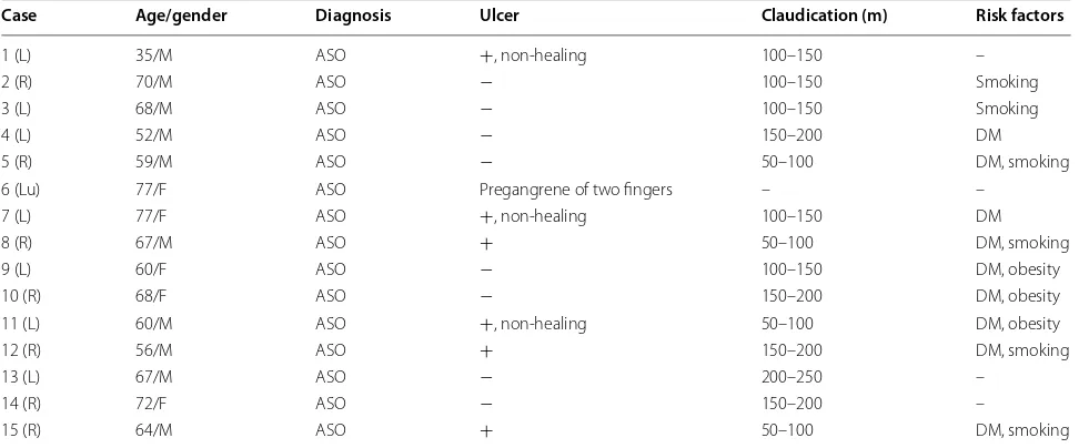
Table 1 Patients’ characteristics
Lu left upper limb, L left lower limb, R right lower limb, ASO arteriosclerosis obliterans, DM diabetes mellitus
Case Age/gender Diagnosis Ulcer Claudication (m) Risk factors
1 (L) 35/M ASO +, non-healing 100–150 –
2 (R) 70/M ASO − 100–150 Smoking
3 (L) 68/M ASO − 100–150 Smoking
4 (L) 52/M ASO − 150–200 DM
5 (R) 59/M ASO − 50–100 DM, smoking
6 (Lu) 77/F ASO Pregangrene of two fingers – –
7 (L) 77/F ASO +, non-healing 100–150 DM
8 (R) 67/M ASO + 50–100 DM, smoking
9 (L) 60/F ASO − 100–150 DM, obesity
10 (R) 68/F ASO − 150–200 DM, obesity
11 (L) 60/M ASO +, non-healing 50–100 DM, obesity
12 (R) 56/M ASO + 150–200 DM, smoking
13 (L) 67/M ASO − 200–250 –
14 (R) 72/F ASO − 150–200 –
[image:2.595.56.542.512.714.2]aspiration force. All adipose tissue was collected from abdomen area, under local anesthesia with lidocaine and adrenaline. Minimum amount of collected tissue was 40 ml.
SVF cell isolation
The lipoaspirate was washed within 12 h of collection with plenty of physiological solution and gentamicin (80 mg/l). Adipose fraction was cut using specially pro-duced blend mesh to avoid usage of collagenase. A mechanical stainless steel two-bladed mill placed in a cyl-inder 5 cm in diameter and equipped with a metal 3 mm diameter mesh was used to mechanically disrupt the adipose tissue. The mill was rotated at speed not exceed-ing 260 rpm. Each fraction was minced three times and remaining homogenous lipoaspirate was centrifuged for 7 min at 850g in 50 ml falcon tubes. The upper fraction containing adipocytes was discarded, and the pellet was washed once with physiological solution and prepared for injections. Cell densities were determined by count-ing in a Neubauer’s hemocytometer, and cell viability was assessed using Trypan blue exclusion assay.
Injection of SVF cells
Cells were prepared in 20 ml luer lock syringes (BD). Cells were diluted in physiological solution and autolo-gous serum of the patient. Minimum amount of viable cells per one syringe applied was 20 million. Application
consisted of at least 30 injections per one 20 ml syringe. Secondary injections were performed 2 months after first application of cells.
Results
Multiple intramuscular SVF cell injections did not cause any complications in any of the patients during 5 days of hospitalization and all follow-up period. Overall, 86.7% of patients showed clinical improvement. Two patients (cases 10, 15) underwent a major amputation, 1 and 2 weeks after SVF cell therapy. The rest of patients reported either diminished or decreased rest pain at 12 weeks after SVF cell treatment. Table 2 shows the out-comes of SVF cell therapy. Ulceration was completely cured or improved in limbs of all patients suffering from ulcers after SVF cell therapy (Figs. 1, 3). No ulcer recur-rence was observed in any of the patients during the fol-low-up period. 86.7% of patients showed improvement in walking distances. The ankle-brachial index (ABI) was improved from 17 to 48% at 12 months after SVF cell therapy, and the ABI was still higher 2 years later for all the patients. Digital subtraction angiography (DSA) per-formed before and after SVF cell therapy showed for-mation of numerous vascular collateral networks across affected arteries (Figs. 1, 2). None of the patients died during the follow-up period. The survival rate and free-dom from major amputation of the limb at 24 months after SVF cell therapy were 100 and 86.7%, respectively.
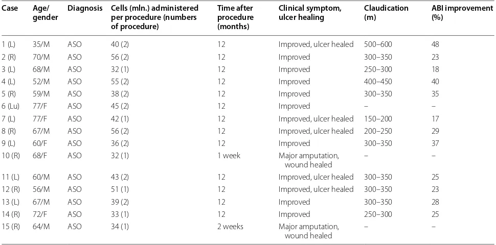
Table 2 SVF cell therapy and outcomes
Lu left upper limb, L left lower limb, R right lower limb, ASO arteriosclerosis obliterans
Case Age/
gender Diagnosis Cells (mln.) administered per procedure (numbers of procedure)
Time after procedure (months)
Clinical symptom,
ulcer healing Claudication (m) ABI improvement (%)
1 (L) 35/M ASO 40 (2) 12 Improved, ulcer healed 500–600 48
2 (R) 70/M ASO 56 (2) 12 Improved 300–350 23
3 (L) 68/M ASO 32 (1) 12 Improved 250–300 18
4 (L) 52/M ASO 55 (2) 12 Improved 400–450 40
5 (R) 59/M ASO 38 (2) 12 Improved 300–350 35
6 (Lu) 77/F ASO 45 (2) 12 Improved – –
7 (L) 77/F ASO 42 (1) 12 Improved, ulcer healed 150–200 17
8 (R) 67/M ASO 56 (2) 12 Improved, ulcer healed 200–250 29
9 (L) 60/F ASO 36 (2) 12 Improved 300–350 37
10 (R) 68/F ASO 32 (1) 1 week Major amputation,
wound healed – –
11 (L) 60/M ASO 43 (2) 12 Improved, ulcer healed 300–350 25 12 (R) 56/M ASO 51 (1) 12 Improved, ulcer healed 300–350 23
13 (L) 67/M ASO 39 (2) 12 Improved 300–350 28
14 (R) 72/F ASO 33 (1) 12 Improved 250–300 25
15 (R) 64/M ASO 34 (1) 2 weeks Major amputation,
[image:3.595.55.540.475.715.2]Discussion
In the last decade cell-based therapies have been inves-tigated as a promising treatment option for patients with CLI who are refractory to other treatment modali-ties. It provides encouraging therapeutic possibilities to enhance the repair of damaged or diseased tissues in CLI patients. Several studies have suggested beneficial effects of autologous BM-MSC based therapies [12, 13]. How-ever, the percentage of MSCs in bone marrow is quite low and decreases with age [14]. Furthermore, after isolation, BM-MSCs need 2–3 weeks of in vitro culture to reach an amount sufficient for transplantation. Moreover, bone marrow suction is an invasive procedure. These drawbacks limit the possibility of wide clinical application of BM-MSCs [15]. In addition, the neovascularization capacity of transplanted BM-MNCs is reduced with aging; there-fore this cell treatment is less appropriate in the older patients [16]. Last but not least, meta-analysis of rand-omized placebo controlled trials showed no advantage of bone marrow derived cell therapy on the primary outcome measures of amputation, survival, and amputation free
survival in CLI patients [17]. Compared with bone mar-row, subcutaneous adipose tissue can provide enough dosage for therapy without cell culture. This tissue is now recognized as an abundant and accessible source of multi-potent stromal cells suitable for regenerative medicine [18]. Nevertheless, adipose tissue is routinely discarded as a medical waste. In this pilot study, we used autolo-gous uncultured adipose-derived SVF cells as a potential treatment option for patients with CLI. Obtained results show the beneficial role of SVF cell therapy in reduc-ing the rate of major amputations and improvreduc-ing quality of life in CLI patients. 86.7% of treated patients avoided the amputation of limbs. Previously it was demonstrated that SVF cell therapy accelerated diabetic wound healing [19]. In our study, complete wound healing occurred in all SVF cell—treated CLI patients. Previous studies have shown that ASCs exert their effects mainly via paracrine mechanisms and make beneficial contributions to tissue repair, regeneration and immunomodulation [11, 20, 21]. We have shown that injection of SVF cells is an effective way to promote healing of ulcer and skin regeneration.
[image:4.595.57.541.86.418.2]Moreover, this study, for the first time, showed that injec-tions of uncultured SVF cells could accelerate angiogen-esis. Digital subtraction angiography performed before and after SVF treatment showed that the transformation of preexistent collateral arterioles into functional collateral arteries occurred in CLI patients. The principle that justi-fies the therapeutic application of stem cells is the resto-ration of vascular cellularity, the control and the support of the newly formed vessels, which ensure an adequate supply of oxygen in critical ischemic areas [22]. Previous researchers have reported that SVF cells could secrete various angiogenic growth factors in vitro and enhance neovascularization of ischemic tissue in vivo [23, 24]. Our data supports previously published reports showing that SVF cells promote angiogenesis and tissue repair. In a study performed by Sheng et al., enhanced angiogenesis and cell proliferation were observed in the tissue treated by transplantation of SVF [15]. We used uncultured SVF cells directly injecting them into ischemic limb. Injections were placed along the occluded native arteries, because the
density of preformed collaterals is highest in parallel orien-tation to the axial arteries. This is the preferred location for collateral growth [22]. Moreover, we suppose that uncul-tured heterogeneous SVF cells can be more effective than a purified cell population due to the fact that heterogene-ous population contains fibroblasts, stem cells, endothelial cells, pericytes, mast cells, preadipocytes, smooth muscle cells, macrophages, and progenitor cells, which are known to accelerate wound healing [15]. SVF cells injected near the wound could not only stimulate host cells around the wound, but also provide growth factors and extracel-lular matrix. The principle of intramuscular injection is the creation of a cell depot with paracrine activity in the ischemic area [22]. Moreover, in order to obtain more ben-eficial effect, we diluted SVF cells with autologous serum, which also contains growth factors and cytokines. The results obtained from this pilot study have demonstrated
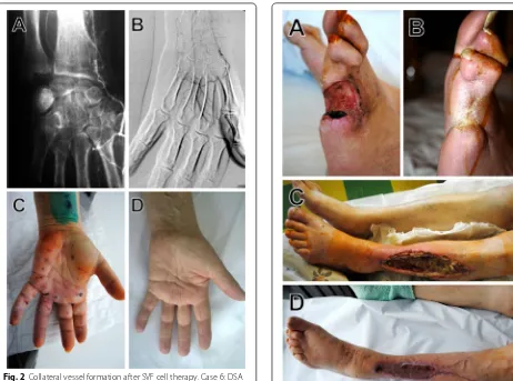
Fig. 2 Collateral vessel formation after SVF cell therapy. Case 6: DSA images before (A) and 10 months after SVF cell injections (B). Images of occluded limb right after SVF administration (C) and 10 months after SVF cell injections (D)
[image:5.595.60.522.86.429.2] [image:5.595.65.296.86.423.2]the beneficial role of SVF cell therapy in reducing the rate of amputations, reducing pain, improving ABI and overall quality of life in CLI patients.
Conclusion
Our data indicate that uncultured SVF cells diluted with autologous serum represent a potent therapeutic combi-nation for CLI patients. The multiple intramuscular SVF cell injections are effective alternative to achieve therapeu-tic angiogenesis in CLI patients for which surgical bypass and/or PTA are not possible, and that this treatment modality is appropriate and safe. Bearing in mind the easy procedure of cell isolation and preparation, SVF cells may provide a promising therapeutic option for CLI. However, to establish this cell therapy as a standard treatment, more investigation with a larger number of patients is necessary.
Abbreviations
ABI: ankle-brachial pressure index; ASCs: adipose-derived stromal/stem cells; ASO: arteriosclerosis obliterans; BM-MNCs: bone marrow-derived mononu-clear cells; BM-MSCs: bone marrow-derived mesenchymal stem cells; CLI: critical limb ischemia; DSA: digital subtraction angiography; MSCs: mesen-chymal stem cells; PAD: peripheral artery disease; PB-MNCs: peripheral blood mononuclear cells; PTA: percutaneous transluminal angioplasty; SVF: stromal vascular fraction.
Authors’ contributions
AD, MP, GA, GV, LL, TI and RR participated in the design of the study and analyzed the data. MP prepared all the figures. AD wrote the manuscript. All authors read and approved the final manuscript.
Author details
1 Laboratory of Immunology, National Cancer Institute, Santariskiu Str. 1,
08660 Vilnius, Lithuania. 2 Department of Vascular Surgery, Vilnius City Clinical
Hospital, Antakalnio Str. 57, 10207 Vilnius, Lithuania. 3 Northway Medical
and Surgical Center, S.Zukausko Str. 19, 08234 Vilnius, Lithuania. 4 Clinics
of Cardiovascular Diseases, Vilnius University, Santariskiu Str. 2, 08661 Vilnius, Lithuania. 5 Department of Plastic and Reconstructive Surgery, Lithuanian
University of Health Sciences, Medical Academy, University Clinics of Kaunas, Eiveniu Str. 2, 50009 Kaunas, Lithuania. 6 Immune Advisors LLC, San Diego, CA
92121, USA.
Acknowledgements Not applicable.
Competing interests
The authors declare that they have no competing interests.
Availability of data and materials
The datasets analyzed during the current study are available from the cor-responding author on reasonable request.
Consent for publication
Informed consent was obtained from all participants.
Ethics approval and consent to participate
This study was approved by the medical ethics committee of Vilnius City Clinical Hospital, Lithuania. Informed consent was obtained from all patients before entry into the study in accordance with the Declaration of Helsinki.
Publisher’s Note
Springer Nature remains neutral with regard to jurisdictional claims in pub-lished maps and institutional affiliations.
Received: 27 April 2017 Accepted: 13 June 2017
References
1. Moriya J, Minamino T, Tateno K, Shimizu N, Kuwabara Y, Sato Y, Saito Y, Komuro I. Long-term outcome of therapeutic neovascularization using peripheral blood mononuclear cells for limb ischemia. Circ Cardiovasc Interv. 2009;2:245–54.
2. Nishida T, Ueno Y, Kimura T, Ogawa R, Joo K, Tominaga R. Early and long-term effects of the autologous peripheral stem cell implantation for critical limb ischemia. Ann Vasc Dis. 2011;4:319–24.
3. Gupta PK, Chullikana A, Parakh R, Desai S, Das A, Gottipamula S, Krishnamurthy S, Anthony N, Pherwani A, Majumdar AS. A double blind randomized placebo controlled phase I/II study assessing the safety and efficacy of allogeneic bone marrow derived mesenchymal stem cell in critical limb ischemia. J Transl Med. 2013;11:143.
4. Samura M, Hosoyama T, Takeuchi Y, Ueno K, Morikage N, Hamano K. Therapeutic strategies for cell-based neovascularization in critical limb ischemia. J Transl Med. 2017;15:49.
5. Liew A, Bhattacharya V, Shaw J, Stansby G. Cell therapy for critical limb ischemia: a meta-analysis of randomized controlled trials. Angiology. 2016;67:444–55.
6. Gimble JM, Guilak F, Bunnell BA. Clinical and preclinical translation of cell-based therapies using adipose tissue-derived cells. Stem Cell Res Ther. 2010;1:19.
7. Gimble JM, Katz AJ, Bunnell BA. Adipose-derived stem cells for regenera-tive medicine. Circ Res. 2007;100:1249–60.
8. Bourin P, Bunnell BA, Casteilla L, Dominici M, Katz AJ, March KL, Redl H, Rubin JP, Yoshimura K, Gimble JM. Stromal cells from the adipose tissue-derived stromal vascular fraction and culture expanded adipose tissue-derived stromal/stem cells: a joint statement of the International Federation for Adipose Therapeutics and Science (IFATS) and the Interna-tional Society for Cellular Therapy (ISCT). Cytotherapy. 2013;15:641–8. 9. Han J, Koh YJ, Moon HR, Ryoo HG, Cho CH, Kim I, Koh GY. Adipose tissue
is an extramedullary reservoir for functional hematopoietic stem and progenitor cells. Blood. 2010;115:957–64.
10. Michalek J, Moster R, Lukac L, Proefrock K, Petrasovic M, Rybar J, Capkova M, Chaloupka A, Darinskas A, Michalek J Sr, et al. Autologous adipose tissue-derived stromal vascular fraction cells application in patients with osteoarthritis. Cell Transpl. 2015;20:1–36.
11. Lee HC, An SG, Lee HW, Park JS, Cha KS, Hong TJ, Park JH, Lee SY, Kim SP, Kim YD, et al. Safety and effect of adipose tissue-derived stem cell implantation in patients with critical limb ischemia: a pilot study. Circ J. 2012;76:1750–60.
12. Tateishi-Yuyama E, Matsubara H, Murohara T, Ikeda U, Shintani S, Masaki H, Amano K, Kishimoto Y, Yoshimoto K, Akashi H, et al. Therapeutic angio-genesis for patients with limb ischaemia by autologous transplantation of bone-marrow cells: a pilot study and a randomised controlled trial. Lancet. 2002;360:427–35.
13. Dash NR, Dash SN, Routray P, Mohapatra S, Mohapatra PC. Targeting nonhealing ulcers of lower extremity in human through autologous bone marrow-derived mesenchymal stem cells. Rejuvenation Res. 2009;12:359–66.
14. Stolzing A, Jones E, McGonagle D, Scutt A. Age-related changes in human bone marrow-derived mesenchymal stem cells: consequences for cell therapies. Mech Ageing Dev. 2008;129:163–73.
15. Sheng L, Yang M, Du Z, Yang Y, Li Q. Transplantation of stromal vascular fraction as an alternative for accelerating tissue expansion. J Plast Recon-str Aesthet Surg. 2013;66:551–7.
16. Sugihara S, Yamamoto Y, Matsuura T, Narazaki G, Yamasaki A, Igawa G, Matsubara K, Miake J, Igawa O, Shigemasa C, et al. Age-related BM-MNC dysfunction hampers neovascularization. Mech Ageing Dev. 2007;128:511–6.
• We accept pre-submission inquiries
• Our selector tool helps you to find the most relevant journal
• We provide round the clock customer support
• Convenient online submission
• Thorough peer review
• Inclusion in PubMed and all major indexing services
• Maximum visibility for your research
Submit your manuscript at www.biomedcentral.com/submit
Submit your next manuscript to BioMed Central
and we will help you at every step:
18. Zuk PA, Zhu M, Ashjian P, De Ugarte DA, Huang JI, Mizuno H, Alfonso ZC, Fraser JK, Benhaim P, Hedrick MH. Human adipose tissue is a source of multipotent stem cells. Mol Biol Cell. 2002;13:4279–95.
19. Han SK, Kim HR, Kim WK. The treatment of diabetic foot ulcers with uncultured, processed lipoaspirate cells: a pilot study. Wound Repair Regen. 2010;18:342–8.
20. Kapur SK, Katz AJ. Review of the adipose derived stem cell secretome. Biochimie. 2013;95:2222–8.
21. Gimble JM, Bunnell BA, Guilak F. Human adipose-derived cells: an update on the transition to clinical translation. Regen Med. 2012;7:225–35.
22. Compagna R, Amato B, Massa S, Amato M, Grande R, Butrico L, de Fran-ciscis S, Serra R. Cell therapy in patients with critical limb ischemia. Stem Cells Int. 2015;2015:931420.
23. Rehman J, Traktuev D, Li J, Merfeld-Clauss S, Temm-Grove CJ, Boven-kerk JE, Pell CL, Johnstone BH, Considine RV, March KL. Secretion of angiogenic and antiapoptotic factors by human adipose stromal cells. Circulation. 2004;109:1292–8.