REVIEW
MALDI-TOF mass spectrometry
as a diagnostic tool in human and veterinary
helminthology: a systematic review
Maureen Feucherolles
1,2, Sven Poppert
3,4, Jürg Utzinger
3,4and Sören L. Becker
1,3,4*Abstract
Background:
Matrix-assisted laser desorption/ionization-time of flight (MALDI-TOF) mass spectrometry (MS) has
become a widely used technique for the rapid and accurate identification of bacteria, mycobacteria and certain
fungal pathogens in the clinical microbiology laboratory. Thus far, only few attempts have been made to apply the
technique in clinical parasitology, particularly regarding helminth identification.
Methods:
We systematically reviewed the scientific literature on studies pertaining to MALDI-TOF MS as a
diagnos-tic technique for helminths (cestodes, nematodes and trematodes) of medical and veterinary importance. Readily
available electronic databases (i.e. PubMed/MEDLINE, ScienceDirect, Cochrane Library, Web of Science and Google
Scholar) were searched from inception to 10 October 2018, without restriction on year of publication or language. The
titles and abstracts of studies were screened for eligibility by two independent reviewers. Relevant articles were read
in full and included in the systematic review.
Results:
A total of 84 peer-reviewed articles were considered for the final analysis. Most papers reported on the
appli-cation of MALDI-TOF for the study of
Caenorhabditis elegans
, and the technique was primarily used for identification of
specific proteins rather than entire pathogens. Since 2015, a small number of studies documented the successful use
of MALDI-TOF MS for species-specific identification of nematodes of human and veterinary importance, such as
Trich-inella
spp. and
Dirofilaria
spp. However, the quality of available data and the number of examined helminth samples
was low.
Conclusions:
Data on the use of MALDI-TOF MS for the diagnosis of helminths are scarce, but recent evidence
suggests a potential role for a reliable identification of nematodes. Future research should explore the diagnostic
accuracy of MALDI-TOF MS for identification of (i) adult helminths, larvae and eggs shed in faecal samples; and (ii)
helminth-related proteins that are detectable in serum or body fluids of infected individuals.
Keywords:
Diagnosis, Helminths, MALDI-TOF, Matrix-assisted laser desorption/ionization-time of flight, Neglected
tropical diseases, Parasites
© The Author(s) 2019. This article is distributed under the terms of the Creative Commons Attribution 4.0 International License (http://creat iveco mmons .org/licen ses/by/4.0/), which permits unrestricted use, distribution, and reproduction in any medium, provided you give appropriate credit to the original author(s) and the source, provide a link to the Creative Commons license, and indicate if changes were made. The Creative Commons Public Domain Dedication waiver (http://creat iveco mmons .org/ publi cdoma in/zero/1.0/) applies to the data made available in this article, unless otherwise stated.
Background
In clinical and laboratory diagnostic settings, mass
spec-trometry (MS) has been utilized for several decades as an
approach for protein-centred analysis of samples in
med-ical chemistry [
1
,
2
] and haematology laboratories [
3
]. In
1975, Anhalt & Fenselau [
4
] proposed, for the first time,
the modification of matrix-assisted laser
desorption/ioni-zation time-of-flight (MALDI-TOF) MS as a method to
characterize bacteria. Indeed, it was demonstrated that
different bacterial species show specific protein mass
spectra, which can be used for rapid identification.
During the past decade, MALDI-TOF MS has been
widely introduced as a diagnostic technique in
microbiol-ogy laboratories, where it has replaced most other tools
(e.g. phenotypic tests, biochemical identification and
Open Access
*Correspondence: soeren.becker@uks.eu
1 Institute of Medical Microbiology and Hygiene, Saarland University, Homburg/Saar, Germany
agglutination kits) as the first-line pathogen identification
method due to its high diagnostic accuracy, robustness,
reliability and rapid turn-around time [
5
]. MALDI-TOF
MS is now routinely employed for identification of
bac-teria [
5
–
8
], mycobacteria [
5
,
9
] and some fungi [
8
]. More
recently, MALDI-TOF MS has been applied in research
settings for the detection and identification of viruses
[
10
], protozoans and arthropods [
11
,
12
]. In clinical
practice, a specific quantity is brought on a target plate
(e.g. culture-grown pathogen). Next, the target plate is
pre-treated with a chemical reagent (so-called matrix,
e.g. α-cyano-4-hydroxycinnamic acid) and subjected to
a mass spectrometer for further analysis. The
MALDI-TOF apparatus, which is commercially available through
different manufacturers [
13
,
14
], uses laser to disperse
and ionize the analyte into different molecules, which
move through a vacuum driven by an electric field before
reaching a detector membrane. The time-of-flight of the
various molecules depends on their mass and their
elec-tric charge. The specific time-of-flight data are
assem-bled, resulting in specific spectra that are compared to a
commercial database, which allows for a rapid
identifica-tion of the infectious agent and diagnostic accuracy, the
latter of which is usually expressed as a score.
MALDI-TOF MS has several strengths if compared to
other diagnostic tools, such as polymerase chain reaction
(PCR) assays. Once the mass spectrometer and the
corre-sponding databases are available in a laboratory,
individ-ual pathogen identification is inexpensive, and the sample
preparation procedure does neither require highly skilled
technicians nor complex additional laboratory
infra-structure. Of note, MALDI-TOF MS is considerably less
prone to contamination and results are available within
a few minutes. However, constant power supply is a
pre-requisite, which limits the suitability of the technique in
resource-constrained settings. Yet, it should be noted
that MALDI-TOF MS is no longer restricted to
high-income countries as it is increasingly available in
refer-ence laboratories in sub-Saharan Africa and elsewhere
[
15
–
19
].
MALDI-TOF does not always require culture-grown
colonies of a given pathogen. Instead, it can also be
employed to identify microorganisms directly from
positive blood culture broths [
6
] with high diagnostic
accuracy [
7
]. Recently, Yang et al. [
20
] proposed a new
framework to analyse MALDI-TOF spectra of
bacte-rial mixtures (instead of only a single pathogen) and to
directly characterize each component without
purifica-tion procedures. Hence, this procedure might become
available to be employed directly on other body fluids
(e.g. urine, respiratory specimens and faecal samples),
which would further increase its relevance in clinical
practice [
21
,
22
].
In contrast to clinical bacteriology, little research has
been carried out pertaining to the application of
MALDI-TOF MS for identification of parasites of human or
vet-erinary importance [
23
]. Several studies utilized the
technique on protozoan parasites such as
Leishmania
spp. [
24
–
26
],
Giardia
spp. [
27
],
Cryptosporidium
spp.
[
28
],
Trypanosoma
spp. [
29
],
Plasmodium
spp. [
30
–
32
]
and
Dientamoeba
spp. [
33
]. These studies used
pre-treat-ment with ethanol and acetonitrile before subjecting the
whole pathogens to MALDI-TOF analysis. Additionally,
the technique has been used for identification of
ectopar-asites and vectors, such as ticks [
34
–
37
], fleas [
38
–
41
]
and mosquitoes [
42
–
49
]. In contrast to the experiments
on protozoans, only selected parts of the ectoparasites
and vectors (e.g. legs, thoraxes or wings) were used and
subjected to the same extraction method. A further novel
approach to apply MALDI-TOF MS in clinical
parasitol-ogy is the identification of specific serum peptides that
are detectable in parasite-infected individuals [
50
].
Helminth infections caused by nematodes (e.g.
Ascaris
lumbricoides
, hookworm,
Strongyloides stercoralis
and
Trichuris trichiura
), cestodes (e.g.
Taenia
spp.) and
trematodes (e.g.
Fasciola
spp. and
Schistosoma
spp.)
account for a considerable global burden of disease and
are among the most common infections in marginalized
populations in the tropics and subtropics [
51
]. Indeed,
according to estimates put forth by the Global Burden of
Disease (GBD) Study, 3.35 million disability-adjusted life
years (DALYs) were attributable to intestinal nematode
infections and schistosomiasis in 2017 [
52
].
Diagnosis is pivotal for effective treatment but requires
at least a basic laboratory infrastructure, light
micro-scopes and well-trained laboratory technicians who
might not be available in remote areas of tropical and
subtropical countries. In high-resource settings, in
con-trast, knowledge on microscopic identification of
hel-minths is waning in many laboratories. It is surprising
that the potential applicability of MALDI-TOF MS as a
diagnostic tool for helminths of human and veterinary
importance has not yet been systematically assessed, in
particular because the technique has been successfully
employed for identification of nematode plant
patho-gens [
53
–
58
]. Hence, the goal of this systematic review
was to summarize the available data on MALDI-TOF MS
application for diagnosis of helminths of medical and
vet-erinary importance, and to provide recommendations for
future research needs.
Methods
Search strategy
and/or veterinary helminthology. The research was
performed according to the guidance expressed in the
Preferred Reporting Items for Systematic Reviews and
Meta-Analyses (PRISMA) Statement [
59
].
The following electronic databases were systematically
searched: MEDLINE/PubMed, ScienceDirect-Embase,
Cochrane Library, Web of Science and Google Scholar.
All studies published from inception to 10 October 2018
were eligible for inclusion without language
restric-tions. The bibliographies of all eligible documents were
hand-searched for additional references. Conference
abstracts or book chapters detected through these
data-bases and additional library searches were also
consid-ered. The search strategy comprised keywords related
to the MALDI-TOF MS technique (e.g. “MALDI-TOF”
and “matrix-assisted laser desorption/ionization
time-of-flight”) and helminthology (e.g. “helminth”, “nematode”,
“cestode” and “trematode”). The full search strategies for
every database are provided in Additional file
1
and the
PRISMA checklist in Additional file
2
.
Eligibility screening
After the systematic literature search, all duplicates were
removed. Titles and abstracts of potentially eligible
stud-ies were screened to identify manuscripts relevant to the
research question. Scientific reports on helminths of either
plants or insects as well as studies on symbiotic bacteria of
helminths were excluded for this review. However, we kept
all publications related to the soil nematode
Caenorhabdi-tis elegans
, as it is used as a model organism for biomedical
research. Additionally, studies pertaining to MALDI-TOF/
TOF tandem MS were excluded, as this is a different
modi-fication of the MALDI-TOF MS technique, which is not
routinely employed in clinical microbiology laboratories,
but rather in research laboratory use for accurate
charac-terization or sequencing of components like amino acids,
metabolites, saccharides, etc. [
60
–
62
].
Data extraction and analysis
The literature search was performed by the first author
of this manuscript (MF). All titles and abstracts were
then independently reviewed by the first and the last
author (MF and SLB) for inclusion and any disagreement
was discussed until consensus was reached. All extracted
manuscripts were analysed using a reference manager
software (Mendeley;
http://www.mende ley.com
).
Results
Search results, number and year of publication of eligible
studies
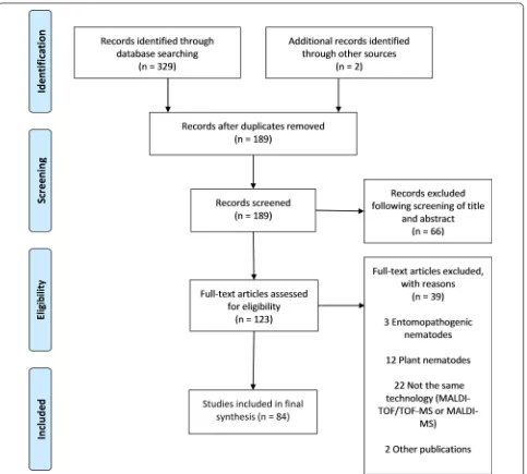
The search procedure and results obtained are shown
in Fig.
1
. In brief, the initial literature search yielded
329 published studies, with an additional two abstracts
identified through further search. Following removal of
142 duplicates, a total of 189 articles were assessed in
more detail, of which 66 studies were excluded based
on the analysis of the respective titles and abstracts.
A full-text analysis was carried out on the remaining
123 studies; 39 articles were finally excluded because
their scope was outside the current research question.
Hence, 84 articles were included, and these were
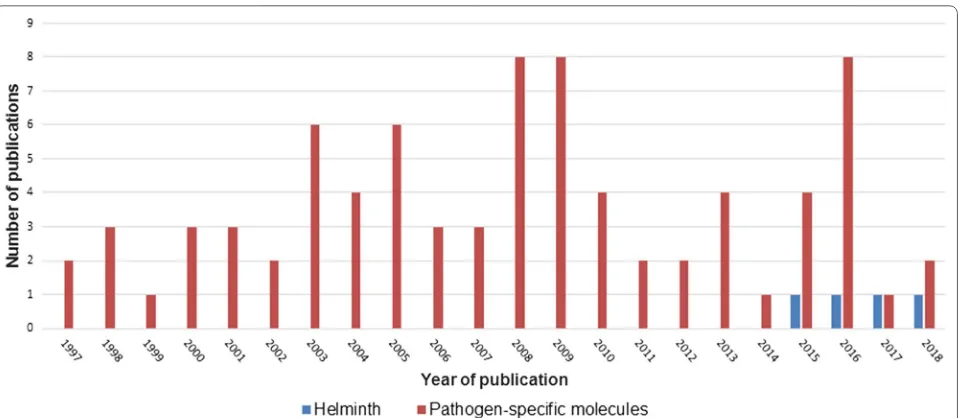
pub-lished between 1997 and 2018. Figure
2
shows the
num-ber of publications, stratified by year of publication. The
heterogeneity of data reported in the articles precluded
any meaningful meta-analysis (Additional file
3
).
Specific applications of MALDI‑TOF MS
The first two manuscripts published in 1997 described
structural analyses of glycosphingolipids found in
Ascaris suum
and
C. elegans
[
63
,
64
]. Indeed, 95% of
all eligible studies used MALDI-TOF MS for
identifi-cation of specific components rather than for the
iden-tification of entire pathogens (Fig.
2
). It was only in
2015 when a report on MALDI-TOF MS as diagnostic
tool for direct identification of
Dirofilaria
spp. became
available [
65
]. Soon thereafter followed a
proof-of-con-cept study utilizing MALDI-TOF MS for identification
and differentiation of
Trichinella
spp. and some
narra-tive reviews mentioning the lack of data on
MALDI-TOF in helminthology [
32
,
66
,
67
]. Yet, most studies
focused on distinct analyses of specific components,
such as peptides [
66
–
86
], proteins [
69
,
87
–
114
], lipids
[
61
,
62
,
115
–
124
], carbohydrates [
125
–
143
] and nucleic
acids [
144
] in a research context. Hence, MALDI-TOF
was mainly applied to study and compare the proteome
or the peptidome of different helminth species, and
most reports focused on
C. elegans
. For example,
Hus-son et al. [
74
] employed a new approach combining
liq-uid chromatography with MALDI-TOF MS to map and
differentiate the neuropeptide profiles of
C. elegans
and
the closely related species
C. briggsae
.
The two studies aiming at an identification of entire
pathogens provided evidence that MALDI-TOF MS could
reliably differentiate between species within the genus
Trichinella
[
67
] and
Dirofilaria
[
65
], respectively. In the
study by Mayer-Scholl et al. [
67
], nine species and three
genotypes of
Trichinella
isolated from mice, domestic
pigs, wild boars and guinea pigs were utilized to create
an in-house database with 27 raw spectra generated per
specimen. All tested isolates could be distinguished with
high diagnostic accuracy. The study by Pshenichnaya
et al. [
65
], which had only been published as a
confer-ence abstract, investigated five
Dirofilaria repens
and five
veterinary dirofilariasis, and reported that these could
be well differentiated by MALDI-TOF MS. However,
data were limited regarding the origin of the study
sam-ples, the quality of the spectra obtained by MALDI-TOF
and the repeatability of the results. Yet, during the
revi-sion of this systematic review, Pshenichnaya et al. [
145
]
published their work on dirofilariasis in a peer-reviewed
journal and provided also data for two different species
of
Ascaris
(i.e.
A. suum
and
A. lumbricoides
). These
hel-minths could be differentiated by MALDI-TOF based on
specific peaks and protein spectra patterns after a cell lysis
using the Sepsityper Kit 50 (Bruker Daltonics; Bremen,
Germany) and a protein extraction with 70% formic acid
and acetonitrile. However, this study has several
limita-tions, and it remains unclear whether calibration steps or
assessments of the repeatability and reproducibility of the
analyses were performed. An additional paper, published
in 2017, reported on MALDI-TOF MS application for
cyathostomin helminths, a very diverse group of
intesti-nal parasites infecting horses [
66
]. These so-called “small
strongyles” show a high degree of resistance against
ben-zimidazole anthelminthics and may lead to severe equine
enteropathy, colic and death [
146
]. The study examined
several species belonging to the cyathostomin helminths
[image:4.595.59.542.86.521.2](e.g.
Coronocyclus coronatus
,
C. labiatus
and
C. labratus
)
and found distinct protein spectra among adult helminths
of different species [
66
]. These findings were recently
con-firmed and substantiated by another study on the
applica-tion of MALDI-TOF for differentiaapplica-tion of cyathostomins,
which was published in April 2019 [
147
].
Discussion
We systematically reviewed the available literature
per-taining to the application of MALDI-TOF MS for
identi-fication of helminthic pathogens of human and veterinary
importance. While the technique has been successfully
employed for many major classes of pathogens (e.g.
bac-teria, mycobacteria and fungi), data on its use in
diagnos-tic helminthology are scarce. Several studies reported on
the differential analysis of specific components, such as
proteins, peptides or lipids with MALDI-TOF MS
tech-niques, but only two recent manuscripts and one
con-ference abstract provided ‘proof-of-concept’ evidence of
its potential utility in diagnosing and differentiating
hel-minth species of medical or veterinary relevance.
The majority of articles identified in this systematic
review focused on protein-centred analyses of helminth
samples. It is important to mention that some of the
MALDI-TOF MS devices employed in these studies had
been subjected to modifications that are not usually
avail-able in routine clinical laboratories. Additionally, these
experiments frequently employed a complex sample
pre-treatment comprising a protein separation by high
pres-sure liquid chromatography (HPLC) or electrophoresis.
Yet, some recent proof-of-concept studies have shown
that MALDI-TOF MS is also capable of diagnosing entire
helminthic pathogens and differentiating similar species
within the same genus based on an analysis of their
indi-vidual protein spectra [
66
,
67
]. Because no helminths are
currently included in commercially available
MALDI-TOF MS identification databases, individual in-house
databases need to be created through generation of main
spectra libraries, ideally following established guidelines
and protocols that are similar to those employed by the
manufacturers of commercially available mass
spectrom-eters [
148
]. Indeed, previous studies have described the
sensitive, reliable and highly reproducible identification
of helminths that cause plant infections and have
con-cluded that MALDI-TOF MS should be more widely
employed as a ‘rapid detection tool’ [
54
–
58
]. Ahmad et al.
[
56
], for example, reported on the suitability of
MALDI-TOF MS to differentiate harmless and juvenile infective
stages of single plant nematodes, as these showed unique,
characteristic protein peak patterns. These studies
should be considered as relevant because plant-parasitic
nematodes can sometimes also be found in human stool
samples [
149
,
150
]. In Brazil, for example, eggs of the
root-knot nematode
Meloidogyne
spp. were detected in
human faeces using a microscopic sedimentation method
[
151
]. Future studies should also employ MALDI-TOF on
serum, as a recent study reported the detection of
spe-cific proteins in serum of mice infected with
Schistosoma
japonicum
[
50
].
While helminth infections pose a considerable burden
on human and animal health [
152
], an accurate
diagno-sis of these conditions is frequently challenging. Indeed,
[image:5.595.60.539.86.295.2]simple diagnostic tools such as stool microscopy for
soil-transmitted helminth infections are of limited value if the
infection intensity is low and highly sensitive diagnostic
techniques such as PCR-based assays are only available
in selected reference laboratories outside endemic areas
[
153
]. In high-income countries, in contrast, knowledge
regarding standard diagnostic parasitology is waning and
differentiation of closely related helminth species based
on their microscopic morphology requires skilled
labora-tory technicians [
154
]. Moreover, some infections
can-not be reliably distinguished with standard diagnostic
techniques. A prominent example are infections caused
by cestodes of the genus
Taenia
[
155
], which may cause
a relatively harmless intestinal infection if cysts of
Taenia
saginata
or
T. solium
are orally ingested with meat of
cat-tle or pig. While eggs of
T. saginata
are not infectious to
humans,
T. solium
eggs can lead to the potentially fatal
disease (neuro-)cysticercosis. While the correct diagnosis
has important implications for treatment, patient
man-agement and potential contact screening (intestinal
car-riage of adult
T. solium
worms poses an increased risk of
cysticercosis for close contacts, such as family members),
it is impossible to distinguish both species based on the
identical morphology of their eggs under a microscope.
Molecular tools can achieve an accurate differentiation
of the two species, but are only available in research
set-tings [
155
–
157
]. Sometimes, proglottids of adult worms
are also passed in the faeces. While a distinct
differen-tiation is possible based on the uterine branches within
a proglottid, misidentification using this approach has
been reported in clinical practice [
158
]. Hence,
achiev-ing a species-specific differentiation based on
MALDI-TOF MS would contribute to an enhanced, more reliable
identification, and future studies should thus address
this issue. Similar considerations hold also true for other
infective agents that can hardly be differentiated by other
methods (e.g. different
Echinococcus
species), novel
spe-cies (e.g. hybrid spespe-cies of
Schistosoma
spp., which have
recently been reported from Corsica, France [
159
]) and
notoriously difficult-to-detect infections (e.g.
strongy-loidiasis). An overview of pathogens for which
develop-ment of MALDI-TOF MS identification protocols would
appear particularly promising is summarized in Table
1
.
It is important to consider the fixative in which a
para-sitological sample is stored. Both formaldehyde and
etha-nol are commonly used to enable a long-term storage
of biological specimens, but this may lead to profound
changes of the protein structure [
160
], which is likely to
influence on the results of MALDI-TOF examinations
carried out on such samples. The virtual impossibility
to amplify nucleic acids from formaldehyde-containing
solutions [
161
] due to fragmentation of the single
com-ponents [
162
] renders most PCR tests useless on these
sample types, but MALDI-TOF analyses of protein
spec-tra might still be possible, albeit with different specspec-tra if
compared to native samples. Hence, future studies should
evaluate this technique on different kinds of fixatives and
on samples that have been stored for prolonged periods.
The present review identified only a few
success-ful studies that employed MALDI-TOF MS to diagnose
helminths. Limitations include the complicated
pre-treatment procedures employed in some studies and the
rather incomplete data presentation in one of the more
clinically oriented research projects [
65
]. New research
is needed to determine whether this technique might
become a clinically meaningful addendum to the current
set of diagnostic options. However, experiences made in
clinical bacteriology, mycobacteriology, mycology as well
as with ectoparasites (e.g. ticks) and vectors (e.g.
mosqui-toes) [
12
,
37
,
163
] are promising. Whereas MALDI-TOF
MS is mainly used on culture-grown colonies for
iden-tification of bacteria and mycobacteria, the goal in
hel-minthology will be to provide a species-specific diagnosis
based on either macroscopic elements or eggs and larvae
that are present in stool samples (or other body fluids and
tissue samples). Hence, specific protocols will need to be
elaborated to this end, which may include sample
prepa-ration, purification and concentration steps, including
guidance on the most appropriate sample preservation.
However, such protocols have been successfully
devel-oped in the past (e.g. for identification of mycobacteria
or moulds) [
164
,
165
]. More recently, specific
pre-treat-ment modifications have even allowed to apply
MALDI-TOF MS on blood culture broths [
166
] and fresh urine
samples for direct identification of bacteria [
167
].
Addi-tionally, detection of parasites in complex samples (e.g.
blood), should be considered (e.g. as an antigen test for
Wuchereria bancrofti
[
168
] or for the detection of
spe-cific serum peptides [
169
]).
Yet, much research and rigorous validation is still
needed before MALDI-TOF MS might be employed
directly on stool samples, and priority should thus be
given to (i) the establishment of in-house main spectra
library databases to allow for species-specific
identifica-tion of selected helminths; (ii) the subsequent
develop-ment of sample treatdevelop-ment protocols; (iii) the validation of
this technique on different clinical sample types; and (iv)
the elaboration of MALDI-TOF MS to be employed on
fixed samples.
Conclusions
Table
1
C
ur
rent parasit
olog
ical t
echniques
, r
elat
ed challenges and r
esear
ch needs f
or a pot
ential application of M
ALDI-TOF MS as diag
nostic t
ool f
or major helminths of human
and v
et
er
inar
y impor
tance
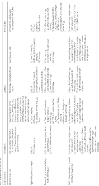
Charac ter istics Nemat odes Cest odes Tr emat odes Helminth species Soil-transmitt ed helminths: Asc aris lumbric oides ; hook -w or m; Str ongyloides ster cor a-lis ; Trichuris trichiur a Tissue nemat odes: D irofi -laria spp .; Trichinella spp .; To xo ca ra spp .; W ucher eria bancr oftiand other agents
causing lymphatic filar
iasis , O nchoc er ca v olvulus , Loa loa ; animal hook w or ms causing cutaneous lar va mig rans Taenia spp .; D iphyllobothrium latum Echinoc oc cus spp . Schistosoma spp .; Fasciola hepatic a
; small liv
er fluk
es
Type of diag
nostic sample (i) St ool; (ii) Ex cr et ed w or ms (i) T issue samples; (ii) S erum; (iii) Ex trac ted w or ms ( e.g . Loa loa ) (i) St ool; (ii) S erum (f or T. solium ); (iii)
Tissue samples in c
ysticer -cosis; (iv) Ex cr et ed pr oglottids (i) St
ool (in animals);
(ii) Tissue biopsies , sur gical samples; (iii) S er ology (i) St ool; (ii) Ur ine; (iii) S erum; (iv) T issue biopsies Parasit olog ical standar d diag -nostic t echniques (i) St ool micr oscop y ( e.g . K at o-Katz t echnique);
(ii) PCR in r
ef er ence laborat or ies (i) Dir ec
t identification on
biopsy samples;
(ii) S
er
ology
;
(iii) PCR in r
ef er ence labora -tor ies; (iv) D et ec
tion of micr
ofilar iae in blood; (v) D et ec
tion of antigen in
blood ( W ucher eria ) (i) Dir ec
t identification of fae
-cally ex cr et ed pr oglottids; (ii) St ool micr oscop y; (iii) S er ology (i) M icr oscop y;
(ii) PCR of c
ysts and tissue
samples; (iii) S er ology (i) St ool/ur ine micr oscop y
(dependent on inf
ec ting species); (ii) R apid diag nostic t est f or cir
culating cathodic antigen
(C
CA) in ur
ine;
(iii) PCR on serum, st
ool or ur
ine; (iv) S er ology Difficulties r elat ed t o cur rently emplo yed diag nostics (i) L ow sensitivit
y in light inf
ec
-tion int
ensities;
(ii) Specific concentration techniques needed f
or S. ster cor alis ; (iii) M
isidentification of hook
-w or m and S. ster cor alis lar vae possible;
(iv) No species diff
er entiation bet w een diff er ent hook -w or
m species possible upon
micr oscop y (i) S er ology fr equently false -negativ e; (ii) F alse -negativ
e PCR r
esults in
case of
‘ne
w
’ species;
(iii) Lack of exper
tise outside specializ ed laborat or ies (i) E
ggs of r
elat ed Taenia spp . ar
e indistinguishable b
y
micr
oscop
y;
(ii) Lack of exper
tise in pr
oglot
-tid diff
er
entiation in man
y laborat or ies (i) S er ology fr equently false -negativ
e in intac
t c ysts; (ii) M icr oscop y (similar mor
phology) and ser
ology (cr oss-r eac tivit y) cannot
reliably distinguish bet
w een Echinoc oc cus spp . (therapeu -tic implications) (i) L ow sensitivit
y in light inf
ec -tion int ensities; (ii) M icr oscop
y and PCR fail t
[image:7.595.131.476.83.729.2]Table
1
(c
on
tinued)
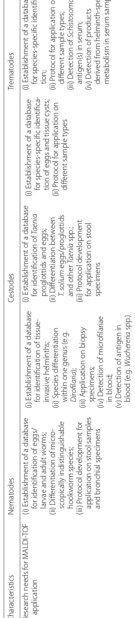
Charac
ter
istics
Nemat
odes
Cest
odes
Tr
emat
odes
Resear
ch needs f
or M
ALDI-TOF
application
(i) Establishment of a database for identification of eggs/ lar
vae and adult w
or
ms;
(ii) Diff
er
entiation of micr
o-scopically indistinguishable hook
w
or
m species;
(iii) P
rot
ocol de
velopment f
or
application on st
ool samples
and br
onchial specimens
(i) Establishment of a database for identification of tissue
-in
vasiv
e helminths;
(ii) Species diff
er
entiation
within one genus (
e.g
.
D
irofilaria
);
(iii) Application on biopsy specimens; (iv) D
et
ec
tion of micr
ofilar
iae
in blood;
(v) D
et
ec
tion of antigen in
blood (
e.g
.
W
ucher
eria
spp
.)
(i) Establishment of a database for identification of
Taenia
pr
oglottids and eggs;
(ii) Diff
er
entiation bet
w
een
T. solium
eggs/pr
oglottids
and other
Taenia
spp
.;
(iii) P
rot
ocol de
velopment
for application on st
ool
specimens
(i) Establishment of a database for species-specific identifica
-tion of eggs and tissue c
ysts;
(ii) P
rot
ocol f
or application on
diff
er
ent sample t
ypes
(i) Establishment of a database for species-specific identifica
-tion;
(ii) P
rot
ocol f
or application on
diff
er
ent sample t
ypes;
(iii) D
et
ec
tion of
Schistosoma
antigen(s) in serum;
(iv) D
et
ec
tion of pr
oduc
ts
der
iv
ed fr
om helminth-specific
[image:8.595.234.369.104.721.2]Preliminary data suggest that MALDI-TOF MS might
hold promise as a future diagnostic tool for direct and
rapid identification of pathogenic helminths in clinical
samples with sufficient diagnostic accuracy. Further
stud-ies are needed to evaluate these concepts and to develop
specific databases for helminth identification, followed
by rigorous validation on well characterised clinical
specimens.
Additional files
Additional file 1. Search strategies employed for our systematic review pertaining to the application of MALDI-TOF mass spectrometry as a diag-nostic tool in human and veterinary helminthology.
Additional file 2. PRISMA checklist for a systematic review examining the application of MALDI-TOF mass spectrometry as potential tool in diagnos-tic human and veterinary helminthology.
Additional file 3. List of references included in the final review (n= 84 articles).
Abbreviations
DALY: disability-adjusted life year; GBD: Global Burden of Disease (Study); MALDI-TOF: matrix-assisted laser desorption/ionization time-of-flight mass spectrometry; MS: mass spectrometry; PCR: Polymerase chain reaction.
Acknowledgements Not applicable.
Authors’ contributions
MF conducted the systematic review of the literature and extracted data. MF and SLB independently assessed published articles for eligibility, analysed and discussed data and drafted the first version of the manuscript. SP and JU contributed to data interpretation and revised the manuscript. All authors read and approved the final manuscript.
Funding
This systematic review received financial support from the ‘Landesforschungs-förderprogramm des Saarlandes’.
Availability of data and materials
The search strategy and all manuscripts included in this systematic review are available within the article and its additional files.
Ethics approval and consent to participate Not applicable.
Consent for publication Not applicable.
Competing interests
The authors declare that they have no competing interests.
Author details
1 Institute of Medical Microbiology and Hygiene, Saarland University, Homburg/Saar, Germany. 2 Luxembourg Institute of Science and Technol-ogy, Environmental Research and Innovation, Belvaux, Luxembourg. 3 Swiss Tropical and Public Health Institute, Basel, Switzerland. 4 University of Basel, Basel, Switzerland.
Received: 5 February 2019 Accepted: 6 May 2019
References
1. Tanaka K, Waki H, Ido Y, Akita S, Yoshida Y, Yoshida T, et al. Protein and polymer analyses up to m/z 100 000 by laser ionization time-of-flight mass spectrometry. Rapid Commun Mass Spectrom. 1988;2:151–3. 2. Marvin LF, Roberts MA, Fay LB. Matrix-assisted laser
desorption/ioniza-tion time-of-flight mass spectrometry in clinical chemistry. Clin Chim Acta. 2003;337:11–21.
3. Rees-Unwin KS, Morgan GJ, Davies FE. Proteomics and the haematolo-gist. Clin Lab Haematol. 2004;26:77–86.
4. Anhalt JP, Fenselau C. Identification of bacteria using mass spectrom-etry techniques. Anal Chem. 1975;47:219–25.
5. Clark AE, Kaleta EJ, Arora A, Wolk DM. Matrix-assisted laser desorp-tion ionizadesorp-tion-time of flight mass spectrometry: a fundamental shift in the routine practice of clinical microbiology. Clin Microbiol Rev. 2013;26:547–603.
6. Carbonnelle E, Mesquita C, Bille E, Day N, Dauphin B, Beretti JL, et al. MALDI-TOF mass spectrometry tools for bacterial identification in clini-cal microbiology laboratory. Clin Biochem. 2011;44:104–9.
7. Singhal N, Kumar M, Kanaujia PK, Virdi JS. MALDI-TOF mass spectrome-try: an emerging technology for microbial identification and diagnosis. Front Microbiol. 2015;6:791.
8. Angeletti S. Matrix assisted laser desorption time of flight mass spec-trometry (MALDI-TOF MS) in clinical microbiology. J Microbiol Methods. 2017;138:20–9.
9. El Khéchine A, Couderc C, Flaudrops C, Raoult D, Drancourt M. Matrix assisted laser desorption/ionization time-of-flight mass spectrometry identification of mycobacteria in routine clinical practice. PLoS One. 2011;6:e24720.
10. Sjöholm MIL, Dillner J, Carlson J. Multiplex detection of human herpesviruses from archival specimens by using matrix-assisted laser desorption ionization-time of flight mass spectrometry. J Clin Microbiol. 2008;46:540–5.
11. Yssouf A, Almeras L, Raoult D, Parola P. Emerging tools for identification of arthropod vectors. Future Microbiol. 2016;11:549–66.
12. Vega-Rúa A, Pagès N, Fontaine A, Nuccio C, Hery L, Goindin D, et al. Improvement of mosquito identification by MALDI-TOF MS biotyp-ing usbiotyp-ing protein signatures from two body parts. Parasit Vectors. 2018;11:574.
13. Faron ML, Buchan BW, Hyke J, Madisen N, Lillie JL, Granato PA, et al. Multicenter evaluation of the Bruker MALDI Biotyper CA System for the identification of clinical aerobic gram-negative bacterial isolates. PLoS One. 2015;10:e0141350.
14. Luo Y, Siu GKH, Yeung ASF, Chen JHK, Ho PL, Leung KW, et al. Perfor-mance of the VITEK MS matrix-assisted laser desorption ionization-time of flight mass spectrometry system for rapid bacterial identification in two diagnostic centres in China. J Med Microbiol. 2015;64:18–24. 15. Fall B, Lo CI, Samb-Ba B, Perrot N, Diawara S, Gueye MW, et al. The
ongoing revolution of MALDI-TOF mass spectrometry for microbiology reaches tropical Africa. Am J Trop Med Hyg. 2015;92:641–7.
16. Lo CI, Fall B, Sambe-Ba B, Flaudrops C, Faye N, Mediannikov O, et al. Value of matrix assisted laser desorption ionization-time of flight (MALDI-TOF) mass spectrometry in clinical microbiology and infectious diseases in Africa and tropical areas. Afr J Microbiol Res. 2017;11:1360–70.
17. Diongue K, Kébé O, Faye MD, Samb D, Diallo MA, Ndiaye M, et al. MALDI-TOF MS identification of Malassezia species isolated from patients with pityriasis versicolor at the Seafarers’ Medical Service in Dakar, Senegal. J Mycol Med. 2018;28:590–3.
18. Taverna CG, Mazza M, Bueno NS, Alvarez C, Amigot S, Andreani M, et al. Development and validation of an extended database for yeast identifi-cation by MALDI-TOF MS in Argentina. Med Mycol. 2019;57:215–25. 19. Chabriere E, Bassène H, Drancourt M, Sokhna C. MALDI-TOF-MS and
point of care are disruptive diagnostic tools in Africa. New Microbes New Infect. 2018;26:S83–8.
20. Yang Y, Lin Y, Qiao L. Direct MALDI-TOF MS identification of bacterial mixtures. Anal Chem. 2018;90:10400–8.
22. Íñigo M, Coello A, Fernández-Rivas G, Rivaya B, Hidalgo J, Quesada MD, et al. Direct identification of urinary tract pathogens from urine samples, combining urine screening methods and matrix-assisted laser desorption ionization-time of flight mass spectrometry. J Clin Microbiol. 2016;54:988–93.
23. Singhal N, Kumar M, Virdi JS. MALDI-TOF MS in clinical parasi-tology: applications, constraints and prospects. Parasitology. 2016;143:1491–500.
24. Cassagne C, Pratlong F, Jeddi F, Benikhlef R, Aoun K, Normand A-C, et al. Identification of Leishmania at the species level with matrix-assisted laser desorption ionization time-of-flight mass spectrometry. Clin Microbiol Infect. 2014;20:551–7.
25. Culha G, Akyar I, Yildiz Zeyrek F, Kurt Ö, Gündüz C, Özensoy Töz S, et al. Leishmaniasis in Turkey: determination of Leishmania species by matrix-assisted laser desorption ionization time-of-flight mass spectrometry (MALDI-TOF MS). Iran J Parasitol. 2014;9:239–48.
26. Lachaud L, Fernández-Arévalo A, Normand AC, Lami P, Nabet C, Donnadieu JL, et al. Identification of Leishmania by MALDI-TOF mass spectrometry using a free web-based application and a dedicated mass spectral library. J Clin Microbiol. 2017;55:2914–33.
27. Villegas EN, Glassmeyer ST, Ware MW, Hayes SL, Schaeffer FW. Matrix-assisted laser desorption/ionization time-of-flight mass spectrometry-based analysis of Giardia lamblia and Giardia muris. J Eukaryot Micro-biol. 2006;53:S179–81.
28. Magnuson ML, Owens JH, Kelty CA. Characterization of Cryptosporidium parvum by matrix-assisted laser desorption ionization-time of flight mass spectrometry. Appl Environ Microbiol. 2000;66:4720–4. 29. Avila CC, Almeida FG, Palmisano G. Direct identification of
trypa-nosomatids by matrix-assisted laser desorption ionization-time of flight mass spectrometry (DIT MALDI-TOF MS). J Mass Spectrom. 2016;51:549–57.
30. Marks F, Meyer CG, Sievertsen J, Timmann C, Evans J, Horstmann RD, et al. Genotyping of Plasmodium falciparum pyrimethamine resist-ance by matrix-assisted laser desorption-ionization time-of-flight mass spectrometry. Antimicrob Agents Chemother. 2004;48:466–72. 31. Gitau EN, Kokwaro GO, Newton CR, Ward SA. Global proteomic analysis
of plasma from mice infected with Plasmodium berghei ANKA using two dimensional gel electrophoresis and matrix assisted laser desorption ionization-time of flight mass spectrometry. Malar J. 2011;10:205. 32. de Dios Caballero J, Martin O. Application of MALDI-TOF in parasitology.
In: Cobo F, editor. The use of mass spectrometry technology (MALDI-TOF) in clinical microbiology. London: Elsevier Inc.; 2018. p. 235–53. 33. Calderaro A, Buttrini M, Montecchini S, Rossi S, Piccolo G, Arcangeletti
MC, et al. MALDI-TOF MS as a new tool for the identification of Dienta-moeba fragilis. Parasit Vectors. 2018;11:11.
34. Karger A, Kampen H, Bettin B, Dautel H, Ziller M, Hoffmann B, et al. Species determination and characterization of developmental stages of ticks by whole-animal matrix-assisted laser desorption/ionization mass spectrometry. Ticks Tick Borne Dis. 2011;3:78–89.
35. Yssouf A, Flaudrops C, Drali R, Kernif T, Socolovschi C, Berenger JM, et al. Matrix-assisted laser desorption ionization-time of flight mass spectrometry for rapid identification of tick vectors. J Clin Microbiol. 2013;51:522–8.
36. Diarra AZ, Almeras L, Laroche M, Berenger J-M, Koné AK, Bocoum Z, et al. Molecular and MALDI-TOF identification of ticks and tick-associ-ated bacteria in Mali. PLoS Negl Trop Dis. 2017;11:e0005762. 37. Dib L, Benakhla A, Raoult D, Parola P. MALDI-TOF MS identification of
ticks of domestic and wild animals in Algeria and molecular detection of associated microorganisms. Comp Immunol Microbiol Infect Dis. 2018;57:39–49.
38. Dvorak V, Halada P, Hlavackova K, Dokianakis E, Antoniou M, Volf P. Iden-tification of phlebotomine sand flies (Diptera: Psychodidae) by matrix-assisted laser desorption/ionization time of flight mass spectrometry. Parasit Vectors. 2014;7:21.
39. Yssouf A, Socolovschi C, Leulmi H, Kernif T, Bitam I, Audoly G, et al. Identification of flea species using MALDI-TOF/MS. Comp Immunol Microbiol Infect Dis. 2014;37:153–7.
40. Lafri I, Almeras L, Bitam I, Caputo A, Yssouf A, Forestier C-L, et al. Iden-tification of Algerian field-caught phlebotomine sand fly vectors by MALDI-TOF MS. PLoS Negl Trop Dis. 2016;10:e0004351.
41. El Hamzaoui B, Laroche M, Almeras L, Bérenger J-M, Raoult D, Parola P. Detection of Bartonella spp. in fleas by MALDI-TOF MS. PLoS Negl Trop Dis. 2018;12:e0006189.
42. Dieme C, Yssouf A, Vega-Rúa A, Berenger JM, Failloux AB, Raoult D, et al. Accurate identification of Culicidae at aquatic developmental stages by MALDI-TOF MS profiling. Parasit Vectors. 2014;7:544.
43. Yssouf A, Parola P, Lindström A, Lilja T, L’Ambert G, Bondesson U, et al. Identification of European mosquito species by MALDI-TOF MS. Parasi-tol Res. 2014;113:2375–8.
44. Niare S, Berenger J-M, Dieme C, Doumbo O, Raoult D, Parola P, et al. Identification of blood meal sources in the main African malaria mos-quito vector by MALDI-TOF MS. Malar J. 2016;15:87.
45. Tandina F, Almeras L, Koné AK, Doumbo OK, Raoult D, Parola P. Use of MALDI-TOF MS and culturomics to identify mosquitoes and their midgut microbiota. Parasit Vectors. 2016;9:495.
46. Raharimalala FN, Andrianinarivomanana TM, Rakotondrasoa A, Collard JM, Boyer S. Usefulness and accuracy of MALDI-TOF mass spectrometry as a supplementary tool to identify mosquito vector species and to invest in development of international database. Med Vet Entomol. 2017;31:289–98.
47. Tahir D, Almeras L, Varloud M, Raoult D, Davoust B, Parola P. Assessment of MALDI-TOF mass spectrometry for filariae detection in Aedes aegypti mosquitoes. PLoS Negl Trop Dis. 2017;11:e0006093.
48. Niare S, Tandina F, Davoust B, Doumbo O, Raoult D, Parola P, et al. Accurate identification of Anopheles gambiae Giles trophic preferences by MALDI-TOF MS. Infect Genet Evol. 2018;63:410–9.
49. Tandina F, Laroche M, Davoust B, Doumbo OK, Parola P. Blood meal identification in the cryptic species Anopheles gambiae and Anopheles coluzzii using MALDI-TOF MS. Parasite. 2018;25:40.
50. Huang Y, Li W, Liu K, Xiong C, Cao P, Tao J. New detection method in experimental mice for schistosomiasis: ClinProTool and matrix-assisted laser desorption/ionization time-of-flight mass spectrometry. Parasitol Res. 2016;115:4173–81.
51. Hotez PJ, Brindley PJ, Bethony JM, King CH, Pearce EJ, Jacobson J. Helminth infections: the great neglected tropical diseases. J Clin Invest. 2008;118:1311–21.
52. GBD 2017 DALYs, HALE Collaborators. Global, regional, and national disability-adjusted life-years (DALYs) for 359 diseases and injuries and healthy life expectancy (HALE) for 195 countries and territories, 1990-2017: a systematic analysis for the Global Burden of Disease Study 2017. Lancet. 2018;392:1859–922.
53. Calvo E, Flores-Romero P, López JA, Navas A. Identification of proteins expressing differences among isolates of Meloidogyne spp. (Nematoda: Meloidogynidae) by nano-liquid chromatography coupled to ion-trap mass spectrometry. J Proteome Res. 2005;4:1017–21.
54. Perera MR, Vanstone VA, Jones MGK. A novel approach to identify plant parasitic nematodes using matrix-assisted laser desorption ionization time-of-flight mass spectrometry. Rapid Commun Mass Spectrom. 2005;19:1454–60.
55. Ahmad F, Babalola OO, Tak HI. Potential of MALDI-TOF mass spectrom-etry as a rapid detection technique in plant pathology: identifi-cation of plant-associated microorganisms. Anal Bioanal Chem. 2012;404:1247–55.
56. Ahmad F, Gopal J, Wu H. Rapid and highly sensitive detection of single nematode via direct MALDI mass spectrometry. Talanta. 2012;93:182–5.
57. Ahmad F, Babalola OO. Application of mass spectrometry as rapid detection tool in plant nematology. Appl Spectrosc Rev. 2014;49:1–10.
58. Ahmad F, Babalola OO, Wu H-F. Potential of MALDI-TOF mass spec-trometry as a detection tool to identify plant-parasitic nematodes. J Nematol. 2014;46:132.
59. Moher D, Liberati A, Tetzlaff J, Altman DG, The PRISMA Group. Preferred reporting items for systematic reviews and meta-analyses: the PRISMA statement. J Clin Epidemiol. 2009;62:1006–12.
60. Yergey AL, Coorssen JR, Backlund PS, Blank PS, Humphrey GA, Zim-merberg J, et al. De novo sequencing of peptides using MALDI/TOF-TOF. J Am Soc Mass Spectrom. 2002;13:784–91.
62. Zhan L, Xie X, Li Y, Liu H, Xiong C, Nie Z. Differentiation and relative quantitation of disaccharide isomers by MALDI-TOF/TOF mass spec-trometry. Anal Chem. 2018;90:1525–30.
63. Gerdt S, Lochnit G, Dennis RD, Geyer R. Isolation and structural analysis of three neutral glycosphingolipids from a mixed population of Caeno-rhabditis elegans (Nematoda: Rhabditida). Glycobiology. 1997;7:265–75. 64. Lochnit G, Dennis RD, Zähringer U, Geyer R. Structural analysis of
neutral glycosphingolipids from Ascaris suum adults (Nematoda: Asca-ridida). Glycoconj J. 1997;14:389–99.
65. Pshenichnaya N, Nagorny S, Aleshukina A, Ermakova L, Krivorotova E. Paper Poster Session II MALDI-TOF application of matrix-assisted laser desorption/ionization mass spectrometry for the identification of dirofilariasis species. 2015. In: Poster presentation at the 25th European Conference on Clinical Microbiology and Infectious Diseases (ECCMID) in Copenhagen, Denmark on 26 April 2015. https ://www.escmi d.org/ escmi d_publi catio ns/escmi d_elibr ary/mater ial/?mid=22817 . Accessed 14 May 2019.
66. Bredtmann CM, Krücken J, Murugaiyan J, Kuzmina T, von Samson-Himmelstjerna G. Nematode species identification—current status, challenges and future perspectives for Cyathostomins. Front Cell Infect Microbiol. 2017;7:283.
67. Mayer-Scholl A, Murugaiyan J, Neumann J, Bahn P, Reckinger S, Nöckler K. Rapid identification of the foodborne pathogen Trichinella spp. by matrix-assisted laser desorption/ionization mass spectrometry. PLoS One. 2016;11:e0152062.
68. Marks NJ, Maule AG, Geary TG, Thompson DP, Li C, Halton DW, et al. KSAYMRFamide (PF3/AF8) is present in the free-living nematode, Cae-norhabditis elegans. Biochem Biophys Res Commun. 1998;248:422–5. 69. Kaji H, Tsuji T, Mawuenyega KG, Wakamiya A, Taoka M, Isobe T. Profiling
of Caenorhabditis elegans proteins using two-dimensional gel electro-phoresis and matrix assisted laser desorption/ionization-time of flight mass spectrometry. Electrophoresis. 2000;21:1755–65.
70. Husson SJ, Clynen E, Baggerman G, Janssen T, Schoofs L. Defective processing of neuropeptide precursors in Caenorhabditis elegans lack-ing proprotein convertase 2 (KPC-2/EGL-3): mutant analysis by mass spectrometry. J Neurochem. 2006;98:1999–2012.
71. Ghaleb AM, Atwood J, Morales-Montor J, Damian RT. A 3 kDa peptide is involved in the chemoattraction in vitro of the male Schistosoma mansoni to the female. Microbes Infect. 2006;8:2367–75.
72. Yew JY, Davis R, Dikler S, Nanda J, Reinders B, Stretton AO. Peptide prod-ucts of the afp-6 gene of the nematode Ascaris suum have different biological actions. J Comp Neurol. 2007;502:872–82.
73. Nagano I, Wu Z, Takahashi Y. Functional genes and proteins of Trichinella spp. Parasitol Res. 2009;104:197–207.
74. Husson SJ, Landuyt B, Nys T, Baggerman G, Boonen K, Clynen E, et al. Comparative peptidomics of Caenorhabditis elegans versus C. briggsae by LC-MALDI-TOF MS. Peptides. 2009;30:449–57.
75. Weinkopff T, Atwood JA, Punkosdy GA, Moss D, Weatherly DB, Orlando R, et al. Identification of antigenic Brugia adult worm proteins by pep-tide mass fingerprinting. J Parasitol. 2009;95:1429–35.
76. Husson SJ, Clynen E, Boonen K, Janssen T, Lindemans M, Baggerman G, et al. Approaches to identify endogenous peptides in the soil nema-tode Caenorhabditis elegans. Methods Mol Biol. 2010;615:29–47. 77. Laschuk A, Monteiro KM, Vidal NM, Pinto PM, Duran R, Cerveñanski C,
et al. Proteomic survey of the cestode Mesocestoides corti during the first 24 hours of strobilar development. Parasitol Res. 2011;108:645–56. 78. Rai R, Singh N, Elesela S, Tiwari S, Rathaur S. MALDI mass sequencing
and biochemical characterization of Setaria cervi protein tyrosine phos-phatase. Parasitol Res. 2013;112:147–54.
79. Konop CJ, Knickelbine JJ, Sygulla MS, Vestling MM, Stretton AOW. Different neuropeptides are expressed in different functional subsets of cholinergic excitatory motorneurons in the nematode Ascaris suum. ACS Chem Neurosci. 2015;6:855–70.
80. Marks NJ, Shaw C, Halton DW, Thompson DP, Geary TG, Li C, et al. Isola-tion and preliminary biological assessment of AADGAPLIRFamide and SVPGVLRFamide from Caenorhabditis elegans. Biochem Biophys Res Commun. 2001;286:1170–6.
81. Konop CJ, Knickelbine JJ, Sygulla MS, Wruck CD, Vestling MM, Stretton AOW. Mass spectrometry of single GABAergic somatic motorneurons identifies a novel inhibitory peptide, As-NLP-22, in the nematode Ascaris suum. J Am Soc Mass Spectrom. 2015;26:2009–23.
82. Mádi A, Mikkat S, Ringel B, Thiesen HJ, Glocker MO. Profiling stage-dependent changes of protein expression in Caenorhabditis elegans by mass spectrometric proteome analysis leads to the identification of stage-specific marker proteins. Electrophoresis. 2003;24:1809–17. 83. Yatsuda AP, Krijgsveld J, Cornelissen AWCA, Heck AJR, De Vries E.
Com-prehensive analysis of the secreted proteins of the parasite Haemon-chus contortus reveals extensive sequence variation and differential immune recognition. J Biol Chem. 2003;278:16941–51.
84. Vepřek P, Ježek J, Velek J, Tallima H, Montash M, El Ridi R. Peptides and multiple antigen peptides from Schistosoma mansoni glyceraldehyde 3-phosphate dehydrogenase: preparation, immunogenicity and immu-noprotective capacity in C57BL/6 mice. J Pept Sci. 2004;10:350–62. 85. Gare D, Boyd J, Connolly B. Developmental regulation and secretion
of nematode-specific cysteine-glycine domain proteins in Trichinella spiralis. Mol Biochem Parasitol. 2004;134:257–66.
86. Yew JY, Kutz KK, Dikler S, Messinger L, Li L, Stretton AO. Mass spectro-metric map of neuropeptide expression in Ascaris suum. J Comp Neurol. 2005;488:396–413.
87. Quinn GAP, Heymans R, Rondaj F, Shaw C, de Jong-Brink M. Schistosoma mansoni dermaseptin-like peptide: structural and functional characteri-zation. J Parasitol. 2005;91:1340–51.
88. Robinson MW, Gare DC, Connolly B. Profiling excretory/secretory proteins of Trichinella spiralis muscle larvae by two-dimensional gel electrophoresis and mass spectrometry. Vet Parasitol. 2005;132:37–41. 89. Loukas A, Hintz M, Linder D, Mullin NP, Parkinson J, Tetteh KKA, et al. A family of secreted mucins from the parasitic nematode Toxocara canis bears diverse mucin domains but shares similar flanking six-cysteine repeat motifs. J Biol Chem. 2000;275:39600–7.
90. Yamauchi S, Higashitani N, Otani M, Higashitani A, Ogura T, Yamanaka K. Involvement of HMG-12 and CAR-1 in the cdc-48.1 expression of Caenorhabditis elegans. Dev Biol. 2008;318:348–59.
91. Monteiro KM, Zaha A, Ferreira HB. Recombinant subunits as tools for the structural and functional characterization of Echinococcus granulo-sus antigen B. Exp Parasitol. 2008;119:490–8.
92. Goldfinch GM, Smith WD, Imrie L, McLean K, Inglis NF, Pemberton AD. The proteome of gastric lymph in normal and nematode infected sheep. Proteomics. 2008;8:1909–18.
93. Ahmad R, Srivastava AK, Walter RD. Purification and biochemical characterization of cytosolic glutathione-S-transferase from filarial worms Setaria cervi. Comp Biochem Physiol B Biochem Mol Biol. 2008;151:237–45.
94. Sahoo MK, Sisodia BS, Dixit S, Joseph SK, Gaur RL, Verma SK, et al. Immu-nization with inflammatory proteome of Brugia malayi adult worm induces a Th1/Th2-immune response and confers protection against the filarial infection. Vaccine. 2009;27:4263–71.
95. Yuan SS, Xing XM, Liu JJ, Huang QY, Yang SQ, Peng F. Screening and identification of differentially expressed proteins between adult female and male worms of Schistosoma japonicum. Zhonghua Yu Fang Yi Xue Za Zhi. 2009;43:695–9.
96. Pak JH, Moon JH, Hwang SJ, Cho SH, Seo SB, Kim TS. Proteomic analysis of differentially expressed proteins in human cholangiocarcinoma cells treated with Clonorchis sinensis excretory-secretory products. J Cell Biochem. 2009;108:1376–88.
97. Sanglas L, Aviles FX, Huber R, Gomis-Rüth FX, Arolas JL. Mammalian metallopeptidase inhibition at the defense barrier of Ascaris parasite. Proc Natl Acad Sci USA. 2009;106:1743–7.
98. Yan F, Xu L, Liu L, Yan R, Song X, Li X. Immunoproteomic analysis of whole proteins from male and female adult Haemonchus contortus. Vet J. 2010;185:174–9.
99. Jarecki JL, Andersen K, Konop CJ, Knickelbine JJ, Vestling MM, Stretton AO. Mapping neuropeptide expression by mass spectrometry in single dissected identified neurons from the dorsal ganglion of the nematode Ascaris suum. ACS Chem Neurosci. 2010;1:505–19.
100. Brophy PM, Jefferies JR. Proteomic identification of glutathione S-trans-ferases from the model nematode Caenorhabditis elegans. Proteomics. 2001;1:1463–8.
101. Kawasaki I, Jeong MH, Shim YH. Regulation of sperm-specific proteins by IFE-1, a germline-specific homolog of eIF4E, in C. elegans. Mol Cells. 2011;31:191–7.
protein kinase (PK-A) catalytic subunit in the nematode, Caenorhabditis elegans. Arch Biochem Biophys. 2012;519:38–45.
103. Millares P, LaCourse EJ, Perally S, Ward DA, Prescott MC, Hodgkinson JE, et al. Proteomic profiling and protein identification by MALDI-TOF mass spectrometry in unsequenced parasitic nematodes. PLoS One. 2012;7:e33590.
104. Ondrovics M, Silbermayr K, Mitreva M, Young ND, Razzazi-Fazeli E, Gasser RB, et al. Proteomic analysis of Oesophagostomum dentatum (Nematoda) during larval transition, and the effects of hydrolase inhibi-tors on development. PLoS One. 2013;8:e63955.
105. Tian F, Hou M, Chen L, Gao Y, Zhang X, Ji M, et al. Proteomic analysis of schistosomiasis japonica vaccine candidate antigens recognized by UV-attenuated cercariae-immunized porcine serum IgG2. Parasitol Res. 2013;112:2791–803.
106. JebaMercy G, Durai S, Prithika U, Marudhupandiyan S, Dasauni P, Kundu S, et al. Role of DAF-21protein in Caenorhabditis elegans immunity against Proteus mirabilis infection. J Proteom. 2016;145:81–90. 107. Henze A, Homann T, Rohn I, Aschner M, Link CD, Kleuser B, et al.
Caenorhabditis elegans as a model system to study post-translational modifications of human transthyretin. Sci Rep. 2016;6:37346. 108. Timm T, Grabitzki J, Severcan C, Muratoglu S, Ewald L, Yilmaz Y, et al.
The PCome of Ascaris suum as a model system for intestinal nema-todes: identification of phosphorylcholine-substituted proteins and first characterization of the PC-epitope structures. Parasitol Res. 2016;115:1263–74.
109. Wang F, Xu L, Song X, Li X, Yan R. Identification of differentially expressed proteins between free-living and activated third-stage larvae of Haemonchus contortus. Vet Parasitol. 2016;215:72–7.
110. Mikeš L, Man P. Purification and characterization of a saccharide-bind-ing protein from penetration glands of Diplostomum pseudospatha-ceum—a bifunctional molecule with cysteine protease activity. Parasitology. 2003;127:69–77.
111. Cheng G-F, Lin J-J, Feng X-G, Fu Z-Q, Jin Y-M, Yuan C-X, et al. Proteomic analysis of differentially expressed proteins between the male and female worm of Schistosoma japonicum after pairing. Proteomics. 2005;5:511–21.
112. Robinson MW, Connolly B. Proteomic analysis of the excretory-secretory proteins of the Trichinella spiralis L1 larva, a nematode parasite of skel-etal muscle. Proteomics. 2005;5:4525–32.
113. Ahn DH, Singaravelu G, Lee S, Ahnn J, Shim YH. Functional and phe-notypic relevance of differentially expressed proteins in calcineurin mutants of Caenorhabditis elegans. Proteomics. 2006;6:1340–50. 114. Grabitzki J, Ahrend M, Schachter H, Geyer R, Lochnit G. The PCome
of Caenorhabditis elegans as a prototypic model system for parasitic nematodes: identification of phosphorylcholine-substituted proteins. Mol Biochem Parasitol. 2008;161:101–11.
115. Acosta D, Cancela M, Piacenza L, Roche L, Carmona C, Tort JF. Fasciola hepatica leucine aminopeptidase, a promising candidate for vaccination against ruminant fasciolosis. Mol Biochem Parasitol. 2008;158:52–64.
116. Mádi A, Mikkat S, Koy C, Ringel B, Thiesen HJ, Glocker MO. Mass spec-trometric proteome analysis suggests anaerobic shift in metabolism of Dauer larvae of Caenorhabditis elegans. Biochim Biophys Acta. 2008;1784:1763–70.
117. Wuhrer M, Grimm C, Dennis RD, Idris MA, Geyer R. The parasitic trema-tode Fasciola hepatica exhibits mammalian-type glycolipids as well as Gal(Β1-6)Gal-terminating glycolipids that account for cestode serologi-cal cross-reactivity. Glycobiology. 2004;14:115–26.
118. Mika A, Gołebiowski M, Szafranek J, Rokicki J, Stepnowski P. Identifica-tion of lipids in the cuticle of the parasitic nematode Anisakis simplex and the somatic tissues of the Atlantic cod Gadus morhua. Exp Parasitol. 2010;124:334–40.
119. Lochnit G, Nispel S, Dennis RD, Geyer R. Structural analysis and immu-nohistochemical localization of tow acidic glycosphingolipids from the porcine, parasitic nematode, Ascaris suum. Glycobiology. 1998;8:891–9. 120. Lochnit G, Dennis RD, Ulmer AJ, Geyer R. Structural elucidation and
monokine-inducing activity of two biologically active zwitterionie glycosphingolipids derived from the porcine parasitic nematode Ascaris suum. J Biol Chem. 1998;273:466–74.
121. Gerdt S, Dennis RD, Borgonie G, Schnabel R, Geyer R. Isolation, charac-terization and immunolocalization of phosphorylcholine-substituted
glycolipids in developmental stages of Caenorhabditis elegans. Eur J Biochem. 1999;266:952–63.
122. Wuhrer M, Rickhoff S, Dennis RD, Lochnit G, Soboslay PT, Baumeister S, et al. Phosphocholine-containing, zwitterionic glycosphingolipids of adult Onchocerca volvulus as highly conserved antigenic structures of parasitic nematodes. Biochem J. 2000;348(Pt2):417–23.
123. Wuhrer M, Berkefeld C, Dennis RD, Idris MA, Geyer R. The liver flukes Fasciola gigantica and Fasciola hepatica express the leucocyte cluster of differentiation marker CD77 (globotriaosylceramide) in their tegument. Biol Chem. 2001;382:195–207.
124. López-Marín LM, Montrozier H, Lemassu A, García E, Segura E, Daffé M. Structure and antigenicity of the major glycolipid from Taenia solium cysticerci. Mol Biochem Parasitol. 2002;119:33–42.
125. Iriko H, Nakamura K, Kojima H, Iida-Tanaka N, Kasama T, Kawakami Y, et al. Chemical structures and immunolocalization of glycosphingolip-ids isolated from Diphyllobothrium hottai adult worms and plerocer-coids. Eur J Biochem. 2002;269:3549–59.
126. Wuhrer M, Grimm C, Zähringer U, Dennis RD, Berkefeld CM, Idris MA, et al. A novel GlcNAcα1-HPO3-6Gal(1-1)ceramide antigen and alkylated inositol-phosphoglycerolipids expressed by the liver fluke Fasciola hepatica. Glycobiology. 2003;13:129–37.
127. Friedl CH, Lochnit G, Zähringer U, Bahr U, Geyer R. Structural elucidation of zwitterionic carbohydrates derived from glycosphingolipids of the porcine parasitic nematode Ascaris suum. Biochem J. 2003;369:89–102. 128. Paschinger K, Rendić D, Lochnit G, Jantsch V, Wilson IBH. Molecular basis
of anti-horseradish peroxidase staining in Caenorhabditis elegans. J Biol Chem. 2004;279:49588–98.
129. Paschinger K, Wilson IBH. Two types of galactosylated fucose motifs are present on N-glycans of Haemonchus contortus. Glycobiology. 2015;25:585–90.
130. Hewitson JP, Nguyen DL, van Diepen A, Smit CH, Koeleman CA, McSorley HJ, et al. Novel O-linked methylated glycan antigens decorate secreted immunodominant glycoproteins from the intestinal nema-tode Heligmosomoides polygyrus. Int J Parasitol. 2016;46:157–70. 131. Veríssimo CM, Morassutti AL, von Itzstein M, Sutov G, Hartley-Tassell L,
McAtamney S, et al. Characterization of the N-glycans of female Angios-trongylus cantonensis worms. Exp Parasitol. 2016;166:137–43. 132. Paschinger K, Wilson IBH. Analysis of zwitterionic and anionic N-linked
glycans from invertebrates and protists by mass spectrometry. Glyco-conj J. 2016;33:273–83.
133. Jiménez-Castells C, Vanbeselaere J, Kohlhuber S, Ruttkowski B, Joachim A, Paschinger K. Gender and developmental specific N-glycomes of the porcine parasite Oesophagostomum dentatum. Biochim Biophys Acta. 2017;1861:418–30.
134. Yan S, Wang H, Schachter H, Jin C, Wilson IBH, Paschinger K. Ablation of N-acetylglucosaminyltransferases in Caenorhabditis induces expression of unusual intersected and bisected N-glycans. Biochim Biophys Acta. 2018;1862:2191–203.
135. Yan S, Vanbeselaere J, Jin C, Blaukopf M, Wöls F, Wilson IBH, et al. Core richness of N-glycans of Caenorhabditis elegans: a case study on chemi-cal and enzymatic release. Anal Chem. 2018;90:928–35.
136. Cipollo JF, Awad AM, Costello CE, Hirschberg CB. N-glycans of Caeno-rhabditis elegans are specific to developmental stages. J Biol Chem. 2005;280:26063–72.
137. Robijn MLM, Koeleman CAM, Hokke CH, Deelder AM. Schistosoma man-soni eggs excrete specific free oligosaccharides that are detectable in the urine of the human host. Mol Biochem Parasitol. 2007;151:162–72. 138. Robijn MLM, Koeleman CAM, Wuhrer M, Royle L, Geyer R, Dwek RA,
et al. Targeted identification of a unique glycan epitope of Schistosoma mansoni egg antigens using a diagnostic antibody. Mol Biochem Parasitol. 2007;151:148–61.
139. Kaneiwa T, Yamada S, Mizumoto S, Montaño AM, Mitani S, Sugahara K. Identification of a novel chondroitin hydrolase in Caenorhabditis elegans. J Biol Chem. 2008;283:14971–9.
140. Tefsen B, van Stijn CMW, van den Broek M, Kalay H, Knol JC, Jimenez CR, et al. Chemoenzymatic synthesis of multivalent neoglycocon-jugates carrying the helminth glycan antigen LDNF. Carbohydr Res. 2009;344:1501–7.