R E S E A R C H
Open Access
The prevalence and impact of
Babesia canis
and
Theileria
sp. in free-ranging grey wolf
(
Canis lupus
) populations in Croatia
Ana Beck
1, Doroteja Huber
1, Adam Polkinghorne
2, Andrea Gudan Kurilj
1, Valerija Benko
3, Vladimir Mrljak
3,
Slaven Relji
ć
4, Josip Kusak
4, Irena Reil
5and Relja Beck
5*Abstract
Background:Babesiaspp. andTheileriaspp. are important emerging causes of disease in dogs. Alongside these domesticated hosts, there is increasing recognition that these piroplasms can also be found in a range of wild animals with isolated reports describing the presence of these pathogen in foxes (Vulpes vulpes) and captive grey wolves (Canis lupus). The prevalence and impact of these infections in free-ranging populations of canids are unknown. To gain a better insight into the epidemiology and pathogenesis of piroplasm infections in free-ranging grey wolves, pathological and molecular investigations into captive and free-ranging grey wolves in Croatia were performed. Results:The carcasses of 107 free-ranging wolves and one captive wolf were the subjects of post-mortem investigations and sampling for molecular studies. A blood sample from one live captured wolf for telemetric tracking was also used for molecular analysis. PCR amplification targeting the 18S RNA gene revealed that 21 of 108 free-ranging wolves and one captive animal were positive forTheileria/BabesiaDNA. Subsequent sequencing of a fragment of the 18S RNA gene revealed that 7/22 animals were positive forBabesia caniswhile the other amplified sequence were found to be identical with corresponding 18S rDNA sequences ofTheileria capreoliisolated from wild deer (15/22). Haematological and cytological analysis revealed the presence of signet-ring shaped or pear-shaped piroplasms in several animals with the overall parasite burden in all positive animals assessed to be very low. Pathological investigation of the captive animal revealed fatal septicemia as a likely outcome of hemolytic anaemia. There was little or no evidence of hemolytic disease consistent with babesiosis in other animals.
Conclusion:Importantly, the presence ofB. canisin free-ranging grey wolves has not been described before but has been reported in a single fox and domestic dogs only. ThatB. canisinfections cause disease in dogs but have little impact on wolf health possibly suggests that the wolf is the natural and the domestic dog is a secondary host. Surprisingly, the frequent finding ofTheileria capreoliin wolves suggests that thisTheileriaspecies is not restricted to ungulates (cervids) but commonly infects also this carnivore species. Nevertheless, the potential role that these asymptomatically infected animals may play in the dispersal of these pathogens to susceptible sympatric species such as domesticated dogs requires further investigation.
Keywords:Grey wolf,Canis lupus, Croatia,Babesia canis,Theileria capreoli, Necropsy, Cytology, Histopathology, Genotyping
* Correspondence:relja.beck@gmail.com
5Department for Bacteriology and Parasitology, Croatian Veterinary Institute, Zagreb, Savska cesta 143, 10000 Zagreb, Croatia
Full list of author information is available at the end of the article
these piroplasms including mammalian species such as lions, lynx, panthers, elephants, giraffes, antelope, buf-falo, several deer species, raccoons, hyena, mongoose, rhinoceroses, and bird species such as seagulls and the kiwi [4, 5].
In Europe, two‘large’canine Babesiaspecies, B. canis and B. vogeli, are the most frequently detected piro-plasms in domestic dogs [1]. Two other ‘small’ canine piroplasms species, referred to as “Babesia vulpes”, “Theileria annae”andBabesiacf. microti, andB. gibsoni, have been confirmed by molecular methods in dogs [6–8]. Non-canine species, B. caballi and T. equi, have been detected by PCR in symptomatic dogs in Spain and Croatia [9, 10]. These piroplasm species have been docu-mented in Croatian symptomatic and asymptomatic dogs [10], while “B. vulpes”, “Theileria annae” andTheileria sp. have been confirmed in the Croatian population of free-ranging red foxes [11].“Babesia vulpes”has also been molecularly confirmed in foxes from Spain, Hungary, Portugal, Italy and Bosnia and Herzegovina [12–17]. Unlike “B. vulpes”, B. canis has been reported only in a single animal during a study of the fox population in Portugal [14] and in a single fox in a similar survey in Bosnia and Herzegovina [17].
Beyond the limited data supporting the presence of piroplasms in foxes, information on the presence and prevalence of piroplasms in other wild carnivores such as grey wolves (Canis lupus) is extremely limited. Two reports have described the presence and pathogenic po-tential of Babesia in captive grey wolves from Poland and Hungary [18, 19]. In the former study, the diagnosis was based exclusively on clinical observations consistent with canine babesiosis (e.g. fever, splenomegaly, icterus, pigmenturia) that was resolved by imidocarb dipropio-nate treatment. Confirmatory diagnosis by detection of piroplasms in blood smear or molecular testing was not performed [18]. Erdelyi et al. [19] molecularly confirmed B. canis as a cause of death in two captive grey wolves suffering from the hemolytic disease. Another molecular epidemiological study conducted on an Italian free-ranging grey wolf population failed to detect piroplasms in any of the seven animals investigated [12]. Larger scale studies are otherwise absent since the distribution of the grey wolf populations in Europe is highly frag-mented and separated by human habitat.
post-mortem cases of asymptomaticBabesia canis infec-tion in two young-adult, free-ranging grey wolves and a fatal infection withB. canisin a captive grey wolf.
Methods Animals
Wolves have been protected by Croatian law since 9 May 1995 [21]. According to the Wolf Management Plan for Croatia [21], all carcasses of wolves found must be submitted to the Faculty of Veterinary Medicine, Zagreb to determine the cause of death following post-mortem analyses. The range of the Croatian population covers about 18,000 km2of the mountainous and Medi-terranean region of Croatia, which corresponds to 31.8% of the total Croatian territory [20]. In addition, the Cro-atian grey wolf can also be sporadically found in an add-itional 6,000 km2 area (transitional habitat) which corresponds to 10.6% of Croatian territory [20]. During the period of this study, the grey wolf population in Croatia was estimated to consist of 209 individual ani-mals distributed in 50 packs, with a density of 1.7 wolves per 100 km2[22].
For each free-ranging wolf investigated, GPS coordi-nates of each carcass found were determined and re-corded on a map of Croatia (Additional file 1: Table S1; Fig. 1). They originated from four geographical regions: Gorski Kotar, Lika, Dalmatia and the aforementioned ‘transitional habitat’. Gorski Kotar is a mountainous re-gion with 63% of its surface covered by forest, mostly composed of deciduous and coniferous trees. The aver-age altitude is 800 m with a moderate, rainy, continental climate zone [20]. Lika is a plateau in mountainous Croatia with similar forest cover and climatic conditions to Gorski Kotar [20]. Dalmatia is the southern part of Dinaric mountain range running from north-west to south-east. This region has a Mediterranean climate with vegetation mostly composed of bush, brush and evergreen plants. The transitional habitat is a region covered by deciduous forest and meadows with a moder-ate, continental climate [20].
prospectively sampled animals (Wolf 2, Wolf 107, Wolf 109) follows:
Wolf 2: A male, 0.5 years old wolf was captured for telemetric research in Plitivička jezera national park, Lika region, Croatia, on 27 November 2014. The wolf was captured by live trap and immobilised with a combination of tiletamine hydrochloride and
zolazepam hydrochloride (Zoletil 100©, Virbac, Virbac, Carros, France). During anesthesia, the animal was measured, equipped with a GPS tracking collar
(Vectronic©, Berlin, Germany) and blood-sampled from the cephalic vein for routine hematologic and biochem-ical analyses. After completing all measurements, the wolf was left in a secure and silent location in the woods prior to recovery. For the following 12 months, the wolf was satellite-tracked with location recording in six-hour intervals, until the collar was automatically dropped from the animal’s neck.
Wolf 107: A male 1.5-year-old wolf was brought to the Clinic of Veterinary Faculty, Zagreb after a car collision outside of the city of Glina in the transitional habitat on 18 November 2015 for emergency surgery. During surgery, blood was sampled for routine haematology and biochemistry. After clinical examination, the spinal cord injury was estimated to be too severe for survival. The wolf was humanely euthanised and delivered for necropsy.
Wolf 109: A 17-year-old, male grey wolf born and kept in captivity in the Zagreb Zoo was delivered for
necropsy on 26 May 2016. Seven months before death, the wolf was separated from the pack because of ag-gression towards other pack members, poor physical condition and poor sight. Three days before death, fe-brile disease with lethargy and anorexia was noted. Due to old age and the poor condition, the animal was hu-manely euthanised.
Postmortem analysis
Carcasses of 107 free-ranging wolves (including Wolf 107) and one captive wolf (Wolf 109) were delivered for necropsy regardless of the degree of autolysis. Body measurements and weight were determined and the age was estimated by teeth inspection using the teeth wear technique [23]. Tissue samples from 26 non-autolytic carcasses were submitted for further routine histopathology. Brain, kidney, liver, lung, myo-cardium and spleen slices were fixed in 10% neutral formalin, dehydrated, paraffin embedded, cut to a thickness of 5 μm and stained with hematoxylin and eosin. Pieces of the brain, kidney, liver, lung, myocar-dium and spleen from each animal were frozen at -20 °C. Blood cloth, kidney, liver, myocardium, lung and spleen cut surface imprints were prepared, air-dried and stained using the May-Grünwald-Giemsa protocol. Due to post-mortal hemolysis, the number of intracellular and extracellular piroplasms was eval-uated in tissue imprints in approximately 4,000 eryth-rocytes and their surrounding areas per each slide.
[image:3.595.58.537.89.330.2]automatic biochemical analyser (OLYMPUS AU 640©, Olympus Chemistry Analyzer, Hamburg, Germany) for the basic metabolic panel using referral intervals for wolves according to published values [24]. A blood ali-quot was frozen at -20 °C for molecular detection of pathogens.
Molecular analysis
Frozen blood from 2 wolves (Wolf 2 and Wolf 107) and spleen samples from all 106 free-ranging wolves and the captive wolf from Zagreb Zoo (Wolf 109) were proc-essed by the same protocol. DNA was extracted from 20 μg of tissue samples or 200 μl of blood using the DNA Blood and tissue kit (Qiagen, Hilden, Germany) in the automatic extraction system Qiacube (Qiagen). In each round of extraction, one sample of DNase/RNase-Free distilled water was included as a blind control for DNA extraction. To detect members of the genera Babe-siaandTheileria, a fragment (~560 bp) of the 18S rRNA gene was amplified and sequenced using the forward pri-mer 5′-GTC TTG TAA TTG GAA TGA TGG-3′ and the reverse primer 5′-CCA AAG ACT TTG ATT TCT CTC-3′[10]. After initial sequencing, a larger fragment ofTheileriasp.-positive samples was re-amplified for the species conformation using a new set primers; forward 5′-AGT TTC TGA CCT ATC AG-3′ and the reverse primer 5′-TTG CCT TAA ACT TCC TTG-3′, that amplifies a 1,090 bp fragment of the 18S rRNA gene, as previously described [25].Babesia-positive samples were further analysed with primers CRIPTOF 5′-AAC CTG GTT GAT CCT GCC AGT AGT CAT-3′and CRIPTOR 5′-GAA TGA TCC TTC CGC AGG TTC ACC TAC-3′ that amplify complete 18S rRNA gene under conditions described by Caccio et al. [26]. Same primer sets were used for sequencing. PCR reaction mixtures of 20 μl were prepared to contain 10 μl G2 GOTaq master mix (Promega, Madison, WI, USA), 7.2 μl of DNase/RNase-Free distilled water (Qiagen), 0.4 μl of 10 pmol/μl each primer and 2μl of sample. The successful amplification of PCR product was confirmed by capillary electrophoresis QIAEXEL (Qiagen) using a QIAxcel DNA Fast Analysis kit, alignment markers (DNA QXAlignmentMarker15 bp/ 3 kb) and QX DNA Size Marker 50–3,000 bp. Amplified PCR products were purified using EXOSAP-it® (USB® Products AffyInc., Ohio, USA) according to
Pathological investigations of grey wolf carcasses
In total, 107 free-ranging grey wolf carcasses were dis-sected with the cause of death determined primarily by gross findings for 100 of these animals (Additional file 1: Table S1). The most common cause of mortality was trauma, followed by organ rupture and exsanguination in 98 free-ranging animals. Lesions were caused by car or train collision or by gunshot. Rarely trauma was in-duced due to intraspecific strife. Two animals that died due to rabies had unremarkable gross findings with a mild to moderate degree of teeth destruction and foreign bodies in the stomach. In seven wolves, the cause of death remained undetermined due to severe decompos-ition postmortem. None of the dissected wolves showed signs of hemolytic disease.
Diffuse histiocytosis and mild erythrophagocytosis within the red pulp of the spleen were the single micro-scopic suggestions of hemolytic disease found in the wild Wolf 107, although round-shaped merozoites measuring 1.5μm were seen within a few erythrocytes in the capil-laries of the brain (Fig. 2) and the myocardium. In the postmortem cytological imprints from blood clots, kid-ney, liver and lungs, round-shaped merozoites measur-ing approximately 1.7 μm and rarely signet-ring shaped merozoites measuring 1.75 μm (Fig. 3) were present. The parasite burden in all postmortem samples was very
[image:4.595.306.537.524.693.2]low (approximately 20 piroplasms detected in 4,000 erythrocytes and their surrounding).
In cytological imprints from the kidneys, skeletal muscle and spleen from the captive wolf (Wolf 109), nu-merous round piroplasms measuring 1.7 μm were evi-dent within erythrocytes or in the background of the cytological sample (approximately 350 piroplasms). Nec-ropsy of the captive wolf revealed pronounced jaundice of mucosa (Fig. 4a), eye sclera and subcutaneous tissue (Fig. 4b). In this case, the cause of death was multiple organ dysfunction syndromes leading to suffocation due to diffuse hemorrhagic lung oedema development. Hepa-tosplenomegaly (Fig. 4b) developed due to severe capil-lary network dilation and blood plasma stasis. Systemic hypoxia was accented by extensive intravascular and extra-vascular hemolysis. Thrombotic lesions connected with sepsis were found in the spleen in the form of occlusive fi-brinous thrombosis of splenic artery segments leading to focally extensive ischemic infarctions. Consequences of myocardial, pulmonary and hepatic disseminated intravas-cular coagulation were seen by disseminated areas of co-agulative necrosis and haemorrhage. Both kidneys had undergone massive acute cortical necrosis most likely due to severe prerenal hypoxia. Discoloration of tubular epithe-lial remnants and formation of intratubular luminal casts developed because of hemoglobinuria and bilirubinuria, also visible by the formation of bile plugs in the liver.
Haematology and biochemistry
Blood smears examined revealed signet-ring shaped or pear-shaped piroplasms within a few erythrocytes in Wolf 2 and Wolf 107. Signet-ring shaped piroplasms measured 2.1 μm in diameter. Pear-shaped piroplasms measured 1.8μm in width and 4.4μm in length.
Wolf 2 showed a mild normochromic normocytic anemia (4.3 × 10¹²/l; reference values 5.6–7.62 × 10¹²/l), slight thrombocytopenia (157 × 109/l; reference values 194–567 × 109/l) and an increase in the plasma value of creatinine kin-ase (2,802 U/l; reference values 95–1,315 U/l). Traumatised Wolf 107 showed normochromic normocytic anaemia (5.6 × 10¹²/l; reference values 6.36–8.19 × 10¹²/l). The bio-chemical profile revealed that only the concentration of the enzyme creatinine kinase (3,577 U/l; reference values 95– 1,315 U/l) was elevated.
Molecular detection of piroplasms
In the current study, samples from 108 free-ranging grey wolves and one captive grey wolf were screened for the presence of Babesia/Theileria DNA. Out of 108 free-ranging grey wolves, 21/108 (19.4%) were positive for Babesia/Theileria spp. DNA. The captive Wolf 109 was also PCR positive forBabesia/Theileriaspp. DNA.
Direct sequencing of the PCR products of these posi-tive cases was used to determine the infecting piroplasm species.B. caniswas detected in three animals that have
Fig. 3aRoundB. canispiroplasms within erythrocytes of a blood clot obtained at postmortem from Wolf 107.bErythrocytes parasitized by ring-shaped piroplasms on lung tissue imprint from Wolf 107. May-Grünwald-Giemsa staining, 1000× magnification.Scale-bars: 5μm
[image:5.595.56.538.88.202.2] [image:5.595.58.539.597.714.2](AY072926). The remaining 15 PCR fragments of 550 bp from free-ranging wolves were also sequenced and, after BLAST search found to be 100% similar to the 18S rDNA sequence of twoT. capreoliand theTheileriasp. 3185/02 isolate. For species confirmation, a larger fragment of 1,005 bp was successfully sequenced from six wolves, re-spectively, and deposited under accession number KY359359 in GenBank. All six sequences were identical to T. capreoli from wild Reeves’ muntjac (KJ451470), T. capreoli from roe deer (AY726011), and theTheileriasp. 3185/02 isolate from red deer (AY421708).
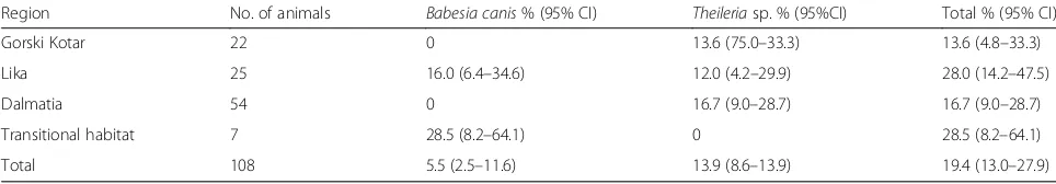
Mapping of the obtained data revealed a diverse distri-bution ofB. canis and T. capreoli (Fig. 1). The putative T. capreoli positive samples were from wolves in all studied areas except the transitional habitat while B. canis was present only in the Lika region at around 600 m above sea level and in two wolves from the transi-tional habitat. The highest prevalence of both piroplasm species was detected in Lika (28%) with the transitional habitat having a similar prevalence (28.5%), albeit the only detected species was B. canis (Table 1). During 12 months monitoring of movement activity,B. canis in-fected Wolf 2 covered area of 563 km2(Fig. 1). The pu-tativeT. capreolipiroplasms were the only ones detected in Gorski Kotar and Dalmatia with a similar prevalence of 13.6 and 16.7%, respectively (Table 1).
Discussion
One of the most important canine vector-borne diseases in Croatia is babesiosis caused mostly by B. canis [10]. Apart from domesticated dogs, B. canis has been mo-lecularly confirmed in the neighbouring countries of Bosnia and Herzegovina from a single fox [17] and two captive grey wolves from Hungary [19] as well as in a single red fox (1/91) from Portugal [14]. Since data on Babesia infection in free-ranging carnivores are scarce,
wolves (5.5%) and a captive animal from Croatia (Wolf 109). As expected, based on analysis of 18S rRNA gene, the sequences were all identical to B. canis from Cro-atian dogs and similar to other European B. canis iso-lates [27, 28]. Interestingly, the prevalence of infected wolves from the Lika region and transitional habitat was almost identical. We were previously aware that the transitional habitat was endemic forB. canisinfection in dogs [7], but the high prevalence in the more southern region of Lika was unexpected. The tick Dermacentor reticulatus, the only known vector of B. canis, was de-tected in wolves in the same region in 2009 (unpub-lished finding), fulfilling conditions for a continuous cycle ofB. canisin wildlife and efficient spreading via in-fected ticks or inin-fected animals [29]. A range of factors such as the ability of free-ranging wolves to cross a dis-tance of several hundred kilometres [30] together with transovarial transmission of B. canis withinD. reticula-tuscould facilitate the spread of the pathogen over long distances. This would appear to be confirmed in this study where Wolf 2, infected with B. canis, originated from the Lika region but moved over a large area and during a 12-month period covered 563 km2. Young indi-viduals leaving the pack and searching for new territories are another aspect of dispersing pathogens [21, 30]. Young, dispersing wolves in search for new habitat can frequently be observed in Croatia, including areas of sporadic occurrence as well as the urban north-west lowland geographic regions [20].
[image:6.595.56.538.648.732.2]Except for Wolf 2 which was still alive at sampling, all free-ranging wolves infected with B. canis died due to trauma or rabies. Regarding the clinical impact, based on clinical assessments and post-mortem investigations in this study, it seems that free-ranging wolves can toler-ate infection with B. canis, the most pathogenic piro-plasm species for canines in Europe, without the
Table 1Prevalence of infected free-ranging wolves from different geographical regions
Region No. of animals Babesia canis% (95% CI) Theileriasp. % (95%CI) Total % (95% CI)
Gorski Kotar 22 0 13.6 (75.0–33.3) 13.6 (4.8–33.3)
Lika 25 16.0 (6.4–34.6) 12.0 (4.2–29.9) 28.0 (14.2–47.5)
Dalmatia 54 0 16.7 (9.0–28.7) 16.7 (9.0–28.7)
Transitional habitat 7 28.5 (8.2–64.1) 0 28.5 (8.2–64.1)
development of clinical signs of severe haemolytic dis-ease. Supporting this assumption is the absence of clin-ical, haematological and biochemical changes consistent with canine babesiosis in Wolves 2 and 107. Even though piroplasms were proven in blood smears of these infected wolves, only discrete normochromic normocytic anaemia, thrombocytopenia and slight elevation of cre-atinine kinase were confirmed. Traumatised Wolf 107 probably showed normochromic normocytic anaemia and creatinine kinase elevation due to internal and ex-ternal blood loss and muscle and spinal cord tissue rup-ture. Although in Wolf 2, slight anaemia and thrombocytopenia could relate toB. canisinfection, ele-vated levels of serum creatinine kinase were expected due to skeletal muscle fibre damage caused by capture in a live trap with subsequent leakage of creatinine kin-ase [31].
Although only a sample size of one, the monitoring carried out in the 12 months following the capture of Wolf 2 also suggests little or no impact on the move-ment of these otherwise healthy animals. In contrast to these findings in free-ranging animals, the captive wolf (109) infected with B. canis showed typical gross and microscopic lesions characteristic for canine babesiosis following post-mortem analysis. Infection with B. canis in dogs often results in a wide range of clinical presenta-tions characterised by fever, lethargy, thrombocytopenia, anaemia, icterus, hemoglobinuria and multiple organ dysfunction syndromes with fatal outcome [32], consist-ent with postmortal findings of the captive wolf in this study. Although dogs can be asymptomatically infected, the number of such cases is very low [10]. It seems that free-ranging wild animals can tolerateBabesia infection better than their domesticated “relatives”. This phenomenon has been described in cheetahs, and do-mestic cats, where cheetahs infected withBabesia lengau stay asymptomatic while infection in domestic cats pro-gress and cause lethal cerebral babesiosis and haemolytic anaemia [33, 34]. The observation suggests that healthy, chronically infected wolves may act as reservoirs for D. reticulatus tick-mediated transmission toB. canis “free” regions and animals including domesticated dogs. A similar reservoir capacity was confirmed in experimental infection studies of young coyotes (Canis lantras) with B. gibsoni where animals were found harbouring para-sites for several months without clinical signs [35].
Another piroplasm, suggested to beTheileria capreoli by 18S rDNA sequence analysis, was an unexpected finding in the current study while also representing the first detection of this pathogen in grey wolves. The para-site was more frequently detected of the two piroplasms (13.9%), and it appears that it was homogeneously dis-tributed in all three regions except the transitional habi-tat. The 18S rDNA sequences amplified in this study
were identical to or similar to the sequence amplified from Theileria strains from red deer (3185/02; 100% similarity, AY421708) and roe deer from Spain (T. capreoli, 100% similarity AY726011), a red fox (3185/02, 99.9% similarity, HM212629) [11], red deer from Poland (ZS T04, 99.9% similarity, DQ520836) [36], and a T. capreoli strain from a wild Reeves’muntjac from China (100% similarity) [37]. Based on this study, it appears that T. capreoli or closely related Theileria sp. ZS T04, Theileria sp. 3185/02 can infect not only wild cervids but also wild canids such as red foxes and wolves. Thei-leriasp. 3185/02 was confirmed in roe deer and red deer from areas where two wolves were harbouring the same parasite (unpublished data). Detection of T. capreoli DNA in Ixodes ricinus ticks from Italy [38] raises the question whether this tick species may act as vectors for this Theileria species. In the current study, two Thei-leria-positive wolves from Gorski Kotar were found that share the same ecological niche with roe deer where I. ricinuswas the only tick species detected [39]. Other in-fected wolves originated from mountainous areas where I. ricinusis the dominant tick species [40]. Further stud-ies will be required to establish the possible role of I. ricinus as a vector for this piroplasm amongst different wildlife species.
Previous studies have shown that deer or roe deer are commonly infected withTheileria spp. without showing clinical signs [30]. Similarly, based on the observations made in this study on necropsied animals, no pathogenic effects of the putative T. capreoli infection (e.g. hemolytic disease) consistent with theilerosis was ob-served in free-ranging grey wolves. Our observations strongly suggest that the putative T. capreoli infections in free-ranging wolves are subclinical.
Conclusions
Abbreviations
CI:confidence interval; GPS: Global positioning system; HPF: high power fields; PCR: polymerase chain reaction
Acknowledgements
The authors thank Marko Poletto, DVM, for technical support.
Funding
This research was funded by GENOTICKTRECK, Croatian Science Foundation Project number: 1957 and VetMedZg, an FP7 project of the European Commission. The field work was supported by Bernd Thies Foundation,
“Plitvice Lakes”National Park, UK Wolf Conservation Trust, EURONATUR, Paradise Wildlife Park,“Sjeverni Velebit”National Park,“Velebit”Nature Park, Croatian State Institute for Nature Conservation and the Croatian Environmental Protection and Energy Efficiency Fund.
Availability of data and materials
The datasets supporting the conclusions of this article are included within the article and its additional file. Representative sequences forTheileria capreoliandBabesia canisare submitted to the GenBank database under accession numbers KY359359 and KY359360.
Authors’contributions
AB performed necropsies with sample collection, analyzed and interpreted gross, cytological and histological findings in necropsied wolves, and drafted the manuscript. DH performed necropsies with sample collection, analyzed and interpreted gross, cytologic and histological findings in necropsied wolves, performed DNA extractions and assisted with preparation of the manuscript. AP analyzed epidemiological and molecular data and assisted with drafting and editing of the manuscript. AGK performed necropsies with sample collection and the with figure preparations. VB performed hematological and biochemical analyses and data interpretation. VM performed hematological and biochemical analyses and data interpretation. JK completed fieldwork with sample collection, GPS tracking and figure preparation. SR fieldwork with sample collection. IR performed DNA extraction and molecular analyses. RB performed parasitological and genotyping analyses, epidemiological data analysis and interpretation, molecular screening and assisted with figure preparation, drafting and editing of the manuscript. All authors read and approved the final manuscript.
Competing interests
The authors declare that they have no competing interests.
Consent for publication Not applicable.
Ethics approval and consent to participate
Autorization for wolf collection was provided by the Ministry of
Environmental and Nature Protection, Zagreb, Croatia under class UP/I-612-07/15-48/47 and number 517-07-1-1-1-15-4.
Publisher’s Note
Springer Nature remains neutral with regard to jurisdictional claims in published maps and institutional affiliations.
References
1. Solano-Gallego L, Baneth G. Babesiosis in dogs and cats - expanding parasitological and clinical spectra. Vet Parasitol. 2011;181:48–60. 2. Schnittger L, Rodriguez A, Florin-Christensen M, Morrison D.Babesia: A
world emerging. Infect Genet Evol. 2012;12:1788–809.
3. Alvardo-Rybak M, Solano-Gallego L, Milian J. A review of piroplasmid infections in wild carnivores worldwide: importance for domestic animal health and wildlife conservation. Parasit Vectors. 2016;9:538.
4. Penzhorn BL. Babesiosis of wild carnivores and ungulates. Vet Parasitol. 2006;138:11–21.
5. Mans B, Pienaar R, Latif A. Areview ofTheileriadiagnostics and epidemiology. Int J Parasitol Parasites Wildl. 2015;4:104–18.
6. Zahler M, Rinder H, Schein E, Goethe R. Detection of a new pathogenic
Babesia microti-like species in dogs. Vet Parasitol. 2000;89:241–8. 7. Baneth G, Florin-Christensen M, Cardoso L, Schnittger L. Reclassification of
Theileria annaeasBabesia vulpessp. nov. Parasit Vectors. 2015;8:207. 8. Hartelt K, Rieker T, Oehme RM, Brockmann SO, Muller W, Dorn N, et al. First
evidence ofBabesia gibsoni(Asian genotype) in dogs in Western Europe. Vector Borne Zoonotic Dis. 2007;7:163–6.
9. Criado-Fornelio A, Martinez-Marcos A, Buling-Sarana A, Barba-Carretero JC. Molecular studies onBabesia,TheileriaandHepatozoonin southern Europe -Part I. Epizootiological aspects. Vet Parasitol. 2003;113:189–201.
10. Beck R, Vojta L, Mrljak V, MarinculićA, Beck A,Živičnjak T, et al. Diversity of
BabesiaandTheileriaspecies in symptomatic and asymptomatic dogs in Croatia. Int J Parasitol. 2009;39:843–8.
11. Deždek D, Vojta L,ČurkovićS, Lipej Z, Mihaljević Ž, Cvetnić Ž, et al. Molecular detection ofTheileria annaeandHepatozoon canisin foxes (Vulpes vulpes) in Croatia. Vet Parasitol. 2010;172:333–6.
12. Zanet S, Trisciuoglio A, Bottero E, de Mera IG F, Gortazar C, Carpignano MG, et al. Piroplasmosis in wildlife:BabesiaandTheileriaaffecting free-ranging ungulates and carnivores in the Italian Alps. Parasit Vectors. 2014;7:70. 13. Farkas R, Takács N, Hornyák Á, Nachum-Biala Y, Hornok S, Baneth G. First
report onBabesiacf.microtiinfection of red foxes (Vulpes vulpes) from Hungary. Parasit Vectors. 2015;8:55.
14. Cardoso L, Cortes HCE, Reis A, Rodrigues P, Simões M, Lopes AP, et al. Prevalence ofBabesia microti-like infection in red foxes (Vulpes vulpes) from Portugal. Vet Parasitol. 2013;196:90–5.
15. Barandika JF, Espí A, Oporto B, Del Cerro A, Barral M, Povedano I, et al. Occurrence and genetic diversity of piroplasms and other Apicomplexa in wild carnivores. Parasitology. 2016;2:1–7.
16. Millán J, Proboste T, de Mera IGF, Chirife AD, de la Fuente J, Altet L. Molecular detection of vector-borne pathogens in wild and domestic carnivores and their ticks at the human-wildlife interface. Ticks Tick Borne Dis. 2016;7:284–90. 17. HodžićA, AlićA, Fuehrer H-P, Harl J, Wille-Piazzai W, Duscher GG. A
molecular survey of vector-borne pathogens in red foxes (Vulpes vulpes) from Bosnia and Herzegovina. Parasit Vectors. 2015;8:88.
18. Karbowiak G, Hapunik J, Miniuk M. The case of babesiosis in farmed wolf (Canis lupusL). Wiad Parazytol. 2008;54:243.
19. Erdelyi K, Mezosi L, Vladov S, Foldvari G. Fatal acute babesiosis in captive grey wolves (Canis lupus) due toBabesia canis. Ticks Tick Borne Dis. 2013;5:281–3. 20. JeremićJ, Desnica S,Štrbenac A, HamidovićD, HuberĐ, Kusak J. Report
on the state of the wolf population in Croatia in 2014. Zagreb: Technical report of Croatian State Institute for Nature Protection; 2014. doi:10.13140/2. 1.4560.2884.
22. Kusak J, Skrbinšek AM, Huber D. Home ranges, movements, and activity of wolves (Canis lupus) in the Dalmatian part of Dinarids, Croatia. Eur J Wildl Res. 2005;51:254–62.
23. Landon DB, Waite CA, Peterson RO, Mech LD. Evaluation of age determination techniques for gray wolves. J Wildl Manag. 1998;62:674–82. 24. Thorensen SI, Arnemo JM, Liberg O. Hematology and serum clinical
chemistry reference intervals for free-ranging Scandinavian gray wolves (Canis lupus). Vet Clin Pathol. 2009;38:224–9.
25. Allsopp BA, Baylis HA, Allsopp MT, Cavalier-Smith T, Bishop RP, Carrington DM, et al. Discrimination between six species ofTheileriausing oligonucleotide probes which detect small subunit ribosomal RNA sequences. Parasitology. 1993;107:157–65.
26. Caccio SM, Antunovic B, Moretti A, Mangili V, Marinculic A. Rafaj RB, et al Molecular characterisation ofBabesia canis canisandBabesia canis vogeli
from naturally infected European dogs. Vet Parasitol. 2002;106:285–92. 27. Schaarschmidt D, Gilli U, Gottstein B, Marreros N, Kuhnert P, Daeppen JA, et
al. QuestingDermacentor reticulatusharbouringBabesia canisDNA associated with outbreaks of canine babesiosis in the Swiss Midlands. Ticks Tick Borne Dis. 2013;4:334–40.
28. Ionita M, Mitrea IL, Pfister K, Hamel D, Buzatu CM, Silaghi C. Canine babesiosis in Romania due toBabesia canisandBabesia vogeli: a molecular approach. Parasitol Res. 2012;110:1659–64.
29. Földvári G,Široký P, Szekeres S, Majoros G, Sprong H.Dermacentor reticulatus: a vector on the rise. Parasit Vectors. 2016;9:314.
30. Kaczensky P, Chapron G, von Arx M, HuberĐ, Andrén H, Linnell J. Status, management and distribution of large carnivores - bear, lynx, wolf & wolverine - in Europe. Part 1- Europe summaries. Report: 1-72. A Large Carnivore Initiative for Europe Report prepared for the European Commission. 2013. http://ec.europa.eu/environment/nature/conservation/ species/carnivores/pdf/task_1_part2_species_country_reports.
31. HuberĐ, Kusak J,Žvorc Z, Rafaj RB. Effects of sex, age, capturing method, and season on serum chemistry values of brown bears in Croatia. J Wildl Dis. 1997;33:790–4.
32. Matijatko V, KišI, Torti M, BrkljačićM, Kučer N, Rafaj RB, et al. Septic shock in canine babesiosis. Vet Parasitol. 2009;162:263–70.
33. Bosman AM, Oosthuizen EC, Peirce MA, Venter EH, Penzhorn BL.Babesia lengausp. nov., a novelBabesiaspecies in cheetah (Acinonyx jubatus, Schreber, 1775) populations in South Africa. J Clin Microbiol. 2010;48:2703–8. 34. Bosman AM, Oosthuizen MC, Venter EH, Steyl JC, Gous TA, Penzhorn BL.
Babesia lengauassociated with cerebral and haemolytic babesiosis in two domestic cats. Parasit Vectors. 2013;6:128.
35. Evers HV, Kocan AA, Reichard MV, Meinkot JH. ExperimentalBabesia gibsoni
infection in coyotes (Canis latrans). J Wildl Dis. 2003;39:904–8. 36. Sawczuk M, Maciejewska A, Skotarczak B. Identification and molecular
characterization ofTheileriasp. infecting red deer (Cervus elaphus) in northwestern Poland. Eur J Wildl Res. 2008;54:225–30.
37. Li Y, Ze C, Zhijie L, Junlong L, Jifei Y, Qian L, et al. Molecular identification of
Theileriaparasites of northwestern Chinese Cervidae. Parasit Vectors. 2014;147:225. 38. Galuppi R, Aureli S, Bonoli C, Caffara M, Tampieri MP. Detection and
molecular characterization ofTheileriasp. in fallow deer (Dama dama) and ticks from an Italian natural preserve. Res Vet Sci. 2011;91:110–5. 39. Pintur K, Beck R, BabićI, PopovićN, FlorijančićT, Krapinec K, et al. Tick
infestation in roe deer from Gorski Kotar area, Croatia. Proceedings of the 47th Croatian and 7th International Symposium on Agriculture; 2012. ISBN: 978-953-7878-05-4.
40. Krčmar S. Hard ticks (Acari, Ixodidae) of Croatia. ZooKeys. 2012;234:19–57.
• We accept pre-submission inquiries
• Our selector tool helps you to find the most relevant journal • We provide round the clock customer support
• Convenient online submission • Thorough peer review
• Inclusion in PubMed and all major indexing services • Maximum visibility for your research
Submit your manuscript at www.biomedcentral.com/submit