0022-538X/05/$08.00⫹0 doi:10.1128/JVI.79.1.289–298.2005
Copyright © 2005, American Society for Microbiology. All Rights Reserved.
The Infectivity and Lytic Activity of Minute Virus of Mice Wild-Type
and Derived Vector Particles Are Strikingly Different
Susanne I. Lang,† Stephanie Boelz, Alexandra Y. Stroh-Dege, Jean Rommelaere,
Christiane Dinsart, and Jan J. Cornelis*
Applied Tumor Virology Program, Deutsches Krebsforschungszentrum, Heidelberg, Germany
Received 21 April 2004/Accepted 24 August 2004
Gene therapy vectors have been developed from autonomous rodent parvoviruses that carry a therapeutic gene or a marker gene in place of the genes encoding the capsid proteins. These vectors are currently evaluated in preclinical experiments. The infectivity of the vector particles deriving from the fibroblastic strain of minute virus of mice (MVMp) (produced by transfection in human cells) was found to be far less (approximately 50-fold-less) infectious than that of wild-type virus particles routinely produced by infection of A9 mouse fibroblasts. Similarly, wild-type MVMp produced by transfection also had a low infectivity in mouse cells, indicating that the method and producer cells influence the infectivity of the virus produced. Interestingly, producer cells made as many full vector particles as wild-type particles, arguing against deficient packaging being responsible for the low infectivity of viruses recovered from transfected cells. The hurdle to infection with full particles produced through transfection was found to take place at an early step following entry and limiting viral DNA replication and gene expression. Infections with transfection or infection-derived virus stocks normalized for their replication ability yielded similar monomer and dimer DNA amplification and gene expression levels. Surprisingly, at equivalent replication units, the capacity of parvovirus vectors to kill tumor cells was lower than that of the parental wild-type virus produced under the same transfection conditions, suggesting that beside the viral nonstructural proteins, the capsid proteins, assembled capsids, or the corre-sponding coding region contribute to the lytic activity of these viruses.
Minute virus of mice (MVM) and H-1 virus belong to the group of autonomous parvoviruses (single-stranded nuclear-replicating DNA viruses) that are endogenous to vertebrates including humans (16). Autonomous parvoviruses are lytic vi-ruses that replicate during the S phase of the cell cycle. The mouse parvovirus MVM and the rat virus H-1 were found to preferentially replicate in and kill oncogenic-transformed and tumor-derived cells of rodent and human origin (for a recent review, see reference 33). Furthermore, a number of reports have described that the infection of laboratory animals with several rodent parvoviruses was associated with the suppres-sion of tumor formation or the rejection of tumor grafts (33). As a consequence, certain of these parvoviruses, including MVM and H-1 virus, have been developed as antitumor vec-tors with the aim of strengthening the antineoplastic effect of the natural parvoviruses. In particular, antitumor vectors car-rying a therapeutic transgene in place of the gene encoding the capsid genes were developed (4, 10, 17, 25, 34). Several of these capsid replacement vectors displayed stronger antineo-plastic effects than the corresponding wild-type or control re-combinant viruses in various mouse and human tumor models (12, 14, 41).
The above-mentioned capsid replacement vectors have thus retained the strong parvovirus early P4 promoter, the
overlap-ping sequences encoding the small nonstructural (NS) viral regulatory protein NS2, and the large viral replication and regulatory protein NS1. The latter polypeptide enables the vector to replicate its genome and totrans-activate the second promoter (P38) that controls expression of the inserted trans-gene (10, 17, 25, 34). For the cell-killing activity of autonomous parvoviruses, including human parvovirus B19, the viral gene product NS1 has been found to be essential (6, 18, 24, 29, 31). Besides the interest in them as antitumor tools, the use of vectors transducing a reporter gene would be of great help for studies aimed at understanding the susceptibility of cells or tissues to parvovirus infection. Yet despite the fact that the first autonomous parvovirus vectors were described about a decade ago (22, 34), little is known about their infectivity and physical properties compared with the wild-type virus. Hence, it is not known whether the above-mentioned vectors have the same stability or efficiency to infect, replicate in, and express viral genes and kill the tumor cells as the parental wild-type virus. Analysis of the transgene itself, the destruction of the capsid gene, or the way virus is produced may uncover as-yet-uniden-tified functions of the viral genome or the capsid that are important for a particular step of the virus cycle. In addition, since contacts were reported between the genome and partic-ular amino acids at the capsid interior (7), the transgene may have an impact on the capsid structure and thus on virus infectivity.
This study was undertaken with the aim to determine how far parvovirus capsid replacement vectors can substitute for wild-type virus. It was observed that MVMp-derived vectors that are routinely produced in transfected human cells were far less infectious than wild-type MVMp produced by infection in
* Corresponding author. Mailing address: Applied Tumor Virology, Abteilung F010 and INSERM U375, Deutsches Krebsforschungszen-trum, Postfach 101949, D-69009 Heidelberg, Germany. Phone: 49 6221 424965. Fax: 49 6221 424962. E-mail: [email protected].
† Present address: Molecular Medicine Partnership Unit, Verfu ¨-gungslabor der Kinderklinik-Abteilung III, 69009 Heidelberg, Ger-many.
289
on November 8, 2019 by guest
http://jvi.asm.org/
A9 mouse cells, cells that are conventionally used to titer MVMp. Our data show that not only recombinant viruses but also wild-type particles produced by transfection in human cells have a low infectivity in mouse A9 cells, an observation that could be traced back to a step of the parvovirus life cycle that takes place prior to viral DNA amplification and gene expression. Moreover, both H-1 virus and MVMp-derived vec-tors were found to be less toxic for the host cells than the corresponding parental wild-type viruses under conditions that allowed equal expression of transduced cytotoxic NS genes. This is the first report that shows that parvovirus infectivity is modulated as a function of the production method and the maintenance of capsid genes.
MATERIALS AND METHODS
Cells. 293T clone 17 (simian virus 40-transformed human HEK293 cells), HeLa (human cervical cancer cells), and Gl261 (mouse glioma cells) cells were maintained in Dulbecco’s modified Eagle’s medium (Life Technologies, GIBCO-BRL, Paisley, United Kingdom) supplemented with 10% fetal calf serum, 2 mM
L-glutamine, and antibiotics. The A9 strain of mouse L cells and NB324K cells
(simian virus 40-transformed newborn human kidney fibroblast cells) were grown in Eagle’s minimal essential medium (GIBCO-BRL) with 5% fetal calf serum, glutamine, and antibiotics.
Virus infection and production.All virus infections were performed for 1 h at 37°C with a small virus inoculum and occasional rocking of the plates. Unless mentioned otherwise, wild-type MVMp virus was derived from virus-infected (multiplicity of infection [MOI] of 0.003 PFU per cell) A9 fibroblasts (9). Wild-type MVMp was also produced by transfection of 293T cells with the molecular clone pdBMVp (17). Recombinant MVMp-derived viruses were routinely pro-duced in 293T cells by cotransfection of recombinant viral DNA clones and a capsid protein-providing helper plasmid (pCMV-VP) by means of a Ca-phos-phate precipitation technique (17, 42). Lipofectamine 2000 (Invitrogen, Karlsruhe, Germany) was used as a transfection agent in two experiments. In one single experiment, two vectors were produced by transfection of NB34K cells. The various wild-type and vector stocks were numbered (see Table 1 and all figures).
The construction of the vector DNA clone pMVMp/MDC transducing the human macrophage-derived cytokine was performed as follows. A 288-bp DNA fragment containing the whole coding sequence of human macrophage-derived chemokine (MDC) was amplified by PCR using primers containing BamHI and SmaI linkers. The PCR product was cloned into pMVMp⌬800 vector DNA (17) digested with BamHI and SmaI restriction enzymes, generating the vector clone pMVMp/MDC. The recombinant virus MVMp/IL-2 carrying human interleukin 2 (IL-2) cDNA was produced from the DNA clone pMVMp/IL-2. The latter plasmid was constructed by inserting the 516-bp-long human IL-2 cDNA (14) into the multiple cloning site of pMVMp⌬800 (17). Similarly, the coding
se-quence of the enhanced green fluorescent protein (EGFP) was inserted into the empty vector clone pMVMp⌬800, generating pMVMp/EGFP. The construction of the vectors transducing the cDNAs of the human chemokines macrophage-inducible protein 1␣(MIP1␣) and its truncated derivative LD78will be re-ported elsewhere. The wild-type H-1 virus and its recombinant derivative trans-ducing human IL-2 cDNA were prepared as previously described (14). The genomes of all viral vectors described are, with the exception of the empty vector MVMp⌬800, close to the size of the corresponding wild-type genome. The viral genome of pMVMp/EGFP has the same size as the wild-type virus, whereas pMVMp/IL-2 is 6% and pMVMp/MDC, ChiMVMp/MIP1␣, and Chi-MVMp/ LD78are 10% smaller than the wild-type genome. The size of MVMp⌬800 is 84% of that of the wild-type.3
H-labeled wild-type and IL-2-transducing viruses were produced as described previously by adding [3
H]thymidine to the medium during the virus production at intervals (8). Unless indicated, stocks of wild-type and vector viruses devoid of empty capsids were obtained by purification over iodixanol gradients (44). The contamination of the vector stocks with replication-competent virus was determined by plaque assays (see below) and ranged from 0.01 to 0.1%.
Virus titrations.Infectious titers of recombinant and wild-type viruses were determined from infected cell hybridization assays performed on A9 and NB324K indicator cells for MVMp-based and H-1 parvovirus-based viruses, respectively, and expressed as replication units (RU) per milliliter (17). An NS-specific DNA probe consisting of the 1,638-bp NcoI fragment of the plasmid pMVMp⌬800 was used for this purpose. Genome titers of the various virus stocks were determined by quantitative dot blot hybridization assays (13), and the total number of full particles per milliliter of virus suspension was deter-mined. In short, virus suspensions were digested with 25 U of DNase I (Promega, Mannheim, Germany)/ml for 1 h followed by a 2-h incubation with 250g of proteinase K (Roche, Mannheim, Germany)/ml at 37°C, and genomic DNA was isolated by phenol-chloroform-isoamylalcohol (25:24:1) extraction. DNA was transferred onto a nylon membrane (GeneScreenPlus; PE Applied Biosystems, Langen, Germany) in fivefold dilutions and hybridized against a radioactive NS-specific probe (see above). Twofold dilutions of a vector plasmid preparation were used as a standard. The contamination of the various vector stocks with replication-competent virus was determined by plaque assays on A9 indicator cells (42). Virus infectivity was expressed by the particle-to-infectivity (P/I) ratio, the quotient of genome and infectious titers.
Analysis of packaged viral DNA. To determine the size of packaged viral genomes, virus samples were first treated with DNase I and then digested with proteinase K and processed by viral DNA extraction as described for dot blot analysis (see above). The DNA samples were electrophoresed through 0.8% agarose gels, blotted onto nitrocellulose membranes, and hybridized against a radioactive NS probe.
Virus uptake and internalization.Cell-associated virus was determined im-mediately after the 1-h infection period with3H-labeled virus by measuring the
percentage of cell-bound input radioactivity (8).
[image:2.585.50.541.80.201.2]Susceptibility of cell-bound viral DNA to extraction.To determine the sus-ceptibility of internalized virus to DNA extraction, A9 cells were infected with wild-type or vector virus at 105genome equivalents per cell. Cells were further
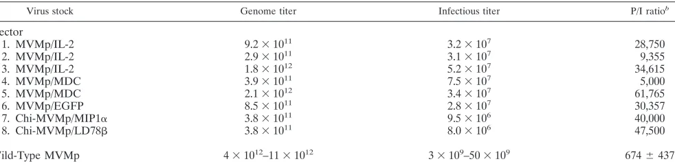
TABLE 1. Infectivity of wild-type MVMp and derived vector particlesa
Virus stock Genome titer Infectious titer P/I ratiob
Vector
1. MVMp/IL-2 9.2⫻1011 3.2⫻107 28,750
2. MVMp/IL-2 2.9⫻1011 3.1⫻107 9,355
3. MVMp/IL-2 1.8⫻1012 5.2⫻107 34,615
4. MVMp/MDC 3.9⫻1011 7.5⫻107 5,000
5. MVMp/MDC 2.1⫻1012 3.4⫻107 61,765
6. MVMp/EGFP 8.5⫻1011 2.8⫻107 30,357
7. Chi-MVMp/MIP1␣ 3.8⫻1011 9.5⫻106 40,000
8. Chi-MVMp/LD78 3.8⫻1011 8.0⫻106 47,500
Wild-Type MVMp 4⫻1012–11⫻1012 3⫻109–50⫻109 674⫾437
a
Compilation of genome and infectious titers from eight independently produced MVMp wild-type and eight recombinant virus stocks is shown. The genome titer is expressed as the number of full particles per milliliter of virus suspension. The infectious titer is given as replication units per milliliter of virus stock. The particle infectivity is defined as the P/I ratio, the quotient of genome and infectious titers. The average P/I ratios of the wild-type MVMp and vector stocks are given with their standard deviations (around 60%). From the averaged P/I values, vector particles are 48-fold less infectious than the wild-type particles. This difference is highly significant(P⬍0.001; Student’sttest).
b
The total P/I ratio for recombinant virus stocks was determined to be 32,168⫾18,715.
on November 8, 2019 by guest
http://jvi.asm.org/
incubated for 2 h and then lysed in 0.6% sodium dodecyl sulfate–10 mM Tris-HCl (pH 7.4)–10 mM EDTA. DNA was extracted by a modified Hirt procedure consisting of proteinase K digestion and selective extraction of low-molecular-weight DNA (10). DNA was electrophoresed through agarose gels, blotted onto nitrocellulose membranes, and hybridized against an NS probe as described above.
Viral genome amplification.Overall viral DNA amplification was determined 30 h postinfection (p.i.) by dispersed cell assays (DCA). In short, 2⫻105
infected cells from a 6-cm-diameter dish were transferred onto a nitrocellulose filter (pore size, 45m; Schleicher & Schu¨ell, Dassel, Germany) and subsequently hybrid-ized against a radioactive NS probe, as described above for virus titration. The radioactivity bound to the filters was measured with a liquid scintillation counter. DNA amplification was also monitored in A9 cells by Southern blot analysis after extraction of low-molecular-weight DNA by the modified Hirt procedure as described above.
Cytotoxicity assays.The cytotoxic effect of parvovirus infection was assessed by colony formation assays (9) as well as Alamar Blue and lactate dehydrogenase (LDH) tests. For the latter two assays, cultures were inoculated with virus at various MOIs, and the survival of the cells was determined 3 days later. Alamar Blue reagent (BioSource Europe, Nivelles, Belgium) is an oxidation-reduction indicator that fluoresces red when it accepts electrons generated through cellular metabolism. Cells grown in 96-well plates (100l of medium per well) were stained with 10l of dye per well. After a 3-h incubation at 37°C, color changes were determined through a double-wavelength (540 and 620 nm) measurement. For LDH tests, a CytoTox96 kit (Promega) was used according to the instruc-tions of the manufacturer. Cell survival was expressed as a percentage of the values measured for noninfected cultures.
Contamination of the vector stocks with replication-competent viruses (14, 42) was too low to have an impact on the cytotoxicity of the vector stocks. Expression of human IL-2 or MDC cytokines did not affect the growth or the survival of the infected cell cultures (data not shown).
Immunoblotting. Cells inoculated with wild-type (MVMp) or recombinant (MVMp/IL-2 and MVMp/MDC) viruses at an MOI of 3 RU/cell were harvested 48 h p.i. After cell lysis, samples were electrophoresed through sodium dodecyl sulfate–10% polyacrylamide gels and electrotransferred to nitrocellulose mem-branes. The blots were probed with the NS1-specific rabbit antiserum SP7 (di-lution, 1:5,000) (5) and processed according to the Amersham (Freiburg, Ger-many) enhanced chemiluminescence protocol.
Immunofluorescence.A9 cells were infected with wild-type or recombinant MVMp virus (103
genome equivalents per cell) and transferred at 6 h p.i. onto multispot slides. After a 48-h incubation period, cells were fixed in
paraformal-dehyde and treated with 0.2% Triton X-100 for 5 min. NS1 present in infected cells was detected by using the mouse monoclonal antibody 3D9 at a 1:40 dilution (3) and visualized with a 1:200 dilution of Cy3-conjugated anti-mouse immuno-globulin G. Stained cells were covered with mounting medium (Vectashield) and examined with a Leica fluorescence microscope.
RESULTS
Viruses produced through transfections are less infectious
than those recovered from infected cells.The parallel infection
of A9 fibroblasts with wild-type MVMp and vector particles at an equal particle-to-cell ratio showed that almost no vector-infected cells were positive for NS1 (Fig. 1, left panel), whereas a majority of wild-type-infected cells expressed detectable amounts of the viral product (right panel). This observation indicated that vector particles were less infectious for A9 cells than wild-type particles. It is noteworthy that in our laboratory, recombinant viruses are routinely produced through cotrans-fections of 293T cells because this cell line yielded the highest vector titers among a series of cells assessed for their produc-tion capacity (data not shown). In contrast, wild-type MVMp stocks are conventionally prepared through infection of A9 cells (9, 19, 36).
In order to confirm these variant virus stock infectivities, the genome titers of eight independently produced wild-type vi-ruses and eight vector batches were determined by dot blot hybridization, and their infectivity was measured by using an infected cell hybridization assay, a technique which is based on the ability of these viruses to amplify their genome in standard indicator A9 cells. The results of these titrations are compiled in Table 1. Virus infectivity was expressed as the ratio of DNA-containing particles to infectious (i.e., DNA replication-competent) virus (P/I). A high P/I ratio thus corresponds to low particle infectivity. Of note, the capsid replacement
recom-FIG. 1. Differential expression of NS1 from wild-type and vector viruses. A9 cells were infected with the same number of wild-type (P/I, 380) or MVMp/IL-2 (Table 1, vector 2) virus particles (103genome equivalents per cell) and analyzed 48 h later for expression of NS1 proteins as
detected by fluorescence microscopy. From the number of NS1-positive cells from several pictures, the vector particle infectivity was calculated to be 22-fold less than that of the wild-type virus. This corresponds to the difference in particle infectivity of the stocks used for infection.
on November 8, 2019 by guest
http://jvi.asm.org/
binant vectors used in this study could all be produced in relatively high titers, in keeping with the fact that overall ge-nome size was close to that of the wild-type virus (4, 17). In some experiments, the empty vector MVMp⌬800 virus was also included, although its titer was consistently found to be lower than that of the vectors listed in Table 1. The genome titers of recombinant and wild-type MVMp virus preparations were about 3.2⫻ 104 and 6.7 ⫻ 102 times higher than the infectious titers, respectively. Thus, around 50 times more vec-tor particles than wild-type particles were needed to achieve the same virus DNA replication in A9 cells. The P/I ratio that was obtained for wild-type MVMp produced through A9 cell infection (674) was close to the values previously published for this virus (19, 20, 36).
These observations raised the question of whether the vari-ations observed in viral infectivity could be traced back to differences in either the production procedure or the virus type. To this end, the wild-type virus was produced in the same way as the vectors, i.e., through transfection of 293T cells with an infectious DNA clone (Table 2). The data from Table 2 show virus-vector productions that were performed in parallel and arrested 30 h posttransfection in order to avoid secondary rounds of wild-type virus infection. Not unimportant for our understanding of the packaging process, the results clearly show that wild-type and vector stocks that are produced in parallel by transfection contain a similar proportion of full virus particles. Hence, the packaging of viral genomes is un-likely to constitute a rate-limiting step in the production of
functional recombinant viruses compared to wild-type MVMp viruses. Interestingly, the specific infectivities of recombinant and wild-type viruses produced through transfection of 293T cells were both considerably reduced in comparison with that of wild-type virus produced through infection of A9 fibroblasts (compare Tables 1 and 2). Although the P/I ratios of the wild-type virus preparations were lower than those of the vec-tor stocks (Table 2), this difference was not statistically signif-icant.
The low infectivity of transfection-yielded virus is not a unique feature of 293T producer cells or of the IL-2-transduc-ing vector that was used in most experiments. Indeed, two different vectors produced through transfection of another hu-man producer cell line (NB324K) were found to be 30-fold less infectious than wild-type MVMp recovered from infected A9 cells (Table 3). Moreover, high P/I ratios of vector particles were also observed after transfection of 293T or NB324K cells by using either Lipofectamine or Ca-phosphate precipitation (Table 3). Altogether, these data indicate that the vector and wild-type virion infectivities depend mainly on the way pro-ducer cells are supplied with the original infectious material (DNA uptake or virus infection) and only to a low extent on the nature of producer cells and viruses. In support of this conclusion, NB324K or 293T cell infection with wild-type MVMp yielded progeny viruses whose infection capability was high and corresponded to that of virions recovered from A9-infected cells (data not shown).
From the data presented in Tables 1 and 2, one may expect that by infecting producer cells with less-infectious wild-type virus particles, progeny virus with a high infectivity for A9 cells will be produced. This was indeed the case, as the further infection of A9 cells with plasmid-derived MVMp generated virus particles that were highly infectious (Table 4).
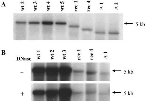
[image:4.585.44.283.90.192.2]The packaging of a greater proportion of incomplete viral genomes may account for the lower infectivity of transfection-produced virus stocks compared to infection-transfection-produced virus stocks. This possibility was not supported, however, by the analysis of the viral DNA extracted from transfection-derived vectors and infection-derived wild-type viruses. As illustrated in Fig. 2A, the genomes of the various virions had the length expected from their respective DNA substitutions. The wild-type particles carry the largest genome, followed by MVMp/ IL-2, MVMp/MDC, and empty vector MVMp⌬800. Samples of the various virus stocks were also treated with DNase prior to viral DNA extraction in order to determine whether they differed in the accessibility of packaged genomes to the nucle-ase. As shown in Fig. 2B, the DNase treatment did not lead to a preferential degradation of vector DNA, indicating that the
TABLE 2. Production of wild-type and vector viruses through transfection of 293T cellsa
Expt Virus Genome titer Infectious titer P/I ratio
1 MVMp 3.7⫻1011 9.8⫻106 3.8⫻104
2 MVMp 6.4⫻1011 1.5⫻107 4.3⫻104
3 MVMp 9.7⫻1011 7.9⫻106 1.2⫻105
4 MVMp 1.4⫻1012 2.3⫻107 6.1⫻104
1 MVMp/IL-2 9.2⫻1011 4.5⫻106 2.0⫻105
2 MVMp/IL-2 5.1⫻1011 4.8⫻106 1.1⫻105
3 MVMp/IL-2 2.3⫻1011 4.4⫻106 5.2⫻104
4 MVMp/MDC 1.1⫻1012 1.5⫻107 7.3⫻104
aA total of 2⫻106293T cells were transfected with 6g of either virus
molecular clone pMVMp/IL-2 or pMVMp/MDC together with 12g of control or helper plasmid, respectively. Treated cells were harvested 30 h posttransfec-tion. Crude extracts (experiments 1 to 3) in 5⫻10⫺2M Tris-HCl–5⫻10⫺3M
EDTA (pH 8.7) were titrated by hybridization assay on A9 indicator cells (in-fectious titers) and by dot blot analysis (genome titers). Experiments 1 to 3 were performed with the same plasmid preparations. The P/I ratio of the various wild-type (6.6⫻104) and vector (1.1⫻105) stocks is not statistically significant
(P⬎0.2; Student’sttest).
TABLE 3. P/I ratios of recombinant MVMp viruses produced by Ca-phosphate or Lipofectamine transfection of NB324K or 293T cellsa
Cell line and virus Method Genome titer Infectious titer P/I ratio
NB324K cells
MVMp/IL-2 CaPO4 1.5⫻1010 7.0⫻105 2.1⫻104
MVMp/MDC CaPO4 1.7⫻10
9 9.0⫻104 1.9⫻104
MVMp/IL-2 Lipofectamine 1.7⫻1010 8.5⫻105 2.0⫻104
293T cells
MVMp/MDC CaPO4 1.1⫻1012 1.5⫻107 7.3⫻104
MVMp/MDC Lipofectamine 5.3⫻1011 1.5⫻107 3.5⫻104
aExperimental conditions were as described for Table 2 except that the virus preparations were purified over iodixanol gradients. CaPO
4, Ca-phosphate.
on November 8, 2019 by guest
http://jvi.asm.org/
[image:4.585.43.541.630.715.2]viral particles assembled in infected cells provided their ge-nome with a similar protection against nuclease digestion com-pared to those assembled in transfected cells. Furthermore, aliquots of wild-type and vector virus stocks were compared for their sensitivity to heat. No differential susceptibility to this treatment (up to 70°C) was observed, as infectious titers were similarly reduced (data not shown). Likewise, the hemaggluti-nation capacity of particles from three wild-type virus prepa-rations did not significantly differ from that of three vector stocks (data not shown).
Viral gene amplification and gene expression.Experiments
were carried out to pinpoint the step of the viral life cycle that determined the lower infectivity of viruses produced through transfection compared to that produced through infection. In a first step, wild-type MVMp from transfected 293T cells and from infected A9 or 293T cells showing more than a 50-fold difference in infectious titers were inoculated to NB324K cells at an equivalent infectious MOI (i.e., DNA replication com-petent) of 10 RU per cell. Overall viral genome amplification was determined 30 h p.i. by DCA. As apparent from Fig. 3A, similar levels of viral DNA amplification were achieved by these three virus stocks under these conditions, suggesting that the intrinsically lower infectivity of viruses obtained through transfection resulted from the impairment of an early step(s) of the viral life cycle, limiting the onset of viral DNA replica-tion but not the amplificareplica-tion of replicative intermediates. Per-missive cells (A9 and NB324K) were infected as described above with wild-type or recombinant MVMp at the same mul-tiplicity of infectious particles, i.e., under conditions erasing the influence of the protocol used to produce viruses. Overall viral genome amplification was again determined 30 h p.i. by DCA. At all MOIs tested, hybridization signals were much higher for the wild-type virus than for the vector virus (Fig. 3B). Similar results were obtained with wild-type H-1 virus and derived vectors in NB324K cells and in several permissive cell lines that are currently used in our laboratory (data not shown). Genome amplification was also determined at 55 h p.i. The extension of the time between virus infection and analysis did not improve the capacity of recombinant genomes to be amplified in A9 cells (data not shown).
A possible reason for the lower production of viral DNA in vector-infected cells may be traced back to the failure of re-combinant particles to produce capsid proteins. Indeed, the capsid or its immediate precursors drive the packaging-cou-pled strand displacement reaction that releases the progeny genome and regenerates a double-stranded intermediate which serves as a substrate for further DNA amplification (30, 32, 40). This defect was verified by Southern blotting analysis allowing viral single-stranded DNA and duplex intermediates to be detected separately. As shown in Fig. 3C, this analysis substantiated the overall greater replication capacity of wild-type virus compared to vector virus as observed in DCAs. While monomer and dimer replicative intermediates were pro-duced by either type of virus, the main qualitative difference between wild-type and vector virus infection was at the level of the single-stranded DNA that was present in the former but undetectable in the latter. When inoculated at equivalent in-fectious doses, wild-type MVMp and two derived recombi-nants achieved similar levels of NS1 protein expression in either A9 or NB324K cells (Fig. 4). This result was consistent with the similar capacity of both types of viruses for accumu-lating monomer- and dimer-length replicative forms in these cells.
Vectors have a lower toxicity than corresponding wild-type
viruses at similar infectious doses.Autonomous parvoviruses
[image:5.585.42.285.444.613.2]are lytic viruses. The capacity of wild-type and recombinant viruses to kill in vitro-transformed cells and tumor cells was determined by using several methods. Alamar Blue staining confirmed the high susceptibility of A9 cells to killing by wild-type MVMp. Surprisingly, the vector was much less cytotoxic
TABLE 4. Infection-mediated particle infectivity switch of plasmid-derived wild-type virusa
Virus and stock Genome titer Infectious titer P/I ratio
Infection-derived MVMp
Initial stock 5.7⫻1012 6.4⫻109 8.9⫻102
Stock after reinfection 5.8⫻1011 9.0⫻108 6.4⫻102
5.3⫻1011 1.3⫻109 4.1⫻102
6.1⫻1011 1.2⫻109 5.1⫻102
Plasmid-derived MVMp
Initial stockb 1.4⫻1012 2.3⫻107 6.1⫻104
Stock after infection 5.5⫻1011 7.5⫻108 7.3⫻102
5.9⫻1011 4.9⫻108 1.2⫻103
5.0⫻1011 4.9⫻108 1.0⫻103
aWild-type virus was produced by the parallel infection of A9 cells either with
a wild-type virus inoculum derived from a highly infectious infection-derived virus preparation or from a plasmid-derived virus stock. Virus was harvested 5 days postinfection, and stocks were concentrated over iodixanol gradients. Av-erage P/I ratios of stocks produced from infection- and transfection-derived virus are 5.2⫻102and 9.8⫻102, respectively.
bThis stock is identical to the preparation described for experiment 4 of Table
2.
FIG. 2. Size and DNase resistance of packaged viral genomes. DNA was extracted from 1010full particles (A) or from 10-l aliquots
of virus stocks (B), electrophoresed through agarose gels, blotted onto filters, and hybridized to a specific probe. In panel B, viral particles were treated (⫹) or not (⫺) with DNase I (0.025 U/l) prior to viral DNA extraction. wt 1 to wt5, independent stocks of wild-type MVMp; rec 1, MVMp/IL-2; rec 4, MVMp/MDC (Table 1);⌬1 and⌬2, two independent stocks of the empty vector MVMp⌬800. The differences in densities (B) correspond to the 12-fold-higher genome titers of the virus stocks. Migration of the genomes correspond to their respective sizes; rec 1, rec 4, and both⌬1 and⌬2 are 94, 90, and 84% of the size of the wild-type genome, respectively.
on November 8, 2019 by guest
http://jvi.asm.org/
than the wild-type virus, even when used at a 10-fold-higher multiplicity of infectious particles (Fig. 5A). Owing to the rel-atively high MOIs used, the greater killing effect of the wild-type virus in A9 cells was unlikely to result from its ability to generate progeny particles that could reinfect cells which es-caped the primary infection. As expected from the results discussed above, wild-type particles derived from transfected 293T cells were as toxic as infection-derived virus when as-sessed at the same infectious multiplicity (data not shown). In order to confirm the differential cytotoxicity of wild-type and recombinant viruses by using another endpoint, the release of LDH from infected cells was determined. The latter assay substantiated the results obtained with the Alamar Blue test (Fig. 5B).
[image:6.585.173.411.68.259.2]To confirm this important observation, the ability of cells to form visible colonies after their infection with MVMp or H-1 virus or their respective vector derivatives was determined by
FIG. 3. Viral genome amplification in A9 and NB324K cells. Cultures were infected with wild-type or recombinant MVMp at a multiplicity of 10 (A) or 3 (C) RU/cell or at different MOIs (B). After incubation for 30 h, infected cultures were analyzed by either DCA for the measurement of the total amount of viral DNA (A, B) or Southern blotting for the identification of replication intermediates (C). Panels A and B: open bars, wild-type virus (wt 1; P/I ratio, 900); striped bar, MVMp wild-type virus stock obtained by infection of 293T cells (P/I ratio, 650); grey bar, wild-type virus from transfection of 293T cells with pdBMVp (P/I ratio, 5⫻104); filled bars, MVMp/IL-2 vector (rec 2). Panel C: ssDNA, single-stranded
DNA; mRF, monomer-length replication forms; dRF, dimer-length replication intermediate. wt 1 to wt3, independent stocks of wild-type MVMp virus; rec 1, MVMp/IL-2; rec 4, MVMp/MDC;⌬1, MVMp⌬800. Values are given with their standard deviations. The viral DNA amplification levels achieved by the various wild-type preparations (A) are not significantly different (P ⬍0.05; Kolmogoroff-Smirnoff test). Viral DNA amplification levels of wild-type and vector preparations (B) are highly significant (P⬍0.001; Student’sttest).
[image:6.585.361.481.363.602.2]FIG. 4. NS1 expression from wild-type and vector virus-infected cells. A9 and NB324K cells were infected with wild-type MVMp (wt 2) and the vectors MVMp/IL-2 (rec 1) and MVMp/MDC (rec 4) at an MOI of 3 RU/cell. NS1 expression was determined 48 h p.i. by immu-noblotting.
FIG. 5. Cytotoxic activity of wild-type parvovirus and derived vec-tors in various susceptible human and mouse cells. The cytotoxic ac-tivity of the wild-type parvoviruses MVMp and H-1 virus and derived vectors was determined by using Alamar Blue staining (A), LDH tests (B), and clonogenicity assays (C). Wild-type MVMp (wt 2) and the vector MVMp/IL-2 (rec 1) were used for all experiments using A9 (A to C), NB324K (C), and Gl261 (C) cells, while HeLa cells were in-fected with wild-type H-1 virus or hH1/IL-2 vector (C). The MOI varied as indicated in A and B or was set at 5 RU/cell (C). Open bars, wild-type viruses; filled bars, recombinant viruses. Survival is expressed as the percentage of uninfected control cultures. Values are given with their standard deviations. Differences between the wild type and vector are highly significant (P⬍0.001; Student’sttest).
on November 8, 2019 by guest
http://jvi.asm.org/
[image:6.585.73.254.584.674.2]colony formation assays. In this assay too, the vector viruses proved to be less toxic for the host cells than the corresponding wild-type viruses (Fig. 5C). Parallel to the clonogenicity assays, larger numbers (105) of infected cells were seeded per 5-cm-diameter dish and inspected daily by light microscopy. The latter analysis showed that the virus-infected cells died, indi-cating that the results from this and the other viability tests were due to cytotoxicity and not to cytostatic effects of the viruses. Among the tested cells (Fig. 5C), HeLa cervical car-cinoma and Gl261 mouse glioma cells do not produce detect-able amounts of infectious H-1 and MVMp progeny viruses, respectively (our unpublished data). This result rules out the possibility that the greater sensitivity of these cells to wild-type viruses compared to vector viruses results from the ability of the wild-type virus to spread and induce secondary infections. Clearly, HeLa and Gl261 cells are both highly susceptible to the killing effect of wild-type viruses but far less susceptible to the killing effect of the recombinant viruses tested at the same infectious titer.
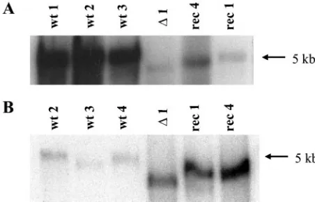
Similar virus entry but varying susceptibility of
intracellu-lar viruses to mild viral DNA extraction. The
above-men-tioned results indicate that the number of virions to be inoc-ulated to achieve a given level of viral DNA replication and expression was much higher for particles recovered from trans-fected cells than that for particles recovered with intrans-fected cells. Hence, virions produced in transfected cells may undergo a block to successful infection early in the virus cycle, prior to or at the onset of viral DNA amplification and expression. In order to identify this block, cell-associated radioactivity from [3H]thymidine-labeled wild-type and IL-2-transducing virions was determined under the standard infection conditions (8). It was found that around 20% of input radioactivity from both wild-type and vector particles were cell associated immediately p.i. (data not shown), a value that was similar to previous determinations (8). Furthermore, almost all of the cell-bound radioactivity was by that time (1 h after the start of the infec-tion) resistant to elution by 1 mM EDTA (19, 20). Thus, these results strongly suggest that the reduction of the infectivity of transfection-derived viruses can be assigned to a large extent to a step between internalization and genome amplification. This led us to assess whether the uncoating of vector particles dur-ing the infection process was less efficient than that of wild-type viruses. To this end, the Hirt extraction method, which is in-efficient in releasing genomic DNA from full particles, was utilized. Virus DNA was extracted 2 h p.i., run through agarose gels, and analyzed by Southern blotting. Yet when cells were infected at the same particle-to-cell ratio, different amounts of viral DNA were recovered. In fact, a large difference between both types of viruses was detected when the intracellular free viral DNA contents were measured. Indeed, on average, 20 times more viral DNA was recovered from cells infected with wild-type virus than that recovered with vector virus (Fig. 6A). The comparison was also made after infection with equivalent numbers of infectious particles. For the viruses used in this experiment, 67 times more vector particles than wild-type par-ticles were needed to achieve the same number of replication centers in A9 cells. The free DNA signal given by vector-infected cells was also much more predominant and yet was only sixfold higher than the one detected with wild-type virus-infected cells (Fig. 6B). Altogether, these results strongly argue
for a preferential release of wild-type DNA compared to vec-tor DNA from incoming infection-generated (as opposed to transfection-generated) particles during the extraction proce-dure. These observations lead us to speculate that the varying sensitivity of viral DNA to the Hirt extraction could reflect a differential intracellular particle susceptibility to destabilizing enzymes, suggesting that vector particles are more rigid than wild-type virions.
DISCUSSION
[image:7.585.307.533.71.216.2]Approximately 1 out of 7⫻102wild-type MVMp particles was able to successfully infect A9 cells, resulting in viral DNA amplification and gene expression. This particle-to-infectivity ratio is close to values previously reported for MVMp (19, 20, 36) and the related H-1 virus (26; our unpublished data). In contrast, vector and wild-type MVMp produced by transfec-tion of human cells were found to have a much lower infectivity towards mouse A9 fibroblasts (P/I ratio of about 3⫻104) in comparison with wild-type virus conventionally produced by infection of these and other permissive cells. This differential infectivity of virus particles produced by two distinct methods applied to several target cells besides the A9 line, suggesting that a common intracellular mediator(s) expressed by various cells can discriminate between the two types of virions. Hence, the way in which the parental viral genetic material serving as substrate for infectious virus production is introduced into host cells (naked DNA transfection versus full virus infection) ap-pears to influence progeny virus infectivity. Accordingly, the transfection-generated parvovirus vector stocks used in previ-ous preclinical studies contained far more particles per infec-tious unit than the wild-type virus stocks obtained through infection (12, 14, 41). The higher particle-to-infectivity ratio of the vectors may become a problem at highly infectious MOIs due to the saturation of all surface receptors for virus binding. This is the reason why all experiments in the present work were
FIG. 6. Differential susceptibility of vector and wild-type virus ge-nomes to Hirt extraction from internalized particles. A9 cells were infected with wild-type MVMp and vector derivatives at the same multiplicity of either full viral particles (105virions/cell [A]) or
infec-tious units (3 RU/cell [B]). Cell-associated virus DNA was isolated by Hirt extraction 2 h after termination of the infection and analyzed by Southern blotting. The average P/I ratios were 250 and 1.7⫻104for wt
2 to wt 4 and rec 1 and rec 4, respectively.
on November 8, 2019 by guest
http://jvi.asm.org/
performed with infectious MOIs of 20 RU/cell at the maxi-mum.
The obstacle to the successful infection of highly susceptible cells with transfection-generated MVMp vectors was found to occur early during the viral life cycle. This early block must be assigned to an inefficient intracellular processing of the vector particles at a stage of infection prior to the onset of viral DNA amplification and gene expression, possibly during the trans-port of incoming particles to the nucleus and/or their uncoat-ing. As the genomes contained in vector and wild-type particles had the expected sizes and were similarly resistant to DNase treatment, the presence of defective particles or the instability of incoming virions are unlikely to be responsible for the lower infectivity of the vectors. Furthermore, no major structural alterations could be detected that would lead to an enhanced heat inactivation or altered hemagglutination capacity of pu-rified vector compared to that of wild-type viruses. Altogether, the present data suggest that intracellular factors required for input virus transport or early modification (e.g., uncoating or other preamplification events) may sense subtle abnormalities in the transfection-generated vector particles, preventing these particles from being processed as efficiently as the wild-type viruses produced through infection. This limitation to the on-set of vector DNA amplification appears to be stochastic, as it can be circumvented by increasing the number of inoculated virions. Consequently, vectors can achieve numbers of DNA replication centers and levels of gene expression similar to those of wild-type viruses, provided that higher-input particle multiplicities are used for the vectors. Interestingly, AAV-2-derived vectors may undergo a limitation to the infection of mouse NIH3T3 fibroblasts similar to that of the MVMp vec-tors, since the lack of AAV-2-transduced gene expression in these cells was ascribed to impaired intracellular trafficking (15).
The virions’ alteration(s) responsible for the lower infectivity of transfection-derived virus particles is at present a matter of speculation. The capsids of viruses produced in transfected cells may be differentially modified compared with those formed in infected cells. This difference may lie in the phos-phorylation of VP proteins (27, 35), which was recently shown to accompany the egress of progeny particles (21). Further-more, certain posttranslational modifications of VP2 proteins were shown to correlate with morphological changes in the capsids (35). This lead to the postulation that some VP mod-ifications might also affect early steps of the virus life cycle, such as virus entry, interaction with intracellular receptors, transport to the nucleus, VP2 cleavage, and VP1 externaliza-tion (21). Transfecexternaliza-tion procedures are known to affect cell physiology in several respects and may thus have an indirect impact on the phosphorylation of capsids. An alternative at-tractive hypothesis is that the natural infection process induces an intracellular physiological milieu that is required for highly infectious viruses to be formed and which cannot be brought about by the mere introduction of naked viral DNA.
The early stage(s) of infection at which transfection-derived MVMp or H-1 parvoviruses get blocked remain(s) to be de-fined. Several postentry barriers to parvovirus infection were identified and contribute to the host range of these agents in cell cultures, tissues, and animal species (38, 39, 43). The best-characterized restriction is the one responsible for the
differ-ential host range of the fibrotropic and lymphotropic strains of MVM, which are infectious for mouse fibroblastic and lympho-cytic cultures, respectively (2, 11, 37). The decision between a restrictive infection and a productive one was shown to be made after entry of the viral DNA into the nucleus and before viral DNA amplification (28). Similarly, phospholipase A2 -de-ficient parvovirus mutants are restricted at a step following virus internalization, endocytosis, and perinuclear accumula-tion but preceding viral replicaaccumula-tion (43). Conformaaccumula-tional mod-ifications at the outer capsid surface are thought to be respon-sible for the restricted infectivity of the host range mutants (1). The limitation to the onset of transfection-derived parvovirus replication reported here is very reminiscent of the phenotype of aforementioned mutants.
In previous studies, the low infectious titers of vector stocks were speculated to result from an impaired packaging of re-combinant genomes compared to that of wild-type genomes (4, 17). The present work argues, at least for the ⌬800 capsid replacement vectors used in this study, against this interpreta-tion by providing strong evidence to assign these low titers to the poor infectivity of full particles produced through trans-fection. In agreement with our conclusion, the genomic titers of vectors and wild-type virus stocks were similar when both types of virions were generated through transfection (Table 2). The similar infectivities of transfection-produced viruses are unlikely to be influenced by possible secondary rounds of in-fection of the wild-type virus, as this procedure was postulated to generate highly infectious particles. Indeed, the finding that similar amounts of wild-type and vector genome particles were generated during the 30-h incubation time argues against this possibility. The fact that secondary rounds of wild-type virus do not play a role may be due to the observation that transfection delays wild-type virus production by several hours compared to infection (our unpublished data). Furthermore, transfection of the 293T producer cells reached almost all cells, making sec-ondary rounds of virus amplification very unlikely. However, recombinant H-1 viruses with increasing transgene sizes or with larger deletions in the capsid genes become gradually less infectious (17). It would be interesting to analyze if the latter vectors have additional deficiencies that distinguish them from the vectors described in this paper, such as packaging prob-lems. Other researchers have speculated about an effect of the nature of the transgene on the virus titer (4, 23) which may be due to altered contacts of the recombinant genomes with the capsid interior (7). As the wild-type viruses produced by trans-fection achieved titers similar to those of vectors with different transgenes, no conclusion can be drawn regarding a possible impact of the transgene structure itself on the vector titer.
When the input multiplicities were matched for equivalent numbers of infectious virions, similar levels of replicative viral genome intermediates, the substrates for viral RNA expres-sion, were produced from wild-type and vector viruses. Indeed, NS1 expression levels were similar at equivalent inputs of in-fectious virus particles. Surprisingly, under these conditions of equal MOIs, the cytotoxic effect of recombinant vectors was much reduced compared with that of wild-type viruses. The expression of NS1 from plasmid DNA was previously shown to correlate with a reduction of the number of transformed cells able to form visible colonies, leading one to assign a cytotoxic function to this viral product (6, 18, 24). However, the lower
on November 8, 2019 by guest
http://jvi.asm.org/
cytotoxicity of vector virus compared to wild-type virus, as observed in the present study, did not correlate with a signif-icant reduction of NS1 expression in cells treated with the former agents. Although mutations within the NS1 gene of vector constructs cannot be fully ruled out, the present results thus provide evidence to suggest that NS1 is not the sole viral effector of parvovirus cytotoxicity and that VP proteins, assem-bled capsids, or the viral DNA region for which the transgene was substituted play a role in these processes. While capsids are indeed required for the concomitant synthesis and dis-placement of progeny genomes (30, 32, 40), the role of VP proteins or genes in cytotoxicity remains to be unraveled.
One of the main objectives of those who intend to bring recombinant parvoviruses to the clinic is to obtain higher in-fectious vector titers. The data presented in this paper show that besides the yields of vector production, the intrinsic infec-tivity of full particles constitutes a parameter that needs to be considered for further optimization. The particle-to-infectivity ratio appears to depend not only on the vector’s structure but also to a large extent on the conditions used to produce the recombinant viruses. In particular, natural infection can be distinguished from experimental transfection by the much higher infectivity of the progeny viruses produced. This argues for the need to engineer suitable packaging cell lines that allow recombinant parvovirus stocks to be amplified through succes-sive rounds of infection.
ACKNOWLEDGMENTS
This work was supported by the Quality of Life and Management of Living Resources Program of the European Union (QLK3-2001-01010).
We greatly acknowledge the Virus Production Unit (B. Leuchs, M. Mu¨ller, K. Ba¨chle, and S. Mu¨nstermann) for providing us with several wild-type and vector stocks.
REFERENCES
1.Agbandje-McKenna, M., A. L. Llamas-Saiz, F. Wang, P. Tattersall, and M. G. Rossmann. 1998. Functional implications of the structure of the murine parvovirus, minute virus of mice. Structure6:1369–1381. 2.Ball-Goodrich, L. J., and P. Tattersall.1992. Two amino acid substitutions
within the capsid are coordinately required for acquisition of fibrotropism by the lymphotropic strain of minute virus of mice. J. Virol.66:3415–3423. 3.Bodendorf, U., C. Cziepluch, J.-C. Jauniaux, J. Rommelaere, and N. Salome´.
1999. Nuclear export factor CRM1 interacts with nonstructural proteins NS2 of parvovirus minute virus of mice. J. Virol.73:7769–7779.
4.Brandenburger, A., E. Coessens, K. El Bakkouri, and T. Velu.1999. Influ-ence of sequInflu-ence and size of DNA on packaging efficiency of parvovirus MVM-based vectors. Hum. Gene Ther.10:1229–1238.
5.Brockhaus, K., S. Plaza, D. J. Pintel, J. Rommelaere, and N. Salome.1997. Nonstructural proteins NS2 of minute virus of mice associate in vivo with 14-3-3 protein family members. J. Virol.70:7527–7534.
6.Caillet-Fauquet, P., M. Perros, A. Brandenburger, P. Spegelaere, and J. Rommelaere.1990. Programmed killing of human cells by means of an inducible clone of parvoviral genes encoding non-structural proteins. EMBO J.9:2989–2995.
7.Chapman, M. S., and M. G. Rossmann.1995. Single-stranded DNA-protein interactions in canine parvovirus. Structure3:151–162.
8.Chen, Y. Q., F. deForesta, J. Hertoghs, B. L. Avalosse, J. J. Cornelis, and J. Rommelaere.1986. Selective killing of simian virus 40-transformed human fibroblasts by parvovirus H-1. Cancer Res.46:3574–3579.
9.Cornelis, J. J., P. Becquart, N. Duponchel, N. Salome, B. L. Avalosse, M. Namba, and J. Rommelaere.1988. Transformation of human fibroblasts by ionizing radiation, a chemical carcinogen, or simian virus 40 correlates with an increase in susceptibility to the autonomous parvovirus H-1 and minute virus of mice. J. Virol.62:1679–1686.
10.Dupont, F., L. Tenenbaum, L. P. Guo, P. Spegelaere, M. Zeicher, and J. Rommelaere.1994. Use of an autonomous parvovirus vector for selective transfer of a foreign gene into transformed human cells of different tissue origins and its expression therein. J. Virol.68:1397–1407.
11.Engers, H. D., J. A. Louis, R. H. Zubler, and B. Hirt.1981. Inhibition of T
cell-mediated functions by MVM(i), a parvovirus closely related to minute virus of mice. J. Immunol.127:2280–2285.
12.Giese, N. A., Z. Raykov, L. DeMartino, A. Vecchi, S. Sozzani, C. Dinsart, J. J. Cornelis, and J. Rommelaere.2002. Suppression of metastatic hemangiosar-coma by a parvovirus MVMp vector transducing the IP-10 chemokine into immunocompetent mice. Cancer Gene Ther.9:432–442.
13.Grimm, D., M. A. Kay, and J. A. Kleinschmidt.2003. Helper-virus-free, optically controllable, and two plasmid-based production of adeno-associ-ated virus vectors of serotypes 1 to 6. Mol. Ther.7:839–850.
14.Haag, A., J. Kestler, P. Menten, J. Van Damme, J. Rommelaere, C. Dinsart, and J. J. Cornelis.2000. High efficient transduction of cytokines to human tumor cells by means of autonomous parvovirus vectors and their antitumor effect in nude mice. Hum. Gene Ther.11:597–609.
15.Hansen, J., K. Qing, H.-J. Kwon, C. Mah, and A. Srivastava.2000. Impaired intracellular trafficking of adeno-associated virus type 2 vectors limits effi-cient transduction of murine fibroblasts. J. Virol.74:992–996.
16.Jacoby, R. O., L. J. Ball-Goodrich, D. G. Besselsen, M. D. McKisic, L. K. Riley, and A. L. Smith.1996. Rodent parvovirus infections. Lab. Anim. Sci. 46:370–380.
17.Kestler, J., B. Neeb, S. Struyf, J. Van Damme, S. F. Cotmore, A. D’Abramo, P. Tattersall, J. Rommelaere, C. Dinsart, and J. J. Cornelis.1999.cis re-quirements for the efficient production of recombinant DNA vectors based on autonomous parvoviruses. Hum. Gene Ther.10:1619–1632.
18.Li, X., and S. L. Rhode III.1990. Mutation of lysine 405 to serine in the parvovirus H-1 NS1 abolishes its functions of viral DNA replication, late promotertransactivation, and cytotoxicity. J. Virol.64:4654–4660. 19.Linser, P., H. Bruning, and R. W. Armentrout.1977. Specific binding sites
for a parvovirus, minute virus of mice, on cultured mouse cells. J. Virol. 24:211–221.
20.Linser, P., H. Bruning, and R. W. Armentrout.1979. Uptake of minute virus of mice into cultured rodent cells. J. Virol.31:537–545.
21.Maroto, B., J. C. Ramirez, and J. M. Almendral.2000. Phosphorylation status of the parvovirus minute virus of mice particle: mapping and biological relevance of the major phosphorylation sites. J. Virol.74:10892–10902. 22.Maxwell, I. H., F. Maxwell, S. L. Rhode III, J. Corsini, and J. O. Carlson.
1993. Recombinant LuIII autonomous parvovirus as a transient transducing vector for human cells. Hum. Gene Ther.4:441–450.
23.Maxwell, I. H., K. L. Terrell, and F. Maxwell.2002. Autonomous parvovirus vectors. Methods28:168–181.
24.Ozawa, K., J. Ayub, S. Kajigaya, T. Shimada, and N. Young.1988. The gene encoding the nonstructural protein of B19 (human) parvovirus may be lethal in transfected cells. J. Virol.62:2884–2889.
25.Palmer, G. A., and P. Tattersall.2000. Autonomous parvoviruses as gene transfer vehicles. Contrib. Microbiol.4:178–202.
26.Paradiso, P. R.1981. Infectious process of the parvovirus H-1: correlation of protein content, particle density, and viral infectivity. J. Virol.39:800–807. 27.Peterson, J. L., R. M. K. Dale, R. Karess, D. Leonard, and D. C. Ward.1978.
Comparison of parvovirus structural proteins: evidence for post-translational modification, p. 431–445.InD.C. Ward and P. Tattersall (ed.), Replication of mammalian parvoviruses. Cold Spring Harbor Laboratory, Cold Spring Harbor, N.Y.
28.Previsani, N., S. Fontana, B. Hirt, and P. Beard. 1997. Growth of the parvovirus minute virus of mice MVMP3 in EL4 lymphocytes is restricted after cell entry and before viral DNA amplification: cell-specific differences in virus uncoating in vitro. J. Virol.71:7769–7780.
29.Rayet, B., J. A. Lopez-Guerrero, J. Rommelaere, and C. Dinsart.1998. Induction of programmed cell death by parvovirus H-1 in U937 cells: con-nection with the tumor necrosis factor alpha signalling pathway. J. Virol. 72:8893–8903.
30.Rhode, S. L., III.1976. Replication process of the parvovirus H-1. V. Isola-tion and characterizaIsola-tion of temperature-sensitive H-1 mutants defective in progeny DNA synthesis. J. Virol.17:659–667.
31.Rhode, S. L., III.1987. Construction of a genetic switch for inducibletrans -activation of gene expression in eucaryotic cells. J. Virol.61:1148–1156. 32.Richards, R., P. Linser, and R. W. Armentrout.1977. Kinetics of assembly of
a parvovirus, minute virus of mice, in synchronized rat brain cells. J. Virol. 22:778–793.
33.Rommelaere, J., and J. J. Cornelis. 2001. Autonomous parvoviruses. Monogr. Virol.22:100–129.
34.Russell, S. J., A. Brandenburger, C. L. Flemming, M. K. L. Collins, and J. Rommelaere. 1992. Transformation-dependent expression of interleukin genes delivered by a recombinant parvovirus. J. Virol.66:2821–2828. 35.Santare´n, J. F., J. C. Ramı´rez, and J. M. Almendral.1993. Protein species of
the parvovirus minute virus of mice strain MVMp: involvement of phosphor-ylated VP-2 subtypes in viral morphogenesis. J. Virol.67:5126–5138. 36.Tattersall, P.1972. Replication of the parvovirus MVM. I. Dependence of
virus multiplication and plaque formation on cell growth. J. Virol.10:586– 590.
37.Tattersall, P., and J. Bratton.1983. Reciprocal productive and restrictive virus-cell interactions of immunosuppressive and prototype strains of minute virus of mice. J. Virol.46:944–955.
38.Tijssen, P., J. Bergeron, R. Dubuc, and B. He´bert.1995. Minor genetic
on November 8, 2019 by guest
http://jvi.asm.org/
changes among porcine parvovirus groups are responsible for major distin-guishing biological properties. Semin. Virol.6:319–328.
39.Truyen, U., and C. R. Parrish.1995. The evolution and control of parvovirus host ranges. Semin. Virol.6:311–317.
40.Tullis, G. E., L. R. Burger, and D. J. Pintel.1993. The minor capsid protein VP1 of the autonomous parvovirus minute virus of mice is dispensable for encapsidation of progeny single-stranded DNA but is required for infectivity. J. Virol.67:131–141.
41.Wetzel, K., P. Menten, G. Opdenakker, J. Van Damme, H. J. Grone, N. Giese, A. Vecchi, S. Sozzani, J. J. Cornelis, J. Rommelaere, and C. Dinsart. 2001. Transduction of human MCP-3 by a parvoviral vector induces leuko-cyte infiltration and reduces growth of human cervical carcinoma cell xeno-grafts. J. Gene Med.3:326–337.
42.Wrzesinski, C., L. Tesfay, N. Salome, J. C. Jauniaux, J. Rommelaere, J. J. Cornelis, and C. Dinsart.2003. Chimeric and pseudotyped parvoviruses minimize the contamination of recombinant stocks with replication-compe-tent viruses and identify a DNA sequence that restricts parvovirus H-1 in mouse cells. J. Virol.77:3851–3858.
43.Zadori, Z., J. Szelei, M. C. Lacoste, Y. Li, S. Garie´py, P. Raymond, M. Allaire, I. R. Nabi, and P. Tijssen.2001. A viral phospholipase A2 is required for parvovirus infectivity. Dev. Cell1:291–302.
44.Zolotukhin, S., B. J. Byrne, E. Mason, I. Zolotukhin, M. Potter, K. Chesnut, C. Summerford, R. J. Samulski, and N. Muzyczka. 1999. Recombinant adeno-associated virus purification using novel methods improves infectious titer and yield. Gene Ther.6:973–985.