A STUDY OF CLINICOPATHOLOGICAL FEATURES
AND PROLIFERATION MARKER KI-67 EXPRESSION
IN OCULAR SURFACE SQUAMOUS NEOPLASIA
Dissertation submitted in
partial fulfilment of the requirements for the degree of
M.D. (PATHOLOGY)
BRANCH - III
INSTITUTE OF PATHOLOGY
MADRAS MEDICAL COLLEGE
CHENNAI – 600 003
THE TAMIL NADU
DR. M.G.R. MEDICAL UNIVERSITY
CHENNAI
CERTIFICATE
This is to certify that this Dissertation entitled “A STUDY OF
CLINICOPATHOLOGICAL FEATURES AND PROLIFERATION
MARKER KI-67 EXPRESSION IN OCULAR SURFACE SQUAMOUS
NEOPLASIA” is the bonafide original work of Dr.GOKULAKANNAN.R,
in partial fulfillment of the requirement for M.D., (Branch III) in Pathology
examination of the Tamilnadu Dr.M.G.R Medical University to be held in
October 2017.
Prof.Dr.RAJAVELU INDIRA, M.D.,
Professor of Pathology,
Institute of Social Obstetrics and Govt Kasturba Gandhi hospital, Madras Medical College,
Chennai – 600003
Prof.Dr.BHARATHI VIDHYA JAYANTHI, M.D.,
Director and HOD, Institute of Pathology, Madras Medical College, Chennai- 600003.
Prof.DR.R.NARAYANA BABU,M.D., D.C.H.,
DEAN,
Madras Medical College and Government General Hospital,
DECLARATION
I, Dr.GOKULAKANNAN.R, solemnly declare that the
dissertation titled “A STUDY OF CLINICOPATHOLOGICAL
FEATURES AND PROLIFERATION MARKER KI-67 EXPRESSION
IN OCULAR SURFACE SQUAMOUS NEOPLASIA” is the bonafide work
done by me at the Institute of pathology, Madras Medical College under the
expert guidance and supervision of Prof.Dr.RAJAVELU INDIRA, M.D.,
Professor of Pathology,Institute of Social Obstetrics and Govt Kasturba Gandhi
hospital, Madras Medical College.. The dissertation is submitted to the
Tamilnadu Dr.M.G.R Medical University towards partial fulfillment of
requirement for the award of M.D., Degree (Branch III) in Pathology.
Place: Chennai
ACKNOWLEDGEMENT
I express my sincere thanks to Prof.Dr.R.NARAYANA BABU, M.D.,
D.C.H.,Dean, Madras Medical College and Rajiv Gandhi Government General
Hospital, for permitting me to utilize the facilities of the Institution.
I take the opportunity to express my thanks to Prof.Dr.BHARATHI
VIDHYA JAYANTHI, M.D.,Director and Professor, Institute of Pathology,
Madras Medical College, Chennai for her opinions, constant encouragement
and valuable suggestions throughout the study.
I am extremely thankful to Prof.Dr.RAJAVELU INDIRA, M.D.,
Professor of Pathology, Institute of Social Obstetrics and Govt Kasturba
Gandhi hospital, Madras Medical College, for her valuable suggestions,
constant support, advice and encouragements throughout the study.
I express my sincere thanks to our former director
Prof.Dr.SARASWATHY.M, M.D., Madras Medical College for her advice
and encouragement during the study.
I am truly thankful to Prof. Dr. R.PADMAVATHI, M.D (Pathology).,
D.G.O., Prof.Dr.GEETHA DEVADAS, M.D., D.C.P.,
Prof.Dr.SUDHAVENKATESH, M.D., Prof.Dr.SELVAMBIGAI, M.D.,
Prof.Dr.RAMAMURTHY, M.D., for their valuable suggestions and
encouragement throughout the study.
I thank the Director of Regional Institute of Ophthalmology and Govt
I express my heartfelt sincere thanks to all my Assistant Professors for
their encouragement, suggestions, and help during the study.
On a personal level, I extend my gratitude to my family and friends for
their help and constant support.
I would like to thank the Institutional Ethics Committee for approving
my study.
I am thankful to my statistician for helping me in statistical analysis.
I thank my Colleagues, Senior Postgraduates, Junior postgraduates,
Technicians and the Staffs of Institute of Pathology , Madras Medical College
for their continuing help, co-operation and support they extended for the
INTRODUCTION
OcularSurface Squamous Neoplasia (OSSN), the most common tumour of
the ocular surface, has a wide geographic variation with an estimated incidence varying
between 0.02 to 3.5 cases per 100,000worldwide.[1]
The term Ocular Surface Squamous Neoplasia (OSSN) wascoined by Lee and
Hirst[2]as a broad term encompassing mild epithelial dysplasia on one endofthe spectrum and invasive squamous cell carcinoma on the other end.
Though OSSN refers to the neoplastic lesions of the epithelium of
conjunctiva, cornea or limbus, it usually begins in the conjunctiva and extendsacross the
limbus to involve the adjacent cornea[3]. OSSN usually occurs in elderly males,
particularly those living in tropics.[2]
ABBREVIATIONS
OSSN - Ocular Surface Squamous Neoplasia
SCC - Squamous Cell Carcinoma
CIN - Conjunctival Intraepithelial Neoplasia
UV-B - UltraViolet-B
HIV - Human Immunodeficiency Virus
DNA - Deoxyribonucleic Acid
MIB 1 - Methylation – inhibited binding protein 1
AJCC - American Joint Committee on Cancer
H&E - Hematoxylin and Eosin
MMC - Mitomycin-C
CONTENTS
S. NO. TITLE PAGE NO.
1 INTRODUCTION 1
2 AIMS AND OBJECTIVES 3
3 REVIEW OF LITERATURE 4
4 MATERIALS AND METHODS 42
5 OBSERVATION AND RESULTS 46
6 DISCUSSION 70
7 SUMMARY 75
8 CONCLUSION 77
ANNEXURES
BIBLIOGRAPHY
INTRODUCTION
Ocular Surface Squamous Neoplasia (OSSN), the most common tumour
of the ocular surface, has a wide geographic variation with an estimated
incidence varying between 0.02 to 3.5 cases per 100,000 worldwide.[1]
The term Ocular Surface Squamous Neoplasia (OSSN) was coined by
Lee and Hirst[2]as a broad term encompassing mild epithelial dysplasia on one
end of the spectrum and invasive squamous cell carcinoma on the other end.
Though OSSN refers to the neoplastic lesions of the epithelium of
conjunctiva, cornea or limbus, it usually begins in the conjunctiva and extends
across the limbus to involve the adjacent cornea[3]. OSSN usually occurs in
elderly males, particularly those living in tropics.[2]
Most of the cases of OSSN go unnoticed by the patient as they are
asymptomatic and slow-growing and hence are undetected at an early stage.
The common presenting symptoms are foreign body sensation, redness,
diminution in vision. It is important to detect it early because of its potential to
cause ocular and even systemic morbidity and mortality.[2]
The Ki-67 antigen, a proliferation-associated nuclear protein is
expressed in all active phases of the cell cycle. Quantitative determination of
the fraction of cells, which stain positive for the Ki-67 nuclear antigen, has
proliferating cells within a given tissue. Estimation of the Ki-67 proliferation
index in tumor cells is also valuable as a prognostic indicator in OSSN.[4][26][27]
This particular study was done to analyze the clinicopathogical
characteristics of OSSN in a tertiary eye care center and to study the expression
AIMS AND OBJECTIVES
This study is done
1) To analyze the clinical and histopathological characteristics of Ocular
Surface Squamous Neoplasia (OSSN) cases, operated during a period of
three years in a tertiary eye care center.
2) To analyze the association of clinical features with the histopathological
type of OSSN.
3) To study the Ki-67 expression and evaluate its usefulness in Ocular
Surface Squamous Neoplasia (OSSN) with respect to the
REVIEW OF LITERATURE
ANATOMY AND HISTOLOGY OF OCULAR SURFACE:
Ocular surface consists of conjunctiva, cornea, and limbus.
CONJUNCTIVA:
The conjunctiva is a transparent mucous membrane which lines the
posterior surface of eyelids and anterior surface of the eyeball upto the limbus.
The three subdivisions of the conjunctiva are: palpebral, forniceal and
bulbar.[10]
The palpebral conjunctiva begins from the mucocutaneous junction of
eyelid edge and lines the eyelid inner surface.
The forniceal conjunctiva is loose and freely mobile in the fornices.
The bulbar conjunctiva, covering the anterior aspect of sclera becomes
[image:14.595.192.423.508.738.2]continuous with the epithelium of cornea at the limbus.[10]
HISTOLOGY OF CONJUNCTIVA
The conjunctival epithelium is non keratinizing stratified squamous
epithelium. It is 5 cell layers thick and consists of basal cells, flattened
polyhedral cells, superficial cells, along with goblet cells, lymphocytes and
occasional dendritic melanocytes .
Figure showing10x view of normal conjunctiva
The conjunctival stroma is made of loose connective tissue with
numerous blood vessels along with lymphatics, nerves, accessory lacrimal
glands, lymphocytes, plasma cells and few mast cells.[11]
The tear film plays an important role in maintaining the homeostasis of
conjunctiva. Conjunctival goblet cells are involved in the production of
mucinous portion of tear film. The tear film provides protection of corneal
surface from microorganisms, foreign bodies and chemicals.[15]
Figure showing Histology of various subdivisions of conjunctiva:-
A-epibulbar; B-palpebral; C-forniceal; D-Periodic acid-Schiff highlighting
CORNEA
ANATOMY OF CORNEA:
Cornea is a specialized avascular transparent structure in the anterior
wall of the globe, involved in refracting the light entering the eye and in
providing structural integrity. The cornea measures 11 to 12 mm horizontally
and 10-11 mm vertically. The cornea is 0.5 mm thick at the centre and 0.7 mm
thick at the periphery.
The most important function of the cornea is to allow light to enter the
eye and focuson the retina. The tear film covering the epithelium of the cornea
is 7 min thickness and regulates the corneal epithelial activity.[15]
HISTOLOGY OF CORNEA:
The cornea is lined by non keratinizing stratified squamous epithelium,
5-7cell layers thick with a thin basement membrane. There are three different
types of cells in corneal epithelium, namely superficial cells, wing cells and
basal cells.
The superficial cells of cornea are flat with microvilli and microplicae
and form two to four layers and do not show any mitotic activity.The wing
cells of cornea are two to three layers thick. They have wing like processes and
do not show mitotic activity.
The basal cells of the cornea are columnar forming a single layer and
show mitotic activity unlike superficial and wing cells. The basal cells are
The layers of cornea are as follows
1) Epithelium
2) Bowman’s layer
3) Stroma
4) Descemet’s membrane
5) Endothelium[10]
Figure showing Histology of cornea
Corneal stroma is made of regularly spaced type 1 collagen to allow
transparency of the cornea. Keratocytes, which are few in number are very
important in maintaining the stromal structure. [15]
EMBRYOLOGY:
The conjunctival epithelium and limbal epithelium are derived from
Surface ectoderm.
The corneal epithelium is derived from surface ectoderm. The stroma is
derived from mesenchyme. The keratocytes and corneal endothelium is derived
Figure showing the Lineage of corneal epithelial cells[15]
LIMBUS
The term ‘limbus’ meaning border, refers to the border zone between
transparent cornea and the opaque sclera. Limbus is not a distinct tissue but
only a zone formed by the junction of the epithelium of cornea and conjunctiva,
thereby separating cornea, conjunctiva, sclera and uvea. The limbus is elliptical
in shape with horizontal orientation of long axis.[13][39]
Histologically a limbal tissue can be represented anteriorly by a line
between the peripheral extremity of Bowman's and Descemet's membrane and
external surface of the globe. The limbus consists of stratified squamous
epithelium and loose connective tissue, resembling conjunctival histology. The
collagen of cornea is little less eosinophilic than that of conjunctiva in
histological sections and it is difficult to clearly establish the demarcation by
conventional staining methods.[39]
The limbus is of considerable interest because it maintains the nutrition
of the cornea in the periphery. It has the pathways for the outflow of aqueous
humour. Limbus also is the site of surgical incisions for cataract surgeries[39]
The ocular surface is made up of constantly restoring epithelial cells,
which are renewed by proliferation of a specific group of cells known as Stem
cells.The stem cells of the cornea are present in the basal layer of the limbus.
The conjunctival stem cells are evenly distributed in the bulbar surface and also
present in the fornices.[13]
The normal limbus protects against vascularization of the cornea by the
conjunctival blood vessels. The limbal stem cells are frequently associated with
tumorigenesis of OSSN. Some studies have reported that the mutated limbal
stem cells lead to disorganized cell growth. The stem cells of the limbus, once
affected, may lead to migration of conjunctival cells over the corneal surface
and subsequent neovascularization. However, the exact mechanism of origin of
Figure showing conjunctival (1), limbal (2), corneal (3) epithelium and
Bowman’s membrane(4)
OCULAR SURFACE SQUAMOUS NEOPLASIA
Ocular surface tumors have wide range from benign non-neoplastic
lesions to highly invasive and aggressive malignancies.[12]
Von Graefe described the first case of Ocular Surface Squamous
Neoplasia (OSSN) as early as 1860. Lee and Hirst coined the term Ocular
Surface Squamous Neoplasia in 1995 to encompass a wide spectrum of
neoplastic lesions affecting the conjunctiva and cornea[2]
CLASSIFICATION OF OSSN
Ocular Surface Squamous Neoplasia (OSSN) has three grades[3] :
1. Benign dysplasia
o Papilloma
o Pseudotheliomatous hyperplasia
2. Preinvasive OSSN
o Conjunctival Intraepithelial Neoplasia/carcinoma in situ
3. Invasive OSSN
o Squamous carcinoma
o Mucoepidermoid carcinoma
EPIDEMIOLOGY:
Ocular Surface Squamous Neoplasia (OSSN), has a wide geographic
variation with an estimated incidence varying between 0.02 to 3.5 cases per
100,000 worldwide.[1] OSSN is the most common primary ocular surface
neoplasm.[12] In the later decades of life, OSSN is the third most common
tumor of the ocular region after Melanoma and Lymphoma.[1]
OSSN is very common in tropical regions and decreases in incidence as
we move towards temperate regions. There is an increasing trend of OSSN in
developing nations with high prevalence of HIV infection. Dark skinned
Caucasians are the most common group of people affected[3].
Most of the cases are undetected as they are usually asymptomatic and
slow growing. Males are most commonly affected as they are more inclined to
be exposed to solar radiation due to outdoor activity.[2]
OSSN is more common in advanced age groups, the average age of
presentation is in sixth decade around 56 years. However the incidence of
preinvasive OSSN (conjunctival intraepithelial neoplasia) is noted in
which may be attributed to the progression of the tumor from pre-invasive
neoplasia towards invasive squamous cell carcinoma[2].
ETIOPATHOGENESIS OF OSSN:
The definite pathogenesis of OSSN is still not known, however many
risk factors are implicated in the development and progression of OSSN.
1) Ultraviolet B Radiation :
Sunlight exposure is one of the most important risk factors in the
pathogenesis of OSSN. This can be attributed to the increased incidence of
OSSN in people living in the tropics, dark skinned individuals, male gender
(exposed to more outdoor occupation and activity than females) and in persons
with pre-existing actinic skin lesions.
Histological features of solar injury which is considered as a major risk
factor, is found in more than half of the cases of OSSN. Ultraviolet B radiation
results in damage to nucleotide excision repair and the resultant DNA repair is
associated with OSSN. UV-B radiation is also implicated in causing p53 (a
tumor suppressor protein regulating cell cycle) mutation which is closely
associated with OSSN[3],[4]
2) Xeroderma Pigmentosum :
In Xeroderma Pigmentosum, there is defective DNA repair mechanism.
It is an autosomal recessive disorder implicated in predisposition to aggressive
cases of OSSN, especially in cases with increased exposure to sunlight. Also it
3) Human Papilloma Virus Infection
HPV genotypes 6 and 11 are associated with both preinvasive as well as
invasive OSSN. DNA of HPV 16 and 18 are seen in conjunctival intraepithelial
neoplasia and invasive OSSN. The possibility of HPV and UV-B radiation
together involved in oncogenesis of OSSN is more likely.
4) Human Immunodeficiency Virus
HIV is emerging as one of the important risk factors in the development
of OSSN. HIV associated OSSN is much common in African population [10].
OSSN tends to occur in an earlier age in HIV affected individuals . OSSN
follows a much aggressive course in HIV affected individuals, in comparision
with normal individuals. Also invasive OSSN is more likely in HIV infected ,
and may require orbital exenteration in such cases[3]
5) Chemical Exposure
Exposure to beryllium, arsenic, trifluridine, petroleum products is also
associated with OSSN.
6) Cigarette Smoking :
Heavy cigarette smoking is an important risk factor in the development
and progression of OSSN, as smoking reduces the quality and amount of tear
film produced, thereby predisposing to squamous metaplasia.
Other risk factors associated with OSSN are
7) Injury to Ocular Surface
8) Vitamin A deficiency
10) Immunosuppression due to other malignancies like lymphoma,
organ transplantation
11) Exposure to dust
12) History of squamous cell carcinoma of the skin of head and neck[12]
The pathogenesis of OSSN is probably due to disorderedepithelial
maturation induced by irritants. Various studieshave not shown any consistent
genetic abnormalities or mutations in tumor cells.[15]
Beta 2 microglobulins and HLA class I antigens are reduced in mild to
moderate dysplasia and are absent in carcinoma in situ and invasive
carcinomas. This implies the possible role of cytotoxic T lymphocytes in
development of noninvasive OSSN. HLA class II antigens are not expressed
ininvasive OSSN, thereby implying the possible role of helper T lymphocytes
in its pathogenesis[15]
CLINICAL FEATURES OF OSSN :
OSSN is usually unilateral and presents as an elevated lesion in the
interpalpebral region near the corneoscleral limbus, either temporally or
nasally. Bulbar conjunctiva is involved most commonly, palpebral conjunctiva
is affected rarely. OSSN presents as a grey white conjunctival growth, at or
near the limbus, with a characteristic blood vessel tufts in the interpalpebral
area. It can also become fleshy if there are dilated feeder vessels. OSSN
presents either as diffuse lesion or well defined lesion. The affected corneal
It is very difficult to clinically distinguish preinvasive OSSN from
Invasive SCC. The presence of extensive leukoplakia, feeder vessels, larger
size favours malignancy. Nodular lesion also favours SCC. Diffuse ill-defined
lesions of OSSN may sometimes mimic as a case of chronic conjunctivitis.
OSSN is usually asymptomatic as it is a slowly growing tumour. It can
also present with symptoms of chronic irritation, redness, foreign body
sensation, decrease in vision.
OSSN rarely metastasise since it is a low grade malignancy. However
preauricular, submandibular and deep cervical nodes may be enlarged in a few
cases.
MORPHOLOGICAL TYPES OF OSSN:
CONJUNCTIVAL OSSN :
Gelatinous:Well defined gelatinous lesions are the commonest type can
present as circumscribed nodular lesions, and are associated with aggressive
clinical course and increased risk of metastasis to the adjacent lymph nodes.
The diffuse type is less common and presents as ill-defined growth, or redness
Figure showing Gelatinous type of Squamous cell carcinoma presenting as a translucent mass in the limbus[12]
Leukoplakic : This type of OSSN usually presents as a superficial ,
opaque-white, focal thickening of the ocular surface epithelium . Leukoplakic
type of OSSN is usually pre-invasive.
Figure showing Leukoplakic squamous cell carcinoma of conjunctiva -opaque white hyperkeratotic plaque[12]
Papilliform: The papillomatous type appears as an exophytic soft tissue
Figure showing Papillomatous squamous cell carcinoma – pinkish
conjunctival growth with numerous dilated blood vessels[12]
The size of the lesion, usually correlates with the malignant histology.
But it is difficult to classify OSSN, based on morphological appearance as
benign, preinvasive or invasive malignancy. Features like spontaneous
bleeding, increased vascularity of lesion, intraocular invasion points towards a
malignant type of OSSN.[2],[3],[12]
There is a marked corkscrew vascular pattern in OSSN. Though surface
keratinization is not specific for OSSN, any ocular surface lesion with surface
keratinization should be carefully evaluated.[10]
CORNEAL OSSN
Corneal OSSN lesions present as mottled ground glass sheet like
appearance. They appear as slightly elevated, well defined, grey white lesions
Figure showing Invasive OSSN with corneal involvement[10]
The corneal lesions are asymptomatic and slow growing. They usually
turn out to be pre-invasive OSSN on histopathological examination and carry
increased risk of recurrence.
CLINICAL DIAGNOSIS AND EVALUATION
CLINICAL EXAMINATION
Patients with a suspected tumor of the ocular surface need a
comprehensive ophthalmologic evaluation which includes the clinical
appearance of the lesion, size, location and assessing the lesion at the slit lamp,
SLIT LAMP ASSESSMENT
Slit lamp examination is part of clinical evaluation which is used to
identify the type of lesion, its appearance, location, to measure the dimensions
of the lesion, and conjunctival vascularity
ULTRASOUND BIOMICROSCOPY (UBM)
This is particularly helpful if corneal orscleral invasion is suspected, as
it useful in imaging of deeper ocular structures.
ANTERIOR SEGMENT OPTICAL COHERENCE TOMOGRAPHY
(ASOCT)
This is an important and recent diagnostic modality for evaluation of
histomorphological features in cases of ocular tumours.
CONFOCAL MICROSCOPY
Confocal Microscopy is an important tool in the diagnosis, treatment
and follow up of OSSN. it is done as an out-patient procedure and aids in
thepathological diagnosis of OSSN. Confocal microscopy also helps to
differentiate between carcinoma in situ and invasive carcinomas. It also is
useful in follow up of OSSN cases, by assessing the recurrence, and in
evaluating the response to chemotherapy.[3],[20]
CYTOLOGY
The maturation of tumour cells in OSSN is abnormal and disorganized
and the study of morphological features of the desquamating ocular surface
The cytological features of dysplastic epithelial cells are pleomorphism,
with enlarged hyperchromatic nuclei with irregular nuclear outline, prominent
nucleoli, increased nucleocytoplasmic ratio and also increased mitotic figures
are also seen.
Desquamating cells of the ocular surface are studied by two methods
namely
1) Exfoliative cytology in which tumourcells using a cytobrush or a
platinum spatula
2) Impression cytology using Biopore membrane or cellulose acteate
papers
Biopore membrane is preferred nowadays due to advantages of better
cell adherence and storage for further analysis.
Figure showing Impression cytology of the ocular surface showing
dysplastic squamous cells with increased nucleus:cytoplasmic ratio,
hyperchromatic nuclei, irregular nuclear membranes, and prominent
ADVANTAGES
1) Easy to perform
2) Non invasive
3) Useful in differentiating between benign and malignant cells
4) Helpful in detection and follow up of cases after treatment,
chemotherapy and in cases of recurrence.
DISADVANTAGES
1) Mild discomfort to the patient
2) Depth of invasion cannot be assessed
3) Cannot be used to differentiate between cases of carcinoma in situ and
invasive carcinoma
4) False negative results may arise due to improper sampling.
HISTOPATHOLOGY OF OSSN :
A high degree of suspicion is needed in any case with discrete
thickening of the conjunctiva or cornea, especially if associated with
prominentconjunctival vasculature and it should probably be excised or
biopsied. Surgical excision is most common procedure done. It is both
Figure showing Squamous cell carcinoma invading the limbus and
the anterior chamber[11]
After surgery, the excised lesion is placed over a filter paper along with
figure representing the exact site of lesion, laterality, with or without margins.
The excised lesion along with filter paper is sent to the histopathology
laboratory in 10% neutral buffered formalin. Margins should be submitted after
mentioning them as medial/lateral/superior/inferior and then processed in
Figure showing method of submission and pictoral documentation of
excised ocular surface lesion, placed on filter paper
Histopathological evaluation is the mainstay in diagnosing OSSN after
incisional or excisional biopsy.[20]
The histological features seen in OSSN are hyperplasia of the stratified
squamous epithelium, loss of goblet cells, loss of polarity, nests or sheet like
pattern of arrangement of neoplastic cells, pleomorphic and hyperchromatic
nuclei, increased nucleo-cytoplasmic ratio, mitotic figures and chronic
inflammatory cell infiltrate.
The most significant histopathological assessment to be made in OSSN
is to assess microscopically, if the tumour is contained within the basement
membrane or the tumour cells have traversed the epithelial basement
HPV associated lesions show koilocytic change. Koilocytes are mature
squamous cells with dense eosinophilic cytoplasm and a distinct halo around
the enlarged nucleus.
The term Conjunctival intraepithelial neoplasia (CIN) is used to describe
lesions limited by the basement membrane. However, the term CIN is not
commonly used nowadays as it is not possible to determine stromal invasion on
clinical examination, so the term OSSN is preferred. CIN is to be regarded
more of a histologic term denoting only noninvasive lesions.
PREINVASIVE OSSN
Preinvasive OSSN is graded as mild, moderate and severe based on
extent of epithelial dysplasia[4],[11],[20]
(i) Mild – CIN grade I : dysplasia involving lower third of the
epithelium
(ii) Moderate – CIN grade II : dysplasia extending into the middle third
of the epithelium
(iii) Severe – CIN grade III : dysplasia involving whole thickness of
epithelium with intact basement membrane – also called as
Figure showing Conjunctival intraepithelial neoplasia with abrupt
transition from normal epithelium (on the right) to the tumour (on
the left)
Figure showing CIN II: moderate epithelial dysplasia
Complete involvement of whole epithelium without any damage to
Fig (18) CIN III: conjunctival carcinoma-in-situ
INVASIVE OSSN
When the basement membrane is breached , the neoplastic cells invade
into the stroma, the term carcinoma is used.
Invasive OSSN is characterized by nests, cords, and sheets of tumour
cells infiltrating into the stroma. Individual tumour cells are large, polygonal
with moderate to scant cytoplasm and hyperchromatic nuclei.
1.SQUAMOUS CARCINOMA :
Squamous cell carcinoma is classified based on degree of keratinization
into well differentiated , moderately differentiated and poorly differentiated.
WELL DIFFERENTIATED SCC :
Well differentiated carcinomas show varying degree of cellular
pleomorphism, hyperchromatic nuclei, prominent nucleoli, moderate to
abundant eosinophilic cytoplasm, few mitotic figures and well developed
keratinization
Figure showing well differentiated squamous cell carcinoma of conjunctiva
MODERATELY DIFFERENTIATED SCC:
Moderately differentiated carcinomas show little less keratinization as
compared to well differentiated ones, and the tumour cells show more
pleomorphism along with features which are between well differentiated and
poorly differentiated SCC.
Figure showing low power (left image) and magnified high power (right
image) view of invasive OSSN ( Moderately differentiated SCC)
POORLY DIFFERENTIATED SCC:
Poorly differentiated squamous carcinoma show more immature tumour
cells with scant cytoplasm, more nuclear pleomorphism, bizarre tumour cells,
giant cells, minimal or absent keratinization and increased atypical mitoses.
2.MUCOEPIDERMOID CARCINOMA
Mucoepidermoid carcinoma is a rare type of invasive OSSN.
Mucoepidermoid carcinoma may be similar to that of squamous cell carcinoma
admixed with mucin producing cells, intermediate cells.
It is a highly aggressive tumour with higher rates of recurrence and
intraocular spread, local spread as well as distant metastasis is rare or occurs
very late. So extended follow up is required in these cases
Figure showing 10x view of mucoepidermoid carcinoma with admixture of
dysplastic squamous cells, mucinouscells, few clear cells and intermediate
cells
RARE VARIANTS
Spindle cell carcinoma, Papillary Squamous cell carcinoma, Acantholytic
squamous cell carcinoma are the uncommon types of Squamous cell carcinoma
ELECTRON MICROSCOPY
Electron microscopy in OSSN show the following findings –increased
number of mitochondria, tonofilaments and endoplasmic reticulum; reduced
desmosomes, altered basement membrane and granular material deposition
between the basement membrane and bowman layer[4]
DIFFERENTIAL DIAGNOSIS :
OSSN may be clinically confused with a lot of other conjunctival
lesions. The differential diagnosis for OSSN are
1) Pterygium
2) Dermoid
3) Pinguecula
4) Choristoma
5) Atypical conjunctival papilloma
6) Actinic keratosis
7) Keratoacanthoma
8) Pyogenic granuloma
9) Pseudo-epitheliomatous hyperplasia
10)Amelanotic nevus
11)Malignant melanoma
METASTASIS AND OSSN :
Metastasis of OSSN though rare is observed in a few cases of advanced
lesions andin highly invasive and aggressive tumours like mucoepidermoid
carcinoma. During the early stages of the disease, the signs of local spread are
not obvious.
Features indicative of ocular and orbital extension are anterior chamber
reaction, circumciliary injection, undetected sclerocorneal perforation,
development of peripheral anterior synechiae which may be found during the
early stages.
AIDS patients with associated invasive OSSN also have an increased
risk of metastasis.
Regional lymph node metastasis is observed in a few cases of malignant
OSSN. Preauricular group of lymph nodes are involved early in tumours which
are located in temporal limbal region. Tumours involving the nasal limbal
region metastasize commonly to the anterior cervical lymph nodes. Metastasis
to the parotid, longs and bone are also rarely noted.[12],[17]
ROLE OF Ki-67 IMMUNOHISTOCHEMISTRY IN OSSN :
The Ki-67 antigen was identified in the 1980s with the help of a
monoclonal antibody used against nuclear antigen from a Hodgkin lymphoma
derived cell line. Ki-67 is named after Kiel university, Germany, where it was
first identified ; the label 67 indicates the clone number on the 96 well plate.
[24],[25]
. Ki-67 is a non-histone protein. The location of Ki-67 gene is human
There is wide variation in the expression of Ki-67 in different cell cycle
phases. While Ki-67 is expressed at low levels in G1 and S phases, the level of
Ki-67 increases to high values in early mitosis and Ki-67 levels significantly
drops in late mitosis. Ki-67 is expressed by proliferating cells in all active
stages of cell cycle (G(1), S, G(2), and mitosis), except resting cells G(0), thus
making human Ki-67 expression as an excellent marker for determining growth
fraction of a given cell population. Since the cellular proliferation is closely
linked to tumor recurrence, Ki-67 is considered as a potential molecular
indicator in the prognosis of a tumour[26],[27]
Ki-67 levels on paraffin sections are done most commonly using MIB-1
antibody[24].Staining is nuclear (usually nucleolar or perinucleolar) and can be
diffuse or granular or mix of both[26]
Figure showing H&E stained conjunctival squamous cell carcinoma (left)
The Ki-67 marker expression is used as an important indicator in
diagnosis and prognosis of various malignancies.[6] Ki-67 expression in
carcinomas of the prostateand the breast are done extensively and in these
tumors, the prognosis and recurrence have been repeatedly showed close
correlation with Ki-67 values [26]. Certain studies have shown that Ki-67 index
can be used as a prognostic marker for OSSN.[5],[6]
The percentage of Ki-67-positive tumour cells (known as the Ki-67
labeling index) often correlates with the clinical course of the disease[26],[27].
TREATMENT:
Treatment of OSSN varies from complete surgical excision of well
delineated tumors to topical chemotherapy in larger unresectable lesions. In
case of squamous cell carcinomas of ocular surface, the surgical procedure
done, may vary from Excision biopsy to Orbital exenteration.
Factors in determining the mode of treatment are
1) Size including depth of the lesion
2) Clinical invasiveness
3) Status of other eye
4) Age of the patient
SURGICAL MANAGEMENT
Complete surgical excision with sufficient margin clearance is the usual
treatment of choice. ‘No touch, wide margin’ technique is currently the
preferred form of surgical excision. Incisional biopsy is done for large ill
defined lesions.
If the cornea is significantly involved, alcohol assisted
keratoepitheliectomy is preferred. Lamellar sclerokeratoconjunctivectomy is
indicated in some cases of invasive OSSN (especially those lesions which are
firmly adherent), so as to enable complete removal of the tumour.[20]
Enucleation is done when a malignant OSSN shows features of
intraocular invasion through the cornea or sclera.
Exenteration is usually done in advanced cases of malignant OSSN
invading into the anterior orbit. Though Exenteration is useful in eliminating
the residual tumour, failure is not uncommon with this procedure.[12]
CRYOTHERAPY :
Cryotherapy is an important procedure, done to the tissue adjacent to
and immediately beneath the lesion. It is done either along with the surgical
excision or in the early postoperative period. Cryotherapy is done in the early
post-operative period, if the surgical margins of the excised lesion are
histopatholgically involved by the tumour.
Cryotherapy is important in reducing the recurrence of OSSN. However,
localized tissue swelling, reduced ocular motility, scleral melting are a few
MEDICAL MANAGEMENT
CHEMOTHERAPY
Topical and intralesional chemotherapy is a simple and cheap treatment
modality, used in OSSN. Chemotherapy is particularly useful in diffuse lesions
with indistinguishable margins, recurrent OSSN, positive margins after surgical
excision and in large tumours as neoadjuvant chemotherapy prior to surgery.
Advantages
1) It treats the entire ocular surface, thereby averts the need for clear
tumour margins.
2) It also reduces the chances of limbal stem cell deficiency
3) Cost effective
Disadvantages
1) Penetration into larger tumours is limited
2) Side effects of individual chemotherapy drugs to ocular surface and
nasopharyngeal epithelium.
MITOMYCIN-C (MMC)
MMC is an anti-tumour drug that inhibits DNA synthesis in G1 and S
phases of cell cycle. MMC causes cell death in cases of OSSN by apoptosis
and necrosis.
MMC is used in dose of 0.02-0.04% , four times a day , 1 week on and
1 week off treatment, for 4 cycles. The drug free period is for repair and
regeneration of healthy cells of ocular surface and to avoid the serious side
Adverse Effects of MMC are redness, sclerocorneal erosions and ulcers,
punctate epithelial keratopathy, limbal stem cell deficiency, tissue necrosis ,
dry eye, uveitis, cataract and glaucoma[4]
5-FLUOROURACIL (5FU)
5FU is an antimetabolite which acts during the S phase of cell cycle.
The mechanism of action is by prevention of DNA and RNA synthesis
5FU is given in dose 1%, four times a day, 1 week on and 1 month off
treatment, the benefit being better efficacy and tolerance.
Adverse effects of 5FU are similar to that of MMC, but less common
due to the longer drug free interval between cycles[4]
IMMUNOTHERAPY
Immunotherapy is one of the latest treatment modalities used in the
treatment of OSSN. Interferon alpha 2b (IFN- b) is a low molecular weight
glycoprotein, produced by leukocytes. It attaches to the cell surface receptors,
thereby inhibiting intracellular events and producing antineoplastic and
antiviral properties.
ADVANTAGES :
1) Can be used in recurrent cases
2) Can be used in Chemotherapy resistant OSSN
3) Can be used to treat small multiple lesions
DISADVANTAGES :
1) Costly
2) Restricted availability in select specialized compounding pharmacies
Mode of administration of IFN- 2b is topical drops or subconjunctival
injections or intralesionalinjections. The dose recommended is 1million and 3
million IU/ml, applied four times a day as topical drops.
Adverse effects of IFN- 2b are flu like symptoms and irritation over the
ocular surface
PEGYLATED INTERFERON ALPHA 2B
Pegylated Interferon alpha 2b, though rarely used, is derived from
recombinant interferon alpha 2b is less toxic and in some cases more effective
in treatment of OSSN
OTHER DRUGS
Topical and sub-conjunctival injections of anti- vascular endothelial
growth factor (VEGF) shows good results in the treatment ofOSSN.[28]
Topical Bevacizumab also shows considerable reduction in the size in a
few cases after 5 to 14 weeks[29].
RADIOTHERAPY
Radiation therapy for treatment of OSSN is used rarely. Recurrent and
incompletely excised invasive OSSN are treated occasionally with contact
radiotherapy. Sources such as strontium-90, iodine-125, radium were used
earlier. Radiotherapy is useful when tumour shows extensive invasion or if the
Due to higher incidence of complications (corneoscleral break down at
the treatment site, tissue necrosis) and longer duration of treatment required,
radiation therapy is rarely used nowadays.
CLINICAL COURSE AND OUTCOMES
Cases of OSSN in which complete surgical excision has been done and
confirmed by histopathological examination, are generally declared cured.
However, there is a considerable risk of recurrence in cases where the tumour
is incompletely excised.[12]
STAGING OF INVASIVE OSSN:
Staging of OSSN is important in prognosis. Higher stage of tumour is
associated with worse prognosis.[40]
AJCC STAGING OF CONJUNCTIVAL CARCINOMA PRIMARY
TUMOUR(T) STAGE
TX - Primary tumor cannot be assessed
T0 - No evidence of primary tumor
Tis - Carcinoma in situ
T1 - Squamous cell carcinoma 5 mm or less in greatest
dimensions T1 stage and beyond represent invasive
cancer
T2 - Squamous cell carcinoma >5mm in greatest dimension,
carcinomas invading cornea, eye, forniceal conjunctiva,
tarsus, lacrimal punctum, canaliculi, plica, caruncle,
anterior or posterior eyelid lamella, or eyelid margin)
T3 - Squamous cell carcinoma invades adjacent structures but
not orbit (Includes involvement of adjacent structures
excluded in T2)
T4 - Squamous cell carcinoma invading orbit with or without
further extension
T4a - Squamous cell carcinoma invading bone
T4c - Squamous cell carcinoma invading paranasal sinuses
T4d - Squamous cell carcinoma invading brain
RECURRENCE FOLLOWING SURGERY:
Lee and Hirsthad reported a 17% recurrence rate after excision of
conjunctival dysplasia, 40% after excision of Conjunctival Intraepithelial
Neoplasias and 30% for Conjunctival Squamous cell carcinomas.[2]
The following are the prognostic factors for recurrence namely,
1) Older age group
2) Large tumours
3) Positive surgical margins
5) Tumour invasion
6) Absence of cryotherapy.
7) Increased level of positive expression of proliferation marker Ki-67 in
the tumour cells.
8) Absent or inadequate Post- operative adjuvant chemotherapy.
Recurrent cases are treated with combination therapy of surgery with
cryotherapy, followed by Chemotherapy with Mitomycin-C.
PROGNOSIS:
The longterm prognosis of OSSN is good. The treatment modalities
followed currently are very much effective as the recurrence rate with these
procedures is less than 5% and the metastasis to regional lymph nodes is less
then 2%.
Aggressive variants of OSSN (mucoepidermoid carcinoma, spindle cell
MATERIALS AND METHODS
This is a Retrospective & Prospective study done at the department of
Pathology, Regional Institute of Ophthalmology & Government Ophthalmic
hospital, Madras Medical College & Government General Hospital, Chennai
for a period of 3 years from May2014 to April 2017.
Out of the total 260 ocular tumour specimens during the time period at
the department of Pathology, OSSN constituted of about 58 cases (22.31%).
DATA COLLECTION:
This study included all thepatients whose ocular biopsy specimens were
histopathologically confirmed as Ocular Surface Squamous Neoplasia (OSSN).
Patients whose complete data could not be obtained and those cases
whose original tissue blocks and slides could not be retrieved are excluded
from the study. Thus 8 cases were excluded from the study. Benign tumours of
the ocular surface stratified squamous epithelium like squamous papilloma,
pseudoepitheliomatous hyperplasia, benign hereditary intraepithelial
dyskeratosis were also not included in the study.
All the clinical data and findings of the OSSN cases were obtained from
the patient files in the pathology registers. The hematoxylin and eosin stained
All the slides were reviewed without the knowledge of previous grading or
patient outcome.
The tumour sub typing was done according to the histological pattern
seen in the microscopic sections of the tumour. The following histopathological
parameters such as dysplasia, loss of polarity, basement membrane integrity,
pattern of arrangement of tumour cells, keratinization, nuclear pleomorphism,
nucleocytoplasmic ratio, invasion, mitotic figures were analyzed in this study.
Following treatment of OSSN , the appearance of a newly identifiable
tumour lesion at the previous site of the tumour is set as a criteria for
recurrence .
Ki-67 IMMUNOSTAINING AND INTERPRETATION
Paraffin blocks of 50 cases of OSSN (containing cases from all the
grades) were collected for immunohistochemical staining for Ki-67 antigen.
The 5 m full thickness sections were cut from paraffin blocks of the 50 cases
and then stained with mouse monoclonal Ki-67 antibody (MIB-1 clone,
pre-diluted) purchased from pathnsitu.
The immunohistochemical staining procedure for Ki-67 is given in
annexure I.
The slides were then examined with Olympus CX21ilight
analysis was done by ‘real time’ counting Ki-67 positive cells per 500 tumour
cells in 10X high power magnification in 3 randomly selected microscopic
fields of each slide. The counting was done in real time and then quantified
total average number of positive cells stained from each set of 3 randomly
analyzed fields and expressed in percentage as Ki-67 proliferation
index.[7],[24],[26]
Sections from tonsillar tissue stained for Ki-67 were taken as positive
control. Negative controls samples were obtained by avoiding the staining with
the primary antibody step during the staining procedure.Ki-67 characteristically
shows nuclear positivity. Non-specific staining of the connective tissue or the
cytoplasm is considered as negative.
The slides are assessed for the presence and cellular localization of the
Ki-67 immunohistochemical staining. If brown nuclear labeling is observed,
the staining was considered positive.
STATISTICAL ANALYSIS:
Statistical analysis was carried out using SPSS software version 17.
Various tests used in the study were the chi square test for discrete variables,
A significant association between various factors analyzed in the study
was found with a level of significance 95% confidence interval and a P cut off
OBSERVATION AND RESULTS
In this study of Ocular Surface Squamous Neoplasia (OSSN),
50histologically confirmed cases during a period of three years were included.
The mean age of occurrence of OSSN in the study was found to be 53.6
years (range: 25 to 84 years). The age distribution of OSSN is given below.
TABLE – 1 : AGE DISTRIBUTION OF OSSN
AGE GROUP Frequency Percentage
20-40 YEARS 10 20
41-60 YEARS 26 52
>60 YEARS 14 28
TOTAL 50 100
Mean age: 53.6
Maximum number of cases (52%) were seen in fifth and sixth decades
of life taken together. OSSN was least prevalent in less than 40 years of age
accounting for about 20% of cases.
CHART-1: AGE DISTRIBUTION OF OSSN
20%
52% 28%
Age Distribution of OSSN
Among OSSN, male patients accounted for 28 cases (56%) and female
patients accounted for about 22 cases (44%)in the study, with a male: female
ratio of 1.3: 1. There was not a wide variation in the occurrence of OSSN
among males and females.
TABLE – 2 : SEX DISTRIBUTION OF OSSN
SEX Frequency Percentage
MALE 28 56
FEMALE 22 44
TOTAL 50 100
CHART – 2 : SEX DISTRIBUTION OF OSSN
56% 44%
0%
Sex Distribution of OSSN
TABLE - 3: DISTRIBUTION OF OSSN WITH RESPECT TO SIDE OF EYE INVOLVED
SIDE Frequency Percentage
RIGHT 34 68
LEFT 16 32
TOTAL 50 100
Among OSSN,the right eye was found to have higher numbers, with 34
cases (68%) and the left eye was involved in only 16 cases (32%) in the study.
CHART - 3 : DISTRIBUTION OF OSSN WITH RESPECT TO SIDE
OF EYE INVOLVED
68% 32%
Distribution of OSSN with respect to side involved
TABLE – 4 : OSSN DISTRIBUTION BASED ON TEMPORALITY OF
THE LESION
SITE Frequency Percentage
NASAL 34 68
TEMPORAL 16 32
TOTAL 50 100
OSSN was found to be occurring in the nasal quadrant of conjunctiva in
higher numbers with 34 cases (68%). The temporal side was found to be
involved in only 16 cases (32%) in the study.
CHART – 4 : OSSN DISTRIBUTION BASED ON TEMPORALITY OF
THE LESION
All the cases of OSSN in the study were unilateral. The other eye was
normal in all the 50 cases.
68% 32%
OSSN distribution based on temporality of the lesion
TABLE – 5 : OSSN DISTRIBUTION BASED ON SITE AND SIDE OF LESION
Among OSSN, the side of eye involved and temporality of lesion was
compared. Right nasal was found to have the highest numbers with 25 cases
(50%), followed byrighttemporal with 9 cases (18%), left nasal with 9 cases
(18%), and left temporalwhich had least numbers with 7 cases (14%) in the
study. Chi square testwas applied had value of 1.4929 and p value of 0.22.
CHART – 5 : OSSN DISTRIBUTION BASED ON SITE & SIDE OF LESION 25 9 9 7 0 5 10 15 20 25 30 Right Left Nasal Temporal
SIDE SITE TOTAL CHI
SQUARE TEST P VALUE NASAL TEMPORAL 1.4929 0.22
RIGHT 25 9 34
LEFT 9 7 16
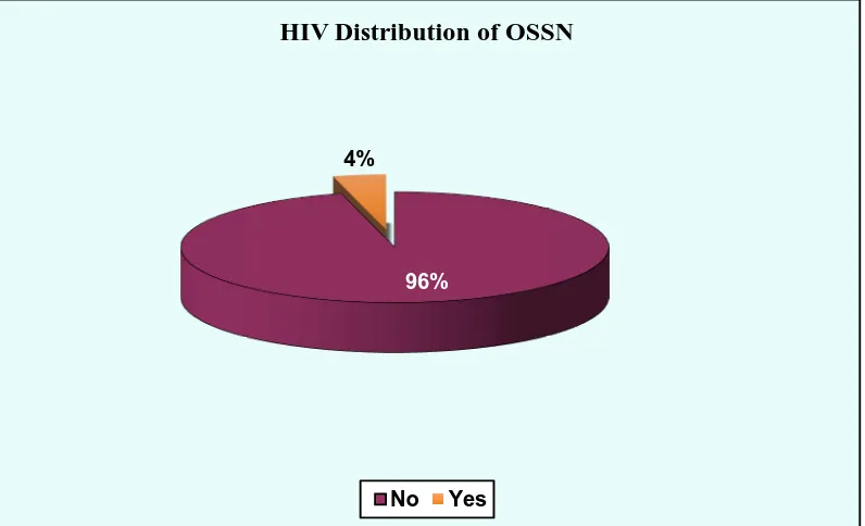
TABLE – 6 : HIV DISTRIBUTION IN OSSN
HIV Frequency Percentage
NO 48 96
YES 2 4
TOTAL 50 100
Among OSSN, only 2 cases (4%) were HIV positive and 48 cases
(96%) were not infected by HIV. Of the HIV positive cases, one was CIN III
and the other was Squamous cell carcinoma, moderately differentiated.
CHART – 6 : HIV DISTRIBUTION IN OSSN
96%
4%
HIV Distribution of OSSN
TABLE – 7 : CORNEAL INVOLVEMENT IN OSSN
Corneal involvement Frequency Percentage
Present 45 90
Absent 5 10
Total 50 100
Cornea was found to be involved in 45 cases (90%) of OSSN and only 5
cases (10%) of OSSN did not show corneal involvement, in the study.
CHART – 7 : CORNEAL INVOLVEMENT IN OSSN
90% 10%
Corneal Involvement of OSSN
TABLE – 8 : MOST COMMON PRESENTING SYMPTOM OF OSSN
SYMPTOM Frequency Percentage
Growth 40 80
Irritation 4 8
Redness 5 10
Vision loss 1 2
Total 50 100
The presenting symptom of OSSN with highest numbers in the study is
growth with 40 cases, constituting 80% in the study. Redness of the involved
eye is nextcommon symptom with 5 cases (10%), followed by irritation with 4
cases (8%) and vision loss with 1 case (2%).
CHART – 8 : MOST COMMON PRESENTING SYMPTOM OF OSSN
0% 10% 20% 30% 40% 50% 60% 70% 80%
Growth irritation Redness Vision loss
80%
8% 10%
TABLE – 9 : COMPARISION OF PRESENTING SYMPTOM WITH CORNEAL INVOLVEMENT
Corneal involvement
Presenting symptom p
value Total Growth Irritation Redness Vision
loss
Present 38 2 4 1
0.033
45
Absent 2 2 1 0 5
Total 40 4 5 1 50
fisher’s exact test
OSSN patients with corneal involvement(total 45 cases) had presented
frequent symptom as growth in 38 cases, redness in 4 cases, irritation in 2 cases
and lossof vision in 1 case. OSSN patients without corneal involvement (total 5
cases)presented as growth in 2 cases, irritation in 2 cases and redness in 1
case.Fisher’s exact test was applied. The p value obtained was 0.033 was
significant.
CHART – 9 : COMPARISION OF PRESENTING SYMPTOM WITH CORNEAL INVOLVEMENT
38
2 4 1
2 2 1
0 0 5 10 15 20 25 30 35 40
Growth Irritation Redness Vision loss
TABLE – 10 : MORPHOLOGICAL TYPES OF OSSN
Type Frequency Percentage
Gelatinous 32 64
Leukoplakic 13 26
papillomatous 5 10
Total 50 100
Among OSSN, gelatinous type was found to have more numbers with 32
cases (64%), Leukoplakic type of OSSN was found in 13 cases (26%),
papillomatous type having the least number with 5 cases (10%) in the study.
CHART – 10 : MORPHOLOGICAL TYPES OF OSSN
64% 26%
10%
Morphological Types of OSSN
TABLE – 11 : COMPARISION BETWEEN MORPHOLOGICAL TYPE
OF LESION AND PRESENTING SYMPTOM
fisher’s exact test
The common presenting symptom in gelatinous type of OSSN was
found to beGrowth as seen in 26 out of 32 cases, followed by redness seen in 4
out of 32 cases and irritation seen in 2 cases in the study. Loss of vision was
not reported as a presenting symptom in this type.
The common presenting symptom in Leukoplakic type of OSSN was
found to be Growth as seen in 10 out of 13 cases. One case each had presented
with symptoms of redness, irritation and loss of vision.
Symptom
Type Of Lesion P
Value Total Gelatinous Leukoplakic papillomatous
GROWTH 26 10 4
0.56
40
IRRITATION 2 1 1 4
REDNESS 4 1 0 5
VISION
LOSS
0 1 0 1
The common presenting symptom in Papillomatous type of OSSN was
also found to be Growth as seen in 4 out of 5 cases. Irritation was the
presenting symptom in 1 case. Redness and loss of vision were not reported as
presenting symptoms in this type of OSSN in the study.
Fisher’s exact test was applied and p value of 0.56 was obtained.
CHART – 11 : COMPARISION BETWEEN MORPHOLOGICAL TYPE
OF LESION AND PRESENTING SYMPTOM
26
2
4
0 10
1 1 1
4
1
0 0
0 5 10 15 20 25 30
Growth Irritation Redness Vision loss
TABLE – 12 : SURGICAL PROCEDURE DONE IN OSSN
SURGERY FREQUENCY PERCENTAGE
EXCISON 48 96
EVISCERATION 1 2
EXENTERATION 1 2
TOTAL 50 100
In the study, Excision biopsy was the most commonly performed
procedure in 48 cases of OSSN (96%). Evisceration was done in 1 case and
Orbital exenteration was done in other.
None of the cases had regional lymph node involvement or distant
metastasis.
TABLE – 13 : GROSS SIZE OF OSSN
SIZE (mm) Frequency Percentage
0-10 39 78
11-20 9 18
>20 2 4
TOTAL 50 100
The size of lesion as noted by greatest dimension and the cases were
OSSN, 39 cases (78%) were of size less than 10 mm (all 39 of which were less
than 5 mm), 9 cases (18%) were of size between 11-20 mm and 2 cases (4%)
wereof size more than 20 mm.
CHART - 12: GROSS SIZE OF OSSN
78% 18%
4%
Gross size of OSSN
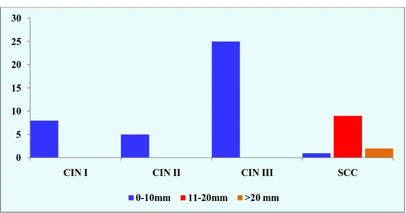
TABLE - 14 : DISTRIBUTION OF DIFFERENT GRADES OF OSSN
BASED ON GROSS SIZE
SIZE
TYPE OF OSSN
P value
CIN I CIN II CIN III SCC
0-10mm 8 5 25 1
<0.001
11-20mm 0 0 0 9
>20 mm 0 0 0 2
Total 8 5 25 13
fisher exact test
The size of OSSN was found to be higher in Invasive OSSN than in pre
invasive OSSN. The mean size of OSSN in the study was 6.3 mm. However,
invasive OSSN (SCC) were much larger lesions with mean size of 13.7 and
pre-invasive OSSN were smaller in size with a mean of 3.24 mm. Fisher’s
exact test was applied with p value obtained being <0.001 being statistically
significant.
CHART – 13 : DISTRIBUTION OF DIFFERENT GRADES OF OSSN BASED ON GROSS SIZE
0 5 10 15 20 25 30
CIN I CIN II CIN III SCC
TABLE – 15 : CORRELATION OF IMPRESSION CYTOLOGY AND
HISTOPATHOLOGY
Cytology Frequency Percentage
Correlated 45 90
Not correlated 5 10
Total 50 100
The impression cytology slides were obtained and diagnosis correlated
with the histopathological examination of excised lesions,in 45 cases (90%) of
OSSN in the study.Non correlation was seen in 5 cases (10%) ,all of which
were pre-invasive OSSN which had been reported as negative for OSSN in
cytology .
CHART – 14 : CORRELATION OF IMPRESSION CYTOLOGY AND HISTOPATHOLOGY
90% 10%
Cytology Correlation
TABLE – 16 : FREQUENCY OF OCCURRENCE OF DIFFERENT
HISTOLOGICAL TYPES OF OSSN
Dysplastic Lesion Type Frequency Percentage
CIN I 8 16
CIN II 5 10
CIN III/CIS 25 50
SCC 12 24
Among OSSN, pre-invasive OSSN (CIN I,II,III) constituted about 38
cases (76%) and invasive OSSN ( all grades of SCC ) account for 12
cases(24%) of the cases.
CHART – 15 : FREQUENCY OF OCCURRENCE OF DIFFERENT
HISTOLOGICAL TYPES OF OSSN
16%
10%
50% 24%
Histological types of OSSN
TABLE - 17: COMPARISION OF DIFFERENT GRADES OF OSSN AMONG THE AGE GROUPS
fisher’s exact test
Among pre-invasive OSSN, CIN I was distributed without much
variation among the three age groups, with a mean age of 50 years. CIN II was
also distributed without much variation among the three age groups, with a
mean age of 45.8 years. CIN III was seen in much higher numbers in the 41-60
age group with 17 cases, and 4 cases each in 20-40 years and more than 60
year age groups with a mean age of 53.6 years.
Invasive OSSN was more common in the more than 60 yearsage group
with 6 cases, followed by 5 cases in the 41-60 year age group, and only 1 case
in the 20-40 year age group. Mean age for invasive OSSN is 57.7 years
Age
group
HPE p value
Total CIN I CIN II CIN III SCC WD SCC MD SCC PD
20-40 3 2 4 0 1 0
0.22
10
41-60 2 2 17 1 3 1 26
>60 3 1 4 0 4 2 14
CHART – 16 : COMPARISION OF DIFFERENT GRADES OF OSSN
AMONG THE AGE GROUPS
In the study, 48 out of 50 cases (96%) of OSSN had uninvolved margins
on histopathological examination. Only 2 cases (4%) in the study had excised
margins showing evidence of tumour infiltration, both being invasive OSSN
(one case being moderately differentiated SCC, other poorly differentiated
SCC)
3
2
4
0 1 0
2 2 17 1 3 1 3 1 4 0 4 2 0 2 4 6 8 10 12 14 16 18
TABLE – 18 : INVOLVEMENT OF MARGINS IN OSSN
Margins Frequency Percentage
Uninvolved 48 96
Involved 2 4
Total 50 100
CHART - 17: INVOLVEMENT OF MARGINS IN OSSN
96% 4%
Margins in OSSN
TABLE – 19 : Ki-67 EXPRESSION IN PRE-INVASIVE AND
INVASIVE OSSN
Ki67 score
TYPE OF OSSN
Total P value
CIN SCC
Score I (0-20%) 23 0 23
<0.001
Score II (20-40%) 15 9 24
Score III (40-60%) 0 3 3
Total 38 12 50
fisher exact test
Ki-67 proliferation index as percentage of tumour cells positive was
grouped intothree with 0-20% assigned as score I; 21-40% assigned as score II;
TABLE - 20: KI-67 EXPRESSION IN DIFFERENT GRADES OF OSSN
fisher’s exact test
The Ki-67 proliferation index in OSSN ranged between 10 -45% with a
mean of 23.48 % in OSSN in the study. Fisher’s exact test was applied. p value
was obtained as <0.001 implying good statistical significance.
Ki-67
score
HPE
p value Total CIN I CIN II CIN III SCC WD SCC MD SCC PD Score I (0-20%)
8 3 12 0 0 0
<0.001
23
Score II
(20-40%)
0 2 13 1 6 2 24
Score III
(40-60%)
0 0 0 0 2 1 3
CHART - 18: KI-67 EXPRESSION IN DIFFERENT GRADES OF OSSN
Among pre-invasive OSSN (total 38 cases), CIN I - all 8 cases were in
score I ; CIN II - 3 cases had score I and 2 cases had score II; CIN III – 12
cases had score I and 13 cases had score II. Among invasive OSSN (total 12
cases), Ki-67 index in 9 cases had score II and 3 cases had score III.
Invasive OSSN (Squamous cell carcinoma) showed the maximum Ki-67
proliferation index with mean of 36.7%. (range 32-45%). Higher grades of
OSSN (poorly differentiated SCC) showing a mean Ki-67 proliferation index
of 40.3%, followed by 37.75% in moderately differentiated SCC and 32% in
the well differentiated SCC. Pre-invasive OSSN (CIN I,II,III) showed
relatively less Ki-67 proliferation index with mean of 18.9%. The p value
showed that the association was statistically significant. 8
3
12
0 0 0
0 2 13 1 6 2
0 0 0 0
2 1 0 2 4 6 8 10 12 14
CIN I CIN II CIN II SCC WD SCC MD SCC PD
TABLE - 21: RECURRENT CASES OF OSSN
Recurrence Frequency Percentage
No 48 96
Yes 2 4
Total 50 100
In the study, it was found that only 2 out of 50 cases (4%) of OSSN
were recurrent cases. In both cases, the histopathological diagnosis was
carcinoma in situ and Ki-67 expression was between 21-40%.
CHART – 19 : RECURRENT CASES OF OSSN
96% 4%
Recurrence in OSSN
Figure – 1 : HPE 195/14 : CIN GRADE I SHOWING MILD DYSPLASIA
[image:80.595.144.475.473.720.2]Figure – 3 : HPE 736/15 : CIN II SHOWING MODERATE DYSPLASIA
[image:81.595.160.460.456.715.2]Figure – 5 : HPE 111/14 : CIN III SHOWING FULL THICKNESS SEVERE DYSPLASIA
[image:82.595.152.469.491.726.2]Figure – 7 : HPE 178/16 : SQUAMOUS CELL CARCINOMA , WELL DIFFERENTIATED
[image:83.595.150.472.484.722.2]Figure – 9 : HPE 368/15 : SQUAMOUS CELL CARCINOMA, MODERATELY DIFFERENTIATED
[image:84.595.154.470.472.730.2]Figure – 11 : HPE 82/15 : SQUAMOUS CELL CARCINOMA, POORLY DIFFERENTIATED
[image:85.595.152.470.487.720.2]Figure – 13 : CONTROL
KI-67 IMMUNOSTAINING OF TONSIL 10X
[image:86.595.142.477.484.721.2]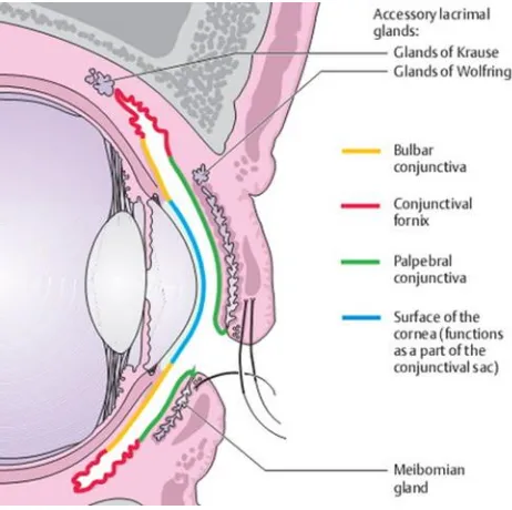
![Figure showing the Lineage of corneal epithelial cells[15]](https://thumb-us.123doks.com/thumbv2/123dok_us/180333.51228/19.595.103.500.139.445/figure-showing-the-lineage-of-corneal-epithelial-cells.webp)
![Figure showing Leukoplakic squamous cell carcinoma of conjunctiva -opaque white hyperkeratotic plaque[12]](https://thumb-us.123doks.com/thumbv2/123dok_us/180333.51228/27.595.150.470.123.338/figure-showing-leukoplakic-squamous-carcinoma-conjunctiva-opaque-hyperkeratotic.webp)