[1]
SPECIATION, VIRULENCE FACTORS DETECTION AND
ANTIFUNGAL SUSCEPTIBILITY TESTING OF CANDIDA ISOLATED
FROM HETEROGENOUS CLINICAL SAMPLES
Dissertation submitted in partial fulfillment of the
Requirement for the award of the Degree of
M.D. MICROBIOLOGY (BRANCH IV)
CHENNAI MEDICAL COLLEGE HOSPITAL AND RESEARCH CENTRE
IRUNGALUR, TRICHY- 621 105
Affiliated To
THE TAMILNADU DR. M.G.R. MEDICAL UNIVERSITY,
CHENNAI, TAMILNADU
[2]
[3]
CERTIFICATE
This is to certify that the dissertation entitled, “Speciation, Virulence Factors
Detection And Antifungal Susceptibility testing of Candida Isolated from Heterogenous
Clinical Samples” by Dr.Shalini.M, Post graduate in Microbiology (2014-2017), is a bonafide research work carried out under our direct supervision and guidance and is submitted to The Tamilnadu Dr. M.G.R. Medical University, Chennai, for M.D. Degree Examination in Microbiology, Branch IV, to be held in April 2017.
Guide: Professor and Head:
Dr. A. Uma M.D, Dr. A. Uma M.D, Professor and Head, Professor and Head,
Department of Microbiology, Department of Microbiology, CMCH&RC. CMCH&RC.
Dean:
Dr. Sukumaran Annamalai M.D, D.H.H.M.,
Chennai Medical College Hospital and Research Centre, Irungalur,
[4]
DECLARATION
I solemnly declare that the dissertation titled “Speciation, Virulence
Factors Detection and Antifungal Susceptibility testing of Candida isolated from Heterogenous Clinical Samples” is bonafide record of work done by me at Chennai
Medical College Hospital and Research centre, Trichy.
The dissertation is submitted to The Tamil Nadu Dr.M.G.R Medical University
towards the partial fulfillment of requirements for the award of M.D Degree (Branch
IV) in Microbiology.
Place: Trichy
Date:
Dr. Shalini.M
Post Graduate Student, M.D Microbiology,
Chennai Medical College Hospital and Research Centre Irungalur,
[7]
[8]
ACKNOWLEDGEMENT
I humbly submit this work to ALMIGHTY, who has given me the
strength, endurance and ability to overcome the difficulties encountered in the process
of compilation of my dissertation work.
I wish to express my sincere thanks to our DEAN, Chennai Medical
College Hospital and Research Centre, Trichy, for permitting me to use the
resources of this Institution for my study.
I feel indebted to Prof & HOD Dr.A.Uma, Department of Microbiology,
Chennai Medical College Hospital and Research Centre Of Microbiology for her
innovative ideas, timely suggestions and valuable guidance during my work. She
was phenomenal in giving new ideas, which paved the way for many new
dimensions in the study. Her immense and indepth knowledge in Microbiology has
helped me to correct the errors. She has a great role in improving my ability to analyze
the study.
I express my thanks and heartfelt gratitude to my co guide
Dr.G.Vazhalavandal, Associate Professor, Department of Microbiology, Chennai
Medical College Hospital and Research Centre, for her valuable guidance and
constant encouragement to complete this study.
I would like to whole heartedly thank our Assistant Professors
Dr.R.Saraswathy, Dr.A.Anupriya, Dr.J.Lalithambigai and Dr.DiegoEdwin,
Department of Microbiology, Chennai Medical College Hospital and Research Centre,
[9]
I would like to whole heartedly thank Dr.Thirumalaikolunthu
Subramaniyan, Professor of Medicine for his invaluable suggestion and constant
advice to me throughout my study.
I also express my sincere thanks to Dr.Rock Britto, Assistant Professor
Department of Community medicine, Chennai Medical College Hospital and Research
Centre, for statistically evaluating the study.
Special thanks to my senior postgraduates for lending a helping hand throughout
the study. I would also wish to thank my junior postgraduates for their help.
I would like to thank all staff of Department of Microbiology, Chennai Medical
College Hospital and Research Centre.
Finally, I am indebted to my family members, my husband and my
children for their everlasting support, encouragement throughout the study
[10]
[11]
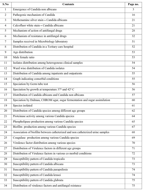
Contents
S.No
Chapters
Page
no.
1
Introduction
1
2
Aims and objectives
6
3
Review of literature
7
4
Materials and methods
33
5
Results
49
6
Discussion
77
7
Summary
95
8
Conclusion
98
Annexure I
Annexure II
Annexure III
[12]
Contents of table
S.No Contents Page no.
1 History of Candida in chronological order 7
2 Taxonomic hierarchy of Candida 8
3 Major pathogenic species of Candida 9
4 Clinical classification of Candida 16
5 Risk factors for invasive Candidiasis 18
6 Clinical features of Candidiasis in HIV 18
7 Macroscopic and microscopic identification of Candida species 21
8 Features that differentiate Candida albicans and Candida dubliniensis 24
9 Evolution of antifungal in chronological order 27
10 Antifungals and their mechanism of action 27
11 Methods for antifungal susceptibility testing of Candida 29
12 Microscopic identification of Candida species by Dalmau technique 40
13 Colours produced by various Candida species 41
14 Fermentation of different sugars by different species 42
15 Sugar assimilation by different species 43
16 Zone size interpretative chart for Candida antifungal susceptibility testing 47
17 Recommended Quality controls for different antifungals 48
18 Analysis of isolates 51
19 Speciation by Dalmau technique 57
20 Speciation by CHROM agar 58
21 Species identification by sugar fermentation 59
22 Species identification by sugar assimilation 59
23 Distribution of Candida from heterogenous clinical samples 63
24 Association of proteinase activity with the clinical samples 64
25 Association of phospholipase activity with the clinical samples 66
26 Association of biofilm with the clinical samples 67
27 Association of coagulase production with the clinical samples 69
28 Susceptibility pattern of Candida species 72
29 Candidiasis in diabetics – in various researches 80
[13]
Table of figures
S.No Contents Page no.
1 Emergence of Candida non albicans 3
2 Pathogenic mechanism of Candida 16
3 Methenamine silver stain – Candida albicans 21
4 Calcofluor white stain – Candida albicans 21
5 Mechanism of action of antifungal drugs 28
6 Mechanism of resistance in antifungal drugs 32
7 Samples received in Microbiology laboratory 51
8 Distribution of Candida in a Teritary care hospital 52
9 Age distribution 53
10 Male female ratio 53
11 Isolates distribution among heterogenous clinical samples 54
12 Ward wise distribution of Candida isolates 54
13 Distribution of Candida among inpatients and outpatients 55
14 Graph indicating comorbid conditions 55
15 Speciation by Germ tube test 56
16 Speciation by growth at temperature 370 and 420 C 56
17 Distribution of Candida albicans and Candida non albicans 57
18 Speciation by Dalmau, CHROM agar, sugar fermentation and sugar assimilation 60
19 Species isolated 61
20 Distribution of Candida species among different age groups 62
21 Proteinase activity among various Candida species 64
22 Phospholipase production among various Candida species 65
23 Biofilm production among various Candida species 67
24 Association of biofilm between catheterized and non catheterized urine samples 68
25 Coagulase production among various Candida species 69
26 Virulence factor distribution among various species 70
27 Distribution of Virulence factors in different age groups 71
28 Distribution of Virulence factors in various co morbid conditions 72
29 Susceptibility pattern of Candida tropicalis 73
30 Susceptibility pattern of Candida albicans 73
31 Susceptibility pattern of Candida parapsilosis 74
32 Susceptibility pattern of Candida krusei 74
33 Susceptibility pattern of Candida glabrata 75
[14]
Abbreviations
C.albicans Candida albicans
C.tropicalis Candida tropicals
C.parapsilosis Candida parapsilosis
C.krusei Candida krusei
C.glabrata Candida glabrata
GTT Germ Tube Test
SDA Sabourauds Dextrose Agar
UTI Urinary Tract Infection
HIV Human Immunodeficiency Virus
AIDS Acquired Immune Deficiency Syndrome
VVC Vulvo Vaginal Candidiasis
SAP Secreted Aspartyl Proteinase Enzyme
IDSA Infectious Disease Society of America
CDC Centre For Disease Control and Prevention
CLSI Clinical and Laboratory Standards Institute
EUCAST European Committee on Antimicrobial Susceptibility Testing
ELISA Enzyme Linked Immune Sorbent Assay
HSP Heat Shock Proteins
GM -CSF Granulocyte Macrophage Colony Stimulating Factor
M- CSF Macrophage Colony Stimulating Factor
[15]
[16]
1.0 INTRODUCTION
Candida species are ubiquitous yeast like fungi associated with human beings for a quite long time. There are about 200 species of Candida, dwelling as
saprophytes in soil and aquatic environment and also colonizing several animal
reservoirs. In about 70% of healthy individuals, Candida exists as commensals of the
gastrointestinal and genitourinary tracts 1.
Whenever there is alteration in the equilibrium between Candida and the host factors, the Candida which is a commensal becomes pathogenic and causes
several diseases. Under such situations Candida species causes superficial, invasive or
disseminated infection by infecting all the sites of the human body. The infections
includes oral thrush, glossitis, vulvovaginitis, intertrigo, paronychia, urinary tract
infection, endocarditis to meningitis 2.
The predisposing factors for candidiasis includes 3
Prolonged administration of antibiotics
Immuno- compromised states such as
o Acquired immuno deficiency syndrome,
o Cancer chemotherapy,
o Immunosuppressants,
o Diabetes mellitus,
o Extremes of age,
o Burns,
o Steroids,
[17]
o Prolonged hospital stay,
o Genetic deficiency syndromes,
o Prolonged Antibiotic therapy,
o Insertion of catheters and unsterile needles.
Candida has emerged as a major cause of human disease and the fungal
infection rates have been increasing over the past 20 years 4.
Candidiasis has worldwide distribution. It is the commonest cause of
hospital acquired blood stream infections in United States 5.
In HIV patients, candidiasis is the second most common infection 6. The
rate of invasive candidiasis have increased with increase in the epidemic of HIV. 80%
of AIDS patients developed oral candidiasis before the advent of Highly Active Anti
Retroviral Therapy regimen. Oro pharyngeal candidiasis is most common among the
HIV patients and it is the important marker for immunosuppression 2.
Candidiasis was found to be the second most common infection in patients
on cancer chemotherapy and transplant recipients 7.
Candida causing nosocomial UTI has become most prevalent with an
increased mortality of about 10- 15% 8.
Eighty percent of women suffer from vulvovaginal candidiasis (VVC)
atleast once in their life time, although all the other organs are also frequently infected
with Candidiasis3.
Candida albicans accounts for 40-60% yeasts isolated in developed
countries, whereas Indian reports show an increased occurrence of infection with
[18]
inanimate materials such as urinary and vascular catheters, and is often involved
in biofilm formation.
From 1970 to 2000 Candida albicans dominated as causal Candida
pathogen worldwide in all forms of candidal infections. Significant changes has
occurred in the last decade have transpired with the progressive important role of
Candida non albicans species imparting a profound influence on selection of
antifungal drugs 9.
Fig 1: Emergence of Candida non albicans
Source:http://link.springer.com/article/10.1007
The extent and severity of these candidial infections depends on the immune status of the host. The Candida species exhibits certain virulence factors that
helps the organism in proliferation, adhesion and invasion of host tissue. The
extracellular hydrolytic enzymes produced by Candida species helps in proliferation,
prevents the phagocytosis and helps in the survival of the organism. Biofilm formation
plays an important role in the pathogenesis. Different species of Candida exhibits
[19]
Infectious diseases society of America (IDSA) guidelines 2016 states that –
candidiasis is a serious, life threatening infection that needs to be treated early,
aggressively and appropriately.
There are various antifungals used in the treatment of candidiasis. These
consist of fluconazole, voriconazole, caspofungin, amphotericin B, and lipid
formulations of amphotericin B. Fluconazole is most commonly used in the treatment
of candidemia.
Resistance to antifungal drugs is increasing in recent years 11. The
resistance of C.albicans, C.tropicalis and C.glabrata to fluconazole is increased
compared to other drugs, due to indiscriminate use of fluconazole for long periods 12.
C.krusei is intrinsically resistant to fluconazole. IDSA also states that, more than 90
% of invasive candidiasis is caused by C. albicans, C. glabrata, C. tropicalis, C.
parapsilosis, and C. krusei, and these have unique virulence potential, antifungal
susceptibility and epidemiology13. All these factors has led to increase in the
mortality and morbidity rates in patients with fungal infections warranting rapid
identification and antifungal susceptibility testing at the earliest.
Therefore due to the variable clinical presentation of Candida infections, it
has become important to identify the Candida species from all the clinical specimens.
Differentiating among Candida species in laboratory is also very important because of
the differences in the virulence of the species and in their susceptibility to anti-fungal
drugs.
Hence the present study was conducted in a tertiary care hospital with 770
[20]
heterogenous clinical samples, speciate it. The virulence factors and the drug
susceptibility pattern was studied for the different species for appropriate and effective
[21]
[22]
2.0 AIMS OF THE STUDY
To isolate and identify Candida from heterogenous clinical samples in a tertiary
care hospital.
To speciate and evaluate their distribution with the age, clinical diagnosis and
co-morbid conditions.
To differentiate between commensal and pathogenic Candida species by
determining the virulence factors of the isolated Candida species.
To determine the in vitro efficacy of antifungal agents of Candida species.
[23]
[24]
3.0. REVIEW OF LITERATURE
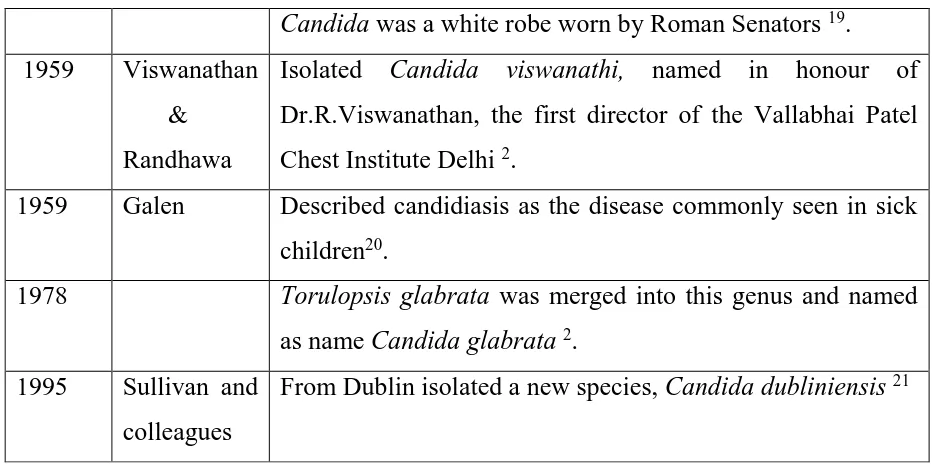
3.1. History:
Candidiasis has a very old history, the disease was described in ancient
times. Hippocrates around 400 B.C. had described oral candidiasis as “mouth affected
with aphthous ulcerations” in “Of the Epidemics”14.
Table:1 History of Candida in chronological order
Year Author Event
1665 Pepy’s Description of the disease was noted in his diary as described
by Galen 2
1751 Hill Isolated the yeast from rotting vegetation and was confused
with the term “Monillia” which is often confused with Candida 15. But the organism isolated was an Aspergillus
species.
1771 Rosen Von
Rosenstein
Defined the invasive form of thrush 16.
1784 Underwood Described oral and gastrointestinal candidiasis as a disease of pediatric age group 17
1844 Bennet Isolated the yeast from the sputum of tuberculosis patient 18
1847 Charles
Philippe Robin
French mycologist, named the fungus causing thrush as Oidium albicans (“to whiten”).
1861 Zenker Isolated from brain of a debilitated patient in whom the yeast
spread from his oral thrush by blood 11
1874 Parrot Noted the first pulmonary infection case caused by Candida
1894 Torulopsis glabrata was isolated from grapes
1954 The name Candida was officially accepted by the 8th
Botanical Congress held at Paris in 2.
[25]
Candida was a white robe worn by Roman Senators 19.
1959 Viswanathan
& Randhawa
Isolated Candida viswanathi, named in honour of
Dr.R.Viswanathan, the first director of the Vallabhai Patel Chest Institute Delhi 2.
1959 Galen Described candidiasis as the disease commonly seen in sick
children20.
1978 Torulopsis glabrata was merged into this genus and named
as name Candida glabrata2.
1995 Sullivan and
colleagues
From Dublin isolated a new species, Candidadubliniensis21
Thus, with the advent of antimicrobial agents in the latter half of the
twentieth century and increase in the immunosuppressed patients in the last few
decades have restored the interest in Candida and candidiasis. These events have led
to an increase of Candida infection particularly the less pathogenic non albicans
species 22.
3.2 Taxonomy 12,23:
[image:25.595.64.535.68.302.2]Candida falls under :
Table 2 : Taxonomic hierarchy of Candida
Kingdom Fungi
Phylum Ascomycota
Subphylum Ascomycotina
Class Ascomycetes
Order Saccharomycetales
Family Sacharomycetaceae
Genus Candida
The genus Candida consists of approximately 200 species out of which 20
[26]
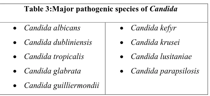
[image:26.595.144.485.96.252.2]The major pathogenic species include the following:
Table 3:Major pathogenic species of Candida
Candidaalbicans
Candida dubliniensis
Candida tropicalis
Candida glabrata
Candida guilliermondii
Candidakefyr
Candida krusei
Candida lusitaniae
Candida parapsilosis
Candida tropicalis is the most common Candida species isolated in
patients with hematologic malignancies and bone marrow transplant recipients 25.
Candida parapsilopsis is frequently isolated from pathological lesion of the
nails and skin. It is reported as the causative agent in endocarditis, endophthalmitis,
septic arthritis and peritonitis.
Candida glabrata is relatively a non pathogenic and normal flora of healthy
individuals. But following the wide spread and increased use of immunosuppressives
together with broad spectrum antifungal therapy, the frequency of mucosal and
systemic infections caused by Candida glabrata among the old age have been
increased 25.
The prevalence of Candida albicans and Candida non albicans has changed.
The prevalence of C.glabrata increases with age and common among patients aged ≥
70 years, whereas in C.parapsilosis it is the other way 26.
In a study, done by Kothari et al. C.tropicalis causes 45% of infections,
C.albicans 23% and other species of Candida 23% 27.
A study done by Chakrabarti et al reported that Candida non albicans is
[27]
3.3. Physiology :
Candida species metabolise glucose via the Hexose Monophosphate
pathway under aerobic conditions (assimilation) or via the Embden Meyerhof
pathway in anaerobiosis (fermentation). Mitochondrial oxidative phosphorylation, Kreb’s cycle and protein synthesis are similar to those of eukaryotic cells.
Candida enzymes are significant as they may be directly involved in pathogenesis.
Growth temperature has an important influence on morphogenesis. Temperatures
around 25°C promote the formation of chlamydospores in C.albicans and higher
temperatures around 37°C promote formation of pseudohyphae 12.
3.4. Epidemiology
Fungi are ubiquitous in plants, mammals and insects. Humans are
continually exposed to numerous genera of fungi through a variety of routes. Candida
are commensals of humans and are commonly found on skin, gastrointestinal tract and
genito urinary tract29.
In United States, the colonization of Candidaalbicans in the oropharynx can be
found in almost 30-55% of young adults 30. Also, the presence of C. albicans in
normal fecal sample is about 40-65% 30. Candidemia is the fourth most common cause
of bloodstream infection, Almost 6.9 out of every 1000 intensive care unit patients are
suffering from candidemia 31.
C. albicans was found to be the most frequently isolated pathogen in Northern
and Central Europe and the USA, whereas Candida non albicans species were found
[28]
The highest proportion of C.glabrata isolates were found in Northern and
Central Europe, whereas C.parapsilosis was most commonly found in Slovakia,
Southern Europe, South America and Asia and C.tropicalis predominated in Eastern
Asia and Argentina32.
Nosocomial transmission or 'cross-infection' of Candidiasis poses significant
problems. Recent literatures have reported the isolation of Candida from the hands of
health care workers in intensive care units and cross- infection. Epidemiologic studies
showed that Candida non albicans are more frequently isolated from the urine
samples than the other clinical samples, this may be probably due to urine
composition and or pH. Mixed isolates were found in patients with nosocomial
Candiduria. The emergence of Candidanon albicans species may represent selection
of more resistant species like C. glabrata and C.krusei33.
3.5. Pathology and pathogenesis:
Candida is a commensal in different site of the body. So the source is
mainly endogenous34 and rarely can be introduced exogenously 35. The exogenous
source includes insertion of catheters or prosethetic valves 36. The repeated isolation
from the same patient and in significant number indicates infection or colonization.
But any isolation from sterile body fluids is significant. In immunocompetent
individuals immune system combats the infection.
3.5.1. Immunity:2,37.
Classical T- cell immunity protection from persistent high level
[29]
Polymorpho nuclear cells damages and phagocytoses the pseudohyphae
protects from deep seated candidiasis
Cytokines (INF-α, IL-3, GM-CSF, M-CSF, G-CSF) phagocytoses the yeast.
Serum factors, and heat labile opsonins increased ingestion of yeast;
thereby B-lymphocytes and antibodies also have a role.
3.5.2. Virulence factors:
There are many virulence factors which plays important role at different
sites and stages of infection. Virulence factors characterized in terms of function and
not considering the antigenicity2.
Virulence factors are 2:
Adhesion
Enzymes
Toxins
Complement receptors
Phenotype switching
3.5.2. a) Adhesion:
Ability of Candida to adhere to the host cell correlates with their virulence
and plays a major role in pathogenesis. Adhesion present on the surface of fungus
interacts with the receptor of the host cell thereby helps in binding with the epithelial
and the endothelial cells. Candida has an ability to bind with the host cells both in
vivo and in vitro, tried in experimental animals 38. In vitro Candida is capable of adhering to exfoliated human epithelial cells, human tissue, cell lines (endothelial,
[30]
3.5.2 .b) Enzymes:
There are about 14 hydrolytic enzymes but only two plays a role in
pathogenesis. They include proteases, lipases, phospholipases, esterases, phosphatases
etc. Secretion of secreted aspartyl proteinase enzyme (Sap) plays a major role in
pathogenesis39. Sap was produced by pathogenic Candida species after adhesions to
epithelial cells40. There are three types of Sap and two types of serine proteinases which are involved in growth control of yeast remodeling and they also facilitate
hyphal invasion leading to disseminated candidiasis 2,41. Mutants which are deficient
in Sap are unable to invade the host tissue42. Phospholipase enzyme is secreted during
the tissue penetration and is essential for the virulence39.
3.5.2. c) Toxins:
The glycoprotein extracts of Candida cell wall resembles endotoxins of
bacteria. These are lethal and pyrogenic anaphylactic shock.
3.5.2. d) Complement receptors:
It binds to the complement derived opsonins plays a role in virulence.
3.5.2. e) Phenotypic Switching:
It is the ability of a single strain to switch reversibly at high frequencies among
different colony phenotypes. This evades the host immune response.
3.5.3. Candida Antigens:
The antigens of Candida is divided into 2 main groups:
[31]
ii. Cytoplasmic antigens
3.5.3 i) Cell wall antigens :
Fungal surface molecules include mannan, glucans and chitin contributes to
the virulence of Candida species. It causes suppression of immune response and
adherence of the host tissue by changing the hydrophobicity 43. The mannoproteins
released from Candida albicans binds with the red blood cells therby causing
hemolysis of red cells. These hyphal cells uses hemoglobin as iron source 44.
These organisms attaches and grows in colonies and causes biofilm
formation45. The biofilm contains extracellular materials like proteins, carbohydrates
etc 46. The antimicrobials have poor penetration in biofilms 47.
3.5.3 ii) Cytoplasmic antigens:
A number of antigenic components in extracts of broken cells are also
considerably significant. These antigens have not yet been associated with specific
cytoplasmic components of the cell.
3.5.4. Virulence factors contributing to Pathogenesis:
yeast cells encounter the host tissue
Colonization takes place – local site
Invades the deeper tissues
adhesion
[32]
The pathogenicity of Candida depends on complex array of micro
organism related putative virulence factors. These include2
Yeast to mycelium transition
Antigenic variability
Phenotypic switching
Adhesion to host cells and tissue
Cell surface hydrophobicity
Molecular mimicry
Production of extracellular enzymes
Most of the biological functions related to pathogenicity and virulence are confined to
the cell wall 2.
Transformation into hyphal forms (in active infection)
Phospholipase (present in hyphal tips)
invasion
hyphae being larger resists phagocytosis
[33]
[image:33.595.129.475.486.749.2]
Source:
Fig 2 : Pathogenic mechanism
Virulence. 2013 Feb 15; 4(2): 119–128.
3.6.Clinical features:
The clinical manifestation of Candida is varied, ranging from acute,
subacute, chronic and episodic2. Involvement may be localized or systemic. The pathologic process vary from irritation and inflammation to chronic and acute
suppuration or granulomatous response 2. Clinical classification of candidiasis 2:
Table 4: Clinical classification of candidiasis
I) Infectious disease
A) Mucocutaneous manifestation
Oral48,49,50 : thrush, stomatitis, glosssitis, cheilitis
Alimentary 51: esophagitis, gastritis.
Vulvovaginitis52, balanitis, balanoposthitis.
Chronic mucocutaneous candidiasis12
Ocular candidiasis 53
B) Cutaneous manifestation
Intertriginous 54and generalized
[34]
Diaper dermatitis2
Candidial granuloma
C) Systemic manifestations
Urinary tract – candiduria56,57
Endocarditis 58,59
Pulmonary candidiasis
Meningitis 35
Candidemia
Dissemination
Arthritis and osteoarticular candidiasis
Osteomyelitis
Endophthalmitis 61
Invasive candidiasis
II) Allergic diseases
Candidids
Eczema
Asthma
Gastritis
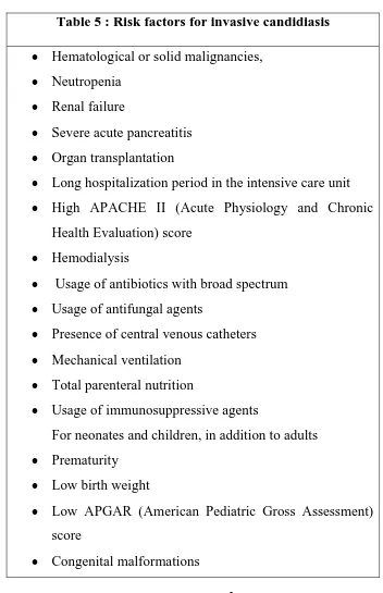
3.6.1. Invasive candidiasis and candidemia:
Invasive candidiasis is a multiorgan infection. Candida is not isolated
from blood culture 12. It is common among acute leukaemia patients, post operative
patients, cancer patients, transplant patients, prolonged ICU admission and also in
drug addicts 12. Patients with candidemia presents with nodular lesions of the skin and
white lesions of the retina. These signs are important in the diagnosis even when the
[35]
[image:35.595.144.496.90.635.2]Risk factors for the development of invasive candidiasis 60.
Table 5 : Risk factors for invasive candidiasis
Hematological or solid malignancies,
Neutropenia
Renal failure
Severe acute pancreatitis
Organ transplantation
Long hospitalization period in the intensive care unit
High APACHE II (Acute Physiology and Chronic
Health Evaluation) score
Hemodialysis
Usage of antibiotics with broad spectrum
Usage of antifungal agents
Presence of central venous catheters
Mechanical ventilation
Total parenteral nutrition
Usage of immunosuppressive agents
For neonates and children, in addition to adults
Prematurity
Low birth weight
Low APGAR (American Pediatric Gross Assessment)
score
Congenital malformations
3.6.2. Clinical forms of candidiasis in HIV patients 2:
Table 6 :Clinical features of candidiasis in HIV
Asymptomatic oral carriage
Oropharyngeal thrush
[36]
Perleche (angular cheilitis)
Leukoplakia
Oesophagitis
Laryngitis
Vulvovaginitis, balanitis
Hematogenous dissemination
3.6.3.Fungal infections in ICU:
Invasive candidiasis is more common in immunosuppressed condition. But
recent studies have shown that invasive candidiasis can occur even in
immunocompetent patients who are critically ill62. They have non specific clinical presentation and high mortality rates. The risk factors associated with invasive
candidiasis are compromised immune function either due to debilitating clinical
condition or use of immune suppressing medications and also prolonged stay in
Intensive care units.
Other risk factors include 62:
Candida colonization at various body sites
Broad spectrum antibiotics
Immunosuppressive therapy like cytotoxic chemotherapy
Corticosteroids
Malnutrition
Malignancy
Neutropenia
Severe burns and
[37]
3.7. Diagnosis of candidial infection :
There are nearly 20 species of Candida which are pathogenic to humans. The
difficulty in the diagnosis lies due to the absence of specific symptoms and signs as
well as the opportunistic nature of the yeast. Candida species identification plays a
major role in successful management.
3.7.1 Specimen:
Specimens are collected depending upon the site of infection. Sputum,
bronchial aspirate, exudates, scrapings from mucosal, dermal or nail lesions etc. are
collected.
3.7.2 A) Direct examination:
The samples are examined in KOH wet mount or normal saline preparation
for the visualization of yeast cells and pseudohyphae.
Gram staining is performed for the presence Gram positive budding yeast
cells and pseudohyphae. The yeast cells are approximately 4-8µm. They are
non-capsulated. The pseudohyphae show regular point of constriction. The microscopy
indicates only candidiasis and the etiology can be established only by culture12. The
pseudohyphae shows colonization and tissue invasion, hence their demonstration
indicates clinical importance.
[38]
The biopsy specimen are kept in the tube containing 10% KOH for an
overnight period at 370C and after mincing they are examined under microscope for
yeast cells and pseudohyphae2.
Table 7: Macroscopic and microscopic identification of Candida species
Species Macroscopy Microscopy
C.albicans Creamy smooth colonies. In old stocks it becomes
waxy, soft, smooth sometimes
reticulated, old cultures are wrinkled and folded with spicules3.
Yeast cells are short and ovoid (5-7μm).
Sometimes elongated yeasts which are smaller and larger cells are also seen 3.
C.tropicalis Creamy white, smooth colonies. Older colonies- white to cream colored, dull soft and wrinkled, often
Yeast cells are ovoid or short ovoid cells (4-8x5-11μm) 3.
Fig 3:Methenamine silver stain- Candida albicans
Source: http://www.microbiologybook.org/mycology
[39]
3.7.2.B)Fungal culture:
Candida grows well in bacteriological basal or simple media like with overgrowth of mycelium 3.
C.glabrata Smooth, soft, glossy and cream colored colony 3.
Yeast cells are smaller round to oval yeasts 2.5-to 4.5x 4-6μm 3.
C.parapsilosis Younger colonies are smooth, soft sometimes lacy.
Older colonies are creamy yellowish, glistening smooth or sometimes wrinkled 3
Yeast cells are short ovoid to long ovoid (3-5x6-
20μm) 3.
C.krusei Colonies are flat, dull and dry and older colonies are
greenish yellow, dull, wrinkled with heavy growth of mycelium around the
colonies 3.
Yeast cells are cylindrical, some of them ovoid to long (3-5x620μm) 3.
C.guilliermondii Colonies are thin, flat, glossy, cream to pink colour. In old stocks colonies become yellowish cream to pink, glistening dull or smooth and wrinkled 3.
Yeast cells are ovoid and cylindrical (2-4x
3-7 μm) 3.
C.kefyr Colonies are cream, smooth colony. In old cultures cream to yellow dull, soft and smooth 3.
Yeast cells are mostly small ovoid with few elongated cells (2.5 to 5 X 5 to 10μm) 3.
C.viswanathi Younger colonies are cream-colored, soft, glistening; older colonies are
creamy soft to membranous,
wrinkled and dull 3.
Yeast cells are globose ovoid to cylindrical (2.5-7x4-1μm)3.
C.lusitaniae Cream-colored, soft, glistening 3. Yeast Cells are ovoid (2 to 6
[40]
nutrient agar and blood agar. The routinely used mycological media for Candida isolation is Sabouraud’s dextrose agar (SDA) with added antibiotics (chloramphenicol
or gentamycin) with pH adjusted to 5.6 63. SDA with cycloheximide should be
avoided as it prevents the growth of C.krusei, C.parapsilosis and C.tropicalis. Most
pathogenic species of Candida grows at 25 and 37°C 63. C.albicans can also grow at
45°C.
The LPCB can be performed from the colonies for the presence of budding
yeast cells and pseudohyphae. Gram staining also can be performed from the culture
colonies.
For the detection of systemic Candidiasis blood culture can be done in
biphasic medium like brain heart infusion agar – broth and incubated at both 250C and
370C. The colonies will be apparent by 24 hours or within 2- 3 days. The colony
morphology of Candida species is smooth and creamy with fine differences. Some
species produce dry colonies.
The growth of the Candida species is also seen on Tetrazolium
Reduction Medium (TRM) and are compared with the standard colours.
3.7.2.C)Germ tube production 63:
A germ tube is a filamentous extension from a parent cell. The true germ
tube has no constriction at the neck. C.albicans and C.dubliniensis produce germ
tubes when incubated with various substances like human or sheep serum, rabbit
plasma, egg albumin, tissue culture medium, thioglycolate soya broth etc. at 370C for
2 hours. Not all strains of C.albicans produce germ tube. The demonstration of germ
[41]
3.7.2. D) Corn meal agar inoculation
The commonly used differential media for identification of Candida upto
species level. Speciation is done by the presence and position of chlamydospores,
blastospores, hyphae and pseudohyphae.
3.7.2.E) Candida CHROM agar63:
It is a rapid, plate based test for the isolation and identification of species.
Principle of the test is that different colour is formed due to the reaction between the
enzymes in the organism and the chromogenic substrate in the medium.
Table :8 Features that differentiate C.albicans and C.dubliniensis
Characteristics C.albicans C.dubliniensis
Germ tube + ++
Chlamydospores + +++
Growth on CHROM agar Light green Dark green
Growth at 42- 450 C + -
3.7.2.F) Biochemical tests:
Candida species are characterized by the pattern by which they use
specific carbohydrate and nitrogen substances. The process by which the Candida
species oxidatively utilize the carbohydrates is called as assimilation and anaerobical
utilization of the carbohydrates is called as fermentation. If given yeast possesses the
ability to ferment a carbohydrate it can also assimilate it.
1) Sugar fermentation tests:
This test involves peptone water added with various carbohydrates, a
[42]
2) Sugar Assimilation tests:
Based on the ability of the Candida to utilize a specific carbohydrate in
the presence of oxygen helps in species identification. The different techniques for
detecting assimilation are as follows:
a. Classical Wickerham and Bruton method 65
b. Auxonographic technique 63
3.7.2.G) Commercial Yeast identification systems:
API 20C system
API ID32C
API Candida kit
VITEK (Biomerieux Vitek, Inc.,Hazelwood,MD)
Minitek (Becton Dickinson Microbiology System,Cockeysville,MD)
Uni-Yeast-1ck (Flow laboratories, Woodcock,UK).
3.7.2.H) Immunodiagnosis:
Antibody against C.albicans molecules or Candida derived molecules
whose presence in sera indicates tissue invasion are identified.
a) Detection of Antibody 12
Gel immunodiffusion
Counter immune electrophoresis
ELISA and Latex agglutination tests.
b) Detection of Antigens 66
[43]
Latex Agglutination systems using monoclonal and polyclonal
antibodies.
Glycoprotein detection using latex agglutination test.
c) Cell mediated immunity can be assessed by:
Skin tests to detect delayed hypersensitivity to candidal antigen.
In vitro assay like lymphocyte transformation test.
3.7.2.I) Molecular techniques
i) Detection of Candidial DNA by PCR using specific probes
ii) 26S rRNA for identification of yeast from blood culture bottles. It detects
the organism within 2-3 hrs 67.
iii) Restriction fragment length polymorphism
iv) Southern hybridization pattern
3.7.2.J) MALDI-TOF:
It is a proteinomics used for rapid identification of Candida species. It is a
powerful method for detection and identification of proteins by molecular weight of
individual specific fragments68. 3.7.3. Candida score:
It helps in early diagnosis of invasive candidiasis and helps the clinician to
start early antifungal treatment 69. It is calculated at the onset of sepsis or shock using
total paraenteral nutrition, multifocal colonization, surgery and severe sepsis.
3.8. Treatment:
3.8.1. Evolution of antifungals70:
[44]
[image:44.595.67.532.96.628.2]discovered only in 1939.
Table 9: Evolution of antifungals in chronological order
Year Discoverer Discovery
1939 Oxford et al Griseofulvin was isolated and discovered from Penicillium griseofulvum
1944 Woolley et al Azoles
1958 Griseofulvin used for clinical purpose
1958 Azoles was used as antifungal drug
1957 Duschinsky et al 5-Flurocytosine was used as anti tumor drug
1960 Clotrimazole, econazole, micanozole
1963 Greenberg & co workers
5-Flurocytosine was used as antifungal drug
1968 Micanozole was used for parenteral injection
1981 Heeres & co workers Ketoconazole
1992 Itraconazole
2001 Itraconazoles – parenteral forms
2002 Voriconazole
2006 Posaconazole
2001 Caspofungin
2005 Micafungin
2006 Anidulafungin
3.8.2.Antifungal agents and their Mechanism of action 71:
Table 10. Antifungals and their mechanism of action
Chemical class Drug Target
Azoles Miconazole Ketoconazole Fluconazole Itraconazole Teroconazole
[45]
[image:45.595.161.457.277.509.2]
Fig 5 :Mechanism of action of antifungal drugs
Source: http://www.doctorfungus.org/thedrugs
Treatment of candidiasis depends on the site of the infection and the virulence
of the organism 12.
Mucocutaneous candidiasis – topical or systemic depending on the site
Candidemia and deep seated infections – Systemic treatment 35.
The choice of the antifungal also depends on the host factors. Voriconazole
Posaconazoles Polyenes Amphotericin B
Nystatin
Ergosterol membrane function
Pyrimidine Flucytosine DNA & RNA synthesis Echinocandins Caspofungin
Micafungin Anidulafungin
[46]
The treatment should be initiated only after the antifungal susceptibility
testing. But for the patients in ICU the empirical therapy can be started in whom
the presence of multifocal Candida colonization posses a high risk for invasive
candidiasis and also in patients who has fever of unknown origin that is refractory
to broad spectrum antimicrobials12.
The selection of antifungal agents mainly depends on the species. As
certain species are intrinsically resistant to certain antifungals 25. Eg:
C.krusei – intrinsically resistant to fluconazole and ketaconazole
C.lusitaniae to amphotericin B
Standardization of in vitro susceptibility tests by the Clinical Laboratory
Standards Institute (CLSI) and the European Committee for Antimicrobial
[image:46.595.81.518.468.678.2]Susceptibility Testing (EUCAST) are highly useful 72.
Table 11 : Methods used for antifungal susceptibility testing of Candida.
Broth macro & micro dilution73 Calorimetric microdilution74 Sensititre yeast one test panel 75 Agar macrodilution
Agar diffusion
o Disk diffusion
o E- test
o Neo sensitabs76
Flow cytometry77.
Antifungal susceptibility has become important in clinical laboratory due to: Increase in the incidence of candidial infection in the last 15 yrs
[47]
Increased emergence of resistance to the antifungals
3.9.Antifungal resistance 78:
The emerging phenomenon of antifungal resistance is primarily a concern
for invasive candidiasis. The knowledge about the antifungal resistance is less when
compared to that of the antibiotic resistant bacterial infections, as they are the threat to
public health. Thus, importance for understanding the emergence of antifungal
resistance, awareness among medical and public health communities about these
infections, and methods used to prevent and control them should be highlighted.
The changing epidemiology of Candida infection has been partly attributed to the selection of less sensitive Candida strains by the widespread use of the azole
fluconazole as a prophylactic and therapeutic agent.
A study by Pfaller et al states that flucanozole is appearing resistant in
Candidaalbicans79.
Centre for Disease Control and Prevention (CDC) states that 7% of all
Candida bloodstream isolates most of which are Candida glabrata. CDC’s
surveillance data indicate that the proportion of Candida isolates that are resistant to
fluconazole has remained fairly constant over the past twenty years. In contrast,
echinocandin resistance appears to be on the rise, with approximately 1% of
all Candida isolates tested at CDC showing echinocandin resistance80. The antifungal resistance is divided into
Clinical resistance
[48]
3.9.1.Clinical resistance
It occurs in patients with severe immunosuppression like HIV. It is a result
of low level of drugs in serum and or tissues caused by poor patient adherence,
drug interaction that decreases antifungal levels.
3.9.2. Cellular or in –vivo resistance
It is independent of host. The pathogenic strains are less responsive to the
standard dose of the drug.
It is divided into
Primary or intrinsic
Secondary or acquired
3.9.2a) Primary resistance:
It is demonstrated in organisms which are naturally resistant to antifungals. Eg :
C.krusei intrinsically resistant to fluconazole
3.9.2b) Secondary resistance:
Initially the isolate is susceptible but later becomes resistant to an antifungal
agent. Most commonly seen in HIV patients.
3.9.3. Mechanisms of resistance:
Resistance to azoles:
It is due to
Changes in the sterol components of plasma membrane
Genetic changes in ERG11gene encoding Lanosterol demethylase
Alteration of enzymes involved in Ergosterol biosynthesis
[49]
Resistance to Amphotericin B:
It is poorly understood, probably due to changes in sterol components of
plasma membrane and lipid composition of cell membrane.
Resistance to Flucytosine:
Deficiency of cytosine permease, cytosine deaminase, UMP pyrophosphorylase
and loss of feedback regulation leading toincreased synthesis of pyrimidines.
Resistance to Echinocandins :
Due to mutations in FKS genes encoding 1,3 -D glucan synthase.
Fig 6: Mechanism of resistance in antifungal drugs
[50]
[51]
4.0 MATERIALS AND METHODS
4.1.1. Design of study:
Cross sectional analytical study
4.1.2. Setting:
Department of Microbiology of a tertiary care hospital.
4.1.3. Period of study:
February 2015 –May 2016
4.1.4. Material:
All clinical samples like blood, urine, sputum, nail scrapings, high vaginal
swab, tracheal aspirate and other body fluids received in the laboratory for culture.
4.1.5. Inclusion criteria:
All samples clinically suspected of candidiasis
4.1.6. Exclusion criteria:
Samples from patients with history of antifungal treatment during the last 6
months.
4.1.7. Data Collection:
All data were entered into Microsoft xcel spread sheet.
4.1.8. Statistical analysis:
Statistical analysis was carried out using SPSS software version 16.0.
Variables are analysed for frequencies and percentages. Chi- square test was used as
test of association, for which p value was calculated. Significance level was set as p value of 0.05. Fisher’s Exact was used as appropriate.
[52]
Institutional Ethical committee approval was obtained before the start of the
dissertation and the approval certificate is enclosed.
4.3. Collection of various clinical samples:
4.3.1. Respiratory tract samples:
Sputum sample was collected in a wide neck dry sterile container after proper
instruction to the patient. Using the inoculation loop, purulent portion of the sputum
was smeared on a clean, grease free, scratch free glass slide. The smear was then air
dried, fixed and stained by the Gram technique. Samples were then microscopically
examined for the Gram positive budding yeast cells and pseudohyphae.
The endotracheal tube tips were received in sterile containers. The tips
including the bore were washed with ~0.5ml sterile peptone water. This was vortexed
thoroughly and the resulting suspension was used to inoculate the plate for culture.
By instilling a small amount of sterile physiological saline into the bronchial
tree and withdrawing the fluid, the broncho alveolar washings were obtained. A
deeper sampling of desquamated host cells and secreations were also obtained via
bronchoscopy by BAL.
For all these samples Grams smear was made and examined microscopically
for the Gram positive budding yeast cells and pseudohyphae.
4.3.2. Pus & wound swabs:
Using a sterile technique, 5 ml of pus was aspirated with syringe and needle
and transferred to a leak-proof sterile container.
For wound swab, the area was wiped with sterile normal saline or 70%
[53]
taken. One swab was used for culture and other for direct smear examination by Gram’s stain.
Grams smear was made and examined microscopically for the Gram positive
budding yeast cells and pseudohyphae.
4.3.3. Urine:
After washing the hands with soap and water, the female patients were
instructed to clean the area around the urethral opening with soap and water, after
cleaning, the area was dried by using a sterile gauze pad, and then the mid stream
urine was collected with the labia held apart. In case of male patients, the same
procedure was followed after retracting the foreskin.
Clean catch midstream urine was collected in a sterile, dry, leak proof,
transparent screw capped container. The urine was processed within 2 hrs.
Catheter Collection:
The area was disinfected before proper collection of samples. Urine samples
were aspirated using a sterile syringe and needle (gauge no.28), through the soft
rubber connecter between catheter and collecting tubing.
The macroscopic appearance of the urine was noted. Then the urine is
examined for the pus cells, budding yeast cells and pseudohyphae by wet mount
preparation.
4.3.4. Blood:
Blood culture bottles containing Brain Heart Infusion Broth was examined daily
[54]
4.3.5. High vaginal swab:
Mucus was removed by gently rubbing the area with the cotton ball. The vaginal
swab were made of cotton or rayon that has been treated with charcoal. The swab was
inserted into the vaginal canal and rotated and moved from side to side for 30 seconds
before removal. Two swabs were taken. One swab was used for culture and other for direct smear examination by Gram’s stain.
4.3.6. Nail scrapings:
The affected nail was cleaned with 70% alcohol and allowed to dry for few
[55]
4.4. Specimen processing :
Clinical samples received in microbiology laboratory
Direct smear-Gram stain
Wet mount
Gram positive budding yeast like organisms with or without pseudohyphae
No yeast like cells Budding yeast like cells
Inoculated in SDA at 37˚C
Creamy yeast like pasty colonies
speciation Virulence factors detection Antifungal susceptibility Not processed further.
[56]
4.5.1 Wet mount:
Wet mount was done on urine and vaginal swab samples. Specimen
was transfered on to the glass slide and cover slip was placed over it. The slide was
examined first under the low power 10x and then high power 40x.Budding yeast cells
with or without Pseudohyphae and yeast were looked for in the samples and then were
processed.
4.5.2. Gram stain:
Gram stain was done using standard method. With the inoculation loop,
clinical sample was transferred on to a clean glass slide and a thin smear was made.
Then the smear was heat fixed by passing the slide over the flame. The smear was stained by Gram’s Method and examined under 100X oil immersion field. Gram
positive oval yeast like budding cells with pseudohyphae were visualized in relevant
samples.
Then those samples were inoculated on SDA with chloramphenicol. A)Species identification tests
5.Germ tube test.
6. Dalmau technique
7. Growth at 450C
8. CHROM agar
9. Sugar fermentation & assimilation.
B)Virulence factor detection 1. Biofilm formation – test tube
method
2. Proteinase activity
3. Phospholipase activity - egg yolk medium
4. Coagulase activity
[57]
Clinical details about the patient were obtained and recorded. History, clinical
findings, Co-morbid conditions, Surgical interventions and results of relevant
investigations were also recorded. Ethical clearance was obtained.
4.6 Culture methods 3,12:
The samples were processed on Sabourauds dextrose agar (SDA) with
Chloramphenicol.
The clinical samples were inoculated in the SDA slopes and incubated at 37°C.
The slopes were examined regularly from 2nd day onwards up to 3 weeks to give a
negative report.
Colonies which appeared cream coloured, pasty and smooth were examined for Gram positive budding yeast cells by Gram’s stain to confirm them as Candida
species.
4.7 Species Identification method:
4.7.1 Germ tube test (Reynolds Braude Phenomenon):
A small portion of an isolated colony was suspended in a test tube
containing 0.5 ml of rabbit or human plasma or serum. The test tube was incubated at
37°C for 2 hours. A drop of yeast suspension was placed on a microscope slide,
overlaid with a coverslip and examined microscopically for the presence of germ
tubes which are long tube like projection from yeast cells. Isolates producing germ
tubes were presumptively identified as C.albicans or C.dubliniensis 81,82. 4.7.2. Growth at 45 °C:
Colonies inoculated in SDA were kept at 45°C in a waterbath. C.albicans
[58]
4.7.3. Cornmeal agar (CMA) 3:
The media was prepared as per the manufacturer’s instruction, autoclaved
and poured in Petri dish. Heavy inoculums of yeast were streaked across the plate and
cover slip was placed over it. The streak line should project beyond the cover slip. The
cover slip was placed to create partial anaerobic environment. Plates were incubated
for 48 hours at 25°C. Colonies were observed at the interface of the cover slip and line
of inoculum under low power and high power subsequently for chlamydospores,
[image:58.595.79.522.331.734.2]blastospores, pseudohyphae and hyphae.
Table 12. Microscopic identification of Candida species by Dalmau technique
C.albicans Pseudomycelium is produced. Large clusters of blastoconidia are formed in grape like clusters, along the length of hyphae. Sessile, intercalary and many terminal chlamydospores are seen 3.
C.tropicalis Abundant pseudohyphae with lots of branching and blastoconidia either singly or in clusters are seen 3.
C.krusei Long, slender, elongated tree like branching pattern are seen. Branching occurs from the junction between cells resembling “crossed match sticks3.
C.glabratta Absence of pseudohyphae Yeast cells measuring 2-4μm forms a distinctive feature of this species 3.
C.guilliermondii
Budding cells spherical to broadly ellipsoidal 3-6x2-4 μm. True hyphae are absent and pseudomycelium may be present.
Blastoconidia are seen in small chains or in clusters and ovoid in shape 3.
C.parapsilosis Pseudohyphae are long thin curved with clusters of
[59]
The findings were recorded and tabulated.
4.7.4. CHROM agar 12:
It is a selective media for the isolation and identification of different species
of Candida. Media was prepared as per the manufacturer’s instruction and was
dispensed in petri dishes after being allowed to cool slightly. Isolates were plated
directly from SDA to HI-Chrom agar and incubated at 30°C for 48 hours. The
various species of Candida were identified by their colony color, size, texture, and
presence of color diffusion into the surrounding agar presumptively in 48hrs
C. albicans ATCC 90028 was used as the control strain.
Table13: Colours produced by various Candida species
Candida species Colours
C.albicans Green
C.dubliniensis dark green
C.tropicalis dark blue
C.krusei Dry pink
C.parapsilosis white to pale pink
C.glabrata white to pink
The findings were recorded and tabulated.
4.7.5. Sugar fermentation and sugar assimilation:
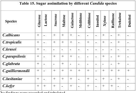
i) Sugar fermentation 12:
Liquid medium containing peptone, 2% sugar and indicator was poured in a test tube (~5ml) and Durham’s tube was placed into each tube. Heavy inoculums of
yeast colonies were suspended into each tube and incubated at 25°C for 1 week. The
[60]
tubes were examined at 48- 72 hours interval for acid (pink color) and gas (in durham’s) production. C.albicans ATCC 90028 was used as the control strain. Species
identification was done by referring the table given below
The findings were recorded and tabulated.
ii) Sugar assimilation (Auxonographic techniques) 65:
18ml quantities of agar and distilled water was dispensed in screw
capped tubes. Autoclaved at 121°C for 20 minutes and stored at 4°c. A heavy
inoculums yeast suspension was prepared from 24 hours old culture in 2ml Yeast
Nitrogen Broth. The prepared suspension was poured into 18 ml of molten agar cooled
at 45°C, and then poured into a 90mm Petri plate. The Petri plate was set at room
temperature until the agar surface hardens. Sugar discs were placed in a circle with
sterile forceps, such that they were at least 30mm was present between centers of each
disc and was incubated at 37°C 3-4 days. Presence of growth around each disc Table 14 :Fermentation of different sugars by different species
Sugars C .albi can s C .tropi cali s C .kru se i C .parapsi losi s C .glabr ata C .gu il llermon dii C .lu sit a n iae C .ke fy r
Glucose AG AG AG AG AG AG AG AG
Lactose - - - - - - - AG
Sucrose AG - - - - AG AG AG
Maltose AG AG - - -
-Galactose AG AG - - - AG AG AG
[61]
indicates assimilation of respective sugars. C.albicans ATCC 90028 was used as the
control strain. Species identification was done by referring the table given below.
The findings were recorded and tabulated.
4.8. Virulence factor detection:
4.8.1.Biofilm formation 83:
Biofilm formation was detected in all the isolates by using a method
proposed by Branchini et al. A loopful of 24-48 hours old organism were taken from
the SDA plate and inoculated into a tube containing 10 ml sabouraud's liquid medium
added with glucose -final concentration of 8%. The tubes were incubated at 37°C for
48 h after which the broth is aspirated out and the walls of the tubes were stained with
1% safranin solution. Biofilm formation was graded as:
Negative – 0
[image:61.595.77.518.135.437.2] Weak positive – 1+
Table 15. Sugar assimilation by different Candida species
Species Gl u cose L ac tose S u cr ose M altose Gal ac tose M eli b iose Ce ll ib iose In ositol Xylose Raf fin ose T re h alose Dul citol
C.albicans + - + + + - - - + - + -
C.tropicalis + - + + + - + - + - + -
C.krusei + - - - -
C.parapsilosis + - + + + - - - + - + -
C.glabrata + - - + - - - + -
C.guilliermondii + - + + + + + - + + + +
C.lusitaniae + - + + + - + - + - + -
[62]
Moderate positive – 2+
Strong positive – 3+
4.8.2. Proteinase factor