COMPARISON OF CRESTAL BONE LEVEL
AROUND ONE-PIECE AND TWO-PIECE
IMPLANTS WITH IMMEDIATE
NON-OCCLUSAL LOADING-A SPLIT MOUTH
DESIGN.
A Dissertation submitted to the
THE TAMILNADU DR. MGR MEDICAL UNIVERSITY
In partial fulfillment of the requirements for the degree of
MASTER OF DENTAL SURGERY
(BRANCH – I)
(PROSTHODONTICS AND CROWN & BRIDGE)
COMPARISON OF CRESTAL BONE LEVEL AROUND
ONE-PIECE AND TWO-PIECE IMPLANTS WITH
IMMEDIATE NON-OCCLUSAL LOADING-A SPLIT MOUTH
DESIGN
A Dissertation submitted to the
THE TAMILNADU DR. MGR MEDICAL UNIVERSITY
In partial fulfillment of the requirements for the degree of
MASTER OF DENTAL SURGERY
(BRANCH – I)
(PROSTHODONTICS AND CROWN & BRIDGE)
CERTIFICATE
This is to certify that DR.S.NASREEN,Post Graduate student (2015 - 2018) in the Department of Prosthodontics and Crown and Bridge, has done this dissertation titled
“COMPARISON OF CRESTAL BONE LEVEL AROUND ONE-PIECE AND
TWO-PIECE IMPLANTS WITH IMMEDIATE NON-OCCLUSAL
LOADING-A SPLIT MOUTH DESIGN ” under my direct guidance and supervision in partial fulfillment of the regulations laid down by The Tamil Nadu Dr. M.G.R. Medical University, Guindy, Chennai – 32 for M.D.S. in Prosthodontics and Crown & Bridge (Branch I) Degree Examination.
Guided by Head of the department
Dr.K.VINAYAGAVEL,M.D.S.,
Professor, Department Of Prosthodontics, Tamil Nadu Govt. Dental College
&Hospital, Chennai-600 003.
DR.C.SABARIGIRINATHAN.M.D.S.,
Professor and Head Of The Department
Department of Prosthodontics
TamilNadu Govt.Dental College
And Hospital, Chennai – 600 003
Head of the institution
Dr.B.SARAVANAN, M.D.S., Ph.D.,
PRINCIPAL
Tamil Nadu Govt. Dental College & Hospital,
DECLARATION
I Dr.S.NASREEN do hereby declare that the dissertation titled
“COMPARISON OF CRESTAL BONE LEVEL AROUND ONE-PIECE AND
TWO-PIECE IMPLANTS WITH IMMEDIATE NON-OCCLUSAL LOADING-A
SPLIT MOUTH DESIGN” was done in the Department of Prosthodontics, Tamil
Nadu Government Dental College and Hospital,Chennai-600 003. I have utilized the
facilities provided in the Government Dental College and Hospital for the study in
partial fulfillment of the requirements for the degree of Master of Dental Surgery in
the speciality of Prosthodontics and Crown & Bridge (Branch I) during the course
period 2015-2018 under the conceptualization and guidance of my dissertation guide
Professor Dr.K.VINAYAGAVEL.M.D.S.,
I declare that no part of the dissertation will be utilized for gaining financial
assistance for research or other promotions without obtaining prior permission from the
Tamil Nadu Government Dental College and Hospital.
I also declare that no part of this work will be published either in the print
or electronic media except with those who have been actively involved in this
dissertation work and I firmly affirm that the right to preserve or publish this work rests
solely with the permission of the Principal, Tamil Nadu Government Dental College
and Hospital, Chennai- 600 003, but with the vested right that I shall be cited as
author(s).
Signature of Student Signature of HOD
TRIPARTITE AGREEMENT
This agreement herein after the “Agreement” is entered into on this day,
________________between the Tamil Nadu Government Dental College and
Hospital represented by its Principal having address at Tamil Nadu Government Dental
College and Hospital, Chennai-3, (hereafter referred to as, ‘the College’)
And
Dr.K.VINAYAGAVEL,M.D.S., aged 45 years working as Professor,
Department of Prosthodontics and crown and bridge at Tamil Nadu Government Dental
College and Hospital, Chennai-3 having residence address at 45/M,K-9/4-TNHB HIG
Officers Apartment,Natesan Nagar West,Virgambakkam,Chennai-92,herein after
referred to as the ‘Researcher and Principal investigator’)
And
Dr.S.NASREEN aged 31 years currently studying as Post Graduate student in
the Department of Prosthodontics and Crown & Bridge, Tamil Nadu Government
Dental College and Hospital, Chennai-3 having residence address at 18/1 Vignesh
illam,State Bank Officers Colony 2nd street, Perambur, Chennai - 12 (herein after
referred to as the ‘PG/Research student and Co- investigator’).
Whereas the ‘PG/Research student as part of her curriculum undertakes to research on
the study titled“COMPARISON OF CRESTAL BONE LEVEL AROUND
ONE-PIECE AND TWO-ONE-PIECE IMPLANTS WITH IMMEDIATE NON-OCCLUSAL
LOADING-A SPLIT MOUTH DESIGN” for which purpose the Researcher and
Principal investigator shall act as Principal investigator and the College shall provide
the requisite infrastructure based on availability and also provide facility to the
Whereas the parties, by this agreement have mutually agreed to the various
issues including in particular the copyright and confidentiality issues that arise in this
regard.
Now this agreement witness as follows ;
1. The parties agree that all the research material and ownership therein shall
become the vested right of the college, including in particular all the copyright
in the literature including the study, research and all other related papers.
2. To the extent that the College has legal right to do go, shall grant to licence or
assign the copyright do vested with it for medical and/or commercial usage of
interested persons/entities subject to a reasonable terms/conditions including
royalty as deemed by the college.
3. The royalty so received by the college shall be equally by all the parties.
4. The PG/Research student and PG/Principal Investigator shall under no
circumstances deal with the copyright, confidential information and know how
generated during the course of research/study in any manner whatsoever, while
shall sole vest with the manner whatsoever and for any purpose without the
express written consent of the college.
5. All expenses pertaining to the research shall be decided upon by the Principal
investigator/Co-investigator or borne sole by the PG/Research student
(Co-investigator).
6. The College shall provide all infrastructure and access facilities within and in
other institutes to the extent possible. This includes patient interactions,
introductory letters, recommendation letters and such other acts required in this
regard.
7. The principal investigator shall suitably guide the student research right from
selection and conduct of research, topic and area research by the student
researcher under guidance from the principal investigator shall be subject to the
prior approval, recommendations and comments of the Ethical Committee of
the college constituted for this purpose.
8. It is agreed that as regards other aspects not covered under this agreement, but
which pertain to the research undertaken by the Student Researcher, under
guidance from the Principal Investigator, the decision of the college shall be
binding and final.
9. If any dispute arises as to the matters related or connected to this agreement
herein, it shall be referred to arbitration in accordance with the provisions of the
Arbitration and Conciliation Act, 1996.
In witness whereof the parties herein above mentioned have on this the
day month and year herein above mentioned set their hands to this agreement in
the presence of the following two witnesses.
College represented by its Principal Student Guide
Witnesses PG Student
1.
MY ACKNOWLEDGEMENT AND SINCERE THANKS
TO MY GUIDE
With immense pleasure and honour I take this opportunity to express my humble
and heartfelt gratitude to my mentor, a relentless source of inspiration and dissertation
guide Dr.K.VINAYAGAVEL,M.D.S., Professor, Department of Prosthodontics,
Tamil Nadu Government Dental College and Hospital, for his able guidance and
support. I am grateful for his help at various stages of the dissertation. Without his help
this dissertation would not have come out in a befitting manner. Each word said to
describe the experience as his student, which was a boon in disguise, would be an
understatement. His esteemed and able guidance made this dissertation a possibility.
His dedication to work which made us realizes the worth of discovering our own
capabilities. His unprecedented calm and patience personality, an unfailing, caring and
understanding demeanor made each endeavor easier.
ACKNOWLEDGEMENT
I am extremely thankful to DR.C.SABARIGIRINATHAN.M.D.S., Professor and
Head of the Department, Department of prosthodontics, Tamil Nadu Government
Dental College and Hospital, Chennai, for his wholehearted support, constant guidance,
help, encouragement, valuable suggestions and support he has rendered at various
stages of the dissertation. I also thank him for the valuable guidance; he has given
throughout my post graduation. Without him immense help this dissertation would not
have come out in a befitting manner.
My sincere thanks to Prof.DR.B.SARAVANAN M.D.S.,Ph.D Principal, Tamil
Nadu Government Dental College and Hospital for his kind help, valuable suggestions
in this study and permitting to use all the facilities in the institution. I also thank him
for the valuable guidance; he has given throughout the period of my post graduate
course.
With immense pleasure I take this opportunity to express my humble and heartfelt
gratitude to my dissertation guide DR.K.VINAYAGAVEL.M.D.S., Professor,
Department of Prosthodontics, Tamilnadu Government Dental College and Hospital for
his valuable guidance and support throughout this dissertation work.
My sincere thanks to DR.A.MEENAKSHI.M.D.S., Professor, Department of
Prosthodontics, Tamilnadu Government Dental College and Hospital for her valuable
guidance throughout my study.
I am thankful to my Associate Professors, Dr.P.Rupkumar.M.D.S.,
Dr.G.Sriramaprabu.M.D.S., Dr.M.Rajakumar.M.D.S., for guiding and helping me
at different stages of this study.
I am thankful to my Assistant Professors, Dr.T.Jeyanthikumari.M.D.S.,
Dr.M.Kanmani.M.D.S., Dr.S.Vinayagam.M.D.S., Dr.V.Harishnath.M.D.S.,
Dr.V.Parimala.M.D.S., Dr.J.GandhimathiM.D.S., Dr.PreethiChandranM.D.S.,
Dr.S.SivaSakthiKumar.M.D.S. for helping me at different stages of this study.
I thank Dr.Mohammed Junaid MDS (Community Dentistry), Chennai for
helping me, to carryout statistical analysis of the various test results.
I would like to thank my parents for their prayers.My special thanks to my husband
Dr.M.Shabbir Ahamed M.D.S(Periodontist) for his constant support and motivation I
owe my sincere thanks to all my senior postgraduates and junior postgraduate students
in the department for their constant encouragement and timely help.
Above all I thank the ALMIGHTY for giving me the strength and courage to
Urkund Analysis Result
Analysed Document: final pro.doc (D34582354) Submitted: 1/11/2018 6:58:00 PM Submitted By: [email protected] Significance: 2 %
Sources included in the report:
https://www.duo.uio.no/handle/10852/33071 http://www.ijdr.in/article.asp?
issn=0970-9290;year=2011;volume=22;issue=2;spage=317;epage=323;aulast=Prasad http://publications.qu.edu.sa/ojs/index.php/health/article/download/1036/985
http://www.readbag.com/personal-us-es-segurajj-documentos-revisiones-de-implantologia-implant-dentistry-pisa-consensus
Instances where selected sources appear:
7INTRODUCTION
Implant dentistry has been in constant development since the introduction of dental implants by Branemark in the 1970s1.Over the last two decades, implant treatment has become one of the first options for the prosthetic rehabilitation of edentulous and partially edentulous jaws. Several improvements have been seen in many implant-related aspects, such as surfaces, thread designs, and placement protocols. However,two main designs have remained clearly differentiated:two-piece implants, introduced and developed by Branemark and
colleagues,1,2 and one-piece implants,introduced and developed by Schroeder and colleagues in the 1980s.3-5The one-piece implant is defined as an anchorage unit and contiguous
prosthetic part manufactured as one unit, whereas a two-piece implant is defined as an
anchorage component and a prosthetic part manufactured as two separate units (Laney et al. 2007).
The original Branemark dental implant was designed as a two-piece implant to allow for a two-stage surgical procedure6.During the first surgical procedure, the top portion of the implant body is placed at the level of the alveolar crest and the gingival tissues are
reapproximated .After a healing period of 3 to 6 months, stage-two surgery is performed, in which a healing or restorative abutment is connected to the implant body, leaving an implant abutment interface (microgap) at the bone level.
The submerged approach was believed to be mandatory for obtaining successful
osseointegration by avoiding the influence of the oral flora and mechanical stresses during the healing process.However,clinical studies 7,8demonstrated that osseointegration was equally obtained when connecting the abutment to conventionally submerged two-piece implants during the initial surgery, if primary stability of the implant is achieved with
controlled occlusal loading thus avoiding a second surgical procedure. An animal study has shown that repeated disconnection and reconnection of the transmucosal component might jeopardize the soft tissue seal at the interface between the abutment and the soft tissue 9. By using an one-piece implant, where the transmucosal component are manufactured in one piece, abutment connection/disconnection is avoided 10. The abutment portion of the one-piece implant is preparable, which makes it possible to create an individualized preparation borderline that follows the anatomy of the soft tissue margin without violating the soft tissue seal. A further advantage with a one-piece implant may be that there is no gap or micro movement at the implant-abutment junction; which will have a detrimental effect on the peri-implant soft and hard tissues.11,12 To minimize the risk of soft tissue encapsulation, it has been recommended that implants be kept load-free during the healing period (3 to 4 months in mandibles and 6 to8 months in maxillae13.In general, removable prostheses are used during the healing period; however, many patients find these provisional prostheses rather uncomfortable. In 1990 the first longitudinal clinical trial supporting immediate or early loading in the mandibles of selected patients was published14.
In this study the restorative protocol is immediate non occlusal loading(INOL).Immediate non-occlusal loading was defined as the seating within 48 hours after implant placement of a provisional restoration that would not be in occlusal contact for approximately 2
2
months.Utmost care is taken that the restoration is relieved of all occlusal contacts-both centric and eccentric.15It is the marginal bone surrounding the implants that provide the stable hard tissue foundation for the soft tissue.The changes occurring on the interproximal bone are the one that have been used to qualify the success off an implant treatment protocol in most of the studies ,with reference to the criteria established by Albrektsson et al 16.
For evaluation of marginal bone levels, perpendicular intraoral radiographs were taken using the long-cone paralleling technique. Standardized Radiographs were taken at implant
placement, at the 3 rdmonth and 6th month follow up visits .Marginal bone levels on both the mesial and distal aspects of the implant were measured. The apical edge of the implant was used as a reference point for the marginal bone level measurements.Film radiographs were digitalized displayed in a software programme.Measuring tool within the software was used to make measurement taking magnification in to account10.
This study aims at evaluation of crestal bone level changes around one piece and two piece implants restored with immediate non occlusal loading in molar region over a six month follow up study using conventional intraoral periapical Radiographs.
Aim of the study:
The aim of this prospective study is to compare the crestal bone level changes around One-piece (OPI) and Two-One-piece(TPI) implants placed with immediate non occlusal loading protocol.
Objectives:
The objective of this study is
• To evaluate radio graphically the crestal bone level changes around one piece implants at Base line T0, at 3 monthsT0-T1, and at 6 monthsT0-T2.
• To evaluate radio graphically the crestal bone level changes around two piece implants at Base line T0, at 3 monthsT0-T1, and at 6 monthsT0-T2.
• To compare the crestal bone level changes between these two implants.
REVIEW OF LITERATURE
Branemark 18 coined the term Osseointegration.Osseointegration was first defined as a histological concept with direct bone-to-implant contact at the resolution level of the light microscope .
A new definition based on implant stability was given by Zarb & Albrektsson 19
0: https://www.duo.uio.no/handle/10852/33071 90%
a process whereby clinically asymptomatic rigid fixation of alloplastic materials is achieved and maintained within bone during functional loading.
3
This
Literature review is presented in five parts,
1. Studies on One- Piece Implants.
2. Studies on Two –Piece Implants placed in single stage.
3. Effect of Insertion Torque.
4. Studies regarding Immediate Loading Concepts.
5. Surgical, implant macro geometry considerations
6. Crestal Bone loss assessment and its implication.
STUDIES ON ONE- PIECE IMPLANTS
Hermann JS, Buser D et al (2001)19Made a Histometric evaluation of unloaded nonsubmerged and submerged implants in the mandiblular canine to compare the biological width around one piece and two piece implants.Histologically under unloaded condition he has found that the gingival margin (GM) is located more coronally and Biologic Width (BW) dimensions are more similar to natural teeth around one-piece nonsubmerged implants compared to either two-piece nonsubmerged or two-piece submerged implants.
Broggini N, McManus LM, Hermann JS, et al (2003)20The purpose of their study was to determine the changes in abutment connection (submerged vs. non-submerged two-piece implants) or the presence of a microgap (two-piece, non-submerged implants vs. one-piece, non-submerged implants) influences the composition of inflammatory cells immediately
adjacent to the implant.They found that the inflammatory cell infiltration for all corresponding locations was significantly greater for two-piece implants as compared with one-piece
implants. They stated that the creation of a microgap at the bone level leads to microbial leakage and a persistent bacterial presence at the peri-implant location. The chemotactic stimuli originating from the microgap then promote sustained neutrophil accumulation. In parallel, mononuclear cells are recruited to the implant surface. The combined and sustained activation of inflammatory cells promotes osteoclast formation/growth and activation to result in alveolar bone loss.
Jack A.Hahn et al (2007) 21made Clinical and Radiographic evaluation of one-piece implants used for immediate function where he placed 47 machined titanium implants which
incorporate a screw shaped implant body and fixed abutment as a single component by both using both flap elevation and flapless placement.Provisional restoration was given
immediately.Radiographs were taken at 1,2, and 3 year,where he found that the mean
marginal bone level was <2mm apical to the reference point in one piece implant(i,e 6%),this found to be less when compared with two piece implants(i,e16%).This may be attributed to the reason that undisturbed healing of the peri-implant soft tissue prevents the disruption of the soft tissue seal.He also found that the flapless placement technique contributed to beneficial
4
marginal bone levels.This may be due to prevention of separation of the periosteum from the tissue thereby maintaining the blood supply.Finally he concluded that one piece implant design resulted in a high cumulative implant survival rate and beneficial marginal bone levels.
Simonis P (2010)22made a study on Long-term implant survival and success rate upon 10-16 year follow-up of non-submerged dental implants and realized that the presence or absence of an implant abutment prosthetic connection may have affected the early bone loss and prosthetic complication.
Sohn, Min-Su Bae et al (2011)23 made a Retrospective Multicenter Analysis of Immediate Provisionalization using One-Piece Implants.In the retrospective analysis, the cumulative implant survival rate was 100% for an average 23 months of follow-up after implant
placement. The results of this study were comparable to other studies reporting low marginal bone loss . Also, the placement and restoration procedures are simple, so the surgical time can be reduced and patient discomfort can be minimized(with less postoperative bleeding, less swelling and pain, and a shorter healing period)Immediate provisionalization and functional loading have shown high success rates in different clinical reports.
José M. Barrachina-Díezet al (2013)24 In his systematic review and meta-analysis
demonstrates excellent implant survival rates for one-piece implants in long-term clinical performance. No statistical differences were observed in the implant survival rates between one-piece implants placed in completely edentulous patients and those placed in partially edentulous patients, including single-tooth edentulism. Furthermore, no differences could be found between one-piece implants placed in the maxilla compared to those placed in the mandible. There is a general trend in the literature for better results in favour of the mandible.
Zeev Ormianer et al (2016 )25 made a study in One- and Two-Piece Implants Placed in the Same Patients and assesed the Clinical Outcomes After 5 Years of Function and they have concluded that the Cumulative 1P and 2P implant survival was 100% with no significant differences in bone loss or prosthetic complications.1P and 2P implants appeared to be equally effective after 5 yrs of function in same patients.
Wolfgang Bömicke et al (2017)26 has stated that the loss of OPI may be due to immediate loading and micromotion during chewing and the prognosis of immediately loaded implants depends on the implant stability.Insertion torque of about 32 to 45Ncm has shown successful results whereas torque of about 20 to 35Ncm has shown failure rates ,so they concluded that primary stability of about 35 Ncm to be good for increasing the implant survival rates.
STUDIES ON TWO –PIECE IMPLANTS PLACED IN SINGLE STAGE
Daniel buser ,et al (1999)27 shared the clinical experience with one-stage, non-submerged dental implants and concluded that, the non-submerged placement of dental implants provide several advantages from biologic, clinical, and biomechanical points of view. It is noticeable that this approach has been utilized more by the clinicians in recent years. It can be expected that this healing modality will become the treatment of choice in implant sites where
5
esthetic is not of much priority, such as posterior implant sites in partially edentulous patients and in fully edentulous patients.
Weber HP, Crohin et al (2000)28 has conducted a study to evaluate the clinical and
radiographic changes in non-submerged dental implant and had found the mean crestal bone loss during the first year was 0.6 mm followed by an annual loss of 0.05 mm approximately.No significant difference was found between the amount of bone loss measured at each of the yearly follow-up visits.
Heydenrijk, Kees; et al (2002)29 Evaluated the feasibility of Two-part implants inserted in a one stage procedure to moniter the microflora in the peri implant area in relation to clinical and radiographic outcome. The short-term results indicate that two-part implants placed in one-stage procedure may be as predictable as inserted in the routine two-one-stage procedure. The peri-implant sulcus can harbour potential periodontal pathogens without any significant signs of tissue breakdown.
Kees Heydenrijk,et al (2003)30 Made Clinical and Radiologic Evaluation of 2-Stage IMZ Implants Placed in a Single-Stage.The results of this Prospective Comparative study reveals that the dental implants placed in a submerged implantation procedure can also be placed in a single-stage procedure and produce the same predictable results as when the same
implants used in a 2-stage procedure or as 1-stage implants.
Esposito M,et al (2010)31Made a review for replacing missing teeth with 1- versus 2-stage implant placement.They concluded that the two procedures did not show clinical significant differences. However the 1-stage procedure might be preferable since it avoids one minor surgical intervention and shortens the waiting time to provide the final restoration.There might be specific situations though, such as when optimal implant stability is not obtained at placement or when barriers are used in conjunction with implants, in which a 2-stage
approach might be preferable.
Minkle Gulati,et al (2015)32Made a study to Evaluate the crestal bone level changes between Two-Piece Implants Placed Following One-Stage and Two-Stage Surgical Protocol in Posterior Mandibular Region. They found that no significant differences were seen in both groups in terms of changes in crestal bone level at the final evaluation. Hence,they concluded that two-piece implants can be placed following one-stage surgical protocol as predictably as when two-stage surgical technique is followed.
EFFECT OF TORQUE
Roland Glauser et al (2004)33analysed the stability of implant by repeated resonance
frequency analysis (RFA)in patients treated with immediate/early-loading protocol.Low RFA denotes failing implants and we can use this information to avoid loading of implants with decreasing degree of stability with time thereby avoiding implant failure.
Jurgen Zix, Dr Med et al (2005)34 have concluded that there was no significant differences in ISQ values between implants with regard to loading period using RFA.
6
Marco Esposito et al(2007)35Made a study to find the difference in success rates between immediately, early, and conventionally loaded implants and he concluded that a high value of insertion torque (primary implant stability)seems to be one of the
important prerequisites for successful immediate or early loading of implants.
Francesco Zuffetti ,Marco Esposito et al (2016)36 Have suggested that for the implants to be immediately loaded it has to be inserted with a torque of at least 30 Ncm, and splinted implants with a torque of at least 20 Ncm.
Daniele De Santis (2016)37 Have studied that implants with self tapping blades allowed higher torque and ISQ values to be achieved at insertion.
De oliverira et al (2016)38 has Concluded that there is not strong evidence that insertion torque of 30 Ncm is enough for implant survival in cases of immediate loading.
Gary Greenstein et al (2017) 39conducted a review to know the role of insertion
torque in attaining primary stability of dental implants.He concluded that the Primary stability is desirable during implant placement, but the absence of micromotion is what facilitates desirable implant osseointegration. Increased in the insertion torque helps to achieve primary stability by reducing implant micromotion.
RATIONALE FOR IMMEDIATE LOADING
H.Weinans et al (1993)40 stated that the micromotion and stresses at the interface are
controlled by various factors such as i)Implant geometry and material properties and ii)patient factors such as magnitude of load applied,bone quality and quantity,resorption
threshold.These two factors control the interfacial resorption of bone.
Matteo Chiapasco1/Silvio Abati2 et al (2001)41Compared the efficiency of delayed and immediate loading of implants with implant-retained mandibular overdentures using Brånemark Implants.They found that the cumulative success rate of both the groups were about 97.5% and there was no detrimental changes in osseointegration with considerable short treatment duration when the implant was immediately loaded and connected with bar.
Antonio Rocci et al (2003)42 conducted a study to compare the Ti-Unite and machined-surfaced Branemark implant system with immediate loading in posterior mandible.The marginal bone resorption was found to be 0.9mm(S D 0.7m)in Ti- Unite system and that of machined surface implant was about 1mm(0.9mm).They demonstrated 10%higher success rate in Ti-Unite system.With machined implant surface the no of failed implants was found to be greater in smokers and with quality type 4.
Degidi ,Piatelli et al (2003)43 When immediate non functional loading was compared with immediate loading in a controlled study immediate non functional loading increased the implant survival rate.
7
Fabrizio Bambini,et al (2004)44Made a study to know the influence of immediate prosthetic loading on peri-implant osteoblastic activity with the help of bone scintigraphy.
Carl Drago et al(2004)45 in his study with a cumulative survival Rate of 97.4%, identified that the success of the immediate restoration protocol include primary implant
stability,elimination of occlusal contacts prior to osseointegration,dietry modifications during the initial healing period.
Massimo Del Fabbro et al (2006)46 Made a systematic review to determine the survival rate of immediately loaded implants and to compare the success rate between rough-surface
implants and machined surface implants.Finally they concluded that survival rate was good enough for IL implants and the implants with rough surface was better than machined surface.
James A.G Hall,Alan G T Payne et al (2006) 47 Made a clinical RCT trial to compare the outcome between conventionally and immediately loaded tapered rough - surface implants and after 1 year they concluded that there is no significant difference between the two group.
Gioacchino Cannizzaro,Marco Esposito et al (2008) 48 They made a study to evaluate the efficacy of immediate loaded versus early loading of implants in bar retained mandibular overdenture after 6 months.Insertion torque for immediate implants was set to be <48 Ncm.Implant stability was measured using Resonance frequency analysis(Ostell)and results expressed in ISQ(Implant Stability Quotient),by using the handles of two metallic instruments and by applying reverse torque of about 20 Ncm.High insertion torque is a pre requisite for immediate loading of implants.They have concluded that mandibular overdenture can be successfully loaded immediately with minimal invasive technique.
Matteo Capelli,Marco Esposito,et al (2010) 49 conducted a randomized clinical trial to compare the clinical efficiency of immediate non-occlusal loaded implants versus early loaded implants in partially edentulous arch they compared the peri-Implant bone and soft tissue changes between two implants. In Immediately loaded cases single piece implants were placed with a torque of <30Ncm and splinted implants with a torque of <20 Ncm.Immediately loaded implants were provided with Non occluding Temporary restoration within 48 hrs and then fully in to occlusion after 2 months.Splinted implants were early loaded implants after 2 months.Final restoration was given after 8 months.They found that there were not of much statistically significant difference between the peri implant bone and soft tissue changes between the two implants.Both the groups had 1.2mm of crestal bone loss and one piece implant had a recession of about 0.2mm after 5 yrs .
Lotte Pull ter Gunne et al (2016) 50 Made a three year RCT and found that the clinical and radiographic results of immediate loading protocol was comparable with that of the early loading protocol.
Iñaki Cercadillo-Ibarguren(2017)51 Made a Retrospective Analysis regarding Early Complications of Immediate Loading in Edentulous Full-Arch Restoration.In Immediate loading protocol the temporary fixed prosthesis must be luted immediately after implant
8
placement.It increases the chance of misfit and undesirable movement of the whole structure, and as the materials employed are weaker, fractures can also occur. Other factors such as bruxism, tobacco and alcohol consumption,diabetes,and a history of periodontal disease may increase the risk of biologic and technical complications. Many controversies exist about what is the most suitable material for provisionalization. Some authors state that all-acrylic resin prostheses have a greater shock absorption capacity due to their low elastic modulus, which reduces the amount of stress transmitted to the bone-implant interface and decreases the risk of overload. Some authors, recommended the use of a metal reinforcement inside the
provisional prosthesis making it easier to repair fractures because, in many cases, the broken piece of acrylic material remains partially adhered to the metal structure. The author
concluded that fracture of provisional prostheses is a common finding, affecting 17% of patients, and is significantly more prevalent in bruxers and in maxillary prostheses.
SURGICAL ,IMPLANT MACROGEOMETRY CONSIDERATIONS
Minimizing bone damage,Maximizing implant stability
Eriksson RA (1985)52 reported bone cell death when a temperature of 40 C was applied for 7 minutes or when a temperature of 47 C was applied for 1 minute.In other words, time and temperature are interrelated critical factors in implant site preparation.
Lee H Silverstein et al (1999)53 stated that the’Ridge expansion technique osteotomes
conserved bone.In addition ,the outcome technique is essentiall heatless and therefore should not destroy the viable bone – forming cells.
Misch et al (2004)54 The implant body design should be more specific for immediate
loading.This is even important for immediate load in single tooth applications and restoration replacing only few teeth.A threaded implant design may have some bone present in the depth of the threads from the day of insertion.Therefore,the functional surface area is greater
during immediate load format.As a result,threaded implants present considerable advantages or immediate load protocols,because their design features do not require integration to resist loads and they also have greater surface area to resist occlusal forces.
Misch et al (2008)55 The density of available bone in the edentulous site has a primary influence on treatment planning,implant design,surgical approach,healing time and initial progressive bone loading.The quality of recipient bone directly influences the amount of trauma generated during osteotomy preparation.
Jack A Hahn (2009)56in discussing the concept of the tapered design,said that the tapered design works in harmony with the anatomical constrictions of the jaws.Tapered shape often requires fewer drilling steps and allows for the placement of a wider cervical diameter implant in more favourable positions,without having to tilt the implant more than an average of 15 degrees.Increasing the cervical diameter offers a wider prosthetic platform for the
restoration,which results in less stress to the crestal bone.Having the ability to place an implant in a more favourable position for the prosthesis is also an important factor in reducing stress.
9
Mangano, Francesco et al (2017)57 made a histologic/histomorphometric study was to
compare the early bone formation around immediately loaded implants with nanostructured calcium-incorporated (NCI) and machined surface,It is a split mouth design where temporary implants were placed in posterior maxilla, After 8 weeks, all temporary implants were
retrieved for histologic/histomorphometric evaluation. The bone-to-implant contact (BIC%) and the bone density (BD%) were calculated.A statistically significant difference was found between the two surfaces with regard to BIC% ( p > 0.001), while no significant difference was found with regard to BD% ( p = 0.09).He concluded that the NCI surface seems to increase the peri-implant endosseous healing properties in the native bone of the posterior maxilla, under immediate loading conditions, when compared with the machined surface.The
nanostructured calcium-incorporated surface has led to better histologic and
histomorphometric results than the machined surface; therefore, the clinical use of implants with nanostructured calcium-incorporated surface may be beneficial.
CRESTAL BONE LOSS ASSESSMENT AND ITS IMPLICATION
Albrektsson et al (1989) 16 established criteria for implant success as follows
An individual unattached implant is immobile when tested clinically. A radiograph does not show any evidence of periimplant radiolucency. Vertical bone loss is less than 0.2mm annually following the first year of service of the implant and not more than 1.5-2mm from the implant abutment junction during the first year. Individual implant performance is characterized by an absence of persistant or Irreversible signs and symptoms such as pain, infections,
neuropathies, paraesthesia, or violation of mandibular canal. In the context of foregoing, a success rate of 85% at the end of a 5-year observation period and 80% at the end of a 10-year period are the minimum criteria for success.
P.J.Henry et al (1988) 58 has stated that the Radiographic examination of marginal bone height changes appears to be an acceptable parameter in the assessment of prognosis for osseointegrated fixtures.
Young-Chul Jung (1996) 59 has concluded that Rapid bone loss occurred in the first 3 months and it might be because of periosteal elevation, surgical trauma during recipient bed
preparation, and stress concentration because of excessive tightening of the implant and he also found that the alveolar bone showed resorption up to the polished neck after which the bone loss got,stabilized at the first thread of the implants.
Chris C. L. Wyatt et al (2001)60 has stated that A computer-assisted measurement technique for measuring bone levels proximal (mesial and distal) to oral implants imaged on
standardized intraoral radiographs provide good accuracy and reliability. There were no differences between bone measurements made directly from bone proximal to implants placed in a dry mandible and those resulting from the computer-assisted measurement technique.
Fredrick Hermann et al (2007) 61has stated that a stable bone level around an implant neck is a prerequisite for achieving support thereby a long term optimal and stable gingival
10
contours.The stable bone serves to support the soft tissue thereby determining the long term esthetic and functional outcome.
C.E.Misch(2008) 55 bone loss has been described in the crestal region of successfully
osseointegrated implants regardless of surgical approach.The effect of crestal bone loss can range from early failure of implants to esthetic complications and periimplant
diseases.According to the author ,the consequences of marginal bone loss are such that all phases of implant dentistry,from diagnosis and treatment planning to final stages of occlusion and prosthesis delivery,must focus on its reduction or elimination.
0:
http://www.readbag.com/personal-us-es-segurajj-documentos-revisiones-de-implantologia-implant-dentistry-pisa-consensus 100%
The International Congress of Oral Implantologists (ICOI) Pisa Consensus Conference,
Carl.E.Misch 62
considered the marginal bone around the crestal region of an implant to be a significant indicator of implant health.
The bone loss assessed should be related to the original bone leval at the implant
surface,rather than to the previous level.The most common method that has been used
0:
http://www.readbag.com/personal-us-es-segurajj-documentos-revisiones-de-implantologia-implant-dentistry-pisa-consensus 87%
to assess the marginal bone loss is with a conventional radiograph.Although this only measures only the mesial and distal bone loss,it is a time tested method.
The consensus on successful implants includes,no pain to be
0: http://publications.qu.edu.sa/ojs/index.php/health/article/download/1036/985 96%
observed with palpation,percussion or function.No clinical mobility is noted in any
of the
0: http://publications.qu.edu.sa/ojs/index.php/health/article/download/1036/985 84%
direction with loads less than 500mg.Less than 2.0 mm of radiographic crestal bone loss is observed compared with the
radiograph during
0: http://publications.qu.edu.sa/ojs/index.php/health/article/download/1036/985 100%
11
implant insertion surgery.The implant has no history of exudates.
Ingemar Abrahamsson et al (2009) 63 In a systematic review stated that no implant system found to be superior in marginl bone preservation.
Xi Ding et al (2009) 64 in an FE model study stated that increasing the length and diameter of implants decreased stress and thereby bone loss on the alveolar crest Diameter had a more significant effect than length..The stress and strain values notably increased under
buccolingual loading as compared with vertical loading.
Francesco Carinci et al (2012) 65 Has stated that OPI incorporate the trans-mucosal abutment which faces the soft tissues as an integral part of the implant.The interface between the trans-mucosal component and the implant is usually located in the neighbourhood of the alveolar bone level.In his report he has shown that the SVR and SCR were 91.7% and 97%, respectively for OPI.
Daniele De Santis
0: http://www.ijdr.in/article.asp?
issn=0970-9290;year=2011;volume=22;issue=2;spage=317;epage=323;aulast=Prasad 100%
et al (2016) 38 conducted a study to find the correlation between
Primary Stability and Crestal Bone Loss of Implants Placed with High Insertion Torque and concluded that implants with self tapping design has higher torque and ISQ values to be achieved at insertion,and resulted in long-term osseointegration without excessive crestal bone resorption..
MATERIALS AND METHODOLOGY
The present clinical study was conducted to evaluate the crestal bone level changes around one piece and two piece implants placed with immediate non occlsual loading protocol in the mandibular molar region of the same patient.This study was performed in the department of Prosthodontics, Tamilnadu Government Dental College and Hospital, Chennai.
ETHICAL COMMITTEE APPROVAL:
The study was carried out after obtaining approval from the Institutional Ethical Committee.
ARMAMENTARIUM:
For clinical examination
• Mouth mirror
• Williams periodontal probe
• Curved explorer
12
• Dental tweezers
• Kidney tray
• Cotton roll
• Sterilized disposable gloves
• Disposable facemask
FOR SURGICAL PROCEDURE :
• Sterile patient drapes
• Sterile towels
• Sterile gauze,cotton
• Mouth mirror
• Williams periodontal probe
• Dental tweezers
• Surgical gloves
• Disposable mouth mask
• Disposable syringe ,needle
• Lignocaine with adrenaline anesthetic solution.
• Bard parker blade no 15 and handle – straight.
• Periosteal elevator
• Implant surgical motor(Confident Dental Equipments,Banglore)
• 20:1High Speed contr-angle hand piece(NSK,Nakanishi Inc,Japan)
• Hand piece sleeve –sterilized.
• Betadine
• Normal Saline and irrigation syringe
• Straight and Curved scissors
• Curved artery forcep
• Dapen dish
13
• Straight artery forceps
• Implant surgical kit
• One piece implant (EQUINOX NO 1)
• Two piece implant(EQUINOX NO 1)
• Suture material – 3-0 black silk braided
• Needle holder.
FOR PROSTHETIC PROCEDURE:
• Permanent abutment.
• Implant hex driver
• Hand piece
• Tungsten carbide bur
• Periodontal probe
• Petroleum jelly
• Tooth coloured autopolymerizing resin
• Autopolymerizing resin monomer
• Wax Knife
• Wax Carver
• Bard Parker blade No1.5 and Handle
• Articulating paper forceps
• Articulating paper
• Temporary cement
• Permanent cement
• PolyVinyl Siloxane(Aquasil,Dentsply Caulk)
• Type IV dental stone
• Instruments for Metal Ceramic crown fabrication
RADIOGRAPHIC ARMAMENTARIUM:
14
• IOPA flim(Kodak Ekta speed Flim Adult size 2)
• Long Cone X Ray tube
• X Ray flim positioner
• Poly Vinyl Siloxane Putty(Aquasil,Dentsply)
• X Ray Grid
SUBJECT SELECTION:
Study participants were selected from the out patient department in Tamilnadu Government Dental College and Hospital with the age group of 20-40 years of both male and female
subjects were included in the study. All the participants were informed about the purpose and methods of the study and signed the informed consent.
SAMPLE SIZE:
Total number of subjects : 8
CRITERIA FOR SELECTION:
Inclusion criteria :
• Educated patient conscious of oral hygiene and willing to undergo restoration with dental implants.
• Adequate patient compliance and availability to meet the follow up schedule.
• Periodontally healthy individual.
• Either sex.
• Systemically healthy individuals
• Age group-20-40yrs.
• Missing mandibular first molars in the same patients with adequate interarch space.
• One piece and two piece implants from the same manufacturer.
• Atleast 30 N/cm of insertion torque at the time of placement.
Exclusion criteria:
• Poor oral hygiene
• Smoking and substance abuse.
• Parafunction, bruxisim or clenching.
15
• Medically compromised patients such as uncontrolled diabetes,immunosuppression, bleeding disorder, cancer, stroke & severe osteoporosis .
• Patient under bisphosphonates medication.
• Pregnancy at the time of inclusion.
• Known allergy or hypersensitivity to any product used in the study.
• Patients with drifted and supraerupted teeth in the edentulous area.
MATERIALS USED IN THE STUDY
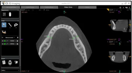
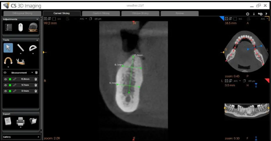
S.No NAME commercial name FORM OF THE MATERIAL MANUFACTURER DETAILS 1. CBCT Machine Computed Axial Tomographic Scans SIRONA DENTAL SYSTEMS GmbH 2.
Physiodispenser Implant surgical motor CONFIDENT DENTAL EQUIPMENTS,Banglore) 3. Myriad –Smart Implant system One-Piece Implant EQUINOX MEDICAL TECH .V THE
NETHERLANDS 4. Myriad-Plus Implant system Two-Piece Implant EQUINOX MEDICAL TECH .V THE NETHERLANDS 5. Aquasil Putty and Light body Poly Vinyl Siloxane Impression Material AQUASIL,DENTSPLY CAULK
6. X-Ray Mesh Gauge Measuring Lead -
7. Dental X ray Flim IOPA radiographic flim SIZE 2 ADULT FLIM KODAK-E SPEED
PREOPERATIVE PREPARATION:
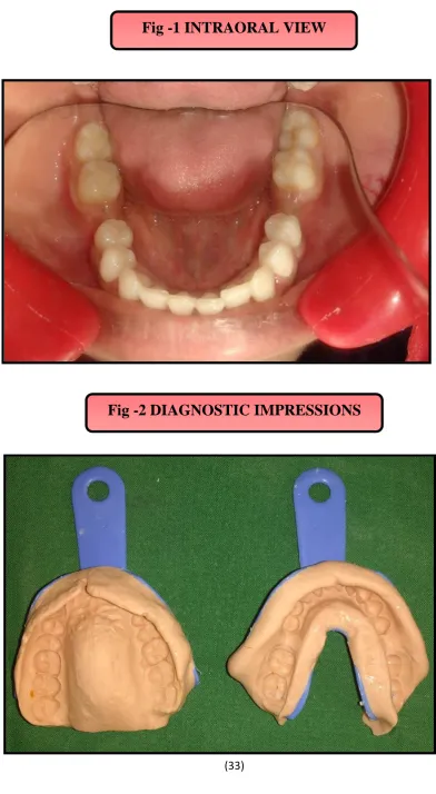
All the patients underwent thorough oral prophylaxis .
Preoperative records
1.Informed consent from the patient
2.Study casts
3.Preoperative photographs
4.Routine blood investigations
5.Orthopantographs
6.Computed Axial Tomographic Scans.
SURGICAL PROCEDURE:
Following screening, all patients are consented to the planned treatment strategy. The patients were advised to start on antibiotic (Amoxicillin 500mg 1 hr before surgery) &
analgesic (Ibuprofen 600mg 1hr preoperatively). Oral disinfection was performed using a 0.2% chlorhexidine digluconate mouthwash.
16
The patient’s face is disinfected with 7.5% povidone iodine. The oral cavity is prepared with 5% povidone iodine and the patient is draped as per the routine surgical procedure.
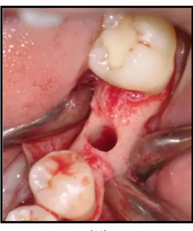
After giving local anaesthesia to the patient a mid-crestal incision was made and a mucoperiosteal flap was elevated.
Initial access of the osteotomy site was made using the D2.0mm pilot drill.The pilot drill was used to establish depth and axis/direction of drilling. Once the depth was established with the pilot drill it is not advisable to change or vary drill depth with subsequent drills but only to progressively widen the osteotomy site. Direction or axis of drilling can be varied slightly if required with subsequent drills. Recommended drill speeds with pilot drill is between 800-1000rpm.Pilot drill depth was decided according to the length of the implant planed.
The osteotomy site was then prepared with the subsequent drill of increasing diameter with that of same length .Drill speed recommended here was 500-800 rpm.The implant can be inserted /turned into place using the torque ratchet or the handpiece.Final tightening is recommended to be done by the torque ratchet with an optimal insertion torque of between 30-40Ncm. After achieving a desired primary stability one piece implant was placed on the desired quadrant in which it was planned and checked for clearance by asking the patient to close his/her mouth and if satisfactory the flap is closed by means of interrupted sutures if the clearance is insufficient then modification of the abutment is done and then the flap is closed. Same procedure is followed on the other side of the arch and after preparing the osteotomy site for two piece implants ,the root fixture is placed and immediately the abutment was also placed by means of two piece one stage loading protocol and then the flap was closed by interrupted sutures.
Postoperative instructions include avoidance of the surgical site while brushing and eating, the use of a 0.2% chlorhexidine mouthwash two times a day for 2 weeks , antibiotic therapy for 5 days (Amoxicillin 500 mg three times a day) and analgesic (Ibuprofen 600 mg maximum two times a day) was continued.
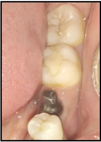
PROSTHETIC PROTOCOL;
The restorative protocol in this study was Immediate Non Occlusal Loading[INOL]protocol. Also known as Immediate Restoration of the implants wherein the implant is placed and the abutment connected on the same day in a single stage in a single stage in case of two piece implants, whereas it is unibody design in one piece implants. This abutment is utilized to support a provisional prosthesis out of occlusal contact that is luted in place within the first 48hrs after surgery. Utmost care is taken that the restoration is relieved of all occlusal contacts-both centric and eccentric.
This modality has the clinical advantage of increasing patients acceptance of oral implants as the fixture was provisionalized immediately.It also involves a single surgical phase, thereby eliminating the additional surgical trauma the patient is subjected to in the second surgical phase.The overall treatment time is reduced in terms of soft tissue healing and
17
maturation.After loading of the implants post-operative radiographs [standardizationdone] were taken for both the sites for initial i,e baseline assessment[T0].
Follow up was done and crestal bone level assessed with the help of radiographs
[standardization done]at three months[T0-T1] on both the sides. Provisional restoration was removed and final impression made with the acrylic special tray with putty and light body (ELASTOMER;AQUASIL;ADDITION SILICONE)impression material for fabricating a cement retained prosthesis. An abutment level impression was made and working model poured with Type IV dental stone and provisional restoration again luted in patient’s mouth. Wax pattern was made and the metal framework tried in patients mouth.Approprite shades of porcelain were fired in dentin and enamel layers and glazed to complete the final restoration.Final prosthesis was luted .[cement retained prosthesis].Regular follow-up was done and crestal bone level assessed after six months of placement [T0-T2]with the help of radiographs [standardization done].
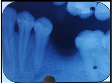
RADIOGRAPHS TO EVALUATE OF CRESTAL BONE LOSS
The radiographic technique used in this study was Intraoral Panoromic Radiographs using long cone (paralleling)technique with a Rinn positioning device.The exposure parameters were kept standardized at 70 kVp,10 Ma ,0.2 seconds.In the long cone imaging technique ,receptor and the object are parallel to each other.Owing to this parallelism ,image shape distortion is minimized.Standardisation of radiographs were done with X-ray lead grid in which each squares corresponds to 1mm2 and it is affixed over the flim .The bite block is relation to the existing natural teeth and was indexed with putty material (Additional Silicone) to ensure maximum positional reproducibility.
Both reference and final radiographs were made,Reference radiographs were taken on the day of placement of implant,at 3months ,and at 6months with definitive restoration.The vertical components of the periimplant Crestal bone loss was assessed on the mesial and distal sides separately near the implant by having the apical end of the implant as the reference point.The radiographic image was digitalized using the (IMAGE J) software
programe and the measuring tool within the software was used to make measurements.The radiographs can be adjusted for brightness,contrast,and zoom of the images to obtain optimal measuring condition.
METHODOLOGY:
STUDY DESIGN:
SURGICAL PROTOCOL – IMMEDIATE RESTORATION
PROSTHETIC PROTOCOL – IMMEDIATE NON OCCLUSAL LOADING
RESULTS
This study evaluated 8 patients for a period of six months making a detailed note on the hard tissue parameters i,e the crestal bone level changes around one piece and two piece implants
18
placed with immediate non occlusal loading.With regard to complications and failures ,no implant was excluded from the study as all the 8 implants which were placed showed good stability and osseointegration. All patients showed good compliance and healing period was uneventful. The observations and results of various other parameters are summarized in the tables and figures.
Data analysis was performed using the patient as the experimental unit.For all parameters, the mean values per subject and per visit were calculated. The statistical analysis was done using the computer software program SPSS version 16.0 (Statistical Package for Social Science, Version 16).All the descriptive data are presented as mean ± SD and range values. This
software package allows the user to carryout statistical analyses and help education
reasearchers, among others, to understand the value of the obtained data during a study.
Intergroup comparison between one piece and two piece implants based on the crestal bone level changes at various time period was carried out by using Independent Sample t-test. Paired sample t-test is a statistical technique that is used to compare two population means in the case of two samples that are correlated. Percentage difference between one piece and two piece implants at various time intervals has been calculated by using Mann-Whitney Test.Intra group comparison i,e within one piece and two piece implants was done by using repeated measures ANOVA.Multiple comparisons of crestal bone level changes in one piece and two piece implants at various time frame was assessed using Bonferroni. Post hoc test.
P value
The P value or calculated probability was the estimated probability of rejecting the null hypothesis (H0) of a study question when that hypothesis was true. The smaller the p-value, the more significant the result will be. All P-values are two tailed, and confidence intervals were calculated at the 95% level. Differences between the two populations were considered significant when the P value is ≤ 0.05.
Finally the mean P value calculated does not show much of significance
with a P value of about 0.274, 0.194, 0.162 at T0, T1, T2 respectively.However the intragroup comparison within the group shows a P value of about 0.000 which was found to be highly significant.
Values tables:
Table 1:gives all the details regarding the patients,the implants used ,the insertion torque achieved and the site of placement of implant in 8 subjects.
Table 2:gives the measured bone height around one piece implant at the mesial and distal sites during various time interval in 8 subjects.
Table 3:gives the measured bone height around two piece implant at the mesial and distal sites various time intervals in 8 subjects.
19
Table 4:gives the difference in the crestal bone level heights in relation to one piece implants at various time intervals in 8 subjects.
Table 5:gives the difference in the crestal bone level heights in relation to two piece implants at various time intervals in 8 subjects.
Statistical tables:
Table 6:shows the mean and S.D values of crestal bone height changes around one piece implants which was found to be 12.126+0.697 , 11.528+0.665 , 11.108+0.200,at T0(Baseline),T1 (At 3 months),T2(At 6 months)respectively .It also shows the mean an S.D values of bone height changes around two piece implants which was found to be 12.574+0.866, 12.084 +0.940, 11.756+1.102 at T0(Baseline),T1(At 3 months),T2(At 6 months) respectively.The mean difference between OPI and TPI was found to be -.447,-.555.-.648 with a P value of
0.274,0.194,0.162 respectively which shows no significant difference between one piece and two piece implants.
Table 7:shows the mean difference and S.D values of crestal abone level changes around OPI and TPI. The mean difference between OPI and TPI was found to be 0.108,0.200,0.092 with a P value of 0.271,0.224,0.267 respectively which shows no significant difference between one piece and two piece implants
Table 8:shows the mean percentage difference in bone levels around one piece and two piece implants at various time intervals (T0-T1)(T0-T2),(T1-T2).with a P values of about
0.093,0.093,0.294.respectively which shows that no significant difference exist between one piece and two piece implants.
From all the above tables we infer that there is no significant difference in the crestal bone loss between one piece and two piece implants placed in same patients with a six months followup.
Table 9:shows the mean crestal bone level changes within two piece implants at varying time intervals.The mean values found to be 12.57,12.084,11.756 ,at baseline(T0),at 3 months(T1),at 6 months(T2)respectively.The P value found to be 0.00 at all the time period which is found to be highly significant.This table shows that irrespective of the design of the of the implant crestal bone loss will occur after implant placement.
Table 10:shows the mean crestal bone level changes within one piece implants at different time intervals.The mean values found to be 12.126,11.528,11.108 at baseline(T0),at 3 months (T1),at 6 months(T2)respectively.The P value found to be 0.00 at all the time period which is found to be highly significant.This table shows that irrespective of the implant design crestal bone loss will occur over a period of time after implant placement.
Table 11:shows the comparison of difference in mean crestal bone levels between various time frames.For one piece implant the P values at various time frames (T0 Vs T1),(T1 Vs T2),(T0 Vs T2) found to be 0.001,0.002,0.001 respectively. For two piece implants the P values at
various time frames (T0 Vs T1),(T1 Vs T2),(T0 Vs T2) found to be 0.0001,0.0001,0.0001
20
respectively. All the values are found to be highly significant indicating that crestal bone loss will tend to occur over a period of time.
DISCUSSION:
This study involved the method of placing one piece and two piece implants in the molar
region of the same patients and loading it immediately and thereby assessing the crestal bone level changes between these two implants.From the patient point of view he benefits from having a fixed prosthesis that too which has been loaded immediately.From clinical point of view this study helps us to compare the crestal bone level changes taking place between one piece and two piece implants.
SURVIVAL & SUCCESS
Clinical outcomes can be assessed by primary and secondary predictors.The primary primary predictor was the presence /absence of the implant at the end of the observation period which is defined as the implant survival rate(SVR).It simply refers to the no of implants still in place at the end of the followup period.Second predictor was the periimplant bone resorption which is defined as the implant success rate(SCR).
Based on the short term results of this present study which has been conducted for a time period of 6 months it has been considered to be a successful treatment strategy with a cumulative implant survival rate of 100% after 6 months of function.This results are comparable to other short term studies conducted by Zeev Ormianer,Mariusz Duda et al, (2016)25using the same protocol which has been used for this study,where the survival rate rate was 100% with no significant differences in bone loss with a P value of P=0.1952.In a systematic review Literature with meta analysis conducted by Barrachina,Esam et al (2013) 24the longterm outcome shows the survival rates of about 96.79% for one piece implants and about 98.16% for two piece implants respectively.This difference however ,was not clinically significant (P=0.3073).Reviews by Pjetursson et al also support this study.In order to get the expected survival rate strict patient selection criteria is very much important.Comparison of implants with respect to success rates is difficult because of various study protocols
involved.Most of the studies states success criteria as absence of pain,
inflammation,mobility,and peri-implant radiolucency.Thus in this study success of either one piece or two piece implants in terms of osseointegration and biological acceptance is
unquestionable.
SELECTION OF IMPLANT DESIGN
In this study two specific designs of implants were used ,one piece implant and two piece implant designs(MYRAID).This is root shaped tapered implant (Anaform) which is the most proven and versatile shape for both immediate and delayed implantation.High primary stability has been achieved with tapered implant design when compared to cylindrical design.The threaded implanat design may allow some bone to get locked in to the threads thereby increasing the functional surface area which is of greater importance with respect to immediate loading protocol followed in this study.The implant used in this study has a
21
moderately rough surface such as nanopore surface which show stronger bone response than the relatively smoother or rougher surfaces.Rough surfaces such as aggressively etched and blasted or plasma sprayed have increased incidence of peri-implantitis due to increased risk of retaining bacteria when exposed to oral environment.The implant has a uno prosthoteic
design with in-plane switching that controls crestal bone loss around implats.All the one piece implants used in this study had a uno platform of about 4.4mm and the two piece implants had a platform of about 5mm.Studies have been done by Barrachina (2013)24where
comparison has been done between one piece implant with TPS (Titanium Plasma Sprayed and SLA .One piece implants with SLA surface showed a survival rate of 99.23% when compared to 97.94%in implants with TPS surface.
Comments on OPI:In an attempt to simplify original Branemark concept and to establish success in immediate implant placement and immediate loading protocol the concept of one piece implant was evolved.It incorporates transmucosal abutment as an integral part of the implant thereby it eliminates the structural weakness in two piece implant. It can also be used immediately for function and also eliminate the need for second stage surgery which results in psychological trauma to the patient and to the soft tissue.OPI has no microgap or micro movement between implant abutment junction thereby the amount of bacterial colonization and inflammation is reduced.Studies done by Broggini, Manus et al(2003)66 and by Quiryen M ,Steenberghe et al(1993)67support this concept. Broggini et al have stated that there is a significant difference in the inflammatory cells between one piece and two piece
implants.Studies done by Quiryen reported accumulation of microorganism even in the internal surface of the screw hole.Due to the lack of any connecting screw it is possible to design implant of smaller diameter in OPI which can be used in narrow edentulous spaces. Despite of various advantages OPI has some disadvantages,In OPI it is not possible to change the angulation of the abutment after placement so much of precise placement is necessary .The second limitation is that because if immediate restoration the risk of over loading be a drawback during initial healing phase.Similarly there is only limited option for getting finish line configuration.Roynesdal et al (1999)68 have reported not to use OPI in augmentation or GBR(Guided Bone Regeneration)procedure as these procedures may require tight closure of the wound to prevent infection and to prevent bone or membrane exposure.
Comment on TPI:As the name suggest it has two separate parts i,e implant fixture which is submerged during the first surgical procedure and the transmucosal part which is connected to the implant at the second surgical procedure after a period of submerged healing.TPI can be placed in either single stage or two stage procedure.The major reason for inserting implant in two stage procedure is to minimize the risk of infection thereby the periimplant soft tissue is separated from the oral environment during the healing period.Studies done by Bernard et al (1995)69 Erickson et al (1994)70, Barber et al (1996)71,Becker et al(1997)72, Abrahamsson et al (1999)73 have stated that the two part implant palced in single stage procedure offer the same clinical and radiographical outcome when it is inserted in two stage procedure.
In this study we have placed one piece implant and two piece implants of same dimensions in same patient at two molar regions.In this the TPI is palced following the single stage
protocol,so that both the implants (OPI & TPI)were exposed to same loading conditions.
22
SURGICAL PROCEDURE
As far the surgical protocol is considered it can be either flap elevation technique or flapless technique. With respect to flapless technique there is reduction in the surgical trauma,
surgical time, bleeding is also minimized intraoperatively,the blood supply is also maintained and no need for suturing.Inspite of several advantages it has its own limitations such as ,it is dificuult to visualize the anatomical landmarks and the presence of any bony defects such as dehiscence,angulating the implants and to completely visualize the bone morphology is also difficult. Thermal trauma to the bone is more due to lack of irrigating solution reaching the site cannot modify circumferential soft tissue. These shortcomings can be overcome by flap elevation procedure. Another disadvantage in following flapless procedure is that there is possibility of contamination of implant as the epithelial or the connective tissue may get deposited in the surface of the implant thereby interfering with the osseointegration.Studies have been done by Fortin, Bosson et al (2006)74and Nkenke Eitner et al (2007)75regarding the surgical procedures followed during implant placement.
In this study we have followed the flap elevation surgical protocol for the placement of both OPI & TPI.The advantage is that it .helps in assessing the quality and morphology of the bone. The circumferential soft tissue can be manipulated to get ideal dimensions of keratinized mucosa. However the importance of keratinized mucosa around the implant is debated.
Implants with polished neck or implants with machined surface absence of keratinized gingiva is not of much importance but in case of rough – surfaced implants(sand blasted, large grit acid etched), the presence of keratinized gingiva is critical. Wood et al(1972)76 has
demonstrated relation between surgical procedure(flap elevation)used gingival recession and bone resorption.
INSERTION TORQUE
The term dental insertion torque refers to the moment of force used to insert an implant into bone. This force affects primary stability (mechanical stabilization of the implant), which helps achieve osseointegration.Primary stability denotes a lack of implant micro motion (detected with the naked eye). Movement of 100 pm to 150 pm is deleterious to implant
osseointegration,because it induces peri implant bone remodelling, fibro-encapsulation, and implant loss.
Besides the geometrical aspect and surgical aspects of implants, osseointegration also depends upon primary implant stability. Implant stability of about <30 Ncm was pursued in each cases and is considered to be one of the pre requisite for immediate loading. Studies by Greenstein et al(2017)39 states that Primary stability (mechanical stabilization) is attained when an implant is seated in bone and demonstrates no visible movement when forces are applied to it.It depends upon various factors such as bone density (quality), percentage of initial bone-implant interface, bone quality and dimensions, implant geometry, surface
micromorphology, magnitude of insertion torque, and surgical technique used when creating an osteotomy. In contrast, secondary stability (biological stability) occurs when new bone is formed along the implant interface.7 Bone deposition results in biological anchorage of the implant, and this is synonymous with osseointegration.16Primary stability is desirable when
23
placing implants, but the absence of micromotion is what facilitates predictable implant osseointegration. Increased insertion torque helps achieve primary stability by reducing implant micromotion. Trisi et al (2011)77confirmed that increased insertiontorque reduced implant micromotion when the implants were subjected to lateral forces. However, it depends upon bone quality, implant designs and drilling techniques. Akkocaoglu M, Uysal S, et al (2005) 78stated that ,there may be no benefit in increasing insertion torque in dense bone after stability is attained. He also stated that a moderate increase of insertion torque may provide some benefits with respect to reducing micromotion, especially in less dense bone. Alves CC, Neves M(2009)79 stated that another technique to enhance initial stability is to use tapered implants, which develop elevated insertion torque forces.Siepenkothen et al (2007)80 has stated that getting a high degree of primary stability is very much crucial in one-piece
implants,,because the design of the implants does not allow to submerge the implant below the gingiva during early periods of healing.
In this study all the implants OPI & TPI were immediately loaded with an insertion torque of about < 30Ncm,which is the ideal torque value when the implant is to be immediately
loaded.However in some cases where the bone is dense the implants were wrenched to an insertion torque value of about 20 & 25 Ncm.The drilling speed used is abo