A STUDY ON PREVALENCE OF VOIDING AND DEFECATORY
DYSFUNCTION IN POSTMENOPAUSAL WOMEN WITH
PELVIC ORGAN PROLAPSE
DECLARATION
I hereby declare that this dissertation titled “
A study on prevalence of
voiding and defecatory dysfunction in postmenopausal women with pelvic
organ prolapse”
is carried out by me under the guidance and supervision of
Dr.Aruna Nitin Kekre, Professor of Obstetrics and Gynaecology Unit-II,
Christian Medical College, Vellore. This dissertation is submitted in partial
fulfillment of the requirements for the degree of M.S in Obstetrics and
Gynaecology examination of the Tamil Nadu Dr.M.G.R.Medical University to
be held in April 2017.
Vellore
Dr. Nanthini. V
CERTIFICATE
This is to certify that the “
A study on prevalence of voiding and
defecatory dysfunction in postmenopausal women with pelvic organ
prolapse”
is a bonafide work of
Dr. Nanthini.V
that was carried out under my
guidance and supervision for the M.S. (Obstetrics and Gynaecology)
examination of the Tamil Nadu DR.M.G.R. Medical university, to be held in
April 2017.
Dr. Aruna Nitin Kekre
Professor of Obstetrics and Gynaecology –Unit II
CERTIFICATE
This is to certify that the dissertation “
A study on prevalence of voiding
and defecatory dysfunction in postmenopausal women with pelvic organ
prolapse”
is a bonafide work of
Dr. Nanthini.V
that was carried out under my
guidance and supervision for the M.S. (Obstetrics and Gynaecology)
examination of the Tamil Nadu DR.M.G.R. Medical university, to be held in
April 2017.
Dr. Annie Regi
Professor and Head
Department of Obstetrics and Gynaecology
Christian Medical College & Hospital, Vellore-632004.
Tamil Nadu.
The Principal
ACKNOWLEDGEMENTS
I would like to express my sincere gratitude to Dr.Aruna Nitin Kekre, Professor of
Obstetrics and Gynaecology- Unit-II, Christian Medical College and Hospital, for her
valuable help, guidance and support in this endeavor. I am especially grateful to her for
introducing me to this subspecialty of urogynaecology and motivating me to do this work on
postmenopausal women with pelvic organ prolapse.
I am grateful to Dr. Lilly Varghese, Professor and Head of Obstetrics and
Gynaecology- Unit II, Dr. Vaibhav Londhe, Associate Professor of Obstetrics and
Gynaecology- Unit II, for their co-guidance. I am indebted to Dr. Nitin Kekre, Professor and
Head of Unit -II, Urology Department, Christian Medical College, Vellore, for his valuable
guidance and support.
I would like to acknowledge Dr. Thambu David, Professor and Head of Medicine
-Unit II for creating interest in research and Dr. Vishalakshi, Department of Bio-statistics, for
her valuable help in statistical analysis of data. I am thankful to the institute research
committee for their suggestions and financial support.
My heartfelt gratitude to Mr. Madhan, from department of CEU, Mrs. Manimozhi and
Mr. Samuel Jebaraj from Obstetrics and Gynaecology Unit II for their help and assistance. I
am thankful to my patients for their willingness and co-operation, without whom this study
would not be possible. I thank my husband Dr. N. Saravanan and my daughter S. Lithika and
my parents for their support and encouragement for doing this work.
CONTENTS
1.
INTRODUCTION
1
2.
AIMS AND OBJECTIVES
3
3.
REVIEW OF LITERATURE
4
4.
MATERIALS AND METHODS
45
5.
RESULTS
59
6.
DISCUSSION
71
7.
CONCLUSION
76
8.
BIBLIOGRAPHY
77
1
Introduction
International continence society defined pelvic organ prolapse “as the the symptomatic
descent of the anterior vaginal wall, the posterior vaginal wall, the apex of the vagina or the
vaginal vault” [1]. Women with POP may presents with symptoms of urinary incontinence,
fecal incontinence, voiding dysfunction, and defecatory dysfunction, this interrelated group
of conditions collectively describe as “Disorders of the Pelvic Floor” [2]. POP usually have
multifactorial risk factors often harmonize with each other leads to severe illness .The
symptoms of POP can impact her day to day life ,sexual function, adversely have emotional
impact [3].
Management of Pelvic organ prolapse involves significant healthcare resources; In
United States about 300 million dollars are spent on the health care of women with POP
during 2005 [4] The “surgical repair of prolapse” was the common surgery performed in
elderly women during the period of 1979 to 2006 [5]. As age advances POP increased and
result in negative impact on health care [6]. Women with symptomatic POP may present with
a single symptom such as vaginal bulge, pelvic pressure, urinary, bowel and sexual
symptoms.
Understanding of the symptoms attributed to prolapse allows one to counsel patients
appropriately about the surgical repair. Until recently, the association between specific
2 In recent years, several studies have attempted to fill this void. The present study aims to
understand the prevalence of voiding and defecatory dysfunction in postmenopausal women
with pelvic organ prolapse and to correlate with stage and compartment of prolapse. The
available outcome of these measures can be used by clinicians and researchers to assess the
functional outcomes of prolapse and its treatment on patients with an emphasis on symptom
3
Aims and Objectives of the Study:
Aims:
To study the prevalence of voiding and defecatory dysfunction in post- menopausal women with pelvic organ prolapse.
Objectives:
Primary objective
To correlate the stage and compartment of prolapse with voiding dysfunction using maximal flow rate( Q max) and post void residual urine
volume( PVR) and defecatory dysfunction using CRADI- 8 (colorectal
distress inventory-8) bowel subscale score with in PFDI-20 (short form of the
pelvic floor distress inventory).
To correlate stage of pelvic organ prolapse(POP) with Lower urinary tract symptoms(LUTS) using International prostate symptom score (IPSS)
To correlate the stage of POP with quality of life (QOL) using International prostate symptom score (IPSS).
Secondary objective
To correlate stage of prolapse with the preoperative and postoperative maximal flow rate (Qmax) and post void residual urine volume (PVR).
To correlate the pre and postoperative voiding dysfunction
4
Review of Literature
Incidence
The longevity of life is increasing in India. Aged population more than 60 years has
grown from 6% in 1950 to 8% in 2000 and is expected to be 13% in next 10 yrs [8,9]. About
30% of women attending gynecology OPD and 50% of women over 50 years have POP [10].
According to a study done by Oslen et al, there is 11% life time risk for surgical procedure
due to pelvic organ prolapse and 30% will require repeat surgery [11].
The WHO assessments showed 33 % of the total global burden of disease is related
to women health [12,13]. POP world wide prevalence is 2 to 20 % less than 45 years of age
[14]. In India women suffer from POP is about ,1 million and more in reproductive age
group[15]. The POP present in 50% of women and only 20% taken treatment. [16].
In this hospital CMC& H(Tertiary care hospital located in Vellore, SouthIndia).
Every month more than 500 patients attending Gynaecology Outpatient department and
about 150 surgeries are done for gynecological Problems per month and nearly 10 surgeries
5 History:
Pelvic organ prolapse (POP) was first described in medical history by “Ebers
Papyrus in 1500 B.C”. pelvic organ prolapse remains a common and devastating female
pelvic floor disorder, with significant effect on quality of life. Recommendations “to repair
the displaced womb" were described in the Ebers Papyrus. Hippocrates described
pomegranate pessaries to reduce POP. Succussion (tying a woman upside down by her feet
until the prolapse reduced) was described in history. Leonardo Da Vinci (1452-1519)
contributed to texts after extensive cadaveric pelvic dissection. Andrea Vesalius enlightened
the entire female genital tract and uterine ligaments. By the end of the 16th century, pessaries
were being widely used, evolving from lint balls to those made of brass, cork, wood or metal,
then rubber in 1844.
During the latter half of the 19th century dramatic advances occurred in surgical
instruments. James Sims propagated the use of silver sutures in pelvic repair surgery in 1852.
The first vaginal hysterectomy surgery for POP was reported by Choppin, of New Orleans,
in 1861. Alwin Mackendrot described the pelvic connective tissue ,cardinal ligaments in
1895. Archibald Donald and William Fothergill established the Manchester operation to
counter POP. Le Fort developed partial colpocleisis in 1877, a technique still used today. In
1898, Thomas Watkins, not believing in removal of the uterus ,with out any disease, and
described interposition surgery. In 1971 two American gynecologists, Randall and Nichols
reported the surgical outcomes of transvaginal sacrospinous fixation in women with vault
prolapse. Two major changes have occurred in POP surgery: introduction of vaginal mesh
and advanced endoscopic surgery. Graft use in pelvic reconstructive surgery can be
6
ANATOMY OF PELVIC SUPPORT —The muscles, connective tissue of pelvic floor and
and its attachment bony pelvis gives anatomic support to pelvic organ (fig 1)
[image:13.612.133.516.171.398.2]
7 The levator ani muscle complex, contains pubococcygeus, puborectalis and iliococcygeus
[image:14.612.78.536.179.470.2]muscles, which gives primary support to the pelvic organs. (Figure 3 and Figure 4).
8 Figure 3. Pelvic diaphragm - view into the pelvic floor illustrating the muscles of the pelvic
diaphragm and their attachments to the bony pelvis.
The endopelvic fascial attachments, referred to as the uterosacral and cardinal
ligaments, steady the pelvic organs in the correct position so that the pelvic muscles can
10
Levels of pelvic organ support — The levels of vaginal support was described by De
[image:17.612.134.559.150.460.2]Lancey [19,20]. The endopelvic fascia connects levels of support .
Figure 5. DeLancey levels of vaginal support.
●Level 1 – complex of Uterosacral/cardinal ligament supports the uterus and upper vagina
this support represents vertical fibers of the paracolpium that are a continuation of the
uterosacral/cardinal ligament complex which inserts variably into the cervix and vagina
11 In a magnetic resonance imaging (MRI) study of asymptomatic women, the uterosacral
ligaments were found to originate on the cervix in 33 percent, cervix and vagina in 63
percent, and vagina alone in 4 percent [22]. Loss of level 1 support contributes to the
prolapse of the uterus and/or vaginal apex.
●Level 2 – Paravaginal attachments along the length of the vagina to the superior fascia of
the levator ani muscle and the arcus tendineus fascia pelvis (also referred to as the “white
line”). Loss of level 2 support contributes to anterior vaginal wall prolapse (cystocele).
●Level 3 – Perineal body, perineal membrane, and superficial and deep perineal muscles,
which support the distal one third of the vagina. Anteriorly, loss of level 3 support can result
in urethral hypermobility. Posteriorly, loss of level 3 support can result in a distal rectocele or
perineal descent.
Nerve supply — The innervation of the pelvic region derives from the S2, S3, and S4
segments of the spinal cord, which fuse to form the pudendal nerve. The pudendal nerve
innervates the external anal sphincter, whereas the levators, coccygeus muscles, and
urogenital diaphragm appear to be innervated by a direct connection of S2, S3, and S4 nerve
fibers [23].
Pelvic floor dysfunction
Refers to a wide range of symptoms that occur when muscles of the pelvic floor are weak,
tight, or impairment of the sacroiliac joint, lower back, coccyx, or hip joints. Symptoms can
12 organ protrusion [24]. Tissues adjoining the pelvic organs may have increased or decreased
sensitivity or irritation resulting in pelvic pain. Pelvic floor dysfunction may comprise group
of clinical conditions that includes urinary incontinence, fecal incontinence, pelvic organ
prolapse, voiding dysfunction due to motor and sensory abnormalities of the lower urinary
tract, defecatory dysfunction, sexual dysfunction and several chronic pain syndromes,
including vulvodynia. The three most definable conditions faced clinically are urinary
incontinence, anal incontinence and pelvic organ prolapse [25].
RISK FACTORS
The pelvic organ prolapse has multifactorial etiology and results from a combination of risk
factors, which vary from patient to patient. Established risk factors for POP include parity,
advancing age, and obesity [26,27]. To understand the etiology of prolapse is to prevent and
treat the problem in early stage.
The site-specific defect theory is based on the fact that tears in the ‘‘endopelvic
fascia” surrounding the vaginal wall permit herniation of the pelvic organs. The vaginal wall
is composed of squamous epithelium, smooth muscle muscularis, and adventitia. All
elements are embedded in anextracellular matrix that includes collagenand elastin fibers and
smooth muscle. Abnormalities of any of these components may contribute to vaginal
dysfunction andthe development of pelvic organ prolapse.
Vaginal delivery is the greatest risk factor for pelvic organ prolapse, due to neuromuscular
injury that leads to pelvic organ prolapse . Snooks performed a studies to document the
13 muscles of women with prolapse can be documented by pelvic magnetic resonance imaging
as well as on muscle biopsy [11,12 ].
A r study by DeLancey documented injury to the levator ani muscles in vaginally parous
women.[11,12].The evidence for neuromuscular injury contributing to pelvic organ prolapse
is playing an important role in the development of prolapse in women.
Parity: The risk of POP increases with increasing parity [28,29]. The Oxford Family
Planning study, a prospective cohort study of more than 17,000 women followed for 17 years
found that, compared with nulliparity, the risk of hospital admission for POP increased
markedly after the first (4-fold) and second birth (8-fold), and then increased less rapidly for
subsequent births (third: 9-fold; fourth: 10-fold) [29]. Among parous women, it has been
estimated that 75 percent of prolapse can be attributed to pregnancy and childbirth [30].
Obstetric factors in addition to parity can influence the risk of prolapse. POP can
develop during pregnancy prior to delivery. Vaginal delivery is associated with a higher
incidence of POP than cesarean. In the Pelvic Organ Support Study (POSST), increasing
parity was associated with advancing prolapse[29].
Age: The POP more in Older women [31]. 100% increased risk of prolapse for each decade
of life by POSST [29]. Olsen showed the total incidence of primary operations for
prolapse and incontinence increased from 0.1% at 20- to 29-age group to 11.1% in 70- to
79-age group.
Aging is a complex process and the increased incidence of prolapse may be the result
of the combination of physiological aging, hypoestrogenism, and an increased incidence of
14 Women who develop prolapse at early age have a more severe form of the disease
than older women[31]. Women with spina bifida and weak pelvic floor muscles may
present at a young age with advanced pelvic organ prolapse.
A progressive increase in the rate of prolapse with age was reported in one study
among 1000 women presenting for an annual gynecological exam; every additional 10 years
of age conferred an increased risk of prolapse of 40 percent [32]. In contrast, in the NHANES
study described above, the proportion of women with symptomatic prolapse was lowest in
young women and then remained fairly constant over age 40 years: ages 20 to 39 (1.6
percent); 40 to 59 (3.8 percent); 60 to 79 (3.0 percent); and ≥80 (4.1 percent). women likely
to have a neuromuscular etiology for their prolapse.
Obesity: Overweight and obese women (body mass index >25) have a two-fold higher risk
of having prolapse than other women [32]. While weight gain is a risk factor for developing
prolapse .However, it is controversial whether weight loss results in prolapse regression. A
study of 16,608 postmenopausal women found no association with weight loss and
regression of POP [33]. However, there are reports of POP regression in women after
bariatric surgery [34].
Numerous studies have identified obesity as an independent risk factor for stress
urinary incontinence. [34].However, the association between obesity and the development of
pelvic organ prolapse is less clear. In some studies, increased body mass index has been
associated with pelvic organ prolapse 3; however, other studies have failed to find a
15
Menopause: Genitourinary structures are rich in estrogen receptors. The female genital and
lower urinary tracts share a common embryological origin arising from the urogenital sinus.
And both the systems respond to the female sex steroid hormones [35]. The effect of
endogenous estrogen on the urogenital tissue is mediated through ER Alpha & Beta,
[image:22.612.219.478.229.415.2]Progesterone & Androgen receptors [36].
Fig.6. Diagram of development of urogenital sinus.
The anatomical and physiological changes occur in the Perimenopausal and
immediate postmenopausal period. And hence Estrogen deficiency in postmenopausal affects
the urogenital organs and leads to genitourinary syndrome
Estrogen affects the continence mechanism by increasing the urethral pressure.
Estrogen receptors are present in the the proximal and distal urethra and improve the
maturation index. Estrogen causes increase blood flow to the urethra and sphincter and
16 It increases the urethral closure pressure and improves pressure transmission to proximal
urethra an action that promotes continence. In postmenopausal women with estrogen
deficiency a positive urethral pressure cannot be maintained and with increased
Intra-abdominal pressure there is leakage of urine result in Stress urinary incontenance(SUI)[37].
[image:23.612.156.419.228.499.2]Histology of urethra
Figure 7.Histology of urethra
During the menopausal transition, voiding symptoms, like frequency, Urgency, incontinence
can potentially worsen in the perimenopausal period, and SUI is more common in the
postmenopausal women and this study had showed, testosterone may have a role in voiding
17
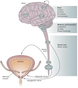
[image:24.612.140.450.131.482.2]Estrogen and bladder function
Figure 8.Neural control of bladder.
Sex hormone influences central neurological control of micturition.Estrogen receptors
are present in the cerebral cortex, limbic system hippocampus and cerebellum.
Estrogen increases the Sensory threshold of bladder, enhance the Alpha Adreno
receptor sensitivity in the urethral smooth muscles and causes Beta -3 Adreno receptor
18
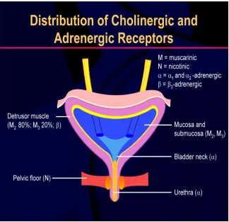
Figure 9. Distribution of cholinergic and adrenergic receptors in bladder
Estrogen receptors are present in the dome of the bladder, trigone that undergo squamous
metaplasia. Estrogen directly affects detrusor functions through modification in the
muscarinic receptors and reduces the involuntary detrusor contraction[37]. Withdrawal of
estrogen in post menopausal women is a common cause for voiding dysfunction.
Effect of estrogen on periurethral tissues
Genital tract Connective tissue consists mainly of collagen and structural glycoprotein and is
considered as an important factor responsible to support the pelvic organs. Estrogen
19 tract. Estrogen is essential to maintain the tensile strength and elasticity of the connective
tissue in order to support the genital organs and urethra[36].
Lack of estrogen decreases the volume of the vaginal muscles, resulting in weakness of the
ligaments holding the uteric pelvic floor and the bladder, resulting in the development of
prolapse of the internal genitalia leading to Pelvic organ prolapse and stress urinary
incontinence[37].
.
Hysterectomy: The incidence of clinically significant post-hysterectomy vaginal vault
prolapse requiring surgical intervention has been estimated at 36 per 10,000 person years .
Marchionni et al, showed the total incidence of post-hysterectomy vault prolapse as 1.8%,
but may increase to 11.6% in women who had hysterectomy for Pelvic organ prolapse. [38].
This indicates that postoperative vault prolapse is likely to result from preexisting poor
function of supporting structures.
The uterine cervix and the vagina are stabilized by the endopelvic fascia and its
extensions, the cardinal and uterosacral ligaments. As already mentioned above the vaginal
axis is directed horizontally and is supported by the levator plate. vaginal birth cause
damage to these supporting structures and leads to a change in the vaginal axis and descent
of the pelvic organs through the levator hiatus. These events result in pelvic organ prolapse.
Hysterectomy is associated with an increased risk of apical prolapse. Factors that may
influence the risk of prolapse after hysterectomy are age and the surgical route (abdominal or
20 careful consideration of the benefits and limitations of surgery must be undertaken before
intervention. The patient’s age, life expectancy, future strain acting on the pelvic floor, coital
function and co morbid medical conditions should be the part of preoperative evaluation.
The patient’s perception of her symptoms will be influenced by her understanding of
the benign nature of pelvic organ prolapse, the effect on quality of life, and the probability
that the symptoms troubling her will improve after surgery[39]..
Race and ethnicity: Data suggest that African-American women have a lower
prevalence of symptomatic POP than other racial or ethnic groups in the US [40]. In a
prospective cohort study of 2270 women, the risk in Latina and white women was four- to
five-fold higher than in African-American women [40]. In contrast, other studies have found
no relationship between POP and race or ethnicity [41].
Other risk factors: Chronic constipation , results in repetitive increases in intraabdominal
pressure [42,43]. Data conflict regarding whether the risk of prolapse is increased in women
with occupations that involve heavy lifting [43]. Some connective tissue disorders (eg,
21
Symptoms associated with pelvic organ prolapse
Bulge symptoms
Bulge or herniation symptoms that have been attributed to worsening pelvic organ
prolapse include a sensation of bulging or protrusion in the vagina, a sensation of
‘‘something falling out’’ of the vagina, actually seeing or feeling a vaginal or perineal Bulge Symptoms
Sensation of vaginal bulging or protrusion
Seeing or feeling a vaginal or perineal
bulge
Pelvic or vaginal pressure
Heaviness in pelvis or vagina
Urinary Symptoms
Urinary incontinence
Urinary frequency Urinary urgency
Weak or prolonged urinary stream
Hesitancy
Feeling of incomplete emptying
Manual reduction of prolapse to start or
complete voiding
Position change to start or complete
voiding
Bowel Symptoms
Incontinence of flatus or liquid/solid stool
Feeling of incomplete emptying
Hard straining to defecate
Urgency to defecate
Digital evacuation to complete defecation
Splinting vagina or perineum to start or complete
defecation
Pelvic floor dyssynergia
Feeling of blockage or obstruction during defecation
Sexual Symptoms Dyspareunia
Decreased lubrication
Decreased sensation
Decreased arousal or orgasm
Pain
Pain in vagina, bladder, or rectum
Pelvic pain
22 bulge, as well as pelvic pressure, fullness, and heaviness. Samuelsson et. al. evaluated pelvic
organ support in 487 women presenting for a regular gynecologic health examination . Some
degree of prolapse was noted in 30.8% of subjects, with only 2% having prolapse that
reached the introitus. A sense of heaviness in the lower abdomen was noted in 9.7%
of women with some degree of vaginal prolapse compared with 7.5% in the group with
normal vaginal support, a non-significant difference. [45].
Urinary symptoms
Urinary bladder and urethra are well supported by anterior vaginal wall. loss of this
support results in cystocele formation, which cause stress urinary incontinence. It is
therefore pelvic organ prolapse and stress urinary incontinence often coexist. Urinary
symptoms can be grouped under three category as follows,
Abnormal storage Abnormal voiding Abnormal sensation
Incontenance: urge,stress
Frequency /nocturia
Nocturnal enuresis
Staining to void
Hesitancy
Incomplete emptying
Poor stream
Post micturition dribble
Urgency
Dysuria
Absent sensation
Painful bladder
Urinary symptoms are not always attributed to the prolapse. But some of the
symptoms in prolapse may be due to urethral kinking which includes urinary hesitancy,poor
stream of voiding,Incomplete voiding, Need to reduce the vaginal bulge manually to
23 changes made to void,post void urinary dribble,urinary urgency,frequency,incontenance and
urinary retension.
Urinary incontinence: This is defined as the complaints of any involuntary loss of urine.
Urge and urge urinary incontinence: “compelling sudden desire to pass urine” .
Urge urinary incontinence – “involuntary leakage of urine that is preceded by urgency”
Frequency :day time frequency is the number of times a women voids during working
hours.Normal diurnal frequency is considered to be between 4-7 voids per day
Nocturia :Defined as the complaint that the patient has to wake up one or more times during
night to void.
Nocturnal enuresis is the complaints of urine occurring during sleep.
Stress urinary incontinence: “Defined as the involuntary loss of urine with exertion or
effort or with coughing and sneezing”
Mixed urinary incontinence: women with mixed urinary incontinence statisfy the definition
for both stress and urge incontinence
Coital incontinence: IUGA/ICS defined as “ involuntary loss of urine associated with
coitus”. This is further subdivided in to “incontinence with penetration or with orgasm”. And
24
Insensible incontinence: The IUGA/ICS defines insensible in continence as the complaints
of urinary incontinence where the women is unaware of how it occurred
Hesitancy :Defined as “difficulty in initiation of micturition resulting in a delay of the onset
of voiding after she is ready to pass urine”.
Straining to void Describes “the muscular effort used to initiate ,maintain,or improve the
urinary stream”.
Incomplete emptying: “Feeling of incomplete emptying experienced by the individual after
passing urine”
Post micturition dribble: Is defined as “involuntary loss of urine immediately after passing
urine”
Slow stream: Defined as “the individual perception of reduced urine flow”
Position dependent micturition: Defined as the complaint of having to adopt specific
positions to be able to micturate spontaneously or improve bladder emptying.(squatting,
leaning forward, leaning backward)
Pelvic organ prolapse often co-exits and may contribute to lower urinary tract symptoms.
Women may describe the learned behavior of using their fingers to reduce the bulge in order
to successfully micturate or defecate which is referred to as digitation. Over 40% of women
with urethral sphincter incompetence will also have significant cystocele. Vaginal prolapse
can mask urethral sphincter incompetence and when the prolapse is reduced the urethral
25 Women with higher stage III or IV of pelvic organ prolapse are more likely to have greater
symptoms of voiding dysfunction than those with stage I or II POP (44% vs. 9%).
Urodynamic evaluation showed that 72% of women with advanced prolapse had urethral
obstruction compared 6% in stage 1 or 2 POP. None of the patients with Stage 3 or 4 POP
demonstrated urodynamic stress incontinence, whereas 86% of those with stage1or 2 POP
did. As many as 30% of women with stage 3 or 4 prolapse have elevated postvoid residuals
(PVR>100 cc) [46].
Women with advanced prolapse have lower mean and maximum flow rates than women
with urinary incontinence. Despite this objective evidence of voiding dysfunction in women
with advanced prolapse. The correlation between specific voiding symptoms such as
hesitancy, weak stream, and feeling of incomplete emptying and severity of prolapse is weak.
Voiding dysfunction symptoms, particularly the need to splint to urinate, appears to resolve
after prolapse surgery in the majority of women who report these symptoms preoperatively.
[47].
.
Defecatory symptoms:
POP patients frequently report symptoms related to bowel dysfunction, includes “feeling of
incomplete rectal emptying, hard straining to defecate, the need to apply digital pressure to
the vagina or perineum (splint) to start or complete defecation, fecal urgency, and
incontinence of flatus or stool”[48].
The most common bowel symptom associated with prolapse is constipation .Defecatory
symptoms may be present in women with any anatomic site of prolapse, although they tend
26 of women presenting to a pelvic floor clinic described above, women with stage I prolapse
were the least likely to require splinting to defecate (8 to 15 percent), but the likelihood of
splinting symptoms did not continue to increase with advancing prolapse (stage II: 21 to 38
percent; stage III to IV: 2.6 to 29 percent) [51,52].
Studies evaluating the relationship between pelvic organ prolapse and defecatory symptoms
have several limitations.
Assessment of posterior compartment support is challenging There is no clear guidelines whether physical examination or radiologic evaluation should be used to
assess the anatomic and functional relationship of the posterior compartment. Studies
that based on physical examination alone to evaluate posterior vaginal support and
compared to defecography have demonstrated high sensitivities(91%–94%) for the
detection of rectocele. However, physical examination fails to detect enteroceles or
sigmoidoceles detected during proctography in approximately 50%of cases in patients
with severe prolapse.[53].
The presence of enterocele or sigmoidocele causing bowel disorders in women with
posterior vaginal prolapse is currently unknown. The ‘‘gold standard’’ method for
anatomic evaluation posterior compartment defects is defecography . However this is
not ideal, as it has lost of false positive results and normal asymptomatic women
27 The radiographic criteria used to define significant rectocele at proctography (≥3 cm)
also did not correlated defecatory symptoms.
The second limitation was to evaluate the effect of prolapse surgery on bowel function, due to poorly defined terms such as ‘‘constipation’’ or ‘‘defecatory
dysfunction,’’ and non-availability of standard definitions of constipation or its
subtypes in women with pelvic floor disorders
Constipation
Constipation is a symptom with many different definitions “based on stool frequency,
consistency, the need for straining, incomplete emptying”, Classification of constipation
based on the ROME II criteria .
Functional constipation demonstrated delay in transit times on colonic transit
studies. The outlet-type constipation, was described as constipation—predominant IBS or
functional constipation along with one of the following symptoms constipation—predominant “irritable bowel syndrome “
functional constipation
28 1.A sensation that stool cannot be passed when having a bowel movement, a need to
press on or around their bottom or vagina to try to remove stool to complete a bowel
movement,
2. Difficulty relaxing or letting go to allow the stool to come out at least one fourth
of the time.
Cleveland Clinic experience, outlet-type constipation found to be the predominant
subtype seen in patients with pelvic organ prolapse and urinary incontinence. Interestingly,
the prevalence of all types of constipation, and outlet-type in particular, were not different
between those patients with stage 3 or 4 pelvic organ prolapse and those with urinary
incontinence without prolapse [54]. A case–control study compared women with
uterovaginal prolapse and urinary incontinence with normal control women reported that
61% of womenwith uterovaginal prolapse complained straining at stool as a young adult
compared with 30% of women with stress incontinence and 4% of normal control
women.[55].
Outlet-type constipation is commonly associated with pelvic organ prolapse and urinary
incontinence. Multiple causes for defecatory symptoms to be considered in women with
pelvic organ prolapse because defecatory dysfunction may be a cause of prolapse rather than
a consequence, and specific bowel symptoms correlate poorly with worsening prolapse.
Furthermore, its important that women with pelvic organ prolapse should be counseled prior
29
Effects on sexual function:
Pelvic organ prolapse is common in elderly age group, assessing the relationship between
pelvic organ prolapse and sexual function, it is difficult to separate the effects of pelvic organ
prolapse from the normal changes associated with aging and menopause . Literature showed
that sexual function assessment in women with pelvic organ prolapse are mostly limited by
their small sample size, retrospective nature, and failure to use validated sexual function
questionnaires. Very few papers have compared sexual function in women with prolapse
with similarly aged women without prolapse. However, existing evidence suggests that, in
general, women with pelvic organ prolapse have similar rates of sexual activity as similarly
aged women without pelvic organ prolapse [56].
The most common reason for sexual inactivity in this population is absence of a male
partner.[57,58 ]Of those with a partner, male sexual dysfunction, particularly erectile
dysfunction, is the most often cited reason for inactivity[59] In 1 study, women with prolapse
or detrusor instability were more likely to cite their pelvic floor symptoms as a reason for
sexual inactivity than women with stress urinary incontinence or mixed incontinence[59] .
Among sexually active women with prolapse, approximately one third report that their
prolapse interferes with their sexual function [57,58 ] .
Ellerkmann et al found that impairment of sexual activity was moderately associated with
worsening prolapse in all 3 vaginal compartments, with the apical prolapse being the most
pronounced[48 ]. However, the only study thus far that has compared sexual function in
women with prolapse with that of women without prolapse using a validated sexual function
questionnaire found no difference in frequency of intercourse, libido, vaginal dryness,
30 Furthermore, there is a high rate of sexual satisfaction (81%–84%) in women with
pelvic organ prolapse who are in an intimate relationship. [57,59] . The majority of studies of
sexual function in patients with prolapse have focused on the effect of vaginal reconstructive
surgery on vaginal length and caliber or on postoperative sexual functioning, particularly
dyspareunia. Most prospective studies demonstrate that sexual function either does not
change or improves in the majority of women after vaginal reconstructive surgery for pelvic
organ prolapse [58,59] . Vaginal length and caliber appear to have little relationship with
postoperative sexual satisfaction. However women who undergo posterior colporrhaphy
especially in conjunction with Burch colposuspension, are at increased risk of developing
dyspareunia, with rates as high as38%. [58]
Prolapse does not appear to be associated with decreased sexual desire or with
dyspareunia, although reports vary according to whether POP is associated with adverse
effects on orgasm or sexual satisfaction [58]. Some women report that they avoid sexual
activity because of fear of discomfort or embarrassment associated with POP, particularly
those with urinary or fecal incontinence during sexual activity [59].
Various Questionnaires are used to assess the voiding and defecatrory dysfunctiions in
women with POP
Questionnaires can be divided into 3 categories:
1) Measure of presence of particular symptoms and their
severity (Symptom questionnaires);
2) Measure quality of life (Quality-of-Life [QOL] questionnaires),
31 Questionnaire development is a complex process that is governed by the principles of
psychometrics. Psychometrics is the science of the measurement of responses to phenomena
that are not easily quantifiable.
Questionnaire must demonstrate 3 important psychometric properties: validity,
reliability, and responsiveness. The validity of a questionnaire is simply whether it
measures what is intended. The reliability of a questionnaire refers to its ability to measure
in a reproducible fashion. Responsiveness refers to a questionnaire’s ability to reliably detect
the overall effect of treatment and ability to detect clinically meaningful change.
Questionnaire with good psychometric properties, said to be ‘‘validated.’’ Other
characteristics that are desirable in a questionnaire include being easy to understand and
feasible to implement. Symptom questionnaires are used to assess the presence, severity,
and impact of particular symptoms or groups of symptoms.
Pelvic Floor Distress Inventory questionnaire is the valid and reliable symptom
questionnaire designed for women with pelvic organ prolapse .This comprehensive
symptom questionnaire is intended for women with all forms of pelvic floor disorders. It
assesses 46 pelvic floor symptoms and has 3 scales: a urinary scale (identical to the UDI), a
colorectal scale, and a pelvic organ prolapse scale. Patients are asked to indicate if they have
a particular symptom and if so, they are asked to assess how much it bothers them on a
4-point scale.
Short version of the PFDI: The Pelvic Floor Distress Inventory short form 20 (PFDI-20),
which also has urinary, colorectal, and pelvic organ prolapse scales. The urinary scale of the
32 validity and have demonstrated excellent responsiveness in patients undergoing surgery for
pelvic organ prolapse [60].
.
The International Prostate Symptom Score (I-PSS): is based on the answers to
eight questions in which 7 questions are regarding to urinary symptoms and 1 question about
quality of life. Each question related to urinary symptoms were allowed to choose by the
patients, one out of six answers indicating increasing severity of the concern symptom. The
answers are assigned points from 0 to 5. The total score ranges from 0 to 35 (asymptomatic
to very symptomatic).
The questions refer to the following urinary symptoms as follows;
Questions Symptom
1. Incomplete emptying
2. Frequency
3. Intermittency
4. Urgency
5. Weak Stream
6. Straining
7. Nocturia
33 8th Question refers to the patient’s quality of life. The first seven questions of the
I-PSS are similar to the questions appearing on the American Urological Association (AUA)
Symptom Index which currently classifies symptoms as follows:
IPSS 7-urinary symptom score
The International Scientific Committee (SCI), World Health Organization (WHO)
and the International Union Against Cancer (UICC) guidelines recommends the use of single
question to assess the QOL. The answers to this question range from 0 to 6 implies
“delighted” to “terrible” [61].
Scarpero et al. reported that the IPSS precisely explains both the female LUTS as
well as male LUTS and predict the degree of bother and effect on quality of life (QOL) [62].
Recently, the IPSS has been proven to have excellent internal consistency and good
constructive validity in assessing female LUTS [61].
The pelvic organ prolapse Quantification (POP-Q) classification system involves the
description of dimension of the vagina and perineum and of the topography of the anterior
and posterior vaginal walls. POP-Q measurements corresponds to stages (0-4) of prolapse in
each compartment (anterior, apical, and posterior) of vagina.
symptoms score
Mild 0-7
Moderate 8-19
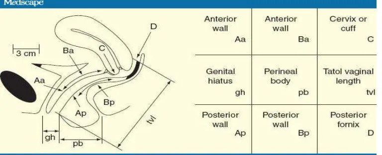
34 Figure 10. POP-Q points
Point Aa: Point that lies in the midline of anterior vaginal wall and is 3cm proximal to the external urethral meatus.
Point Ba: represent the most distal portion the remaining upper anterior vaginal wall
Point Ap: Posterior vaginal wall 3 cm proximal to the hymen
Point Bp: Most distal position of the remaining upper posterior vaginal wall others
Point C : Cervix or vaginal cuff
Point D:Posterior fornix or vaginal apex
Genital hiatus (gh):External urethral orifice to posterior hymenal remant
Total vaginal length(TVL):Hymenal ring to point D
Perineal body (pb):Posterior hymen to middle of anal opening
STAGES OF PROLAPSE:
STAGE 0 : No prolapse. Apex can descend within 2cm above the hymen.
STAGE I : Leading edge is >1 cm above the level of hymen
35 STAGE III :Leading edge descends > 1cm beyond hymen but no further than 2 cm less than the total vaginal length.
STAGE IV: Complete eversion .Leading point > + (TVL-2) cm.
[image:42.612.89.461.222.608.2]
36 Several studies have shown anterior wall defects associated with voiding dysfunction and
perineal, posterior vaginal support defect with defecatory dysfunction in urogynaecology
patients. The POP-Q Bp points, not the pb measurement, correlates with symptoms of
defecatory dysfunction .
Uroflowmetry
International continence society defined Urodynamics as observation made during
Urodynamic studies. And Uroflowmetry should always be interpreted with clinical
information. European Association of urology guidelines recommends Uroflometry for
women with significant pelvic organ prolapse.
ICS working party on urodynamic equipment described three methods of
Uroflow meter as follows
1.The gravimetric method
2.The Electronic dip stick method
3.The rotating Dip stick method
Following parameters are calculated in uroflowmetry
Voided volume: The volume of total voided urine expelled from the bladder
Residual urine volume: Amount of urine remaining in the bladder after complete void
Flow time: The time during which measurable flow actually occurs
37
Time to maximum flow: The time between the onset of flow to the point of maximum flow
Mean flow rate: Volume voided divided by flow time.
Normal flow curve is a bell shaped, continuous, smooth curve with a increasing flow rate
[image:44.612.96.543.207.461.2]Voided volume more than 150 ml and Qmax more than 15ml/sec considered normal.
Fig12:Uroflow curve
In Pelvic organ prolapse maximal flow rate and average flow rate were significantly less
compared to normal healthy individuals.
Several studies have shown that women with severe POP have abnormal
urodynamics despite the absence of subjective symptoms. Most of the patients with
high-stage cystocele have shown that obstructive uroflow which implies high urethral resistance
38 Detrusor hypocontractility may be more common in patients with high-stage
cystocele because the detrusor function gradually becomes weaker as a long-term effect of
voiding against resistance [51,63].
PVR (Post void residual urine)
Measuring amount of residual urine in the bladder after a voluntary void is
defined as post void residual urine volume (PVR). Portable bladder scanners are
convenient, accurate, cost-effective and non-invasive screening test for evaluating
voiding dysfunction [64]. Threshold values for abnormal post void residue are not
clearly defined. The volumes of 50 ml to 100 ml are considered as abnormal residual
urine volume by most urologists.
Fig 13: Ultrasound measurement of bladder volume in sagittal and transverse
39 There is an increased risk of upper urinary tract dilation and renal insufficiency with
very large post void residue (>300 ml) .Large post void residue may be caused by bladder
hypo contractility. Poor bladder contractility may result from neurogenic, myogenic,
psychogenic, or pharmacologic causes[64].
Management: Treatment is indicated for women with symptoms of prolapse or associated
conditions (urinary, bowel, or sexual dysfunction). Obstructed urination or defecation or
hydronephrosis from chronic ureteral kinking are all indications for treatment, regardless of
degree of prolapse [65]. Treatment is generally not indicated for women with asymptomatic
prolapse [66].
Management options: Women with symptomatic prolapse can be managed expectantly, or
treated with conservative or surgical therapy. Both conservative and surgical treatment
options should be offered. There are no high quality data comparing these two approaches.
The choice of therapy depends upon the patient’s preferences, as well as the ability to
comply with conservative therapy or tolerate surgery. Some data suggest that age, the degree
of POP as measured by descent of leading edge of prolapse, preoperative pelvic pain scores,
and prior prolapse surgery are independently associated with treatment choices. In a study of
152 women, older patients and those with increased preoperative pelvic pain scores were
more likely to choose pessary over surgery [66]. On the other hand, the likelihood of
choosing surgery was increased in women with more severe prolapse or a prior POP repair.
Expectant management — Expectant management is a viable option for women who can
tolerate their symptoms and prefer to avoid treatment. Women with symptomatic or
40 evaluated on a regular basis to assess for the development or worsening of urinary or
defecatory symptoms and/or findings.
Conservative management : In POP Conservative therapy is the first line choice since
surgical treatment suffers the risk of complications and recurrence [67]. However, prolapse is
naturally a chronic problem, and many women ultimately prefer surgery to conservative
therapy since effective surgery does not require ongoing maintenance.
Vaginal pessary:The main stay of non-surgical treatment for POP is the vaginal pessary. In
elderly women with co morbid medical conditions confers a significant risk for surgical
[image:47.612.229.511.318.621.2]morbidity and mortality.
41 The reported surveys showed 87% to 98% of gynecologists were using pessaries for
conservative management[68-69] . Pessaries are silicone devices and are available in various
shapes and sizes, which support the pelvic organs. About half of the women who use a
pessary continue to do so in the intermediate term of one to two years. Pessaries needs to be
removed and cleaned on a regular basis. Gynaecologist have reported the decrease in
prolapse stage after long-term pessary use and the prosperous role of pessaries in preventing
progression of prolapse.
Pelvic floor muscle exercises: Pelvic floor muscle exercises (PFME) appear to
result in improvements in POP stage and POP-associated symptoms. Studies have
demonstrated the benefit of PFME, particularly with individualized training and/or
supervision [70-71]. As an example, one trial included 109 women with stage I to III
prolapse who were assigned to either PFME for six months with regular supervision by a
physical therapist or to a control group [70-71]. Women in the PFME group had significant
reductions in the frequency and bother of most prolapse, bladder, and bowel symptoms
(exceptions were urgency urinary incontinence symptoms, difficulty with stool emptying,
and solid stool fecal incontinence). Improvement in POP stage was found more frequently in
the PFME group (19 versus 8 percent). Further study is needed of the effect of PFME on
POP.
Surgical treatment: Surgical candidates include women with symptomatic prolapse not had
improvement in their symptoms on conservative management of their prolapse There are
many surgeries for prolapse which includes vaginal and abdominal approaches with and
without graft materials. Surgical prognosis depends upon the severity of symptoms, extent of
42 Surgical management of pelvic organ prolapse can be challenging, because multiple
support defects frequently coexist. It is rare to find isolated defects in the anterior or posterior
compartments without also finding an apical defect.
Apical compartment
Many experts would argue that adequate suspension of the apex is the cornerstone of
a successful prolapse repair. If the muscularis layer of the vaginal tube is intact and the apex
is well suspended, many anterior and posterior defects will also resolve. This is evident by
the numerous reports of successful anatomic outcomes after repair of stage IV prolapse by
[image:49.612.126.574.351.618.2]simply suspending the apex at the time of colpopexy without concomitant repairs[72]..
43
Anterior compartment
Evaluation and repair of the anterior vaginal wall represents a surgical challenge for
the pelvic reconstructive surgeon. Careful evaluation evaluation of defects preoperatively
must be coupled with intraoperative assessment for optimum repair. Often multiple sites
must be addressed to reestablish support and bladder function must always be considered
along with anatomic reconstruction.
Surgery has traditionally been associated with a recurrence/reoperation rate of up to 30
percent after the initial surgery [73], with some centers reporting reoperation in over 50
percent of patients who have undergone at least two prior surgical procedures for prolapse
[74].
Posterior compartment
Compared with the traditional colporrhaphy, which assumes that the anatomic defect
is the result of stretching of the rectovaginal septum or vaginal muscularis, the site-specific
technique is based on the assumption that discrete tears of these layers result in posterior wall
prolapse. Possibly as a resultof the general idea that plication of tissue to the midline does not
restore ‘‘normal anatomy,’’many surgeons have abandoned the colporrhaphy technique in
favor of the site-specific defect repair[75]. Like with many surgical conditions, the most
important preoperative goal should be to help each patient to have realistic preoperative
44
ABBREVIATIONS
POP-PELVIC ORGAN PROLAPSE
VD- VOIDING DYSFUNCTION
DD –DEFECATORY DYSFUNCTION
POP-Q-PELVIC ORGAN PROLAPSE QUANTIFICALTION SYSTEM
QMAX-MAXIMAL FLOW RATE
PVR-POST VOID RESIDUAL URINE
IPSS-INTERNATIONAL PROSTATE SYMPTOM SCORE
PFDI-20-PELVIC FLOOR DISTRESS INVENTORY SCORE
CRADI-8 -COLORECTAL DISTRESS INVENTORY SCORE
LUTS-LOWER URINARY TRACT SYMPTOMS
QOL-QUALITY OF LIFE
VH-VAGINAL HYSTERECTOMY
PFR-PELVIC FLOOR REPAIR
P VALUE- CALCULATED PROBABILITY VALUE
45
MATERIALS AND METHODS
Research design: This is a cross sectional study done in Christian Medical College and
Hospital Vellore, India.
Study period October 2015 To august 2016
Study Setting: This study was conducted in the department of obstetrics and
Gynaecology in patients at Christian Medical College and Hospital Vellore.
The Study was approved by institutional review board and ethics Committee of the
Hospital.
Inclusion criteria:
Postmenopausal women with Stage 2, 3 and 4 POP
Vault Prolpase
Exclusion criteria:
Women with neurological disorder
Women with prior anti-incontinence or bowel surgery
46
During the study Period ,all the patients who fulfilled the eligibility criteria were invited to
participate. All the patients were explained about the nature of the study. An informed written
consent was obtained from each patients . Postmenopausal women with POP stage 2, 3 ,4 and vault
prolapse were admitted in gynaecology ward for pelvic reconstructive surgery were recruited and
given a study number .
A detail history was taken and women are interviewed by the principle investigator to complete the short form of pelvic floor distress inventory (PFDI-20) and International
prostate symptom questionnaire I-PSS score for Lower urinary tract symptoms(LUTS).
socio-demographic and clinical details were also collected .All the patients maintained a 24 hours
voiding diary.
Voiding diary parameters includes intake volume, voided volumes, and incontinent and urgency episodes, usage of pads, day and night frequency and type of activities performed
during these episodes documented by patients. 24 hrs urine production, nocturnal urine
volume and maximum voided volume were assessed from voiding dairy.
pelvic examination done for all women with POP-Q staging system.Definition, and descriptions conformed to the standards recommended by the international continence
society(ICS)
POP-Quantification
Point Aa: Point that lies in the midline of anterior vaginal wall and is 3cm proximal to the external urethral meatus.
Point Ba: Represent the most distal portion the remaining upper anterior vaginal wall
47 Point Bp: Most distal position of the remaining upper posterior vaginal wall
Point C : Cervix or vaginal cuff
Point D: Posterior fornix or vaginal apex
Genital hiatus (gh): External urethral orifice to posterior hymenal remant
Total vaginal length(TVL): Hymenal ring to point D
Perineal body (pb): Posterior hymen to middle of anal opening
STAGES OF PROLAPSE:
STAGE 0 : No prolapse. Apex can descend within 2cm above the hymen.
STAGE I: Leading edge is >1 cm above the level of hymen
STAGE II: Leading edge descends to within 1cm of plane of hymen ( above or
below)
STAGE III :Leading edge descends > 1cm beyond hymen but no further than 2 cm
less than the total vaginal length.
50 The following investigations were done
Serum creatinine
Blood sugar fasting and post prandial
Urine microscopy
Urine culture was done in those women with significant white cell counts on urine
microscopy
Uroflow and post void residual urine volume were measured
PVR
Post void residual volume was measured with in 10 mints of voiding using a transabdominal
scan.by measuring three orthogonal diameters of the bladder, the post void residual bladder volume
was estimated and calculated by the formula
Volume = (π × D1×D2×D3)/6
D1- Widest diameter in the transverse scan (cm)
D2- Antero posterior diameter in the longitudinal scan.(cm)
D3- Cephalocaudal diameter in the longitudinal scan (cm)
51 All patients who had high post void residual bladder volume were assessed again for PVR.
Residual volume more than 100 ml was taken as significant. This information enabled us to
evaluate function as well as to find out an obstruction of lower urinary tract.
Uroflow:
Gravimetric method is used in our institute for uroflowmetry.
This method measures the urine flow rate by measuring the weight of the collected fluid. The
output electrical signal is directly related to the mass of fluid collected.
[image:58.612.164.475.332.582.2]Diagram of normal uroflow curve
52
Voiding Dysfunction : Was defined as the presence of at least one of the following criteria
1. Maximal flow rate below 15ml/sec
[image:59.612.90.565.197.568.2]2. PVR >=100ml
Fig17.. Diagrammatic representation of
53
The International Prostate Symptom Score (I-PSS): is based on the answers to eight questions in
which 7 questions are regarding to urinary symptoms and 1 question about quality of life. Each
question related to urinary symptoms were allowed to choose by the patients, one out of six answers
indicating increasing severity of the concern symptom. The answers are assigned points from 0 to 5.
The total score ranges from 0 to 35 (asymptomatic to very symptomatic).
The questions refer to the following urinary symptoms as follows;
Questions Symptom
1. Incomplete emptying
2. Frequency
3. Intermittency
4. Urgency
5. Weak Stream
6. Straining
7. Nocturia
8th Question refers to the patient’s quality of life. The first seven questions of the I-PSS are
similar to the questions appearing on the American Urological Association (AUA) Symptom
54
IPSS 7-urinary symptom score
symptoms score
Mild 0-7
Moderate 8-19
Severe 20-35
The International Prostate Symptom Score (I-PSS)-for Quality of life assessment
The International Scientific Committee (SCI), under the patronage of the World
HealthOrganization (WHO) and the International Union Against Cancer (UICC),
recommends the use of only a single question to assess the quality of life. I-PSS - Question
eight refers to the patient’s perceived quality of life. The answers to this question range from
“delighted” to “terrible” or score 0 to 6.
In our study we defined quality of life as “satisfied”if the score is less than 3 and the
quality of life as “unsatisfied” if the score is 3 and above .
Pelvic Floor Distress Inventory (PFDI-20)
The short form of the Pelvic Floor Distress Inventory (PFDI-20) ,a condition-specific,
validated, health related quality of life questionnaire with 3 subscales,which is designed to
evaluate distress caused by specific pelvic floor symptoms including urinary ,bowel and
pelvic organ prolapse symptoms.items on the PFDI-20 form first ask wheather each symptom
is experienced or not(“yes” or “no” response),and if “yes”, the degree of bother is assessed
55
Colorectal Anal Distress Inventory (CRADI-8)
The overall degree of bother attributed to 8 bowel symptoms in women with pelvic organ
prolapse is described by “the CRADI8(colorectal distress inventory-8)bowel subscale score
with in PFDI-20(short form of the pelvic floor distress inventory)”.CRADI-8 form first ask
whether each symptom is experienced or not(“yes” or “no” response),and if “yes”, the
degree of bother is assessed on the scale from 1(not at all)to 4(quite a bit).
In our study we defined a negative response as either the report of “no “for the symptom or
the report of “yes” but with a degree of bother specified as “not at all “or somewhat”
A positive response was defined as the report of “moderately “or “quite a bit” of bother.
In this study we defined defecatory dysfunction as “The women who had given positive
response for 2 or more questions out of 8 questions “in CRADI_8 bowel subscale score with
in PFDI-20(short form of the pelvic floor distress inventory).
All the study patients Underwent surgery as per the discretion of the surgeon.
All the patients were followed up on their 6th post-operative day and uro flow and PVR
were done . pre and post-operative uroflow and PVR correlated .
The information was computerised and confidentiality was maintained.
Sample size calculation
The required sample size to show that about 40% of POP women will develop voidal
56 precision. The 40% was considered as there were studies that showed voidal dysfunction to
range from 30 to 50%. [76].
Ref: European Journal of Obstetrics & Gynecology and Reproductive Biology 174
2014) 146–149.
Formula:
Reference for formula:
Lemeshow S, Hosmer D. W, Klar J, Lwanga S. K, Adequacy of Sample Size in
Health Studies. John Wiley and Sons, 1990.
Expected Proportion 0.4 0.4 0.4
Precision (%) 10 7.5 9.5
Desired confidence level (1- alpha) % 95 95 95
57 Presence or absence of voidal dysfunction was calculated using maxium flow rate and/or
PVR. Descriptive measures such as mean with SD and/or median with IQR was presented for
all continuous variables whereas frequencies and percentages was presented for all
categorical variables.
All the categorical variables were associated with voidal dysfunction using Chi-square test
with continuity correction and for small sample size Fisher’s exact test was used.
All continuous variables were compared across women with and without voidal dysfunction
using independent t-test or Mann Whitney U test. The decision of choosing independent t-test
or Mann Whitney U test was done on the basis of assumption of normality which was
assessed by plotting QQ plot and histogram along with Shapiro Wilk test of significance for
normality.
All those significant in the Chi-square test or independent t-test at p value ≤ 0.2 were taken
for logistic regression analysis. SPSS version 21 was used for analysis and p value < 0.05
58
Post op follow
up with flow & PVR
Surgery
VH
with PFR
Pre op work up, Stage of POP ,Symptom scoring Flow & PVR
Patients with
POP
59
RESULTS
This cross sectional study was conducted in the Department of Obstetrics and
Gynaecology, Christian Medical College Hospital, Vellore between October 2015 to August
2016. The study was approved by the institutional review board and ethical committee of the
hospital.
The calculated sample size for this study was 120 cases but this interim analysis was
done for 60 cases. Sixty post-menopausal women with pelvic organ prolapse stage II, III and
IV admitted in the ward for surgery were recruited after informed consent. All the sixty
women were examined and interviewed with international prostate symptom score (IPSS)
questionnaire and short form of pelvic floor distr