DEXMEDITOMIDINE VERSUS CLONIDINE AS AN ADJUVANT TO 0.75% ROPIVACAINE
IN LOWER SURGERIES
THE TAMILNADU Dr. M.G.R MEDICAL
In partial fulfilment of the requirements for
M.D
DEXMEDITOMIDINE VERSUS CLONIDINE AS AN ADJUVANT 0.75% ROPIVACAINE FOR EPIDURAL ANAESTHESIA
LOWER ABDOMINAL AND LOWER LIMB SURGERIES IN A TERTIARY CARE CENTRE
A COMPARATIVE STUDY
Dissertation
Submitted to
THE TAMILNADU Dr. M.G.R MEDICAL UNIVERSITY
In partial fulfilment of the requirements for
the award of the degree of
M.D. ANAESTHESIOLOGY
Branch X
MAY 2018
DEXMEDITOMIDINE VERSUS CLONIDINE AS AN ADJUVANT EPIDURAL ANAESTHESIA
LOWER LIMB IN A TERTIARY CARE CENTRE -
This is to certify that this dissertation entitled “Dexmeditomidine versus Clonidine as an Adjuvant to 0.75% Ropivacaine for Epidural Anaesthesia in Lower Abdominal and Lower Limb Surgeries in a Tertiary Care Centre - A Comparative Study” is a bonafide record of the work done by Dr. Sathesh Kumar B M, under guidance and supervision in the Department of Anaesthesiology during the period of her postgraduate study for M.D Anaesthesiology [Branch-X] from 2015-2018.
Dr. Rommy Geever, MD
[Co-guide]
Asst. Professor
Department of Anaesthesiology Sree Mookambika Institute of Medical Sciences
Kulasekharam, Kanyakumari District: Tamil Nadu - 629161
Dr. A Thavamani,
MD[Guide]
HOD and Professor
Department of Anaesthesiology Sree Mookambika Institute of Medical Sciences
Kulasekharam, Kanyakumari District, Tamil Nadu 629161
Dr. Rema. V. Nair,
M.D., D.G.O.,Director
Sree Mookambika Institute of Medical Sciences
This is to certify that this dissertation work titled “Dexmeditomidine versus Clonidine as an Adjuvant to 0.75% Ropivacaine for Epidural Anaesthesia in Lower Abdominal and Lower Limb Surgeries in a
Tertiary Care Centre - A Comparative Study” of the candidate Dr. Sathesh Kumar B M, with registration Number 201520552 for the
award of DOCTOR OF MEDICINE in the branch of Anaesthesiology [Branch-X]. I personally verified the urkund.com website for the purpose of plagiarism Check. I found that the uploaded thesis file contains from introduction to conclusion pages and result shows 6 percentage of plagiarism in the dissertation.
In the following pages is presented a consolidated report of the study on “Dexmeditomidine versus Clonidine as an Adjuvant to 0.75% Ropivacaine for Epidural Anaesthesia in Lower Abdominal and Lower Limb Surgeries in a Tertiary Care Centre - A Comparative Study” cases studied and followed up by me at Sree Mookambika Institute of Medical Sciences, Kulasekharam from 2016-2017. This thesis is submitted to the Dr. M.G.R. Medical University, Chennai in partial fulfilment of the rules and regulations for the award of MD Degree examination in Anaesthesiology.
Dr. Sathesh Kumar B M, Junior Resident
Department of Anaesthesiology, Sree Mookambika Institute of Medical Sciences,
Kulasekharam,
Kanyakumari District. Tamil Nadu 629161.
Date:
I wish to express my sincere thanks to Dr. Velayutham Nair, Chairman and Dr. Rema V. Nair, Director of Sree Mookambika Institute of Medical Sciences for having permitted me to utilize the facilities of the hospital for the conduct of the study.
My heartfelt gratitude to Prof. Dr. A. Thavamani, Professor and Head, Department of Anaesthesiology, Sree Mookambika Institute of Medical Sciences and Hospital for his motivation, valuable suggestions, expert supervision, guidance and for making all necessary arrangements for conducting this study.
I thank Prof. Dr. Jayaprakash, for his constant motivation and support apart from providing immense help and valuable suggestions in carrying out this study. His continuous support and ideas are the key factors for the construction of the study.
I thank Prof. Dr. Bala Krishnan, for his constant motivation, guidance and valuable suggestions in carrying out this study.
I thank Prof. Dr. Paramasivan, for his constant support and encouragement throughout the study.
in my work. He was a perfectionist, absorbed in work wanting his students excel in the field of anaesthesia.
I wish to thank Dr. Prasandan, Associate Professor, very knowledgeable teacher who is always eager to teach and share his knowledge.
I extend my gratefulness to Dr. Ravi Shankar, Assistant Professor, a good mentor and guide who tirelessly guided and encouraged me to strive and work hard.
I express my heartfelt gratitude to Assistant Professors Dr. Rommy, Dr. Beula, and Dr. Saji, who had evinced constant and keen interest in the progress of my study right from the inception till the very end and was instrumental in the successful completion of the study.
I thank Dr. Anitha R, my co-pg, for her valuable and timely help to complete my study on time. I also thank my colleagues Dr.Sheen, Dr.Shrey, Dr.Ashwini, Dr.Karikalan for helping me in various technical aspects of my study.
Sl. No.
Contents
Page No
1.
Introduction
1-3
2.
Aims and Objectives
4
3.
Hypothesis and Scientific Justification
5-6
4.
Review of Literature
7-47
5.
Materials and Methods
48-56
6.
Results
57-74
7.
Discussion
75-80
8.
Conclusion
81
9
Bibliography
i-vi
Sl. No Tables Page No
1 Gender Distribution 58
2 ASA Physical Status Distribution 59
3 Weight Distribution 60
4 Height Distribution 61
5 Age Distribution 62
6 Distribution of Surgery 63
7 Duration of Surgery 64
8 Efficacy of the drug 65
9 Peak level of sensory blockade 66
10 Time to complete Motor Blockade (Motor Grade 3) 67 11 Duration of Blockade-Time to Rescue Analgesia 68
12 Time to 2 Dermatome Regression 69
13 Sedation Score at 10 Minutes 70
14 Sedation Score at the end of surgery 71
Sl. No Figures Page No
1 Parts of Vertebral Bone 29
2 Boundaries of Epidural Space 33
3 Midline Approach for Epidural Space 39
4 Tecnicniques to find out Epidural Space 43 5 Paramedian Approach for Epidural Space 45
6 Gender Distribution 58
7 ASA Physical Status Distribution 59
8 Weight Distribution 60
9 Height Distribution 61
10 Age Distribution 62
11 Distribution of Surgery 63
12 Duration of Surgery 64
13 Efficacy of the drug 65
14 Peak level of sensory blockade 66
15 Time To Complete Motor Blockade 67
16 Duration of blockade - time to rescue analgesia 68
17 Time to 2 dermatome regression 69
18 Sedation score at 10 minutes 70
19 Sedation Score at the end of surgery 71
હ – Alpha mcg – Microgram kg – Kilogram mg – Milligram ml – Millimeter
ASA – American Society of Anesthesiology min – Minutes
L2-L3-L4 – Lumbar C1 – Cervical T1 – Thoracic S1 – Sacral
PCA pumps – Patient Controlled Analgesia Pump SpO2 – Partial Pressure of Saturation
ADRS – Adverse Drug Reactions MAP – Mean Arterial Pressure RL – Ringer Lactate
HR – Heart Rate
Background:
The quality and duration of analgesia is improved when a local anaesthetics is combined with an alpha 2 adrenergic agonists. Both Clonidine and Dexmedetomidine are alpha 2 adrenergic agonists which have analgesic properties have been extensively studied and it has been established that Clonidine as an adjuvant, effectively prolongs the duration of action of local anaesthetics when given epidurally. There are limited studies demonstrating the effects of epidural Dexmedetomidine and its effects of local anaesthetics.
Aims and objectives:
The aim of this study was to compare the effect of Clonidine and Dexmedetomidine in terms of anaesthesia, analgesia, sedation and side effects when used as an adjuvant to epidural Ropivacaine in lower abdominal and lower limb surgeries.
Methodology:
using Ramsay sedation scale, intra operative hemodynamic parameters and complications if any- nausea, vomiting, bradycardia, hypotension were noted.
Results:
Both groups were comparable demographically with respect to age and sex distribution, height and weight characteristics. The onset and duration of sensory blockade was found to be significantly shorter in the RD group (p<0.005). The sedation in group RD was found to be significantly better than group RC (p<0.005). There was no significant difference found between the two groups in terms of onset of motor blockade and hemodynamic changes. Both groups had a similar incidence of hypotension and bradycardia which was not found to be significant. The side effects in both groups were minimal and comparable between the two groups.
Conclusion:
1 | P a g e
INTRODUCTION
Epidural anaesthesia is a versatile technique used for providing both anaesthesia and analgesia in the post operative period. It may be combined with regional anaesthesia or other forms of general anaesthesia. It can provide intra operative hemodynamic stability and has been proven to reduce perioperative stress response thus causing a decrease in the complications and help in improving patient outcome. It also helps in early mobilization of the patient by providing relief to post operative pain and decreases the incidence of thromboembolic events.(1-5)
A number of agents such as
be used as adjuvants to local anaesthetics that act synergistically thereby increasing the efficacy of the l
required dose and toxic side ef
The duration and
anaesthetic is combined with an alpha 2 adrenergic agonist as adjuvant. Dexmedetomidine and
local anaesthetic effects
Clonidine is an alpha 2 adrenergic agonist that enhances the action of local anaesthetic drugs on
It acts by blocking
thereby decreasing the intensity and duration of analgesia. It is known to cause sedation and the side effects of its use are
hypotension.(13-16)
A number of agents such as opioids, ketamine, and alpha agonists can be used as adjuvants to local anaesthetics that act synergistically thereby increasing the efficacy of the local anaesthetic drugs, decreasing the total required dose and toxic side effects of both groups of drugs.(9,10
The duration and the quality of analgesia can be improved when a local anaesthetic is combined with an alpha 2 adrenergic agonist as adjuvant.
exmedetomidine and Clonidine are alpha 2 adrenergic agonists potentiates local anaesthetic effects and have analgesic properties.(10-12)
Clonidine is an alpha 2 adrenergic agonist that enhances the action of local anaesthetic drugs on administration via the epidural or intrathecal route. the A and C fibres and it causes local vasoconstriction thereby decreasing the intensity and duration of analgesia. It is known to cause sedation and the side effects of its use are b
2 | P a g e opioids, ketamine, and alpha agonists can be used as adjuvants to local anaesthetics that act synergistically thereby ocal anaesthetic drugs, decreasing the total
9,10)
the quality of analgesia can be improved when a local anaesthetic is combined with an alpha 2 adrenergic agonist as adjuvant. Both lonidine are alpha 2 adrenergic agonists potentiates
3 | P a g e Dexmedetomidine is a newer alpha 2 adrenergic agonist and is about 8 times more selective α2 adrenoreceptor agonist than Clonidine and hence
allows the use of higher drug dosage with less alpha1 effect. It has been found to have hemodynamic stability, anxiolytic, analgesic, sedative, neuroprotective and anaesthetic sparing effect. It causes intense motor blockade and co-operative sedation without increasing the incidence of the adverse effects.(7-19)
Clonidine and Dexmedetomidine act on both pre and post synaptic sympathetic nerve terminals and also has central action which causes a decrease in the sympathetic outflow, leading to its analgesic, sedative, and hemodynamic effects.(13,15,16,18) The effects of Clonidine as an adjuvant with local anaesthetics has been studied extensively and it effectively prolongs the duration of action of the local anaesthetics when given epidurally.
4 | P a g e
AIMS AND OBJECTIVES
To study and compare the effect of Dexmedetomidine and Clonidine in epidural anaesthesia when given as an adjuvant to 0.75% Ropivacaine
1. The onset and duration of sensory blockade. 2. The onset of motor blockade.
3. The change in intra operative hemodynamic parameters. 4. The level of sedation.
5 | P a g e
HYPOTHESIS AND SCIENTIFIC JUSTIFICATION
Hypothesis
Epidural Dexmedetomidine prolongs the duration of anaesthesia with hemodynamic stability and improve post-operative analgesia.
Scientific Justification
Neuraxial block for lower abdominal surgeries are becoming popular as its advantages are many as compared to general anaesthesia. Epidural
anaesthesia consists of the interruption of nerve transmission temporarily in the epidural space, an effect produced by the injection of a local anaesthetic (Ropivacaine 15ml) solution in the epidural space. The role of an anesthesiologist is to render pain free surgical procedures.
6 | P a g e Dexmedetomidine is a highly selective, potent α₂ - adrenoreceptor
agonist that acts centrally with a short duration of action. It has the capability to sedate, hypnotize and provide analgesia that prolongs the duration of sensory and motor blockage obtained with epidural anaesthesia while still maintaining patient arousability and respiratory function. It is used epidurally with Ropivacaine.
7 | P a g e
REVIEW OF LITERATURE
Role of epidural anaesthesia:
Moraca et al(5) reviewed a number of meta analysis, retrospective and prospective studies to assess the benefits and complications of epidural anaesthesia and analgesia. They concluded that benefits of epidural anaesthesia includes suppression of surgical stress by sympatholysis, stable hemodynamics, reduction in postoperative pulmonary complications and effective pain relief, thus improving patient cooperation for physiotherapy and early mobilization leading to permitting earlier extubation, and reducing the length of stay. They also found 30% reduction of cardiac morbidity, 40% reductions in pulmonary complications and 30% reduction in blood loss. This review indicated significant reduction in perioperative complications and postoperative morbidity were associated with the use of epidural anaesthesia and postoperative analgesia. They also found that adding opioids to local anaesthetics provided better analgesia with less toxicity than either of the drugs alone.
8 | P a g e Significantly fewer complications were seen with epidural anesthesia and hence concluded that epidural anesthesia and analgesia eliminate the perioperative physiological stress responses. This lead to decrease in surgical complications by providing better analgesia, decreased ICU stay and improving outcomes.
John Riggs et al(4) studied 915 high risk patients undergoing major abdominal surgery in their MASTER anaesthesia Trial Study. They found no significant advantage in combining epidural with general anaesthesia. However there was improvement in analgesia, reduction in respiratory complications and the lower risk of adverse consequences that suggests high-risk patients undergoing major intra abdominal surgery may substantially benefit from effect of combined general and epidural anaesthesia intra operatively and continued postoperative epidural analgesia.
Ropivacaine in epidural anaesthesia:
9 | P a g e Shalina(6) et al compared 0.75% Ropivacaine and 0.5% Bupivacaine in epidural anaesthesia in patients undergoing lower limb orthopaedic surgeries. The patients were randomized into groups to receive Ropivacaine 0.75% and Bupivacaine 0.5%. Epidural block was performed using an 18G Touhys needle and test dose of 2% Lignocaine with 1:200000 Adrenaline was given as test dose after which 20ml of the test solution was injected into the epidural space. The onset of sensory and motor block time to two dermatome regression, duration of analgesia and time to complete motor blockade and side effects were studied. They found that the block parameters were comparable and showed no significant difference.
Clonidine as an adjuvant to local anaesthetics:
10 | P a g e peak within 1 to 2 hours and last for 6 to 8 hours. The direct action of Clonidine on the spinal cord causes inhibition of the pre ganglionic nerve fibres causing decrease in the sympathetic outflow resulting in hypotension. Clonidine causes bradycardia by increasing vagal activity and baroreceptor reflexes and also by direct action on the heart. These side effects are dose dependent and are observed at doses above 150 mcg. Sedation after epidural administration is due to systemic absorption and redistribution to the higher centers. Clonidine causes inhibition of the locus coeruleus which is the center of regulation of sleep and wakefulness. It is known to cause dose dependent sedation with an onset of around 20 minutes.
A study was conducted by Bapista et al(21) in 80 patients undergoing hemorrhoidectomy under epidural anaesthesia to assess the safety, pain intensity correlated with age and body mass index, epidural anaesthesia with Ropivacaine and Clonidine, one group received 14ml of Ropivacaine 0.75% alone and the other 14ml of Ropivacaine 0.75% with Clonidine 4mcg/kg. VAS score (p=0.0000) was significantly lower in the Clonidine group. They concluded that Ropivacaine 0.75% along with Clonidine(4mcg/kg) provided better analagesia with fewer hemodynamic changes.
11 | P a g e achieved was a minimum of T6-T7 dermatome. The onset of analgesia was much shorter in RC group (8.64±2.56 minutes) along with prolonged duration of analgesia (173.50±32.44). Analysis of results revealed that the incidence of bradycardia and hypotension was significantly higher in the Clonidine group (p>0.05). The dose requirement for postoperative pain relief was significantly lesser in the RC group. The authors concluded that addition of 75mcg of Clonidine to isobaric Ropivacaine resulted in longer and more effective analgesia.
Alves et al(23) conducted a double blinded study in 70 patients who were undergoing lower abdominal, perineum or lower limb surgeries. The study candidates were divided into 2 groups. In the one group, 0.75% Ropivacaine(150mg) was used and in other, Clonidine(300mcg) and 0.75% Ropivacaine (150mg) was given as epidural anaesthesia. Onset of sensory block at T10 was 18±9 and 17±07 minutes in the control group respectively. But, an increase in the duration of motor and sensory block along with more sedation in the Clonidine group was noticed which was statistically significant (p<0.001). Significant bradycardia was seen in the Clonidine group. There was also decreased incidence of tremors.
12 | P a g e sympathoadrenal hyperactivity. Dry mouth, constipation and transitory drowsiness were the most common side effects. They diminished with time, even when the dose was progressively increased. Bradycardia was produced by inhibition of cardiac sympathetic innervation, but no serious dysrhythmias occurred.
The drug can be used to advantage as a replacement for Guanethidine or Methyldopa but must be given with a diuretic agent. Orthostatic hypotension was rare. Addition of hydralazine or reserpine in conventional dosage decreased blood pressure very moderately. No change was observed when alpha Methyldopa was added. On the contrary, administration of Clonidine to a patient exhibiting partial adrenergic blockade with Guanethidine augmented the effects of such blockade, causing a further decline in both standing and recumbent blood pressure.
13 | P a g e due to the necessary personalization of prescribed drugs. Central acting alpha agonists are useful as "adjuvant" therapeutic drugs increasing the proportion of patients with controlled disease.
Nakayama M, et al. in 2001 studied analgesic effect of epidural Neostigmine after abdominal hysterectomy. They administered epidural Bupivacaine (10mg) with either saline (control group), 5mcg/kg(5mcg group) or 10 mcg/kg Neostigmine (10mcg group). They concluded that epidural Neostigmine of 10mcg/kg in Bupivacaine provides longer duration of analgesia than does Bupivacaine alone with 5mcg/kg of Neostigmine after Neostigmine.
Shoji K, Saito H and Masaki E in 2004 conducted a study to determine whether pre incisional epidural Neostigmine co administered with Ropivacaine modulates stress response and postoperative pain status. In 20 patients for abdominal hysterectomy either 10ml of 0.75% Ropivacaine alone or along with 300mcg Neostigmine was administered before induction of general anesthesia. They concluded that epidural Neostigmine coadministered with Ropivacaine does not change stress responses and fails to improve the postoperative analgesic effects of the local anesthetic.
14 | P a g e Ropivacaine and 0.5ml/kg of 0.2% Ropivacaine plus 2mcg/kg Neostigmine to Ropivacaine given by the caudal route to children improves the quality of anaesthesia and postoperative analgesia more than Ropivacaine alone.
Dexmedetomidine as an adjuvant to local anaesthetics:
15 | P a g e concluded that Dexmedetomidine acts synergistically with 0.75% Ropivacaine as an effective and a safe epidural anesthetic and analgesic.
Yu-Nan Lin et al(25) studied the effects of adding Dexmedetomidine to Ropivacaine for cervical plexus block. Forty patients who underwent thyroid surgery were divided into two groups randomly. Patients in Dexmedetomidine group received 30ml of 0.375% Ropivacaine with 1mcg/mg of Dexmedetomidine while, those in the control group received 30ml of 0.375% Ropivacaine with saline. Onset of sensory blockade was 4.72 minutes in group D and was 6.64 minutes in group C, which was statistically significant. There was also a significant increase in the duration of blockade in group D in comparison to group C. The degree of sedation was also higher in the group D patients. The mean arterial pressure and heart rate in group D was found to be significantly lower than in group C in the dexmedetominidine, two patients were found to have bradycardia and were treated with atropine. Based on these findings, they inferred that the addition Dexmedetomidine to Ropivcaine for cervical plexus block could shorten the onset and extend the duration of analgesia, while providing adequate sedation.
16 | P a g e scale and hemodynamic changes, degree of analgesia were assessed and recorded at 5, 10, 15 and 30 minutes, subsequently every half hour until the end of the anesthetic and surgical events. 17% of the patients had a sedation score of 3 within 5 minutes and 90% of the patients had a sedation level of 3-4 from 15 to 90 minutes, 3-4 patients had a sedation score of 5 from 30-60 minutes. They concluded that adequate sedation was maintained between 10 to 120 minutes with a single epidural bolus dose of Dexmedetomidine and may be considered as an alternative to achieve active sedation with reduced respiratory depression which may arise on using large dose of intravenous sedatives.
17 | P a g e surgery. Hence they concluded that addition of Dexmedetomidine epidurally not only prolongs the duration of analgesia and provide adequate sedation but also decreases the requirement of rescue analgesics with associated hypotension and bradycardia.
Bajwa et al(11) compared Dexmedetomidine and Clonidine as adjuvants to Ropivacaine in epidural anesthesia in patients undergoing vaginal hysterectomy. One group received 17ml of Ropivacaine 0.75% with dexmeditomidine 1.5mcg/kg and the other 17ml of Ropivacaine 0.75% with 2mcg/kg Clonidine and the block characteristics were observed. There was an early onset of sensory block in the patients receiving Dexmedetomidine as compared to Clonidine which was not statistically significant. The sedation was significantly better (p<0.05) in the Dexmedetomidine group when compared to Clonidine group. The mean time for two sensory dermatome regression and motor regression was prolonged in the Dexmedetomidine group. The time for first rescue analgesia was also significantly prolonged in the Dexmedetomidine group. The side effect between two groups was similar with an increased incidence of nausea and dry mouth. When compared to Clonidine, Dexmedetomidine was a better adjuvant to Ropivacaine.
18 | P a g e duration of surgery. There was a significantly longer duration of motor and sensory blockade in the group of patients receiving Dexmedetomidine as compared to the group receiving Clonidine. It was also found that, there was a significant increase in the duration of analgesia in the Dexmedetomidine group in comparison to the Clonidine group. The quality and intensity of block was higher in Dexmedetomidine group than in the Clonidine group. Hence they concluded that Dexmedetomidine increased both the quality and the duration of brachial plexus block in comparison with Clonidine when used as an adjuvant to Ropivacine.
19 | P a g e El-Hennawy28 et al compared the effects of Clonidine and Dexmedetomidine as adjuvants to Bupivacaine in caudal anaesthesia. Caudal anaesthesia was induced with 0.25%Bupivacaine at 1ml/kg with 2mcg/kg of either Dexmedetomidine or Clonidine. They found no significant difference between the two groups in terms of duration of analgesia hemodynamic profile and side effects (p=0.796).They observed that there was no significant difference between Dexmedetomidine and Clonidine as adjuvants to local anaesthesia.
20 | P a g e currently insufficient data in the clinical setting to support the advantage and safety of perineural and neuraxial Dexmedetomidine with local anaesthetics.
21 | P a g e provide the same sensory and motor blockade as Bupivacaine 0.5 and 0.75%. In patients about to undergo upper limb surgery, 30 to 40ml Ropivacaine 0.5% produced brachial plexus anaesthesia broadly similar to that achieved with equivalent volumes of Bupivacaine 0.5%, although the time to onset of sensory block tended to be faster and the duration of motor block shorter with Ropivacaine. Ropivacaine had an adverse event profile similar to that of Bupivacaine in clinical trials. Several cases of CNS toxicity have been reported after inadvertent intravascular administration of Ropivacaine, but only 1 case of cardiovascular toxicity has been reported to date. The outcome of these inadvertent intravascular administrations was favourable. Ropivacaine is a well tolerated regional anaesthetic with an efficacy broadly similar to that of Bupivacaine. However, it may be a preferred option because of its reduced CNS and cardiotoxic potential and its lower propensity for motor block.(38)
22 | P a g e epidural and peripheral perineural injections, using single-shot injections and continuous infusions. Differential sensory/motor block is only apparent at low concentrations (0.2% and less). A significant amount of recent literature focuses on its use for peripheral blocks of the lower limbs, i.e, sciatic and femoral nerve blocks. The primary benefit of Ropivacaine is its lower toxicity, mainly lower cardiotoxicity, following accidental intravascular injection. This higher therapeutic index leads to an improved safety profile as compared with potent local anaesthetics such as racemic Bupivacaine. For that reason, Ropivacaine is a good choice for both intraoperative and postoperative regional anaesthesia and analgesia.(39)
Caudal ropivacine has been shown to cause less motor blockade and longer duration of analgesia in the postoperative period than Bupivacaine in children. This study was undertaken to compare the total venous plasma concentrations of similar doses of Ropivacaine and Bupivacaine following caudal administration.
The plasma concentrations of Bupivacaine were significantly lower than for Ropivacaine at 60, 90 and 120 min after the block Absorption and tissue distribution of Ropivacaine is slower than for Bupivacaine following caudal administration in children.(40)
24 | P a g e atropine for the treatment of bradycardias and hypotension of more than 20%. No significant differences were found in frequency of analgesia (pin-prick) between both groups. One of 22 patients in the ro group and 6 of 22 in the bu group required additional analgesics or general anaesthesia. With 2 patients after ro and 4 patients after bu the relaxation was insufficient for good operating conditions. Ropivacaine 1% produced a longer duration of analgesia and better clinical efficacy than Bupivacaine 0.75%. The clinical difference in motor blockade was not statistically significant. The Bromage scale is not representative for a substance with good analgesic effects and moderate motor blocking properties as has been shown in sophisticated studies on Ropivacaine motor blockade.(41)
25 | P a g e complete sensory regression was longer with Bupivacaine than with Ropivacaine. Bupivacaine’s motor block onset in the lower extremity was significantly faster than Ropivacaine. Time to peak lower extremity motor block was shorter with Bupivacaine than with Ropivacaine. Duration of lower extremity motor blockade was longer with Bupivacaine than Ropivacaine. There were no significant difference between the two groups of drug for changes in RAM scores. Peak motor block scores using the modified Bromage score and surgeon’s satisfaction with the operating conditions also did not have a significant difference between the two groups.(42) Both (0.75% Ropivacaine and 0.75% Bupivacaine) provide adequate surgical anaesthesia for lower abdominal surgery when administered via epidural route. However, lower extremity motor blockade with Ropivacaine is significantly shorter with slower onset and sensory blockade shorter at these concentrations than Bupivacaine.(43)
Kampe et al., (1999) assessed the analgesic efficacy of Ropivacaine 0.1% with or without Sufentanyl 1µg/ml in a prospective randomized double blind study involving 30 ASA I-III patients undergoing elective hip replacement surgery with epidural infusion at 5-9 ml/hour. They found that motor blockade was negligible in both groups and addition of Sufentanyl 1microgrm/ml to Ropivacaine 0.1% decreased the opioid requirement by six folds.(18)
26 | P a g e postoperative analgesia through PCA pumps after major abdominal surgery. They used a basal epidural infusion of 4ml/hour with incremental dose of 1.5ml and 20 minutes lockout interval. They found no differences in pain relief, motor block, degree of sedation and recovery of gastrointestinal motility between the two groups. However they reported the request for incremental doses and more analgesic solution consumption in patients receiving Ropivacaine alone than patients receiving the Ropivacaine/ Fentanyl mixture and also a significant decrease in peripheral SpO2, lasting up to 48 hours after surgery in the latter group. They concluded that 0.2% Ropivacaine with or without Fentanyl provided adequate pain relief in most patients with a very low degree of motor blockade and adding 2 microgram/ml Fentanyl to 0.2% Ropivacaine reduced total consumption of local anesthetic and also need for incremental doses. But there was no 10 clinically relevant advantages in quality of pain relief and incidence of motor block with addition of Fentanyl.(17)
Pouzeratte et al (2001) compared analgesic efficacy, adverse effects of Ropivacaine with Bupivacaine, both combined with Sufentanyl and also the efficacy of Ropivacaine 0.2% alone in 60 patients who underwent abdominal surgery. It was a prospective, randomized, double-blinded study. The patients were randomly allocated into three groups to receive 0.25% Bupivacaine with 1mcg/ml Sufentanyl 0.25%
27 | P a g e Sufentanyl, 0.125%Ropivacaine 0.5mcg/ml Sufentanyl and 0.2% Ropivacaine alone for postoperative analgesia. They found that the local anaesthetic consumption was less among Bupivacaine with Sufentanyl group when compared with that of Ropivacaine with Sufentanyl and Ropivacaine alone (P = 0.0003 and P =0.0003, respectively). Addition of Sufentanyl to Ropivacaine optimized its analgesic efficacy as denoted by decreased consumption of local anaesthetics in Ropivacaine with Sufentanyl group. They concluded that addition of 0.5 mcg/ml Sufentanyl to 0.125% Ropivacaine provided more effective postoperative epidural analgesia than 0.2% Ropivacaine alone. In combination with Sufentanyl, Ropivacaine was less potent than Bupivacaine by a factor of 0.75% in reliving the pain(21)
Kida K, et al in 2008 conducted a study to determine whether intraoperative systemic Dexmedetomidine improves postoperative pain and interacts with epidural Neostigmine to produce analgesic effects. They concluded that the intraoperative systemic infusion of Dexmedetomidine alone at doses causing sedation does not result in postoperative analgesic effects. However, the co-administration of systemic Dexmedetomidine and epidural Neostigmine at higher doses may be a useful method to improve postoperative pain management.
28 | P a g e 100mcg fentanyl and 0.1% Ropivacaine with 100mcg fentayl with either 500mcg or 750mcg. They noted long lasting and effective analgesia. They concluded that 500mcg Neostigmine is as effective epidurally as 750mcg.
Taspiner V, Pala Y, Diker S et al in 2012 studied pre emptive analgesia and hemodynamic efficacy of epidural Neostigmine. They concluded that pre emptive 8mcg/kg epidural Neostigmine reduced analgesic consumption with low frequency of adverse effects.
ANATOMICAL ASPECTS OF VERTEBRAL COLUMN
Anatomy holds a central position in regional anesthesia because of obvious necessity of correctly delivering the therapeutic solutions to the target neural structures.
Vertebral Bones
The vertebral column is a flexible and flexuous column, formed by series of bones called vertebrae.
The spine consists of 33 vertebrae.
• 7 cervical (C1-7)
• 12 thoracic (T1-12)
• 5 lumbar (L1-5)
• 5 sacral (S1-5 fused into one)
29 | P a g e The upper 3 regions remain distinct throughout life, and are known as true or movable vertebrae; those of the sacral and coccygeal are termed fixed or false vertebrae. Although the vertebrae differ in design at different levels of vertebral column, common elements can be defined.
The cervical, thoracic and lumbar vertebrae have certain differentiating features. Cervical vertebrae differ from the thoracic and lumbar vertebrae in having foramina in their transverse process. The thoracic vertebra differs from the lumbar and cervical vertebrae in that they have articular facets for ribs on their bodies.
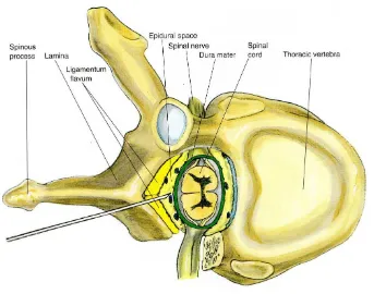
Atypical lumbar vertebra is made up of the following parts: 1. The body
2. Vertebral arch
3. Transverse processes 4. Spinous process
[image:46.595.109.524.536.707.2]5. Superior and inferior articular processes.
30 | P a g e Body:
It is kidney shaped. They are weight bearing. The flat articular surfaces are covered with hyaline cartilage, which is firmly united to the fibrocartilagenous intervertebral discs (annulus fibrosus and nucleus pulposus). The anterior and posterior longitudinal ligaments reinforce the union the between the bodies. The broad anterior longitudinal ligaments is firmly attached to the intervertebral discs and loosely attached to bodies. The posterior longitudinal ligament is narrower and is similarly attached.
Vertebral arch:
It is composed of pedicles and laminae, that surround the spinal cord and its coverings and protect it. Each half of the vertebral arch is divided into two parts by the root of the transverse process. Anteriorly, the arch is formed by the powerful rounded pedicle whose function is to transmit stress. Posteriorly, it is completed by the lamina, which is flat and is mainly protective in function. From the vertebral arches 4 articular processes project, 2 upward and 2 downward to articulate with similar processes of the adjacent vertebrae.
Transverse processes:
31 | P a g e Spinous process:
It is almost horizontal, quadrangular and thickened along its posterior and inferior borders. They act as levers for muscles which control posture and active movements of the vertebral column.
Spinous processes of the cevical, the first two thoracic, and the last four lumbar vertebrae are all particularly horizontal and are therefore opposite the bodies of their respective vertebrae. The other spinous processes are inclined in the downward direction, with their tips opposite the bodies of the vertebrae below. Exception is the tip of the first lumbar spinous process, which is opposite the intervertebral disc.
Superior and inferior articular processes:
The superior articular processes spring from the junctions of pedicles and laminae. They project upwards behind the pedicles and come to lie just above the level of transverse processes and the articular facets on their posterior surfaces face backwards and medially. The inferior articular processes extend downwards from the infero lateral aspects of the laminae. They lie below the level of the transverse process and the articulate with the facets on the superior articular processes of the vertebra below.
Intervertebral Foramina:
32 | P a g e The areolar tissue around these foramina is soft and loose in the young individual and the anesthetic solution and catheter may also pass through one of these foramina. For this reason lesser amount of local anesthetic solution is required to produce an epidural block in the elderly as compared to young individuals.
The sacral and coccygeal vertebrae fuse at puberty. Marellously exact system of ligaments, interposed cartilages and muscles at with synergestic and antagonistic precision to hold these vertebrae together and to keep the vertebra column from collapsing.
There are 4 anatomical curvatures in the vertebral column of which, the thoracic and the sacral are primary and concave anteriorly and the cervical and the lumbar are secondary and convex anteriorly. These curves have a significant influence on the spread of local anesthetic in the sub arachnoid and the epidural space.
LIGAMENTS:
The vertebral column is bounded together by several ligaments, which gives it stability and elasticity
1. The Supraspinous ligament 2. The Interspinous ligament 3. The Ligamentum flavum
Fig 2.
Supraspinous Ligament
It is a strong thick fibrous band connecting the apices of the spines from the 7th cervical vertebra o
broad. In cervical region, it blends into the neck ligaments, where it is specialized as the ligamentum nuchae and extends from 7
to occipital protuberance.
Interspinous Ligament
It is a thin fibrous structure connecting adjacent spines. The fibres are almost membraneous and extends from the apex and upper surface of the lower spine towards the root and inferior surface of the next higher vertebrae.
Fig 2. Boundaries of Epidural Space
Supraspinous Ligament
It is a strong thick fibrous band connecting the apices of the spines cervical vertebra of the sacrum. At lumbar region it is thick and broad. In cervical region, it blends into the neck ligaments, where it is specialized as the ligamentum nuchae and extends from 7th cervical vertebra to occipital protuberance.
Interspinous Ligament
in fibrous structure connecting adjacent spines. The fibres are almost membraneous and extends from the apex and upper surface of the lower spine towards the root and inferior surface of the next higher vertebrae.
33 | P a g e It is a strong thick fibrous band connecting the apices of the spines
the sacrum. At lumbar region it is thick and broad. In cervical region, it blends into the neck ligaments, where it is cervical vertebra
34 | P a g e They meet the supraspinous ligament posteriorly and tend to blend with ligamentum flavum in front.
Ligamentum Flavum
It consists of yellow elastic tissues. The fibres are perpendicular in direction. They extend from the anterior inferior surface of upper lamina downward to the anterior superior surface of lower lamina. Ligament exsist as a right and left half. Internal surface of left and right ligamentum flavum form an acute angle with its vertex in contact with the interspinous ligament. The dorsomedian connective tissue band extends from the apex of the ligamentum flavum and the periostium through the extradural space to the spinal dura matter.
Posterior Longitudinal Ligamnent
Runs within the vertebral canal and posterior surfaces of body of vertebrae from which it is separated by the basivertebral veins.
Anterior Longitudinal Ligament
Runs along the front of vertebral bodies, it is adherent also to the intervertebral discs.
Spinal Meninges
35 | P a g e extradural space. It is connected by fibrous slips to posterior longitudinal ligament, especially near lower end of vertebral canal. A strong fibrous layer forms a tubular sheath attached above to margins of foremen of magnum and ending below at lower border of second sacral vertebra.
Arachnoid mater: this is a thin transparent sheath closely applied to the dura it surrounds the cranial and spinal nerves as far as their point of exit from the skull and vertebral canal.
Pia mater: this is separated from the arachnoid by the sub arachnoid space, filled with cerebrospinal fluid. The pia mater closely invest the cord and sends delicate septa into its substance. From each lateral surface of the piamater a fibrous band, the denticulate ligament, projects into the subarachnoid space, and is attached by a serious of pointed processes to the dura as far down as the first lumbar nerve. Pia mater ends as a prolongation, the filum terminate which pierces the distal end of the dural sac and is attached to the periostium of the coccyx.
36 | P a g e NERVE SUPPLY OF MENINGES
The posterior aspect of the dura and arachnoid contains no nerve fibres and so no pain is felt on dural puncture. The anterior aspect is supplied by spinovertebral nerves. Each of these enters the intervertebral foramen and passes up for one segment and down for 2 segments.
Spinal nerves:
These are 31 pairs in number and are as follows: 1. 8 cervical
2. 12 thoracic 3. 5 lumbar 4. 5 sacral 5. 1 coccygeal
Anterior root: is efferent and motor. Sympathetic pre ganglionic axons arise from cells in the intermediolateral horn of the spinal cord from T1 to L2. Posterior root: is larger than anterior. All the afferent impulses from whole body including viscera pass into the posterior roots.
Each posterior roots has a ganglion and conveys fibers of 1.pain 2.Tactile, 3.Thermal, 4.Deep or muscle sensation from bones, joints, tendons etc, 5.Afferent from the visera and 6.Vasodilator fibres.
37 | P a g e foramina to form the main spinal nerve trunks, which soon divide into anterior and posterior primary divisions-mixed nerves.
EPIDURAL SPACE Definition:
Epidural space is a potential space, ellipitical, surrounds the dural sac, that extends from the foramen magnum to the coccyx and laterally, communicates with the paravertebral space through the intervertebral foraminae.
Boundaries: the epidural space is bounded superiorly by the foremen magnum where the periosteal and spinal layer of dura fuse, inferiorly by the sacrococcygeal membrane, anteriorly by the posterior longitudinal ligament covering the posterior aspect of the vertebral bodies and the intervertebral discs, posteriorly by the ligamentum flavum and the anterior surface of the vertebral laminae, and laterally by the pedicle of the vertebrae and the intervertebral foramina.
EPIDURAL SPACE ANATOMY
38 | P a g e To reach the epidural space in the midline sagital plane, the following structures are to be penetrated :
• Skin
• Subcutaneous tissue
• Supraspinous ligament
• Interspinous ligament
• Ligamentum flavum
The ligamentum flavum is an important landmark for the technical identification of the epidural space during induction. The first three tissues offer little resistance to the advancing needle, but the resistance increases when the ligamentum flavum is reached. As the needle passes through this structure there is a sudden give way of the resistance. While performing epidural analgesia, it is important to recognize this point as little further advancement may result in subarachnoid penetration.
CONTENTS OF EPIDURAL SPACES:
39 | P a g e The roots vary greatly in size and thickness. The thoracic roots are thin, while the cervical and lumbosacral roots subserving the limbs are thick. The great difference in size and neural populations within the roots are interrelated. The very large diameter and high neural population of the dorsal and ventral roots of the first sacral segments are associated with great resistance to epidural blockade. Prolonged latency and poor analgesia of S1 segment are due to poor penetration of local anesthetic and it deserves a special mention as they have an important role in the mechanism of the action to epidural anesthesia.
[image:56.595.179.449.455.708.2]The arachnoid villi and granulations invaginate the epidural veins in the region of the dural cuff and drain the CSF from the vessels, drain the CSF into the epidural fat, from where it is drained by lymphatics.
40 | P a g e EPIDURAL VESSELS:
The branches of the subclavian, aortic and iliac arteries cross the epidural space and enter the subarachnoid space in the region of the dural cuffs. These branches provide blood supply as far as the spinal roots. Apart from the cervical region, the entire blood supply to the spinal cord passes through the epidural space.
The epidural veins are arranged in the form of longitudinal plexuses on either side of the line. They do not posses values. These veins although divided into anatomical groups, all interconnect and form a series of horizontal segmental anastomosis. They connect with intervertebral foramina and communicate with the vertebral, ascending cervical, deep cervical, intercostal, iliolumbar and lateral sacral veins. As the epidural veins have no valves they afford a connection between the pelvic veins below and the intracranial veins above.
The epidural veins become distended during coughing and straining and also when the inferior vena cava is obstructed by large abdominal tumors or in late pregnency. This distension of epidural veins diminishes the effective volume of the epidural space. Under these circumstances the requirement of the local anesthetic is markedly decreased, as a small volume of drugs tends to spread over a wide area in the epidural space.
41 | P a g e between the fatty areolar tissues. The epidural fat constitutes an important pharmacological space and depot for injected local anaesthetics and drugs and it is one of the three competitors for its share of the drug. The other two competitors being nervous tissue of spinal roots and cords and blood vessels within the spinal canal.
Drugs with high lipid solubility and lipoprotein binding charactristics will tend to enter the fat phase and remain there for a period of time, depending on their pharmacodynamics and on the briskness of the local blood flow competing for uptake. The compliance of the epidural fat varies from person to person and with age. In children and young adults it offers very little resistance.
LYMPHATICS:
Surrounding and draining the dural sac, lympatics run anteriorly from each intervertebral foramen and empty into the longitudinal channals in front of the vertebral column.
42 | P a g e The dorsomedial connection between the dura and the ligamentum flavum can explain some of the results occurring during clinical epidural anesthesia. The epidural needle must separate the dorsomedian fold during insertion. Insertion of a catheter may result in its disposition slightly to either side of the midline. When a true dorsomedian band or membrane exists, a patchy and/or a unilateral type of block can result. The Dorsomedian connections also explain the difficulty and the effort needed to advance the epidural catheter freely into the epidural space.
SIZE OF THE EPIDURAL SPACE:
Regional epidural space width and dural thickness:
• Cervical
• Upper thoracic
• Lower thoracic
• Lumbar
IDETIFICATION OF EPIDURAL SPACE: a) Negative pressure methods:
• Hanging drop technique
• Capillary tube technique
• Manometer technique b) Loss of resistance methods
• Syringe technique
43 | P a g e
• Ballon technique
• Brooks device technique
• Vertical tube of Dawkins technique c) Other techniques:
• Ultrasonic localization
[image:60.595.157.469.282.566.2]• And the oxford- epidural space indicator.
Fig. 4.Tecnicniques to find out Epidural Space
Summary of fate of injected solution in epidural space:
44 | P a g e Superiorly the spread is to magnum. There is possibility of diffusion across dura at base to cerebral CSF with possibility of blockade of cranial nerves, vasomotor and respiratory centres and other vital centres.
Inferiorly to sacral hiatus, caudal canal and through anterior sacral foramina. Laterally through intervertebral foramina to paravertebral space, to produce paravertebral neural blockade. There is rapid access to CSF at dura cuff region to produce spinal nerve root blockade and also subsequent access to spinal cord.
Anteriorly is the thin epidural space between dura and posteriorly longitudinal ligament. There is also access for injection solution to CSF by slow diffusion into the subarachnoid space. Vascular absorption by way of epidural veins may convey drug directly to brain and epidural fat also takes up the drug.
The longitudinal spread in epidural space leads to:
• Leakage by vascular absorption.
• Leakage through intervertebral foramina – paravertebral block of nerve trunks in young subjects – centripetal spread – subpial spread – spinal root and peripheral cord block.
• Diffusion through dural root sleeves – subdural spread – spinal root block.
45 | P a g e Fig 5. Paramedian Approach for Epidural Space
Factors affecting epidural spread:
• Spread increases with age: Escape from the epidural space is relatively less due to the invertebral foramina being more fixed and the epidural vessels being less penetrable.
• Spread is greater in pregnant ladies.
• In arteriosclerotic patients and patients with occlusive arterial disease the spread is greater than normal.
• Spread decreases in dehydration, shock and in cachexia.
• The extent of anesthesia is greater with concentrated solutions.
46 | P a g e PHYSIOLOGICAL CONSIDERATIONS :
Negative pressure in the peridural space is greatest at the points of firm attachment. It is also greatest in the thoracic region, less in the lumbar region, and least or absent in the sacral area. There are two theories explaining the negative pressure are:
1. The Cone Theory: This theory considers that the needle introduced into peridural space depresses the dura and consequently creates a larger epidural space. It is thus considered as an artifact caused by indentation of the dura by the advancing needle.
2. The Transmission Theory: this theory considers that the negative pressure in the epidural space is the transmission of the intrapleural negative pressure via the intervertebral foramina to the peridural space.
EFFECTS ON ORGAN SYSTEM Cardiovascular system :
47 | P a g e such as fluid administration and the use of vasoconstrictors. Sympathetic outflow extends from T1-L2 and blockade of nerve roots below this level, as with, for example, knee surgery, is less likely to cause significant sympathetic blockade, compared with procedures requiring blockade above the umbilicus. Respiartory system:
Usually unless blockade is high enough to affect intercostal muscle nerve supply(thoracic nerve roots) leading to reliance on diaphragmatic breathing alone. This is likely to cause distress to the patient, as they may feel unable to breath adequately.
GIT system:
Blockade of sympathetic outflow(T5-L1) to the GI tract leads to predominance of parasympathetic (vagus and sacral parasympathetic outflow) leading to active peristalsis and relax the sphincters, and a small conracted gut which enhances surgical access. Splenic enlargement 2-3 fold occurs.
Endocrine system:
Nerve supply to the adrenals is blocked leading to a reduction in the release of catecholamines.
Genitourinary tract:
48 | P a g e
MATERIALS AND METHODS
This study design is comparative study.
Study setting in patients undergone lower abdominal and lower limb surgeries under epidural anaesthesia in the department of anaesthesia in Sree Mookambika Institute of Medical Sciences, Kulasekharam. The study period was 12 months.
Total sample size of 70 was selected and divided into two groups which are described below
Group I : Patients receiving 15ml of 0.75% of Ropivacaine with 1 mcg/kg of Dexmedetomidine
Group II : Patients receiving 15 ml of 0.75% of Ropivacaine with 1 mcg/kg of Clonidine
SAMPLING:
Sample size of each group: 35 Total sample size of the study: 70
Scientific basis of sample size used in the study: Formula comparative and descriptive study.
݊= 2ܵଶ ( ܼ
ଵ+ܼଶ)ଶ
(ܯ
49 | P a g e S1 = Standard deviation of Dexmedetomidine = 3.96
S2 = Standard deviation of Clonidine = 4.86 Z1 = Z value associated with alpha = 1.64 Z2 = Z value associated with beta = 0.84 M1 = mean of Dexmedetomidine = 13.14 M2 = mean of Clonidine = 15.8
Sample size = 34.34 ~ 35
sample size of Dexmedetomidine group = 35 Sample size of Clonidine = 35
Total sample size = 70
S= pooled standard deviation = 4.43
Sampling Technique: Convenient sampling.
Inclusion criteria:
• Patients giving valid consent.
• Patients under ASA (American Society of Anaesthesiology) physical status 1 and 2 (ASA 1 – normal and healthy patients, ASA 2 – patients with mild systemic disease without any functional limitation.
• Patients undergoing lower limb and lower abdominal elective surgeries under epidural anaesthesia.
50 | P a g e Exclusion criteria:
• Refusal by the patient.
• Patients with ASA physical status 3 or more.
• Patients posted for emergency surgeries and caesarean section.
• Patients with history of alcohol or drug abuse.
• Patients who are allergic to any of the test drugs.
• Contraindication to spinal anaesthesia.
Formulation of the drug used:
Dexmedetomidine hydrochloride as liquid for injection, (S)-4-[1-(2,3-dimethyl phenyl) ethyl]-1 H-imidazole hydrochloride. Clonidine as liquid for injection.
Dugs used : Dexmedetomidine, Dextomed, Neon laboratories. Clonidine
Dose of the drug used: 1mcg/kg Dexmedetomidine with 0.75% Ropivacaine for epidural, 150mcg Clonidine with 0.75% Ropivacaine for epidural
Frequency of the drug used: a loading dose of 1mcg/kg of Dexmedetomidine with Ropivacaine or 1mcg/kg Clonidine with Ropivacaine during intraoperative period
Route of the drug used: Epidural
Duration of the drug used: as intraoperative.
51 | P a g e Mode of management: Hypotension (MAP below 20% of the baseline, systolic pressure < 90mmHg) will be treated with incremental doses of Mephentermine 3mg i.v and additional RL solution as appropriate. Bradycardia (HR < 50bpm) will be treated with Atropine 0.6m.g i.v and respiratory depression (RR < 12 breaths per min) by administering supplemental oxygen – 4-6L/min.Inj. Hydrocortisone 100mg i.v stat, Inj. Pheniramine maleate 1 ampoule (45.5mg) i.m stat, Inj. Adrenaline 1:1000.
Agreement of compensation: As per the rules of this institution.
Registration with Clinical Trial Registry of India [CTRI]: Yes, registration copy attached herewith.
Clinical trial design: comparative and descriptive study.
Clinical trial done at: single site
Allocation ratio of different groups: 1:1
Randomization: Yes
Type of randomization: randomized control trial
Method used to generate random sequence numbers: randomized purposive sampling technique
Allocation concealment mechanism: not applicable
52 | P a g e Parameters to be studied: heart rate per min, Blood pressure in mmHg, SpO₂ in percentage.
Instruments used: Multiparameter, Schiller, made in Switzerland-India.
Procedure in brief:
After approval of the study protocol by our institutional committee, written informed consent will be taken from each patient. ASA status I and II patients of either sex, aged between 18-65 years, weighing 50-70kgs, undergoing lower limb or lower abdominal surgery under epidural anaesthesia will be enrolled in this study. All the patients will be visited on the day prior to surgery, explained in detail about the anaesthetic procedure and informed written consent will be obtained.
The patients will be kept nil orally 6hrs prior to the day of surgery. Patients with a history of alcohol or drug abuse, diabetes mellitus, cardiac cases, hypertension, COPD, psychological disease, hepatic and/or renal disease, spinal deformities or any contraindication to spinal anaesthesia (for eg: coagulation defects, infection at the puncture site, pre-existing neurological deficits in the body, etc.) and patients allergic to amide type of local anaesthetics are excluded from the study.
53 | P a g e Pressure and SpO₂ probe was attached and baseline parameters recorded and
Inj. Ranitidine 50mg i.v and Inj. Metoclopramide 10mg slow i.v half an hour before the surgery. Group I was given Dexmedetomidine 1mcg/kg with Ropivacaine epidurally, whereas Group II will be given 1mcg/kg of Clonidine with Ropivacaine epidurally. Patients will then be positioned and 15ml 0.75% Ropivacaine was administered epidurally in L3-L4 interspace through a standard midline approach using a 18-G tuohy needle and all patients will be supplemented with oxygen - 4L/min via a face mask throughout the procedure after positioning the patient. Sensory block will be assessed using sterile pin prick method in the mid-axillary line on both sides of the chest, motor block was assessed using a modified Bromage scale (grade 0 ⇾ no paralysis; grade I ⇾ unable to raise extended leg; grade 2 ⇾ unable to
flex the knee, grade 3 ⇾ unable to flex the ankle)
Sensory and motor block was assessed every minute for the first 10mins and thereafter every 10mins during the surgery and every 15mins postoperatively and be recorded. The highest dermatome level of sensory blockade and motor blockade will be recorded. Recovery time for the sensory blockade is considered as two dermatome regression of anaesthesia from the maximum level; motor block duration is the time to return to grade 1on the modified Bromage scale. Postoperative pain was assessed by using the Visual Analog Scale (VAS 0 ⇾ no pain and VAS 10 ⇾ worst possible pain) at 4, 8, 12
54 | P a g e 50mg slow i.v. The time of patient’s first request for postoperative analgesia after the surgery was recorded as duration of postoperative analgesia.
The Ramsay sedation score was used to assess sedation (1⇾ Anxious or
agitated; 2⇾ co-operative and tranquil; 3⇾ drowsy, but responsive; 4 ⇾asleep,
but responsive to glabellar tap; 5⇾ asleep with a sluggish response to tactile
stimulation and 6⇾ asleep, with no response). The score was re-evaluated
every 10mins after the administration of the drug for up to 180mins and every 15mins thereafter. Score 5/6 will be considered as excessive sedation.
The vital data viz. heart rate (HR), mean arterial pressure (MAP), oxygen saturation (SpO₂), respiratory rate (RR) was recorded immediately
before and 60secs after dural puncture, every 10mins after epidural anaesthesia intraoperatively and every 15mins in the postoperative period.
STATISTICAL ANALYSIS:
1. The study parameter was entered in Microsoft excel work sheet 2013 version and data was analysed by using statistical package of social science(SPSS) trial version 20.0
2. Test of significance used – student t test 3. Level of confidence is 95%.
4.2 ASSESSMENT OF PARAMETERS
55 | P a g e every minute till onset of block at T10.
Duration of analgesia is defined as the time taken from the onset of sensory blockade at the T 10 level to the time of sensation of pain at the surgical site with a VAS score of >3.
Peak sensory level was defined as the highest dermatome level of sensory blockade achieved after administration of study drug.
Time to two dermatome regression was defined as the time interval from the sensory block at the highest dermatome to the regression of sensory blockade by two dermatomes. The sensory level was assessed every 15minutes after 2 hours of epidural bolus injection till 2 dermatome regression of sensory level was observed.
The time to motor blockade was defined as the time interval from the administration of epidural study drug to the achievement of grade 3 motor blockade in the lower limbs.
The degree of motor block was assessed using the modified Bromage scale.
The assessment for motor block was done every 5 minutes after administration of study drug till a block of modified Bromage grade 3 motor blockade was achieved.
57 | P a g e
RESULTS
The study group consisted of 70 patients, 35 in group RC who received 15ml of 0.75% of Ropivacaine with 1mcg/kg Clonidine and 35 in group RD who received 15ml of 0.75% of Ropivacaine with 1mcg/kg Dexmedetomidine. Both groups were comparable demographically with respect to age and age distribution, height & weight characteristics. The distribution of the types of surgery and the duration of surgery was comparable between the two groups.
58 | P a g e DEMOGRAPHIC PROFILE
Table 1 Gender Distribution
Gender
Category
Total
Group 1 Group 2
N % N % N %
Male 25 71.4 21 60.0 46 65.7
Female 10 28.6 14 40.0 24 34.3
Total 35 100.0 35 100.0 70 100.0
p=0.314
Fig 6. Gender Distribution
Gender between the 2 groups. The group RC had males of 71.4±60 and the group RD had females of 28.6±40. There was no significant difference in the gender composition between the two groups. (p=0.314)
0% 10% 20% 30% 40% 50% 60% 70% 80% 90% 100%
Group 1 Group 2
59 | P a g e Table 2 ASA Physical Status Distribution
ASA
Category
Total
Group 1 Group 2
N % N % N %
Grade 1 25 71.4 27 77.1 52 74.3
Grade 2 10 28.6 8 22.9 18 25.7
Total 35 100.0 35 100.0 70 100.0
[image:78.595.108.517.117.283.2]p=0.299
Fig 7.ASA Physical Status Distribution
ASA grading between the 2 groups. The group RC had ASA1 of 71.4±77.1 and the group RD had ASA2 of 28.6±22.9. There was no significant difference in the ASA grading between the two groups. (p=0.299)
0% 10% 20% 30% 40% 50% 60% 70% 80% 90% 100%
Group 1 Group 2
ASA
60 | P a g e Table 3 Weight Distribution
N
Weight in Kg
T P
Mean sd
Group 1 35 61.14 9.239
1.199 0.235
[image:79.595.134.498.210.504.2]Group 2 35 58.63 8.282
Fig 8. Weight Distribution
Weight of persons in the group RC had a mean weight is 61.14±9.239 and the group RD had a mean weight is 58.63±8.282.
61.14
58.63
0 10 20 30 40 50 60 70
Group 1 Group 2
W
e
ig
h
t
in
K
61 | P a g e Table 4 Height Distribution
N
Height in cm
T P
Mean SD
Gro