White Rose Research Online URL for this paper:
http://eprints.whiterose.ac.uk/98064/
Version: Published Version
Article:
Brüning, Jan-Gert, Myka, Kamila Katarzyna and McGlynn, Peter
orcid.org/0000-0001-8629-4713 (2016) Overexpression of the Replicative Helicase in
Escherichia coli Inhibits Replication Initiation and Replication Fork Reloading. Journal of
Molecular Biology. pp. 1068-1079. ISSN 0022-2836
https://doi.org/10.1016/j.jmb.2016.01.018
[email protected] https://eprints.whiterose.ac.uk/ Reuse
This article is distributed under the terms of the Creative Commons Attribution (CC BY) licence. This licence allows you to distribute, remix, tweak, and build upon the work, even commercially, as long as you credit the authors for the original work. More information and the full terms of the licence here:
https://creativecommons.org/licenses/
Takedown
If you consider content in White Rose Research Online to be in breach of UK law, please notify us by
Overexpression of the Replicative Helicase in
Escherichia coli
Inhibits Replication Initiation
and Replication Fork Reloading
Jan-Gert Brüning, Kamila Katarzyna Myka and Peter McGlynn
Department of Biology,University of York, Wentworth Way, York YO10 5DD, United Kingdom
Correspondence toPeter McGlynn:[email protected].
http://dx.doi.org/10.1016/j.jmb.2016.01.018
Edited by B. Connolly
Abstract
Replicative helicases play central roles in chromosome duplication and their assembly onto DNA is regulated via initiators and helicase loader proteins. TheEscherichia colireplicative helicase DnaB and the helicase loader DnaC form a DnaB6–DnaC6 complex that is required for loading DnaB onto single-stranded DNA.
Overexpression ofdnaCinhibits replication by promoting continual rebinding of DnaC to DnaB and consequent prevention of helicase translocation. Here we show that overexpression of dnaB also inhibits growth and chromosome duplication. This inhibition is countered by co-overexpression of wild-type DnaC but not of a DnaC mutant that cannot interact with DnaB, indicating that a reduction in DnaB6–DnaC6concentration is responsible for the phenotypes associated with elevated DnaB concentration. Partial defects in theoriC-specific initiator DnaA and in PriA-specific initiation away from oriC during replication repair sensitise cells to dnaB
overexpression. Absence of the accessory replicative helicase Rep, resulting in increased replication blockage and thus increased reinitiation away fromoriC, also exacerbates DnaB-induced defects. These findings indicate that elevated levels of helicase perturb replication initiation not only at origins of replication but also during fork repair at other sites on the chromosome. Thus, imbalances in levels of the replicative helicase and helicase loader can inhibit replication both viainhibitionof DnaB6–DnaC6complex formation with excess DnaB, as shown here, andpromotionof formation of DnaB6–DnaC6complexes with excess DnaC [Allen GC, Jr., Kornberg A. Fine
balance in the regulation of DnaB helicase by DnaC protein in replication in Escherichia coli. J. Biol. Chem. 1991;266:22096–22101; Skarstad K, Wold S. The speed of theEscherichia coliforkin vivodepends on the DnaB:DnaC ratio. Mol. Microbiol. 1995;17:825–831]. Thus, there are two mechanisms by which an imbalance in the replicative helicase and its associated loader protein can inhibit genome duplication.
© 2016 The Authors. Published by Elsevier Ltd. This is an open access article under the CC BY license (http://creativecommons.org/licenses/by/4.0/).
Introduction
The structural complexity of the genetic material of a cell demands complex copying systems to achieve high-fidelity DNA replication. Replicative helicases are at the leading edge of replication forks, both driving strand separation and acting as a protein interaction hub at the heart of the replisome[3]. These helicases are active as hexamers that form toroidal quaternary structures and bind a single strand of nucleic acid in their central channel[4]. Unwinding of duplex DNA is achieved by NTP-driven translocation along this bound single-stranded DNA (ssDNA) and steric exclusion of the complementary strand creating ssDNA templates that can then be copied by DNA polymerases[5].
The replicative helicase needs to be loaded at an origin of replication before it can begin to unwind DNA. In Escherichia coli, the initiation of replication starts with binding oforiCby the ATP-bound initiator protein DnaA that leads to melting of the DNA duplex and creation of a ssDNA bubble[6]. Helicase loading also requires the helicase loader protein DnaC in complex with a DnaB hexamer in a 6:6 ratio [7]. During initiation, the circular DnaB hexamer is opened within this complex, allowing the ssDNA to be passed into the central channel of the helicase[8]. ATP hydrolysis by DnaC results in its dissociation, allowing DnaB to translocate along ssDNA towards the fork junction and subsequent association of other enzymes with DnaB to form the active replisome [9]. However, if DnaC is in excess over DnaB, then DnaB helicase and
thus replisome activity are inhibited [1,2]. Inhibition results from the ATP-bound form of DnaC continually reassociating with DnaB to form a DnaB6–DnaC6
complex that cannot translocate[10,11].
Once a replication fork is translocating along the template DNA, then potential barriers such as DNA damage or nucleoprotein complexes are encoun-tered frequently[12–15]. Some of these barriers can be bypassed or cleared by the original replisome, allowing replication to continue [16,17]. However, some nucleoprotein barriers, especially those asso-ciated with transcription, need to be cleared ahead of the fork by accessory replicative helicases such as Rep inE. coli[18,19]. This removal of nucleoprotein barriers minimises replisome pause time and hence reduces the probability of replisome inactivation since replisomes lose activity as a function of pause time [20–22]. In E. coli, this minimisation of nucleoprotein-induced fork pausing by Rep is promoted by a physical interaction between the Rep C-terminus and DnaB[18,23,24].
Replisome inactivation still occurs in spite of mechanisms that reduce the probability of loss of replisome function[13,25]. In such circumstances, the replisome must be reloaded back onto the chromo-some in order to complete genome duplication. Replisome reloading is triggered by the reassembly of the replicative helicase back onto the chromosome in a reaction that, as withoriC, requires DnaC [26]. However, the presence of the ssDNA binding protein SSB prevents DnaB loading onto ssDNA and replication initiator proteins are need to overcome this inhibition[6]. DnaA-mediated replication initiation is sequence specific and occurs only at the origin, and thus, additional factors are required away fromoriCto overcome this SSB-dependent barrier. InE. coli, two pathways that facilitate reloading of the replisome back onto the chromosome away from oriC exist
[25,27]. One pathway for replication fork reloading involves the helicase PriA. PriA binds to DNA forks possessing a 3′OH group of a nascent leading strand close to the fork branch point in an orientation allowing PriA translocation along the lagging strand template
[28,29]. PriA binding to the fork results in recruitment of the additional reloading factors PriB and DnaT and subsequent loading of DnaB onto the lagging strand template via a DnaB–DnaC dodecamer [30–34]. Alternatively, DnaB reloading can be catalysed by PriC. PriC has complementary forked DNA binding specificity to that of PriA, targeting forks lacking a 3′
OH group of a nascent strand close to the fork branch point, but the outcome again is replicative helicase reloading onto the lagging strand template[25,27]. If a ssDNA region is absent on the lagging strand template, additional DNA unwinding by the 3′-to-5′
helicases Rep or PriA is required to provide sufficient ssDNA for PriC-directed DnaB loading[25,35].
We have found that overexpression of DnaB in
E. colihas a modest inhibitory effect on chromosome
duplication. This inhibition is alleviated upon co-over-expression of the helicase loader DnaC indicating that a reduction in the concentration of DnaB6–DnaC6
complexes, needed for DnaB loading during replica-tion initiareplica-tion and reinitiareplica-tion [7], is responsible for inhibition of chromosome duplication by excess DnaB. Partial defects in DnaA-dependent replication initiation at oriC or in PriA-dependent replication reinitiation away from oriC act synergistically to increase the toxicity of DnaB overexpression. Therefore, overex-pression of the replicative helicase impacts on repli-some assembly at and away from oriC. However, absence of PriC does not hypersensitise cells to excess DnaB indicating that, in otherwise unstressed cells, PriA-directed fork repair predominates or that PriC-specific substrates can be targeted by PriA either with or without further processing of the forked DNA. The inhibitory effect of excess DnaB is also exacer-bated in cells lacking the accessory replicative heli-case Rep that correlates with the elevated replication blockage and thus replication reinitiation needed in
Δrep cells [19,36]. These data demonstrate that elevation of replicative helicase levels can result in inhibition of chromosome duplication. Thus, paradox-ically, inhibition of replication can occur both by promotion of DnaB6–DnaC6 complex formation via
excess DnaC[1,2]and by reducing the probability of DnaB6–DnaC6complex formation via excess DnaB as
shown here. Such imbalances could conceivably result in replicative stress regardless of the organism.
Results
Overexpression of DnaB inhibits colony formation and this inhibition is exacerbated by the absence of Rep
levels of DnaB inhibit growth of an otherwise wild-type strain and that inhibition is greatly exacerbated in the absence of Rep.
We then tested which function of Rep was responsible for hypersensitivity ofΔrepcells todnaB
[image:4.595.129.463.90.255.2]overexpression. Rep has been implicated in promot-ing fork movement along protein-bound DNA and also in promoting PriC-dependent reloading of DnaB
[18,25,35]. However, ΔpriC cells displayed only a modest decrease in colony size similar to that seen in wild type (Fig. 1D). Therefore, a defect in PriC-directed replisome reloading was not responsible for thednaB
hypersensitivity of Δrep cells. Thus, absence of the only other known function of Rep, promotion of fork movement along protein-bound DNA, may be the missing function that hypersensitises cells to dnaB
overexpression.
A helicase closely related to Rep, UvrD, promotes replisome movement along protein-bound DNA in the absence of Rep[18,19]. However, UvrD cannot compensate fully for the absence of Rep [18,24]
resulting in Δrep but not ΔuvrD cells displaying increased replisome pausing at nucleoprotein com-plexes [36]. dnaB overexpression in ΔuvrD cells again gave only a modest decrease in colony size similar to that seen in the wild type (Fig. 1C). These data indicate that the partial ability of UvrD to compensate for the absence of Rep-promoted fork movement is not sufficient to ameliorate the effects ofdnaBoverexpression.
dnaCcounters dnaBtoxicity
A 1:1 DnaB:DnaC ratio is needed for maximal DnaB helicase and replisome activity[1]. Therefore,
we analysed whether the effects of dnaB overex-pression reflected a substoichiometric level of DnaC with respect to DnaB.
dnaCwas cloned into the same arabinose-induci-ble overexpression plasmid as that used for dnaB, creating pBADdnaC.Growth ofrep+andΔrepcells containing pBADdnaC inhibited growth in the pres-ence of arabinose, as expected[1,2](Fig. 2a-iii and b-iii). In contrast, pBADdnaBC in which wild-type
dnaC had been cloned downstream of dnaB
suppressed the growth defects caused by overex-pression of dnaB, a suppression that was most apparent inΔrep cells (Fig. 2b, compare ii and iv). Suppression of thednaB-dependent growth defect in pBADdnaBC was not due to lack of elevated DnaB levels as SDS polyacrylamide gel electrophoresis revealed similar levels of DnaB in cells containing pBADdnaB and pBADdnaBC (Fig. 2c, compare lanes 7 and 11 in i and ii). However, we could not detect increased intracellular levels of DnaC with either pBADdnaCor pBADdnaBC(Fig. 2c, compare lanes 9 and 11 in i and ii). This was surprising given the ability of pBADdnaC to inhibit growth, as expected when DnaC concentrations are elevated
[1,2]. It is possible that overexpressed DnaC was degraded rapidly but it is also possible that suppres-sion of DnaB toxicity can be achieved by substoi-chiometric elevation of DnaC levels. A third formal possibility is that suppression by dnaC is not dependent on formation of DnaB–DnaC complexes and is thus not dependent on the DnaB:DnaC ratio, but it occurs via some other unidentified mechanism. This alternative mechanism cannot be absence of DnaB overexpression due to the cloning of dnaC
downstream of dnaB as DnaB was overexpressed
pBAD
pBADdnaB
pBAD
pBADdnaB
pBAD
pBADdnaB
pBAD
pBADdnaB
A) wild type
B)
∆
rep
C)
∆
uvrD
D)
∆
priC
- arabinose
+ arabinose
10-1 10-6
10-1 10-6
from pBADdnaBCclones (Fig. 2c-i and c-ii, lanes 11 and 15). Furthermore, suppression was dependent on the interaction of DnaC with DnaB. DnaC(R10P) is deficient only in DnaB binding and, thus, does not inhibit growth when overexpressed [37] [see also pBADdnaC(R10P) in Fig. 2a-v and b-v]. Cloning of
dnaC(R10P) downstream ofdnaBfailed to alleviate inhibition of growth by dnaBinΔrep cells (Fig. 2b, compare iv and vi). We conclude that DnaC counters the growth defects associated with elevated DnaB via a DnaB–DnaC interaction.
Hypersensitivity ofΔrepcells todnaB overexpression is alleviated by reducing nucleoprotein barriers to replication
Minimisation of replisome pausing and breakdown at nucleoprotein complexes is dependent on Rep helicase activity and is also promoted by a physical interaction between DnaB and the C-terminus of Rep
[18,19,36]. repK28R encodes a helicase-deficient Rep that lacks all accessory helicase function but retains the DnaB interaction domain [24]. Cells
(a)
(b)
[image:5.595.102.498.91.493.2](c)
bearingrepK28Rdisplayed hypersensitivity todnaB
overexpression similar to that seen in Δrep cells (Fig. 3a-iii). The repΔC33 allele encodes Rep that retains helicase activity but lacks the C-terminal DnaB interaction domain, resulting in a reduction in rather than abolition of accessory helicase activity [18,24].
dnaBoverexpression inrepΔC33cells did not lead to the large reduction in colony numbers seen inΔrep
andrepK28Rcells but did confer a large decrease in colony size (Fig. 3a-iv). Thus, Rep helicase function is essential for tolerance of dnaB overexpression and the Rep–DnaB interaction promotes this tolerance, a pattern similar to that found for minimisation of replisome pausing by Rep[36].
We also tested whether mutations known to decrease, rather than increase, replisome pausing and breakdown countered sensitivity to dnaB over-expression. RNA polymerase mutations that desta-bilise transcription complexes or inhibit backtracking suppress genome duplication defects by reducing nucleoprotein barriers to replication [14,36,38,39]. We tested two such mutations, rpoB(G1260D) and
rpoB(H1244Q)[40]. Bothrpomutations suppressed the toxicity of dnaB overexpression in Δrep cells (Fig. 3b). Suppression of toxicity under high levels of
dnaBinduction was greater forrpoB(G1260D) than forrpoB(H1244Q) (Fig. 3b, compare v and vi with iv on 0.2% arabinose), reflecting patterns of suppres-sion of genome duplication defects conferred by these mutations [36,40]. However, bothrpoB muta-tions provided robust suppression at an intermediate arabinose concentration (Fig. 3b, 0.02% arabinose). Therefore, mutations known to reduce replicative
barriers suppress the hypersensitivity ofΔrepcells to
dnaBoverexpression.
Taken together, these data indicate that inhibition of growth in Δrep cells by dnaB overexpression is related to increased frequency of replisome pausing and breakdown at protein–DNA complexes.
Elevated levels of DnaB inhibit chromosome duplication inΔrepcells
We probed the impact of dnaB overexpression upon genome duplication by monitoring chromo-some content using flow cytometry under run-out conditions. These conditions allow cells to complete ongoing rounds of replication but prevent reinitiation of replication and inhibit cell division [41]. Such conditions provide an indication of the numbers of origins per cell and the ability of cells to complete chromosome replication during the 2-h course of the run out.
Overexpression ofdnaBresulted in a decrease in the median number of chromosome equivalents from 4 to 2 in wild-type cells, indicating that elevated levels of DnaB cause a decrease in oriC numbers per cell (Fig. 4, compare a and b). This decrease in the median number of origins per cell suggests some perturbation of replication initiation atoriC. However, the majority of cells overexpressingdnaBcontained an integral number of chromosomes (Fig. 4b). Therefore, chromosome duplication was achieved within the 2-h run out indicating completion of the elongation and termination phases of replication.
(a)
[image:6.595.85.499.475.677.2](b)
In Δrep cells, the number of origins per cell was higher than inrep+cells even in the absence ofdnaB
overexpression (Fig. 4, compare a and c). This effect is due to increased chromosome duplication time in
Δrep cells that results in more frequent reinitiation prior to termination of replication [24,36,42]. Induc-tion ofdnaBoverexpression inΔrepcells caused a major defect in chromosome metabolism evinced by the inability to generate intact, discrete chromo-somes (Fig. 4d). Therefore, elevated DnaB concen-trations severely inhibit completion of chromosome duplication in the absence of Rep.
We imaged rep+andΔrep cells without and with
dnaBoverexpression. Overexpression inrep+cells did not result in significant perturbation of nucleoid structure (Fig. 5, compare a and b with c and d). Occasional chains of cells were observed in therep+
strain with elevated DnaB but the nucleoids in these chains appeared similar in structure to those found in the absence ofdnaBoverexpression (Fig. 5, compare b and d). Δrep cells formed occasional elongated cells even in the absence of dnaB
overexpression but nucleoid structure was similar to that observed inrep+cells (Fig. 5, compare a and b and e and f). In contrast,dnaBoverexpression in
Δrep cells resulted in mainly filamentous cells (Fig. 5g and h). Within these filaments, the nucleoids were extended but these filaments also contained significant volumes lacking DNA (Fig. 5g and h),
2 4 8 16
(a)
(b)
(c)
rep+/
pBAD
Chromosome equivalents
rep+/
pBADdnaB
∆rep/
pBAD pBAD∆repdnaB/
(d)
2 4 8 16
[image:7.595.66.292.84.314.2]2 4 8 16 2 4 8 16
Fig. 4. Overexpression ofdnaBperturbs chromosome replication in wild type and in Δrep cells. Strains were grown in LB in the absence of arabinose and then shifted into LB plus 0.2% arabinose for 2 h. Initiation atoriCand cell division were then inhibited for 2 h (“runout condi-tions”) and DNA content was monitored by flow cytometry. Strains (a) and (b) were TB28 harbouring pBAD and pJGB143, respectively, whilst strains (c) and (d) were N6577 with pBAD and pJGB143.
rep
+∆
rep
pBAD
pBAD
dnaB
pBAD
pBAD
dnaB
phase contrast
DAPI
(a)
(c)
(e)
(g)
(b)
(d)
(f)
(h)
[image:7.595.139.466.430.686.2]indicative of an inability to complete chromosome duplication and/or segregation.
We conclude that elevated levels of DnaB in an otherwise wild-type cell result in a decrease in the number of initiation events at oriC per cell cycle. However, once initiation has occurred, then chromo-some duplication and segregation can be completed successfully. In contrast, elevated DnaB levels in the absence of Rep result in failure complete chromo-some duplication.
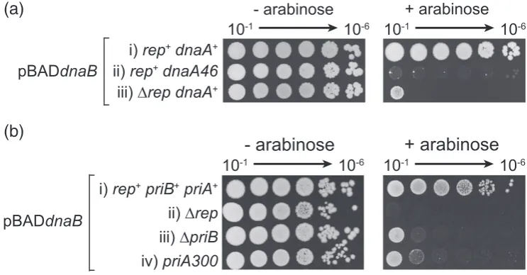
Defects in replication initiation at or away fromoriCincrease sensitivity to dnaBoverexpression
Loading of DnaB onto ssDNA at oriC occurs via DnaA whilst loading of DnaB away from oriC is catalysed by PriA and PriC [6,13]. Therefore, we tested whetherdnaBoverexpression caused defects inoriC-dependent and/ororiC-independent replica-tion initiareplica-tion by screening for synergy betweendnaB
overexpression and replication initiator mutations partially defective in loading of DnaB.
dnaA46 is a temperature-sensitive allele of the
oriC-specific replication initiator that can sustain
oriC-dependent replication initiation and therefore cell division at 30 °C but not at 42 °C. However, even at 30 °C, this allele is not fully functional[43,44]. We testeddnaBoverexpression in adnaA46strain at the permissive temperature and found that, although colonies could form, they displayed significant growth defects as compared with dnaA+ cells (Fig. 6a, compare i and ii).
PriC-dependent replisome reloading is not impor-tant in countering the impact of dnaB overexpres-sion, as indicated by the lack of hypersensitivity in
ΔpriC cells (Fig. 1D). We could not test ΔpriA
hypersensitivity due to the already poor viability of cells lacking PriA[45]. Therefore, we tested a strain that contains a mutant PriA that can still bind forked DNA structures and load DnaB but that is partially defective in replisome reloading due to the absence of PriA helicase activity[25,46,47]. A strain bearing thispriA300allele displayed hypersensitivity todnaB
overexpression (Fig. 6b-iv). This hypersensitivity was not as extreme as that shown by Δrep cells (Fig. 6b, compare ii and iv), but this intermediate growth inhibition could be the result of the partial defect in, as opposed to absence of, PriA-dependent DnaB reloading displayed by priA300. ΔpriB cells also have a partial defect in PriA-directed replisome reloading[48]and were also hypersensitive todnaB
overexpression (Fig. 6b-iii).
We conclude that cells with partial defects in either DnaA-dependent replication initiation atoriCor PriA-dependent replication reinitiation away from oriC
display elevated sensitivities todnaBoverexpression.
Discussion
We have demonstrated that overexpression of the replicative helicase DnaB inE. coliinhibits growth and that this inhibition can be countered by the helicase loader, DnaC. Overexpression of DnaB impacts upon both DnaA-dependent replication initiation atoriCand
(a)
[image:8.595.108.480.474.668.2](b)
PriA-dependent replication reinitiation away fromoriC. Absence of the accessory replicative helicase Rep also sensitises cells todnaBoverexpression, an effect that correlates with an increased need for replication reinitiation away from oriC in Δrep cells. These findings indicate that elevation of DnaB concentration inhibits both the initiation of chromosome duplication and replication reinitiation after replication forks break down. Therefore, both elevated DnaB, as shown here, and elevated DnaC [1,2] can inhibit chromosome duplication, highlighting the importance of maintaining appropriate ratios of replicative helicase and helicase loader.
The mechanisms behind the effects of overexpress-ing eitherdnaB(Fig. 1) ordnaC[1,2]must differ since the causative molecular species formed in each case must be different. Excess DnaC promotes the reassociation of ATP-bound DnaC with ssDNA-bound DnaB and these DnaB–DnaC complexes cannot translocate, inhibiting replication fork move-ment [10,11]. How might overproduction of DnaB perturb chromosome metabolism? DnaB-associated growth inhibition is enhanced by defects in initiation of replication at and away fromoriC(Fig. 6a and b) and by the absence of Rep (Figs. 1, 3 and 4). Cells lacking Rep display increased replisome pausing and break-down that elevates the need for replisome reloading away from oriC [18,19,36,49]. Mutations that have defects in the initiation of replication (dnaA46,priA300
orpriBstrains) or that increase the need for initiation (Δrep) therefore hypersensitise cells todnaB overex-pression. Synergies withΔrep,priA300orpriBcould potentially be explained by aberrant binding of excess DnaB onto the chromosome leading to inhibition of the elongation phase of chromosome duplication. Such DnaB binding could promote replisome blockage by forming nucleoprotein barriers and/or catalysing harmful unwinding of DNA structures. However, binding of excess DnaB to double-stranded regions of the chromosome is unlikely since DnaB cannot load onto double-stranded DNA either in the absence or in the presence of DnaC[5,11]. Furthermore, inhibition of the elongation phase of chromosome duplication would be predicted to cause an increase in origin numbers [24,50], which is the opposite of what is observed upondnaBoverexpression (Fig. 4a and b). Inhibition of elongation is also inconsistent with the synergy observed betweendnaBoverexpression and the partial defect inoriC-directed initiation indnaA46
cells (Fig. 6a).
The ability ofdnaCco-overexpression to suppress DnaB toxicity and the need for this co-overexpressed DnaC to interact with DnaB to effect suppression provides an alternative explanation in which excess DnaB inhibits replisome assembly rather than pro-motes replisome breakdown. Suppression by dnaC
indicates that it is the formation of DnaB complexes depleted of DnaC that is toxic (Fig. 2). Formation of a DnaB6–DnaC6 complex induces a conformational
transition within the DnaB hexamer that results in a discontinuity within the DnaB ring, allowing entry of ssDNA and hence loading of DnaB onto chromo-somes[8]. However, DnaB6–DnaC6complexes exist in equilibrium with DnaB hexamers that have fewer than six DnaC monomers bound [51]. Therefore, excess DnaB would reduce the concentration of DnaB6–DnaC6 complexes and, thus, inhibit loading
onto the chromosome. However, whilst it is clear that suppression by dnaC requires a functional DnaB–
DnaC interaction, evinced by the inability of
dnaC(R10P) to suppressDnaB-induced growth de-fects (Fig. 2b), elevation of DnaC levels could not be detected (Fig. 2c). This lack of detection could be due to rapid degradation of DnaC but it might also reflect the ability to suppress excess DnaB toxicity by substoichiometric levels of DnaC. The cooperative binding of DnaC to DnaB[51]could be one factor in allowing modest overexpression of DnaC to counter DnaB toxicity, facilitating the formation of increased numbers of DnaB6–DnaC6 complexes even when
DnaB remains in excess over DnaC.
A second and not mutually exclusive mechanism might be that DnaB complexes not bound to DNA and depleted of DnaC can interact with DnaG primase and the τ clamper loader subunit [52,53]
whereas DnaB6–DnaC6 complexes cannot, effec-tively titrating out other replication enzymes. This titration could inhibit replisome assembly at a step after DnaB loading onto ssDNA. However, regard-less of whether inhibition of replisome assembly occurs at the step of DnaB loading or at a later step, this inhibition would be predicted to occur both at
oriC and away from oriC at sites of replication breakdown. Partial inhibition of replisome assembly by elevated DnaB can explain reducedoriCinitiation events per cell cycle in wild-type cells (Fig. 4a and b) and the hypersensitivity of cells that already bear a partial defect in replisome assembly at (dnaA46) and away fromoriC(priA300andΔpriB) (Fig. 6). Partial inhibition of replisome reloading might also explain the increased illegitimate recombination caused by
Elevated DnaB also acts synergistically with defects in PriA-dependent but not PriC-dependent repair (Figs. 1 and 6). The different sensitivities of
priA300 versus priCcells might be explained by more frequent usage of PriA-directed repair as opposed to PriC that correlates with the severe reduction in viability and increased sensitivity to DNA damaging agents in ΔpriA but not ΔpriC cells [45,55]. Thus, absence of sensitivity to dnaB overexpression in
ΔpriC cells again raises questions about whether PriC-catalysed loading of DnaB, independent of PriA, is a physiologically important reaction. However, it remains possible that PriC does play a significant role in replisome reloading in vivo. The lack of obvious phenotypes inΔpriC cells could reflect an ability of PriA to target damaged forks ordinarily targeted by PriC whereas PriC might not efficiently target substrates normally acted upon by PriA in ΔpriA
cells. The targets and frequencies of use of PriA and PriCin vivoremain poorly defined.
In contrast to the bacterial situation, elevating levels of the eukaryotic replicative helicase MCM2-7 would require co-overexpression of multiple genes
[56] rather than the single gene found in bacteria. Furthermore, an excess of the eukaryotic replicative helicase is present in vivo under normal circum-stances and protects against replicative stress by allowing backup origins to be licensed and then used if forks break down [57]. However, our findings demonstrate that an imbalance between just two enzymes required for replisome assembly can result in severe defects in genome duplication. Indeed, overexpression of single MCM subunit genes can contribute to the development of cancer in higher organisms [58–61]. Our work implies that the phenotypes associated with such overexpression may be dictated by the interacting partners of the subunit whose levels are elevated. Therefore, exquisite coordination in the production of replisome components, especially the replicative helicase and associated enzymes, might be needed regardless of the complexity of the organism.
Materials and Methods
Plasmids and strains
Full-lengthE. coli dnaBwas amplified from a single colony ofE. colithat had been resuspended in 50μl of water and heated at 95 °C for 5 min prior to removal of cell debris by centrifugation. Amplification was performed with a forward primer containing a HindIII site and an NdeI site (dnaB_FW in Supplementary Table 1) and a reverse primer containing an XmaI site (dnaB_RV). The PCR product was digested with HindIII and XmaI and Klenow treated before ligating into NcoI- and XmaI-digested and Klenow-treated pBAD[18]to form pBADdnaB(pJGB143) bearing kanamycin resistance and an arabinose-induciblednaBgene.
pBADdnaBCwas generated by inserting full-lengthE. coli dnaCdownstream ofdnaBinto pJGB143 digested with PstI and HindIII, giving pJGB404. For this,dnaCwas amplified from the genome with a forward primer containing a PstI site plus a 6-bp Shine–Dalgarno sequence up to the Kozak sequence from pBAD24[62](dnaC_FW.1) and a reverse primer containing a HindIII site (dnaC_RV.1). pBADdnaC (pJGB408) is derived from pJGB404 by excisingdnaBvia EcoRI digestion and religation of the 7.1-kb fragment.
DnaCwas also cloned into pSK(−) as a EcoRI-BamHI fragment so that dnaC is inserted in the opposite orientation with respect to the promoter. For this, dnaC was amplified using a forward primer with EcoRI and NdeI sites (dnaC_FW.2) and a reverse primer with a BamHI site (dnaC_RV.2), creating pSK(−)dnaCi n v (pPM202).
pJGB412 is pSK(−) encoding dnaC(R10P)inv generated
by site-directed mutagenesis of pPM202 using comple-mentary 23-mer forward and reverse primers containing a g29c mismatch flanked by 11 bp of the dnaC wild-type sequence [dnaC(R10P)_FW and dnaC(R10P)_RV]. dnaC(R10P) from pJGB412 was PCR amplified with the same primers used for pJGB404, digested with EcoRI and HindIII and cloned into pJGB408 digested with EcoRI and HindIII to generate pBADdnaC(R10P) (pJGB415). pBADd-naBC(R10P) was generated as for pJGB404 except that the mutant dnaC from pJGB412 was used as a PCR template, resulting in pJGB418.
All strains used are listed in Supplementary Table 2.
Spot tests
Strains containing pBAD derivatives were grown in liquid LB containing 5 g l−1NaCl supplemented with kanamycin
(30μg ml−1) overnight. Growth was performed at 37 °C
with the exception of the strains used inFig. 5a that were grown at 30 °C due to the temperature-sensitive nature of the dnaA46 allele. Serial dilutions of overnight cultures were made in 56/2 salts from 10−1 to 10−6 and were
spotted on LB kanamycin (30μg ml−1) agar containing
0%, 0.02% or 0.2% arabinose as indicated. The plates were incubated at 37 °C or 30 °C, as indicated, for 16 h.
SDS polyacrylamide gel electrophoresis
Strains containing pBAD plasmids were grown at 37 °C in LB containing 30μg ml−1kanamycin until theA
Flow cytometry
Culture samples for flow cytometry were grown in LB plus 30μg ml−1kanamycin with shaking at 37 °C until an
A650of 0.4 was reached. Cultures were then diluted to an A650of 0.01 into fresh LB plus 30μg ml−1kanamycin and 0.2% arabinose and growth was continued for 2 h. Afterwards, rifampicin and cephalexin were added to 100μg ml−1 and 15μg ml−1, respectively, and the cells
were grown for another 2 h at 37 °C. Samples were then processed as in Ref.[36]except that flow cytometry was performed on a CyAn ADP Analyser (Beckman Coulter).
Microscopy
Strains containing pBADdnaBwere grown in 10 ml LB supplemented with 30 mg ml−1kanamycin to anA
650of 0.4 at 37 °C prior to diluting to anA650of 0.01 in 10 ml LB with 30 mg ml−1kanamycin without and with 0.2% arabinose.
Incubation at 37 °C continued for about 2–3 h until the cultures reached A650= 0.4, when 1 ml of culture was centrifuged. The pellet was resuspended in 400μl 56/2 salts. The nucleoids were visualised by staining in 10μg ml−14′,6-diamidino-2-phenylindole (DAPI) for 5 min
and laid on 1% agarose pads containing 56/2 salts. Microscopy was performed on a Zeiss Axioskop2 equipped with a QICAM Fast 1394 camera (QIMAGING) and a DAPI (49) filter set (Zeiss).
Supplementary data to this article can be found online at
http://dx.doi.org/10.1016/j.jmb.2016.01.018.
Acknowledgements
The authors would like to thank Bob Lloyd for supplying strains. This work was funded by the Biotechnology and Biological Sciences Research Council (BB/K00168X/1).
Received 11 November 2015; Received in revised form 11 January 2016; Accepted 11 January 2016 Available online 23 January 2016
Keywords: DNA replication; DNA repair; genome stability; transcription; disease
†Present address: J.-G. Brüning, Molecular Biology Pro-gram, Memorial Sloan-Kettering Cancer Center, New York, NY 10065, USA.
Abbreviations used: ssDNA, single-stranded DNA; DAPI, 4′,6-diamidino-2-phenylindole.
References
[1] G.C. Allen Jr., A. Kornberg, Fine balance in the regulation of DnaB helicase by DnaC protein in replication inEscherichia coli, J. Biol. Chem. 266 (1991) 22096–22101.
[2] K. Skarstad, S. Wold, The speed of theEscherichia colifork
in vivodepends on the DnaB:DnaC ratio, Mol. Microbiol. 17 (1995) 825–831.
[3] P. McGlynn, Helicases at the replication fork, Adv. Exp. Med. Biol. 767 (2013) 97–121.
[4] O. Itsathitphaisarn, R.A. Wing, W.K. Eliason, J. Wang, T.A. Steitz, The hexameric helicase DnaB adopts a nonplanar conformation during translocation, Cell 151 (2012) 267–277. [5] D.L. Kaplan, The 3′-tail of a forked-duplex sterically determines whether one or two DNA strands pass through the central channel of a replication-fork helicase, J. Mol. Biol. 301 (2000) 285–299.
[6] M.L. Mott, J.M. Berger, DNA replication initiation: Mecha-nisms and regulation in bacteria, Nat. Rev. Microbiol. 5 (2007) 343–354.
[7] J.A. Kobori, A. Kornberg, TheEscherichia coli dnaCgene product. III. Properties of thednaB–dnaCprotein complex, J. Biol. Chem. 257 (1982) 13770–13775.
[8] E. Arias-Palomo, V.L. O'Shea, I.V. Hood, J.M. Berger, The bacterial DnaC helicase loader is a DnaB ring breaker, Cell 153 (2013) 438–448.
[9] L. Fang, M.J. Davey, M. O'Donnell, Replisome assembly at
oriC, the replication origin ofE. coli, reveals an explanation for initiation sites outside an origin, Mol. Cell 4 (1999) 541–553.
[10]M.J. Davey, L. Fang, P. McInerney, R.E. Georgescu, M. O'Donnell, The DnaC helicase loader is a dual ATP/ADP switch protein, EMBO J. 21 (2002). 3148–3159.
[11] M.K. Gupta, J. Atkinson, P. McGlynn, DNA structure specificity conferred on a replicative helicase by its loader, J. Biol. Chem. 285 (2010) 979–987.
[12]J.G. Bruning, J.L. Howard, P. McGlynn, Accessory replicative helicases and the replication of protein-bound DNA, J. Mol. Biol. 426 (2014) 3917–3928.
[13]J.T. Yeeles, J. Poli, K.J. Marians, P. Pasero, Rescuing stalled or damaged replication forks, Cold Spring Harb. Perspect. Biol. 5 (2013) a012815.
[14]B.W. Trautinger, R.P. Jaktaji, E. Rusakova, R.G. Lloyd, RNA polymerase modulators and DNA repair activities resolve conflicts between DNA replication and transcription, Mol. Cell 19 (2005) 247–258.
[15]A.S. Ivessa, B.A. Lenzmeier, J.B. Bessler, L.K. Goudsouzian, S.L. Schnakenberg, V.A. Zakian, The Saccharomyces cerevisiaehelicase Rrm3p facilitates replication past nonhis-tone protein–DNA complexes, Mol. Cell 12 (2003) 1525–1536. [16]J.T. Yeeles, K.J. Marians, TheEscherichia colireplisome is inherently DNA damage tolerant, Science 334 (2011) 235–238. [17]B.T. Payne, I.C. van Knippenberg, H. Bell, S.R. Filipe, D.J. Sherratt, P. McGlynn, Replication fork blockage by transcrip-tion factor–DNA complexes inEscherichia coli, Nucleic Acids Res. 34 (2006) 5194–5202.
[18]C.P. Guy, J. Atkinson, M.K. Gupta, A.A. Mahdi, E.J. Gwynn, C.J. Rudolph, et al., Rep provides a second motor at the replisome to promote duplication of protein-bound DNA, Mol. Cell 36 (2009) 654–666.
[20] K.J. Marians, H. Hiasa, D.R. Kim, C.S. McHenry, Role of the core DNA polymerase III subunits at the replication fork A is the only subunit required for processive replication, J. Biol. Chem. 273 (1998) 2452–2457.
[21] P. McGlynn, C.P. Guy, Replication forks blocked by protein– DNA complexes have limited stabilityin vitro, J. Mol. Biol. 381 (2008) 249–255.
[22] K.A. Mettrick, I. Grainge, Stability of blocked replication forks
in vivo, Nucleic Acids Res. (2015).
[23] J. Atkinson, M.K. Gupta, P. McGlynn, Interaction of rep and DnaB on DNA, Nucleic Acids Res. 39 (2011) 1351–1359. [24] J. Atkinson, M.K. Gupta, C.J. Rudolph, H. Bell, R.G. Lloyd, P.
McGlynn, Localization of an accessory helicase at the replisome is critical in sustaining efficient genome duplica-tion, Nucleic Acids Res. 39 (2011) 949–957.
[25] S.J. Sandler, Multiple genetic pathways for restarting DNA replication forks in Escherichia coli K-12, Genetics 155 (2000) 487–497.
[26] J. Liu, L. Xu, S.J. Sandler, K.J. Marians, Replication fork assembly at recombination intermediates is required for bacterial growth, Proc. Natl. Acad. Sci. U. S. A. 96 (1999) 3552–3555.
[27] R.C. Heller, K.J. Marians, The disposition of nascent strands at stalled replication forks dictates the pathway of replisome loading during restart, Mol. Cell 17 (2005) 733–743. [28] J.M. Jones, H. Nakai,DuplexOpening by primosome protein
PriA for replisome assembly on a recombination intermedi-ate, J. Mol. Biol. 289 (1999) 503–516.
[29] P. McGlynn, A.A. Al-Deib, J. Liu, K.J. Marians, R.G. Lloyd, The DNA replication protein PriA and the recombination protein RecG bind D-loops, J. Mol. Biol. 270 (1997) 212–221. [30] J. Liu, P. Nurse, K.J. Marians, The ordered assembly of the phiX174-type primosome. III. PriB facilitates complex forma-tion between PriA and DnaT, J. Biol. Chem. 271 (1996) 15656–15661.
[31] J. Liu, K.J. Marians, PriA-directed assembly of a primosome on D loop DNA, J. Biol. Chem. 274 (1999) 25033–25041. [32] J.Y. Ng, K.J. Marians, The ordered assembly of the
fX174-type primosome. I. Isolation and identification of intermediate protein–DNA complexes, J. Biol. Chem. 271 (1996) 15642–15648.
[33] C.J. Cadman, M. Lopper, P.B. Moon, J.L. Keck, P. McGlynn, PriB stimulates PriA helicase via an interaction with single-stranded DNA, J. Biol. Chem. 280 (2005) 39693–39700. [34] M. Lopper, R. Boonsombat, S.J. Sandler, J.L. Keck, A
hand-off mechanism for primosome assembly in replication restart, Mol. Cell 26 (2007) 781–793.
[35] R.C. Heller, K.J. Marians, Unwinding of the nascent lagging strand by rep and PriA enables the direct restart of stalled replication forks, J. Biol. Chem. 280 (2005) 34143–34151. [36] M.K. Gupta, C.P. Guy, J.T. Yeeles, J. Atkinson, H. Bell, R.G.
Lloyd, et al., Protein–DNA complexes are the primary sources of replication fork pausing inEscherichia coli, Proc. Natl. Acad. Sci. U. S. A. 110 (2013) 7252–7257.
[37] A.V. Ludlam, M.W. McNatt, K.M. Carr, J.M. Kaguni, Essential amino acids ofEscherichia coliDnaC protein in an N-terminal domain interact with DnaB helicase, J. Biol. Chem. 276 (2001) 27345–27353.
[38] P. McGlynn, R.G. Lloyd, Modulation of RNA polymerase by (p)ppGpp reveals a RecG-dependent mechanism for repli-cation fork progression, Cell 101 (2000) 35–45.
[39] D. Dutta, K. Shatalin, V. Epshtein, M.E. Gottesman, E. Nudler, Linking RNA polymerase backtracking to genome instability inE. coli, Cell 146 (2011) 533–543.
[40] B.W. Trautinger, R.G. Lloyd, Modulation of DNA repair by mutations flanking the DNA channel through RNA polymer-ase, EMBO J. 21 (2002) 6944–6953.
[41] K. Skarstad, R. Bernander, E. Boye, Analysis of DNA replication in vivo by flow cytometry, Methods Enzymol. 262 (1995) 604–613.
[42] H.E. Lane, D.T. Denhardt, The rep mutation. IV. Slower movement of replication forks inEscherichia coli repstrains, J. Mol. Biol. 97 (1975) 99–112.
[43] A. Lobner-Olesen, M. Slominska-Wojewodzka, F.G. Hansen, M.G. Marinus, DnaC inactivation inEscherichia coli K-12 induces the SOS response and expression of nucleotide biosynthesis genes, PLoS ONE 3 (2008), e2984.
[44] T. Hinds, S.J. Sandler, Allele specific synthetic lethality betweenpriCanddnaAtsalleles at the permissive temper-ature of 30 °C inE. coliK-12, BMC Microbiol. 4 (2004) 47. [45] P. Nurse, K.H. Zavitz, K.J. Marians, Inactivation of the
Escherichia coli priA DNA replication protein induces the SOS response, J. Bacteriol. 173 (1991) 6686–6693. [46] K.H. Zavitz, K.J. Marians, ATPase-deficient mutants of the
Escherichia coliDNA replication protein PriA are capable of catalyzing the assembly of active primosomes, J. Biol. Chem. 267 (1992) 6933–6940.
[47] S.J. Sandler, J.D. McCool, T.T. Do, R.U. Johansen, PriA mutations that affect PriA–PriC function during replication restart, Mol. Microbiol. 41 (2001) 697–704.
[48] R. Boonsombat, S.P. Yeh, A. Milne, S.J. Sandler, A novel
dnaCmutation that suppressespriB repmutant phenotypes inEscherichia coliK-12, Mol. Microbiol. 60 (2006) 973–983. [49] B. Michel, S.D. Ehrlich, M. Uzest, DNA double-strand breaks caused by replication arrest, EMBO J. 16 (1997) 430–438. [50] J. Colasanti, D.T. Denhardt, The Escherichia coli rep
mutation. X. Consequences of increased and decreased rep protein levels, Mol. Gen. Genet. 209 (1987) 382–390. [51] R. Galletto, M.J. Jezewska, W. Bujalowski, Interactions of the
Escherichia coliDnaB helicase hexamer with the replication factor the DnaC protein. effect of nucleotide cofactors and the ssDNA on protein–protein interactions and the topology of the complex, J. Mol. Biol. 329 (2003) 441–465.
[52] K. Tougu, K.J. Marians, The extreme C terminus of primase is required for interaction with DnaB at the replication fork, J. Biol. Chem. 271 (1996) 21391–21397.
[53] S. Kim, H.G. Dallmann, C.S. McHenry, K.J. Marians, Coupling of a replicative polymerase and helicase: A t-DnaB interaction mediates rapid replication fork movement, Cell 84 (1996) 643–650.
[54] T. Yamashita, K. Hanada, M. Iwasaki, H. Yamaguchi, H. Ikeda, Illegitimate recombination induced by overproduction of DnaB helicase inEscherichia coli, J. Bacteriol. 181 (1999) 4549–4553.
[55] S.J. Sandler, K.J. Marians, K.H. Zavitz, J. Coutu, M.A. Parent, A.J. Clark,dnaCmutations suppress defects in DNA replication- and recombination-associated functions inpriB
and priC double mutants in Escherichia coli K-12, Mol. Microbiol. 34 (1999) 91–101.
[56] I. Ilves, T. Petojevic, J.J. Pesavento, M.R. Botchan, Activation of the MCM2-7 helicase by association with Cdc45 and GINS proteins, Mol. Cell 37 (2010) 247–258.
[59]C. Giaginis, M. Georgiadou, K. Dimakopoulou, G. Tsourouflis, E. Gatzidou, G. Kouraklis, et al., Clinical significance of MCM-2 and MCM-5 expression in colon cancer: Association with clinicopathological parameters and tumor proliferative capac-ity, Dig. Dis. Sci. 54 (2009) 282–291.
[60]M. Das, S.B. Prasad, S.S. Yadav, H.B. Govardhan, L.K. Pandey, S. Singh, et al., Over expression of minichromo-some maintenance genes is clinically correlated to cervical carcinogenesis, PLoS ONE 8 (2013), e69607.
[61]X. Zhong, X. Chen, X. Guan, H. Zhang, Y. Ma, S. Zhang, et al., Overexpression of G9a and MCM7 in oesophageal
squamous cell carcinoma is associated with poor prognosis, Histopathology 66 (2015) 192–200.
[62]L.M. Guzman, D. Belin, M.J. Carson, J. Beckwith, Tight regulation, modulation, and high-level expression by vectors containing the arabinose PBAD promoter, J. Bacteriol. 177
(1995) 4121–4130.