White Rose Research Online URL for this paper:
http://eprints.whiterose.ac.uk/110638/
Version: Published Version
Article:
Barilla, Daniela orcid.org/0000-0002-3486-7492 (2016) A three-dimensional ParF
meshwork assembles through the nucleoid to mediate plasmid segregation. Nucleic Acids
Research. pp. 1-14. ISSN 0305-1048
https://doi.org/10.1093/nar/gkw1302
[email protected] https://eprints.whiterose.ac.uk/
Reuse
This article is distributed under the terms of the Creative Commons Attribution (CC BY) licence. This licence allows you to distribute, remix, tweak, and build upon the work, even commercially, as long as you credit the authors for the original work. More information and the full terms of the licence here:
https://creativecommons.org/licenses/
Takedown
If you consider content in White Rose Research Online to be in breach of UK law, please notify us by
doi: 10.1093/nar/gkw1302
A three-dimensional ParF meshwork assembles
through the nucleoid to mediate plasmid segregation
Brett N. McLeod
1, Gina E. Allison-Gamble
1, Madhuri T. Barge
1, Nam K. Tonthat
2, Maria
A. Schumacher
2, Finbarr Hayes
3and Daniela Barill `a
1,*1Department of Biology, University of York, Wentworth Way, York YO10 5DD, UK,2Department of Biochemistry, Duke
University Medical Center, Duke University, Durham, NC 27710, USA and3Faculty of Biology, Medicine and Health,
The University of Manchester, Manchester M13 9PL, UK
Received August 8, 2016; Revised December 12, 2016; Editorial Decision December 13, 2016; Accepted December 16, 2016
ABSTRACT
Genome segregation is a fundamental step in the life cycle of every cell. Most bacteria rely on dedicated DNA partition proteins to actively segregate chro-mosomes and low copy-number plasmids. Here, by employing super resolution microscopy, we estab-lish that the ParF DNA partition protein of the ParA family assembles into a three-dimensional meshwork that uses the nucleoid as a scaffold and periodically shuttles between its poles. Whereas ParF specifies the territory for plasmid trafficking, the ParG part-ner protein dictates the tempo of ParF assembly cy-cles and plasmid segregation events by stimulating ParF adenosine triphosphate hydrolysis. Mutants in which this ParG temporal regulation is ablated show partition deficient phenotypes as a result of either altered ParF structure or dynamics and indicate that ParF nucleoid localization and dynamic relocation, although necessary, are not sufficient per seto en-sure plasmid segregation. We propose a Venus fly-trap model that merges the concepts of ParA poly-merization and gradient formation and speculate that a transient, dynamic network of intersecting poly-mers that branches into the nucleoid interior is a widespread mechanism to distribute sizeable cargos within prokaryotic cells.
INTRODUCTION
The distribution of newly replicated genomes to daughter cells is a inely tuned process that requires high spatial preci-sion and coordination with other cellular events. In eukary-otic cells, a large protein complex, the kinetochore, hitches sister chromatids to microtubules of the mitotic spindle that eventually drag them apart (1). Bacteria rely on a more par-simonious apparatus that consists of a NTPase, a DNA-binding protein and acis-acting DNA site, known as a
par-tition or centromere-like site. Genes encoding the two pro-teins together with the partition site constitute a segregation module. These modules are harbored by low copy number plasmids and chromosomes and fall into three categories according to the NTPase they encode, which can be either Walker-type, actin-like or tubulin-like (2).
Walker-type segregation modules are the most widespread and the only modules found also on chromo-somes (3). The modules encode a Walker-type Adenosine triphosphatase (ATPase), ParA, and a site-speciic DNA-binding protein, ParB, that contacts the centromere-like site and recruits ParA into a ternary nucleoprotein com-plex or segrosome. Within this class, two sub-types are recognizable: one characterized by large ParAs (∼250–400 residues) and canonical ParBs (∼320–360 residues) and the other including shorter ParAs (∼200 residues) accom-panied by smaller DNA-binding proteins (≤100 residues) unrelated to ParB. ParA proteins exhibit weak ATPase activity that is stimulated by ParB and that is essential for DNA segregation (4–10). Some ParAs were shown to assemble into higher order structures in vitro (8,11–16) and form cooperative assembly frameworks in vivo (10,
11,16–20), whereas others do not cluster obviously into higher order oligomers (21–23). ParA proteins also display non-speciic DNA binding activity, which allows them to associate with the nucleoid (9,10,16,22–30). The precise mechanism underpinning plasmid segregation mediated by ParA proteins remains elusive. Current evidence suggests two models, whose unifying theme is the use of the nucleoid as a scaffold for segrosome attachment. One model invokes the formation of a nucleoid-associated ParA ilament whose growing tip stochastically captures a ParB–plasmid complex. Upon adenosine triphosphate (ATP) hydrolysis promoted by ParB, the ParA ilament depolymerizes pulling the plasmid toward a pole of the nucleoid (20). The other model predicts a diffusion-ratchet mechanism in which ParA bridges the ParB–plasmid complex to the nucleoid and then a ParA gradient guides plasmid move-ment (23,27,29,30). Recently, a modiied gradient model
*To whom correspondence should be addressed. Tel: +44 1904 328 715; Fax: +44 1904 328 505; Email: [email protected]
C
The Author(s) 2016. Published by Oxford University Press on behalf of Nucleic Acids Research.
This is an Open Access article distributed under the terms of the Creative Commons Attribution License (http://creativecommons.org/licenses/by/4.0/), which permits unrestricted reuse, distribution, and reproduction in any medium, provided the original work is properly cited.
by guest on January 11, 2017
http://nar.oxfordjournals.org/
that takes into account the elastic force of the chromosome has been proposed forCaulobacter crescentus(28).
The multidrug resistance plasmid TP228 harbors a seg-regation module that includes the parFG genes and up-stream partition siteparH(3). ParF is a Walker-type AT-Pase of the ParA family and ParG is a ribbon-helix–helix DNA-binding protein (31,32) that contacts repeats in the centromere-like site (33). Puriied ParF assembles into ex-tensive ilamentsin vitro(8). ATP binding promotes higher order assemblies, whereas adenosine diphosphate (ADP) antagonizes it. Nucleotide association acts as a molecular switch turning on either assembly or disassembly of the protein. Crystal structures of ParF in different nucleotide-bound states suggest a mechanism that underlies polymer formation. ParF bound to ADP is monomeric, whereas ParF-ATP forms dimers, which assemble into dimer-of-dimers building blocks. These units then pack into linear polymers (34). Mutations that disrupt the ParF interfaces in the polymer ablate plasmid segregation (34). ParG dis-plays an N-terminal lexible tail that carries out two sep-arate functions: it stimulates ParF ATP hydrolysis via an arginine inger-like motif and promotes the formation of ParF polymer bundlesin vitro(35).
Here we show that ParF dynamically relocates between the poles of the nucleoid inEscherichia coli, thereby trans-porting the ParG–plasmid complex. This process is driven by cycles of ATP binding and hydrolysis. Perturbations of the ATP conversion cycle, caused by amino acid changes in either ParF or ParG, disrupt ParF location and dynam-ics. Although necessary, oscillation is not suficient to effect plasmid segregation, as shown by a hyperoscillating ParF mutant, whose ATPase activity is no longer responsive to ParG. This observation is a departure from the canons es-tablished by previous indings on pB171 ParA (17,20) and ParA of plasmid F (36).
Strikingly, super resolution three-dimensional (3D) structured illumination microscopy shows that ParF as-sembles into a 3D meshwork that permeates the nucleoid and traps ParG–plasmid complexes within the chromo-some region. Interestingly, in a mutant background in which the ParG arginine inger-like activity is compro-mised, the wild-type ParF protein no longer cooperatively self-assembles into the meshwork structure, but instead accumulates on the ParG–plasmid complexes. The results indicate that the entire nucleoid space is surveyed by the ParF partition protein and that compromised ATP kinetics undermine ParF structure and function. In view of these indings, a Venus lytrap novel mechanism for plasmid capture and segregation mediated by ParA proteins is proposed.
MATERIALS AND METHODS
Plasmids
The plasmids used for partition assays and microscopy (pBM20 series) are based on the pFH450/pFH554 plas-mids (3) and harbored the TP228 segregation module (∼1.2 kb) with either wild-type or mutant parFor parG genes in which the parG gene was fused to the gene encoding the mCherry protein (Clontech). The copy number of the plasmid is estimated to approximately ive per cell based
on published information (21). The plasmid pBM22 con-tained theparFG-mCherry-parHmodule and alacO120
ar-ray (37). A plasmid encoding the ParF-Emerald fusion was constructed in two steps: irst, parFwas cloned in frame with the gene encoding Emerald in plasmid pPT100 (38). Then theparF-emeraldfusion gene was ampliied by poly-merase chain reaction and cloned into pBAD30 under the control of an arabinose-inducible promoter. Alleles con-taining point mutations inparFwere constructed by swap-ping the fragment carrying the wild-type sequence with the fragments containing the mutations. The plasmid pBAD-LacI-EBFP2 was constructed by cloning a truncated ver-sion of lacI that is missing the last 12 codons in frame with the gene encoding EBFP2 in pBAD-ebfp2 (39). The plasmid expressingparG-mCherryunder the control of Ptac
was constructed by cloning the lacIq-Ptac promoter and the
parG-mCherryfusion gene into pCDFDuet-1 vector
(No-vagen).
Microscopy
Escherichia coli BW25113 transformants were grown in 1
ml of M9 glucose medium supplemented with antibiotic(s) for 1 h at 37◦C. As the selective pressure was maintained,
these experiments were performed to investigate plasmid lo-calization rather than to observe plasmid loss. When LacI-EBFP2 was imaged,E. coliJW0336 was used. Cultures were induced with L-arabinose at a inal concentration of 0.02% and grown for 2–3 h at 30◦C. Cell pellets were resuspended
in 50l of M9 glucose with 0.02% arabinose without
antibi-otics. Less than 0.5l were placed on 1.2% agarose M9
glu-cose pads (with 0.02% arabinose and/or 2g ml−1 DAPI
where indicated) and sealed using a geneframe (ABgene) and a coverslip. Confocal microscopy was performed using a Zeiss LSM710 or LSM780 microscope. Wide-ield luores-cence microscopy was performed with an Olympus IX70 In-verted System Microscope and a Photometrics CoolSNAP HQ CCD. 3D-SIM super resolution microscopy employs patterned illumination to excite the sample. The resulting emission is the product of the structured illumination pat-tern superimposed on that of the sample. This brings into the resolvable range high-resolution information that would otherwise be beyond the diffraction limit. Structured illumi-nation microscopy was acquired on the DeltaVision OMX imaging system V2.2 (Applied Precision Inc.) with four solid-state multimode lasers (405, 488, 593 and 635 nm). The OMX V2.2 had an Olympus UPlanSApo 100B1.4 NA
oil objective. Samples were sectioned using a 125 nm z-step and images reconstructed using Volocity software package (Perkin Elmer).
Plasmid partition assays
Partition assays were performed as described elsewhere (3) using the same medium (M9 glucose) and conditions (±0.02% arabinose) adopted for microscopy. The relevant plasmid-bearing strains were grown for ∼25 generations without chloramphenicol selective pressure. Plasmid reten-tion was then determined by replica plating colonies to agar medium in the presence and absence of antibiotic. The val-ues presented are the means of at least three independent tests.
by guest on January 11, 2017
http://nar.oxfordjournals.org/
Fluorescence polarization
Experiments were performed with a PanVera Beacon 2000 luorimeter at 25◦C using 5′luoresceinated double stranded
oligonucleotides at a inal concentration of 1 nM and in-creasing concentrations of ParF. The oligonucleotides used for ParF binding were: 20-mer (5′ – AATTACTCAATT
ACTCAATT - 3′), 42-mer (5′-CAAGAAATAAACCAAA
AATCGTAATCGAAAGATAAAAATCTG – 3′) and
13-mer (5′- CAAGAAATAAACC – 3′). The studies were
con-ducted in a buffer containing 150 mM potassium gluta-mate, pH 7.5, 5 mM magnesium acetate and 5 mM nu-cleotide (ADP or ATP). The salt-dependency studies were conducted with the 20-mer oligonucleotide in the same buffer, but the potassium glutamate concentration ranged from 50 to 350 mM. Samples were excited at 490 nm and luorescence emission was measured at 520 nm. The data were analyzed with KaleidaGraph and itted to a simple bi-molecular binding model by nonlinear regression.
Western blot
Escherichia coli BW25113 transformants harboring the
double plasmid system were grown in M9 minimal medium at 37◦C for 1 h. The culture was induced with 0.02%
L-arabinose and grown for 3 h at 30◦C. Cells were centrifuged
at 5000 rpm at 4◦C for 10 min, resuspended in 1 ml of
binding buffer (20 mM Tris-HCl, pH 7.5, 500 nM NaCl, 15 mM imidazole, 10% glycerol) and sonicated. The extract was centrifuged at 13 000 rpm, 4◦C for 30 min. A total of
60l samples of the supernatant together with aliquots of
puriied ParF and ParG were loaded onto a 12% sodium dodecyl sulphate polyacrylamide gel. The gel was subjected to immunoblotting and the proteins were detected by using afinity-puriied anti-ParF and anti-ParG antibodies (31).
RESULTS
Tracking thetrans-acting factors of the TP228 segrosome in liveE. colicells
Subcellular localization of ParF and ParG inE. coliwas in-vestigated using a two-plasmid system. One plasmid was a segregation probe vector harboring theparFGHmodule, in whichparG was fused to the gene encoding mCherry lu-orescent protein and expressed from its native promoter. A second plasmid carried parFfused to the gene encod-ing the monomeric green luorescent variant Emerald under control of the arabinose-inducible promoter PBAD. Plasmid
partition assays, performed in the same conditions used for microscopy, established that both fusion proteins were func-tional: plasmids carrying nativeparGor theparG-mCherry
allele were equally stable (∼65% retention after∼25 gen-erations of non-selective growth). ParF-Emerald activity was tested in multiple partition assays. First, when
parF-emeraldwas expressed at the level used in luorescence
mi-croscopy, it partially complemented a parFdeletion rais-ing the retention of the plasmid from 0 to 32%. Second, the fusion improved the partition of an otherwise unstable plasmid harboring a module encoding the ParF-K15Q mu-tant (8) whose retention increased from 1 to 23%. Impor-tantly, in the two-plasmid system,parF-emeraldexpression
does not affect the plasmid carrying the parFG-mCherry-parHmodule, whose stability is∼65% both in the presence and absence of the plasmid harboringparF-emerald. West-ern blots on cells harboring the two-plasmid system showed that the luorescent fusion proteins were full-length and that the level of ParF-Emerald was very similar to that of ParF (Supplementary Figure S1).
ParG coalesces into foci that colocalize withparFGH plas-mids
To determine the subcellular position of ParG, cells ex-pressing the parFG-mCherry-parH locus were imaged by luorescence microscopy and ParG-mCherry foci were ob-served (Supplementary Figure S2A). To investigate where these foci localized in relation to the plasmids, an array of
lac operator sites, lacO120, was inserted into the plasmid
carrying the parFG-mCherry-parH module. The resulting construct was transformed into cells containing a second plasmid expressinglacI-ebfp2 encoding the Lac repressor fused to an enhanced version of Blue Fluorescent Protein 2 from the PBADpromoter. LacI-EBFP2 bound thelacOsites,
forming compact blue foci (Supplementary Figure S2A) that mark the position of plasmids harboring the segrega-tion module. ParG-mCherry foci overlapped with the LacI-EBFP2 blue foci (Supplementary Figure S2A), indicating that the ParG-mCherry signal represents the protein bound to theparHsite on the plasmids. In control experiments per-formed with aparFGHplasmid lacking thelacOarray, no blue foci were observed and LacI-EBFP2 instead bound dif-fusely to the nucleoid (Supplementary Figure S2B). Further evidence supporting the colocalization of ParG with plas-mid foci was provided by a complementation experiment. Cells that harboured two plasmids were imaged: one con-tained a disrupted partition module with a frameshift mu-tation inparG, and another expressedparG-mCherryfrom the Ptac promoter. ParG-mCherry foci were visible in the
presence but not in the absence of the plasmid with the par-tition module (Supplementary Figure S2C and D). In the latter scenario, ParG-mCherry was distributed throughout the nucleoid. Overall these results establish that ParG colo-calizes withparFGH plasmids, which relects ParG bind-ing to theparHsite. Thus, ParG-mCherry foci were used as ‘trackers’ ofparFGHplasmids to investigate their position in cells hosting wild-type or partition-defective plasmids.
ParF deines plasmid positioning in the cell
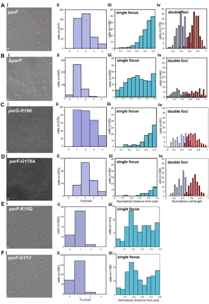
Cells harboring the plasmid with the wild-type
parFG-mCherry-parHmodule mainly exhibited 1–3 plasmid foci
(Figure 1A, i and ii). In cells with one focus, the ParG-mCherry spot was positioned most frequently near mid-cell (Figure1A, iii). When two plasmid foci were present, they were situated at one quarter and three quarter positions on the long axis of the cell (Figure1A, iv). In contrast, cells with a segregation-impaired plasmid due to aparF trunca-tion (parF) displayed a single, randomly located plasmid focus (Figure1B, i–iv). Time-lapse microscopy experiments showed that the plasmid focus was excluded from the nu-cleoid and visible at the tip of the nunu-cleoid or between two nucleoids (Supplementary Movies S1 and 2). Alleles encod-ing ParF with a change in conserved residues of the Walker
by guest on January 11, 2017
http://nar.oxfordjournals.org/
Figure 1. ParF-mediated positioning ofparFGHplasmid. (A–F) Imaging and statistical analysis of ParG foci inEscherichia colicells transformed with a plasmid expressing the indicatedparForparGallele from theparFG-mCherry-parHmodule and a plasmid expressing the sameparF-emeraldallele from the PBADarabinose inducible promoter, or empty pBAD30 vector in the case ofparF. (i) Representative ield of view, scale bar=2m in A,=1m in B–F. (ii) Distribution of ParG foci per cell in a population of individual cells (n) measured from collapsed Z-stack images. (iii) Position of ParG foci along the long axis of cells with a single focus from the population shown in panels A–F ii. Positions are normalized relative to the closest cell pole. (iv) Position of ParG foci along the long axis of cells displaying two foci from the population shown in panels A–D ii. Cells were grown in M9 glucose supplemented with 0.02% L-arabinose in the presence of antibiotics.
by guest on January 11, 2017
http://nar.oxfordjournals.org/
A motif, G11V and K15Q, abolish plasmid partition (8). Plasmid foci number and position in cells grown under se-lective pressure and carrying these mutant genes showed patterns very similar to that of cells harboring theparF
plasmid (Figure1E and F). Notably, most of the cells ex-hibited a single focus, suggesting that post-replication plas-mid separation does not occur. Furthermore, the single fo-cus observed in mutant backgrounds appeared sharper and consistently more compact than foci seen in cells containing the wild-type partition locus (Supplementary Figure S3B and C). These results indicate that both plasmid segregation and positioning are disrupted in the absence of a functional ParF. Thus ParF is necessary to achieve correct localization of the plasmid prior to cell division.
ParF dynamically relocates within the nucleoid
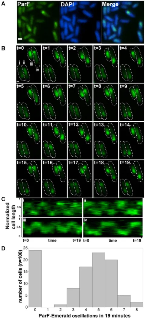
Next, the subcellular localization of ParF-Emerald was ex-amined in populations expressingparF-emeraldin the pres-ence of the plasmid harboring the parFG-mCherry-parH
module. The ParF signal appeared as an extended struc-ture overlapping one pole of the nucleoid (Figure2A). The ParF patches exhibited a compact head followed by a more diffuse tail. The head occupied the extreme edge of the nu-cleoid. In the absence of the plasmid with the partition lo-cus, ParF-Emerald coated the chromosome uniformly, thus retaining its nucleoid localization, but not its asymmetric distribution (Supplementary Figure S3A). In contrast to wild-type, the signal of Walker A mutants ParF-G11V and ParF-K15Q was spread evenly throughout the cell, both in the absence and presence of theparFGHlocus expressing the same mutant allele (Supplementary Figure S3A–C). In-terestingly, the ParG-mCherry signal superimposed on that of wild-type ParF-Emerald, whereas it formed sharp foci silhouetted over the diffuse signal of G11V and ParF-K15Q (Supplementary Figure S3B and C). As ParF self-associates into higher order structuresin vitro(8,34,35), the hazy ParF-Emerald signal may represent dynamic assem-blies in transit over the nucleoid. To test this hypothesis we examined the signal using time-lapse microscopy. ParF patches oscillated between nucleoid poles in a majority of cells (Figure2B and Supplementary Movie S3). Initially the signal appeared compact and round at one pole. Then it be-gan to stretch laterally toward the nucleoid center, migrating soon after across the chromosome and reaching the oppo-site end, where the signal appeared again as a tight struc-ture. Kymographs showing the position of ParF along the nucleoid length over time in one hundred cells revealed that a journey from pole to pole typically occurred every∼4–6 min in most cells (Figure2C and D). No dynamic reloca-tion of ParF was observed in cells expressingparF-emerald
[image:6.612.326.566.69.597.2]in the absence of the TP228 partition locus (Supplemen-tary Figure S4A). ParF-G11V and ParF-K15Q did not ex-hibit redistribution of their diffuse signal over time (data not shown). As these mutants are defective in ATP binding and hydrolysis resulting in disrupted polymerization dynamics (8), the data indicate that ATP association determines nu-cleoid localization of ParF and that impairment of correct assembly-disassembly dynamics abolishes ParF oscillation.
Figure 2.ParF coalesces into a cloud-like structure oscillating between the poles of the nucleoid. (A) Fluorescence microscopy snapshot of Es-cherichia colicells transformed with the plasmid containing the parFG-mCherry-parHmodule and a plasmid expressingparF-emeraldfrom the PBADpromoter. ParF-Emerald (left), DAPI-stained nucleoid (middle) and
merge image (right), scale bar=0.5m. (B) Time-lapse luorescence mi-croscopy images of the sameE. colistrain described in panel A showing the dynamic relocation of ParF-Emerald. Cell boundaries (dashed lines) were overlaid from bright ield images. Time in minutes, scale bar=0.5 m. (C) Kymograph showing the movement of the ParF-Emerald signal along the cell length over time for the four cells (i, ii, iii and iv) shown in panel B. (D) Histogram illustrating the frequency of ParF-Emerald oscil-lations determined by constructing kymographs for 100 individual cells of the strain in panel B.
by guest on January 11, 2017
http://nar.oxfordjournals.org/
Synchronous tracking of ParG in the wake of ParF
The localization of ParG in relation to ParF was investi-gated. In cells expressingparF-emerald, ParG-mCherry ap-peared as patches analogous to and overlapping those ob-served for ParF (Supplementary Figure S3B). These snap-shot images suggest that ParG-plasmid complexes transit with ParF across the nucleoid. To better visualize the move-ment of ParG relative to ParF oscillations, pixel intensities of both ParF-Emerald and ParG-mCherry from time-lapse experiments were rendered into contour plots of individual nucleoids (Figure3A and B). For clarity the two channels are shown separately. As described, the ParF signal oscil-lated across the nucleoid. The images and relative signal quantitations reveal that ParG and the associated plasmid move in synchrony with or lag shortly behind ParF. Cells at time zero exhibited a compact ParF-Emerald signal at one pole of the nucleoid, while the ParG-mCherry signal was comet-shaped with the tail of the comet extending be-yond the contour of the ParF-Emerald signal (Figure3A). However, whereas the ParF-Emerald signal remained es-sentially unchanged in the following four time frames, the ParG-mCherry signal contracted to a round focus overlap-ping the ParF signal. The discrete ParF-Emerald structure then reorganized stretching toward the nucleoid center be-fore migrating to the opposite nucleoid pole. The ParG lu-orescence synchronously tracked ParF with the two pro-teins showing superimposing intensity maxima (Figure3B). Once at the new pole, the overlying signals remained still for a few time frames, before beginning the next race to the opposite nucleoid edge. Overall, a pattern of fully synchro-nized oscillation of the ParF and plasmid-bound ParG sig-nals was apparent. The results indicate that ParG-plasmid complexes move in concert with the ParF patches within the nucleoid boundary.
ParF relocation across the nucleoid is dependent on ParG and partition siteparH
To gain insights into the role played by ParG in ParF oscil-lation, a plasmid carrying a non-functional partition mod-ule was used. This plasmid is segregationally unstable ex-hibiting a retention rate of≤2% in the absence of selective pressure due to truncation of the C-terminus of ParG (3). The ParF-Emerald signal was static and evenly distributed over the nucleoid in cells harboring this plasmid together with the second plasmid expressingparF-emerald (Supple-mentary Figure S4B,top row). This result demonstrates that ParG is a key player in effecting ParF oscillation. To in-vestigate whether providing ParGin transwould reconsti-tute oscillation, a third plasmid expressing parG-mCherry
from the Ptacpromoter was introduced: uneven localization
and oscillation of ParF-Emerald across the nucleoid were restored (Supplementary Figure S4B,middle rowand Sup-plementary Movie S4). In cells expressingparF-emeraldand
parG-mCherry in the absence of the plasmid carrying the
partition siteparH, ParF-Emerald homogeneously coated the nucleoid and did not oscillate (Supplemenatary Figure
S4B,bottom row). Thus, ParF dynamic behavior is
depen-dent on both ParG and theparHsite.
ParG stimulation of ParF ATPase activity mediates ParF os-cillation
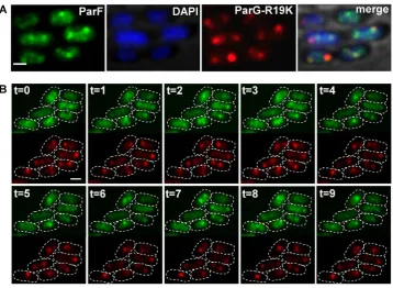
The ParG N-terminus harbors an arginine, R19, that is part of an arginine inger-like motif that stimulates ParF ATP hydrolysis. A ParG-R19K mutant is impaired in stimulation of ATPase activity and results in decreased plasmid stability indicating that this ParG-mediated function is a key regu-latory process for segregation (35). Cells carrying plasmids with theparG-R19Kallele in theparFGHlocus and
express-ingparF-emerald in transwere observed under the presence
of selective pressure. Surprisingly, the ParF-Emerald signal did not assemble into the usual extended structure onto the nucleoid, but distinctively most of the protein coalesced into foci that were not observed in any other mutant background (Figure 4). The ParF signal did not oscillate despite be-ing wild-type (Figure4A and B). The ParF foci colocalized with the ParG–plasmid complexes and showed some mobil-ity accompanied by reorganization of the luorescent signal over time. Most cells displayed one to three ParG-plasmid foci (Figure1C, i and ii). Single ParG-R19K foci localized at midcell as observed in the wild-type background (Figure
1C, iii). In contrast, in cells containing two foci, they were found at all locations along the nucleoid with no bias toward one and three quarter positions. These data indicate that ParF oscillation is dependent on stimulation of its ATPase activity by the ParG–plasmid complex. Lack of stimulation causes accumulation and locking of ParF on ParG–plasmid complexes. In the absence of ParF relocation, plasmid po-sitioning is disrupted.
A hyperactive ATPase ParF mutant displays an increased fre-quency of cross-nucleoid oscillations
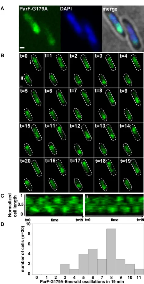
As ParF ATPase activity is pivotal in mediating both ParF relocation and plasmid segregation, we investigated local-ization and dynamics of ParF-G179A, a mutant showing hyperactive ATP hydrolysis and defects in plasmid parti-tioning (40). In terms of kinetic properties, ParF-G179A ATPase activity has a kcat =14, which is almost 10-fold
higher than that of wild-type ParF (kcat=1.6) and the
activ-ity is not stimulated by ParG (40). TheparF-G179A-emerald
allele was expressed in the absence and presence of the plas-mid carrying theparFG-mCherryHlocus with the same mu-tation inparF. In both instances, ParF-G179A localization was identical to that of wild-type ParF: in the absence of the partition module, ParF-G179A homogeneously coated the nucleoid (Supplementary Figure S3A), whereas it as-sociated with one pole of the nucleoid in the presence of the plasmid harboring the segregation components (Figure
5A). Time-lapse experiments revealed that ParF-G179A os-cillates between nucleoid poles like wild-type ParF (Figure
5B and Supplementary Movie S5). Remarkably, the hyper-active ATP hydrolysis induces a higher oscillation frequency compared to wild-type ParF with the majority of signals ex-hibiting pole-to-pole journey times of 2–3 min compared to 4–6 min for wild-type ParF (Figure5C and D). In addition, ParF-G179A oscillation occurred in every cell in a ield of view, whereas ParF did not oscillate in ∼25% of cells. In cultures grown in the presence of selective pressure, most cells contained two ParG-plasmid foci (Figure1D, i and ii)
by guest on January 11, 2017
http://nar.oxfordjournals.org/
Figure 3. Movement of ParG-mCherry in relation to ParF-Emerald oscillation. Pixel intensity contour plots for the ParF and ParG signals from time-lapse microscopy images of a representativeEscherichia colicell harboring the segregation probe vector carryingparFG-mCherry-parHand the plasmid expressingparF-emerald(A) and quantitation of the relative luorescent signals for ParF-Emerald and ParG-mCherry (B). One hundred cells were analyzed and all showed synchronous oscillation of ParF and ParG with the exception of 17 cells that did not show ParF relocation. Time in minutes.
Figure 4. ParF oscillation is dependent on stimulation of ATP hydrolysis by ParG. (A) Fluorescence microscopy snapshot ofEscherichia colicells carrying a plasmid withparG-R19Kmutant allele in the partition module and a plasmid expressingparF-emerald. Individual microscopy channels and a merge image with bright ield are shown. Scale bar=1m. (B) Time-lapse images of cells of the sameE. colistrain as described in panel A. ParF-Emerald and ParG-R19K-mCherry channels (top and bottom, respectively) were imaged at 1 min intervals. Cell boundaries (dashed lines) were overlaid from the bright ield images. Scale bar=0.5m.
by guest on January 11, 2017
http://nar.oxfordjournals.org/
[image:8.612.134.493.419.681.2]Figure 5. Hyperactive ATP hydrolysis results in increased cross-nucleoid oscillation frequency of the ParF-G179A mutant. (A) Fluorescence mi-croscopy snapshot ofEscherichia colicells carrying a plasmid with parF-G179Amutant allele in the partition module and a plasmid expressing
parF-G179A-emerald. ParF-G179A-Emerald, DAPI-stained nucleoid and merge image with bright ield are shown. Scale bar=0.5m. (B) Time-lapse images of the sameE. colistrain described in panel A. Cell bound-aries (dashed lines) were overlaid from bright ield images. Time in min-utes, scale bar=0.5m. (C) Kymograph showing the movement of ParF-G179A-Emerald along the cell long axis over time in cells (i and ii) labeled att=0 in panel B. (D) Populations of cells showing the indicated number of ParF-G179A-Emerald pole-to-pole oscillations over 19 min determined from kymographs of ParF-G179A-Emerald transits in cells of the strain described in A.
that were more compact than those observed with a wild-type partitioning module. Surprisingly, the positions of sin-gle and double ParG foci relative to cell length did not differ between ParF-G179A and wild-type ParF (Figure 1D, iii and iv). It seems that a wild-type-like plasmid positioning
is achieved by ParF-G179A, but the timing of partitioning events is disrupted. This mutant phenotype shows that re-sponsiveness to ParG stimulation and inely tuned ATPase kinetics are key to effect plasmid segregation and that ParF nucleoid localization as well as oscillation are not suficient
per seto warrant plasmid partitioning.
A three-dimensional ParF meshwork permeates the nucleoid to mediate plasmid segregation
More detailed investigation of the ParF structure on the nu-cleoid was hindered by the spatial resolution limit of con-ventional microscopy. To achieve higher resolution of the ParF structure, we imaged cells by 3D structured illumina-tion microscopy (3D-SIM) using an OMX microscope. Im-ages acquired from multiple orientations are subjected to an iterative reconstruction algorithm that achieves a sub-diffraction image with a∼2-fold increase in bothxy- (∼100 nm) andz-axis (∼300 nm) resolution (41). Localization of ParF-Emerald observed by 3D-SIM was the same as that seen using conventional microscopy. In the presence of the plasmid carrying the segregation locus, the ParF signal ap-peared asymmetrically associated with or in transit toward one nucleoid pole (Figure6A and B, Supplementary Figure S5 and Supplementary Movie S6). In the absence of the par-tition locus, ParF uniformly coated the nucleoid (Supple-mentary Figure S5C). However, super resolution 3D-SIM revealed that the ParF comet protrudes into the nucleoid: examination of cell cross sections showed that ParF per-meates the interior of the nucleoid extending through the chromosome in three dimensions (Figure6C). To further conirm the 3D pattern of ParF, Z-stacks were taken and clearly showed that the protein assembles into an organized, meshwork-like structure visible in the different planes of the nucleoid (Figure7A and B). Although the ParF mesh-work is visibly associated with the nucleoid, its pattern does not entirely overlap with that of the chromosome, as projec-tions often shoot out of the nucleoid perimeter (Figure6A and B) or ill up small pockets unoccupied by DNA (Sup-plementary Figure S5B). ParG foci were generally trapped within the ParF meshwork (Figure6B), or were sometimes observed in an area largely devoid of ParF (Figure6A, left-most focus). Supplementary Figure S5A provides an ex-ample of a cell in which ParG foci colocalized with small patches of ParF belonging to the tail of the meshwork clus-tered at one end of the nucleoid. Overall, the super reso-lution microscopy data show that ParF assembles into a structure branching through the nucleoid volume, result-ing in a 3D net through which ParG–plasmid complexes are captured and translocated. Remarkably, no ParF meshwork was observed in cells containing the ATPase stimulation-defective ParG-R19K mutant: in contrast, ParF accumu-lated on ParG foci (Figure8A and B). When the non-fused green luorescent protein’s signal was imaged by 3D-SIM, it lacked speciic localization and was spread throughout the cell (Supplementary Figure S6B and C). The different pat-terns observed for the same ParF-Emerald protein in the wild-type and ParG-R19K mutant context indicate that the ParF meshwork is a physiologically relevant, genuine struc-ture of ParF that is crucial for plasmid segregation. The data also show that the meshwork is not the result of ParF fusion
by guest on January 11, 2017
http://nar.oxfordjournals.org/
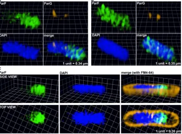
Figure 6. A three-dimensional (3D) ParF meshwork permeates the nucleoid. (A–C) Three-dimensional rendering of 3D-SIM images of liveEscherichia colicells harboring the plasmid containing theparFG-mCherry-parHmodule and the plasmid expressingparF-emerald. The cross section of a cell interior with side and top views is shown in panel C. Individual channels and overlay images are shown.
Figure 7. The ParF meshwork extends throughout the nucleoid interior. (A) Three-dimensional rendering of 3D-SIM images of a liveEscherichia colicell harboring the plasmid containing theparFG-mCherry-parHmodule and the plasmid expressingparF-emerald. Individual channels and overlay images are shown. (B) 3D-SIM Z-stacks of the same cell shown in panel A, the arrows indicate the ParG foci. Scale bar=1m in both panels A and B.
by guest on January 11, 2017
http://nar.oxfordjournals.org/
[image:10.612.62.564.448.697.2]Figure 8. ParF coalesces into foci and does not form a meshwork in the absence of the ATP hydrolysis stimulatory activity of ParG. (AandB) Three-dimensional rendering of 3D-SIM images ofEscherichia colicells that carry a plasmid harboring theparG-R19Kmutant allele in the par-tition module and a plasmid expressingparF-emerald. Individual images for green, red and blue channel are shown as well as the merge image. The nucleoid was stained with DAPI.
to the Emerald protein, because if it were, then the ParF-Emerald pattern would be identical in both backgrounds. ParF-G179A assembled into a structure similar to that of wild-type ParF (Supplementary Figure S6A).
ParF harbors a cluster of positively charged residues and as-sociates with non-speciic DNAin vitro
Microscopy indicated that ParF localizes on the nucleoid. To investigate whether the protein binds DNA, luores-cence polarization experiments were carried out using ParF and luoresceinated double-stranded oligonucleotides har-boring a random sequence. First, ParF bound a 20-mer oligonucleotide with high afinity in the presence of ATP with a Kd of 156 nM, but did not bind in the presence of
ADP (Figure9A). These indings establish that ParF associ-ation to DNA is reliant on ATP binding. Second, we exam-ined the length necessary to allow binding by using 13-, 20-and 42-mer oligonucleotides in the presence of ATP. ParF loosely associated with the 13-bp site, but avidly bound the 20- and 42-bp oligonucleotides (Figure 9B). Thus, ParF DNA binding is dependent on the length of the fragment. Third, the salt dependence of the ParF–DNA interaction was explored. Binding of ParF–ATP to the 20-mer DNA site was measured in the presence of increasing concentra-tions (50–350 mM) of potassium glutamate. ParF bound DNA with a Kd in the nanomolar range up to 150 mM
potassium glutamate (Figure 9C). However, higher ionic strength inhibited binding, which is consistent with non-speciic DNA binding by ParF.
Inspection of the ParF structure revealed that multiple basic residues coat the surface of the protein (Figure9D) and are thus optimally positioned to associate with the DNA and allow ParF to become enmeshed within the nu-cleoid.
DISCUSSION
ParA proteins are responsible for plasmid translocation and maintaining plasmid position during cell growth (2,42). This is a problem of motion, spatial arrangement and tempo. Here we have established that no spatial position-ing of plasmids carryposition-ing theparFGHmodule is attained in the absence of a functional ParF (Figure1). Cells harbor-ing defective ParF-K15Q and ParF-G11V proteins show a single, randomly located ParG–plasmid focus, suggest-ing that post-replication separation of sister plasmids is not achieved. Thus wild-type ParF initiates the segregation pro-cess by splitting paired plasmids and then deining their sub-cellular coordinates. Like other ParA family members, ParF binds DNA non-speciically (Figure9) and localizes to the nucleoid (Figures2and6and Supplementary Figure S3). Due to a cooperative ATP-induced assembly into higher order structures, ParF grows into a 3D meshwork through the nucleoid, as established by 3D-SIM experiments (Fig-ures 6 and7 and Supplementary Figure S5). This allows ParF to survey the nucleoid territory, conining plasmid foci to that region only and demarcating the area of the cell proicient for plasmid segregation. The transient, dy-namic nature of the polymers ensures continuous remod-eling of the ParF structure, so that more distant positions can be reached, upon nucleoid growth and elongation. Mul-tiple cross-nucleoid oscillations may periodically adjust the 3D coordinates of plasmids within the elongating nucleoid. Thus ParF functions as a 3D molecular scanner within the nucleoid perimeter.
ParA ATPase activity is enhanced by its partner pro-tein (4–10,13,28). An arginine inger-like motif in the N-terminus of ParG stimulates ParF ATP hydrolysis and is required for accurate plasmid segregation (35). Here, we have established that no relocation of the ParF meshwork occurs in the absence of a functional ParG andparHsite (Supplementary Figure S4A and B). When ParG is pro-videdin trans, ParF shuttling across the nucleoid resumes (Supplementary Figure S4B and Supplementary Movie S4). Thus, ParG triggers the sequence of events leading to
by guest on January 11, 2017
http://nar.oxfordjournals.org/
Figure 9. ParF associates with non-speciic DNA. Fluorescence polarization studies performed with ParF and luoresceinated oligonucleotides harboring a random sequence. Analysis of DNA binding in the presence of different nucleotides (A), different sizes of oligonucleotides (B) and different salt concen-trations (C). (D) Structures of the the nucleotide sandwich ParF dimer showing the plethora of basic residues that cover the dimer surface that could be used to interact non-speciically with DNA. Basic side chains and the AMP-PNP molecules are shown as sticks.
mid segregation. The most likely mechanism whereby ParG may ignite ParF relocation is through stimulation of ATP hydrolysis that would remove ParF from the DNA lead-ing to dynamic remodellead-ing of the ParF bundles. To inves-tigate this hypothesis, cells that harbored a plasmid encod-ing the stimulation-impaired ParG-R19K mutant were im-aged. Although ParF is wild-type, neither meshwork struc-ture nor oscillation over the nucleoid were detected (Fig-ures 4B and 8). This indicates that the dynamic behavior of ParF is dependent on stimulation of its ATPase activity by ParG. Interestingly, ParF forms discrete foci that overlap those of ParG-R19K, showing that ParF interacts with and accumulates on ParG–R19K–plasmid complexes (Figures
4B and8). However, due to lack of ATP hydrolysis stimu-lation, no ParF meshwork assembly-disassembly cycles oc-cur resulting in lack of transport and positioning of plas-mid complexes. The∼30% of cells harboring a single plas-mid focus at the plas-mid-cell position are likely to be cells in which the ParG–R19K–plasmid cluster is located between separated nucleoids (Figure1C). Signiicantly, the
observa-tion that wild-type ParF displays different patterns in the ParG and mutant ParG-R19K backgrounds demonstrates that the ParF meshwork is a physiologically relevant struc-ture and not a counterfeit resulting from the fusion to the Emerald luorescent protein.
A different insight into the importance of temporal reg-ulation of segregation events was provided by the ParF-G179A mutant that shows hyperactive ATP hydrolysis. Counter-intuitively, ParF-G179A and ParG-plasmid local-ization patterns are indistinguishable from those observed in the wild-type background and ParF-G179A is capable of cooperative assembly into a meshwork apparently equiva-lent to that formed by wild-type ParF (Supplementary Fig-ure S6). However, what is aberrant in this context is the tempo: ParF-G179A oscillation dynamics are faster as a result of its hyperactive ATP hydrolysis (Figure5). ParF-G179A interacts with ParG, but no longer responds to its stimulatory effect (40). The unregulated ATPase kinetics change the tempo of oscillation and abrogate plasmid par-tition. Although plasmid foci are split and apparently
by guest on January 11, 2017
http://nar.oxfordjournals.org/
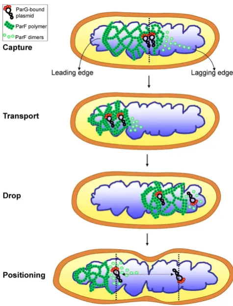
Figure 10. A model for ParF meshwork-mediated plasmid segregation. A pair of newly replicated plasmids carrying ParG dimers bound to the par-tition sites is engulfed into the ParF meshwork, anchored to it via the inter-action of ParG with ParF and transported toward one pole of the nucleoid. The ParF meshwork exhibits a leading edge that is more compact corre-sponding to the advancing front and a looser weave tail that consists of less densely packed oligomers. During the journey across the nucleoid, a net-work of intersecting polymer bundles grows between the plasmids until one plasmid becomes detached from the lagging edge due to ParG-stimulated ATP hydrolysis and dropped at one pole of the nucleoid. The sister plasmid remains anchored to the ParF meshwork and is transported to the oppo-site end of the nucleoid, where it is ultimately released. Subsequent rounds of the ParF meshwork oscillation reset the position of the two plasmids within the elongating nucleoid so that they are driven to the center of the replicated chromosomes.
tioned, they might not be pinned down and held at those locations at the time of cell division due to the very dy-namic nature of ParF-G179A and the altered interaction with ParG. The overall conclusion is that ParG acts as a metronome that dictates the tempo of segregation events, and that nucleoid localization and oscillationper sedo not ensure plasmid segregation in the absence of inely tuned ATPase kinetics.
The current picture of plasmid segregation mediated by ParA proteins is quite fragmented and the mechanism is still elusive. Different models have been proposed whose com-mon denominator is the role of the nucleoid as a platform for segrosome attachment. Based on indings for pB171 partition, one model invokes the formation of a nucleoid-associated ParA ilament whose growing tip stochastically captures a ParB–plasmid complex. Upon ATP hydroly-sis stimulated by ParB, the ParA ilament disassembles
pulling the plasmid toward one pole of the nucleoid (20). The ParA structure is referred to as a ilament, based on
in vitroevidence demonstrating ParA protein
polymeriza-tion and on in vivo microscopy experiments showing an elongated ParA shape. Based on in vitrostudies, another model proposes a diffusion-ratchet mechanism in which ParA recruits the ParB–plasmid cargo to the nucleoid and a ParB-induced ParA gradient guides plasmid movement (23,27,29,30,47). This model postulates movement of ParA and ParB–plasmid molecules on the surface of the nucleoid and numerous studies performed with components of the P1 and F plasmid systems and a lat surface DNA carpet have generated the tenets of this predicted mechanism in a cell-free setup. More recently, a modiied version, desig-nated as DNA-relay model, was proposed (28). In analogy to the Brownian ratchet put forward for plasmid systems, the mechanism envisaged by these authors involves a gradi-ent of chromosomally-encoded ParA inC. crescentuscells that is responsible for the translocation of ParB bound to
parSacross the cell. However, based on simulations of par-tition complex diffusion and mathematical modeling, the DNA-relay model predicts that diffusion alone is not suf-icient to mediate movement of the partition complex and that instead the elastic properties of the chromosome propel the complex across the ParA gradient (28).
Based on our experimental observations, we propose a new model to rationalize the mechanism of plasmid segre-gation by ParA proteins. Using conventional luorescence microscopy, a ilament-like structure for ParF is not appar-ent in live cells, even after deconvolution. Instead a diffuse structure is observed (Figures2and3; Supplementary Fig-ure S3B). A cloud-like signal has been observed for a num-ber of plasmid and chromosome encoded ParAs. Super res-olution 3D-SIM revealed that the ParF structure is a 3D meshwork branching out into the nucleoid (Figures6and
7;Supplementary Figure S5 and Supplementary Movie S6). Biochemicalin vitroinvestigations and structural data have provided evidence of the highly cooperative self-assembly nature of ParF (8,34,35,40). In the super resolution images the ParF signal coalesces into interconnected patches and bundles that permeate the interior of the nucleoid, but also ills in nucleoid gaps and protrudes from its surface. Inter-estingly, a study investigating ParA of plasmid pB171 sug-gested that the protein might form structures within the nu-cleoid rather than homogeneously covering its surface (43). In addition, while this work was being prepared for submis-sion, a paper that provided evidence for plasmid segrega-tion through the nucleoid volume was published (44). Re-cent studies have reported that theE. colichromosome is a low-density ellipsoid, whose radial coninement imparts on it a helical shape (45,46). Super resolution images of the ParF meshwork seem to suggest that the structure has a he-lical pitch that might be a relection of the underlying hehe-lical chromosome shape. However, this feature needs to be fur-ther investigated.
What are the implications of a ParF meshwork for plas-mid segregation? A 3D meshwork acts as a ‘Venus lytrap’ which ensures that ParG–plasmid complexes are readily captured by ParF and is a more effective strategy than ran-dom collisions of single ParA molecules or a linear ilament with plasmid complexes. Given the crowded milieu of the
by guest on January 11, 2017
http://nar.oxfordjournals.org/
cell and the low copy of plasmids, it is crucial that the system is eficient. Based on biochemical and structural data (8,34), we interpret the ParF meshwork as a network of intersecting polymer bundles that result from ParF self-association and ramify in multiple directions, protruding into the nucleoid. A meshwork of polymers imparts higher lexibility as it of-fers multiple attachment points for cargos not only in the XY- but also in the Z-dimension of the nucleoid. Moreover, oligomers at different points of the meshwork can undergo localized disassembly induced by ParG resulting in plas-mid release. Additionally, a 3D structure guarantees sur-veying the entire nucleoid volume, not simply the nucleoid surface as proposed by other models (23,30,47). Pairing of newly replicated plasmids at midcell is thought to be an ini-tial event during partition (48). The plasmid pair may then be engulfed into the ParF meshwork via interaction with ParG dimers bound to the partition site and transported to one pole of the nucleoid (Figure10). The ParF meshwork presents a leading edge that corresponds to the compact, advancing front and a looser-weave, lagging tail that con-sists of less densely arranged oligomers. As plasmids are transported, a network of branched ParF polymers grows between them until one plasmid is dropped due to local-ized ATP hydrolysis mediated by ParG that causes ilament disassembly at the lagging edge of the meshwork. The sis-ter plasmid remains tethered to the compact edge of the ParF meshwork and is shifted to the opposite end of the nucleoid, where eventually it is released again due to ParG-mediated remodeling of the edge of the meshwork. Subse-quent rounds of oscillation of the ParF mesh ensure small-scale adjustments of plasmid position to the middle of the future nucleoids prior to cell division. The repeated cycles of capture and release are important to reset the position of the plasmids over the elongating nucleoid. When the oscil-lation speed of ParF becomes altered due to aberrant ATP kinetics, the separated plasmids might not maintain their i-nal position during the subsequent process of cell division. This model describes a mechanism that is not antithetical to earlier hypotheses, but that amalgamates the concepts of ParA higher-order structures and gradient invoked by previous models. In all cases, the ParA protein deines the territory for plasmid segregation, while ParB/ParG func-tions as timekeeper setting the timing for relocation of ParA molecules across the nucleoid. As numerous ParA proteins involved in DNA and protein traficking (49,50) have been visualized as diffuse, asymmetric clouds by conventional microscopy, we speculate that a 3D meshwork might be a widespread mechanism utilized by Walker type ATPases to distribute and position sizeable cargos in prokaryotes.
SUPPLEMENTARY DATA
Supplementary Data are available at NAR Online.
ACKNOWLEDGEMENT
We are extremely indebted to Joe Pogliano, Marcella Erb and Jennifer Santini (University of California San Diego) for their generous help with the 3D-SIM and to Gopalan Selvaraj for the kind gift of a plasmid encoding gfp Emer-ald.
FUNDING
Medical Research Council [G0801162 to D.B.]; Biotech-nology and Biological Sciences Research Council Doc-toral Training Program Studentship [1312475 to G.E.A.-G.]; Overseas Research Scholarship (to M.T.B.). Funding for open access charge: Medical Research Council.
Conlict of interest statement.None declared.
REFERENCES
1. Tanaka,K. (2013) Regulatory mechanisms of
kinetochore-microtubule interaction in mitosis.Cell. Mol. Life Sci.,
70, 559–579.
2. Hayes,F. and Barill`a,D. (2010) Extrachromosomal components of the nucleoid: recent developments in deciphering the molecular basis of plasmid segregation. In: Dorman,CJ and Dame,RT (eds).Bacterial Chromatin. Springer Publishing, Dordrecht, The Netherlands, pp. 49–70.
3. Hayes,F. (2000) The partition system of multidrug resistance plasmid TP228 includes a novel protein that epitomizes an evolutionarily distinct subgroup of the ParA superfamily.Mol. Microbiol.,37, 528–541.
4. Davis,M.A., Martin,K.A. and Austin,S.J. (1992) Biochemical activities of the ParA partition protein of the P1 plasmid.Mol. Microbiol.,6, 1141–1147.
5. Libante,V., Thion,L. and Lane,D. (2001) Role of the ATP-binding site of SopA protein in partition of the F plasmid.J. Mol. Biol.,314, 387–399.
6. Fung,E., Bouet,J.Y. and Funnell,B.E. (2001) Probing the
ATP-binding site of P1 ParA: partition and repression have different requirements for ATP binding and hydrolysis.EMBO J.,20, 4901–4911.
7. Easter,J. Jr. and Gober,J.W. (2002) ParB-stimulated nucleotide exchange regulates a switch in functionally distinct ParA activities.
Mol. Cell,10, 427–434.
8. Barill`a,D., Rosenberg,M.F., Nobbmann,U. and Hayes,F. (2005) Bacterial DNA segregation dynamics mediated by the polymerizing protein ParF.EMBO J.,24, 1453–1464.
9. Leonard,T.A., Butler,P.J. and L ¨owe,J. (2005) Bacterial chromosome segregation: structure and DNA binding of the Soj dimer––a conserved biological switch.EMBO J.,24, 270–282. 10. Pratto,F., Cicek,A., Weihofen,W.A., Lurz,R., Saenger,W. and
Alonso,J.C. (2008)Streptococcus pyogenespSM19035 requires dynamic assembly of ATP-bound ParA and ParB onparSDNA during plasmid segregation.Nucleic Acids Res.,36, 3676–3689. 11. Lim,G.E., Derman,A.I. and Pogliano,J. (2005) Bacterial DNA segregation by dynamic SopA polymers.Proc. Natl. Acad. Sci. U.S.A.,102, 17658–17663.
12. Ebersbach,G., Ringgaard,S., Møller-Jensen,J., Wang,Q., Sherratt,D.J. and Gerdes,K. (2006) Regular cellular distribution of plasmids by oscillating and ilament-forming ParA ATPase of plasmid pB171.
Mol. Microbiol.,61, 1428–1442.
13. Bouet,J.Y., Ah-Seng,Y., Benmeradi,N. and Lane,D. (2007) Polymerization of SopA partition ATPase: regulation by DNA binding and SopB.Mol. Microbiol.,63, 468–481.
14. Machon,C., Fothergill,T.J.G., Barill`a,D. and Hayes,F. (2007) Promiscuous stimulation of ParF protein polymerization by heterogeneous centromere binding factors.J. Mol. Biol.,374, 1–8. 15. Batt,S.M., Bingle,L.E.H., Dafforn,T.R. and Thomas,C.M. (2009) Bacterial genome partitioning: N-terminal domain of IncC protein encoded by broad-host-range plasmid RK2 modulates
oligomerisation and DNA binding.J. Mol. Biol.,385, 1361–1374. 16. Ptacin,J.L., Lee,S.F., Garner,E.C., Toro,E., Eckart,M., Comolli,L.R.,
Moerner,W.E. and Shapiro,L. (2010) A spindle-like apparatus guides bacterial chromosome segregation.Nat. Cell Biol.,12, 791–798. 17. Ebersbach,G. and Gerdes,K. (2004) Bacterial mitosis: partitioning
protein ParA oscillates in spiral-shaped structures and positions plasmids at mid-cell.Mol. Microbiol.,52, 385–398.
18. Fogel,M.A. and Waldor,M.K. (2006) A dynamic, mitotic-like mechanism for bacterial chromosome segregation.Genes Dev.,20, 3269–3282.
by guest on January 11, 2017
http://nar.oxfordjournals.org/
19. Hatano,T., Yamaichi,Y. and Niki,H. (2007) Oscillating focus of SopA associated with ilamentous structure guides partitioning of F plasmid.Mol. Microbiol.,64, 1198–1213.
20. Ringgaard,S., van Zon,J., Howard,M. and Gerdes,K. (2009) Movement and equipositioning of plasmids by ParA ilament disassembly.Proc. Natl. Acad. Sci. U.S.A.,106, 19369–19374. 21. Sengupta,M., Nielsen,H.J., Youngren,B. and Austin,S. (2010) P1
plasmid segregation: accurate redistribution by dynamic plasmid pairing and separation.J. Bacteriol.,192, 1175–1183.
22. Vecchiarelli,A.G., Han,Y.W., Tan,X., Mizuuchi,M., Ghirlando,R., Biert ¨umpfel,C., Funnell,B.E. and Mizuuchi,K. (2010) ATP control of dynamic P1 ParA-DNA interactions: a key role for the nucleoid in plasmid partition.Mol. Microbiol.,78, 78–91.
23. Hwang,L.C., Vecchiarelli,A.G., Han,Y.W., Mizuuchi,M., Harada,Y., Funnell,B.E. and Mizuuchi,K. (2013) ParA-mediated plasmid partition driven by protein pattern self-organization.EMBO J.,32, 1238–1249.
24. Castaing,J.P., Bouet,J.Y. and Lane,D. (2008) F plasmid partition depends on interaction of SopA with non-speciic DNA.Mol. Microbiol.,70, 1000–1011.
25. Hui,M.P., Galkin,V.E., Yu,X., Stasiak,A.Z., Stasiak,A., Waldor,M.K. and Egelman,E.H. (2010) ParA2, aVibrio cholerae
chromosome partitioning protein, forms left-handed helical ilaments on DNA.Proc. Natl. Acad. Sci. U.S.A.,107, 4590–4595.
26. Sober ´on,N.E., Lioy,V.S., Pratto,F., Volante,A. and Alonso,J.C. (2011) Molecular anatomy of theStreptococcus pyogenespSM19035 partition and segrosome complexes.Nucleic Acid Res.,39, 2624–2637. 27. Havey,J.C., Vecchiarelli,A.G. and Funnell,B.E. (2012) ATP-regulated
interactions between P1 ParA, ParB and non-speciic DNA that are stabilized by the plasmid partition site,parS.Nucleic Acid Res.,40, 801–812.
28. Lim,H.C., Surovtsev,I.V., Beltran,B.G., Huang,F., Bewersdorf,J. and Jacobs-Wagner,C. (2014) Evidence for a DNA-relay mechanism in ParABS-mediated chromosome segregation.Elife,3, e02758. 29. Vecchiarelli,A.G., Hwang,L.C. and Mizuuchi,K. (2013) Cell-free
study of F plasmid partition provides evidence for cargo transport by a diffusion-ratchet mechanism.Proc. Natl. Acad. Sci. U.S.A.,110, E1390–E1397.
30. Vecchiarelli,A.G., Neuman,K.C. and Mizuuchi,K. (2014) A propagating ATPase gradient drives transport of surface-conined cellular cargo.Proc. Natl. Acad. Sci. U.S.A.,111, 4880–4885. 31. Barill`a,D. and Hayes,F. (2003) Architecture of the ParF-ParG protein
complex involved in procaryotic DNA segregation.Mol. Microbiol.,
49, 487–499.
32. Golovanov,A.P., Barill`a,D., Golovanova,M., Hayes,F. and Lian,L.Y. (2003) ParG, a protein required for active partition of bacterial plasmids, has a dimeric ribbon-helix-helix structure.Mol. Microbiol.,
50, 1141–1153.
33. Wu,M., Zampini,M., Bussiek,M., Hoischen,C., Diekmann,S. and Hayes,F. (2011). Segrosome assembly at the pliableparHcentromere.
Nucleic Acid Res.,39, 5082–5097.
34. Schumacher,M.A., Ye,Q., Barge,M.T., Zampini,M., Barill`a,D. and Hayes,F. (2012) Structural mechanism of ATP induced
polymerization of the partition factor ParF: implications for DNA segregation.J. Biol. Chem.,287, 26146–26154.
35. Barill`a,D., Carmelo,E. and Hayes,F. (2007) The tail of the ParG DNA segregation protein remodels ParF polymers and enhances ATP hydrolysis via an arginine inger-like motif.Proc. Natl. Acad. Sci. U.S.A.,104, 1811–1816.
36. Ah-Seng,Y., Rech,J., Lane,D. and Bouet,J.Y. (2013) Deining the role of ATP hydrolysis in mitotic segregation of bacterial plasmids.PLOS Genet.9, e1003956.
37. Lau,I.F., Filipe,S.R., Søballe,B., Økstad,O.A., Barre,F.X. and Sherratt,D.J. (2003) Spatial and temporal organization of replicating
Escherichia colichromosomes.Mol. Microbiol.,49, 731–743. 38. Teerawanichpan,P., Hoffman,T., Ashe,P., Datla,R. and Selvaraj,G.
(2007) Investigations of combinationations of mutations in the jellyish green luorescent protein (GFP) that afford brighter luorescence, and use of a version (VisGreen) in plant, bacterial, and animal cells.Biochim. Biophys. Acta,1770, 1360–1368.
39. Ai,H.W., Shaner,N.C., Cheng,Z., Tsien,R.Y. and Campbell,R.E. (2007) Exploration of new chromophore structures leads to the identiication of improved blue luorescent proteins.Biochemistry,46, 5904–5910.
40. Dobruk-Serkowska,A., Caccamo,M., Rodr´ıguez-Casta ˜neda,F., Wu,M., Bryce,K., Ng,I., Schumacher,M.A., Barill`a,D. and Hayes,F. (2012) Uncoupling of nucleotide hydrolysis and polymerization in the ParA protein superfamily disrupts DNA segregation dynamics.J. Biol. Chem.,287, 42545–42553.
41. Gustafsson,M.G.L., Shao,L., Carlton,P.M., Wang,C.J.,
Golubovskaya,I.N., Cande,W.Z., Agard,D.A. and Sedat,J.W. (2008) Three-dimensional resolution doubling in wide-ield luorescence microscopy by structured illumination.Biophys. J.,94, 4957–4970. 42. Derman,A.I., Lim-Fong,G. and Pogliano,J. (2008) Intracellular
mobility of plasmid DNA is limited by the ParA family of partitioning systems.Mol. Microbiol.,67, 935–946.
43. Ietswaart,R., Szardenings,F., Gerdes,K. and Howard,M. (2014) Competing ParA structures space bacterial plasmids equally over the nucleoid.PLoS Comput. Biol.,10, e1004009.
44. Le Gall,A., Cattoni,D.I., Guilhas,B., Mathieu-Demazi`ere,C., Oudjedi,L., Fiche,J.B., Rech,J., Abrahamsson,S., Murray,H., Bouet,J.Y. and Nolmann,M. (2016) Bacterial partition complexes segregate within the volume of the nucleoid.Nat. Commun.,7, 12107. 45. Fisher,J.K., Bourniquel,A., Witz,G., Weiner,B., Prentiss,M. and
Kleckner,N. (2013) Four-dimensional imaging ofE. colinucleoid organization and dynamics in living cells.Cell,153, 882–895. 46. Hadizadeh Yazdi,N., Guet,C.C., Johnson,R.C. and Marko,J.F. (2012)
Variations of the folding and dynamics of theEscherichia coli
chromosome with growth conditions.Mol. Microbiol.,86, 1318–1333. 47. Vecchiarelli,A., Mizuuchi,K. and Funnell,B.E. (2012) Suring
biological surfaces: exploiting the nucleoid for partition and transport in bacteria.Mol. Microbiol.,86, 513–523.
48. Edgar,R., Chattoraj,D.K. and Yarmolinsky,M. (2001) Pairing of P1 plasmid partition sites by ParB.Mol. Microbiol.,42, 1363–1370. 49. Savage,D.F., Afonso,B., Chen,A.H. and Silver,P.A. (2010) Spatially
ordered dynamics of the bacterial carbon ixation machinery.Science,
327, 1258–1261.
50. Roberts,M.A., Wadhams,G.H., Hadield,K.A., Tickner,S. and Armitage,J.P. (2012) ParA-like protein uses nonspeciic chromosomal DNA binding to partition protein complexes.Proc. Natl. Acad. Sci. U.S.A.,109, 6698–6703.
by guest on January 11, 2017
http://nar.oxfordjournals.org/