Imprinting Mechanisms
Miguel Constaˆncia,
1Benjamin Pickard, Gavin Kelsey, and Wolf Reik
1Programme in Developmental Genetics, The Babraham Institute, Cambridge CB2 4AT, UK
A number of recent studies have provided new insights into mechanisms that regulate genomic imprinting in the mammalian genome. Regions of allele-specific differential methylation (DMRs) are present in all imprinted genes examined. Differential methylation is erased in germ cells at an early stage of their development, and germ-line-specific methylation imprints in DMRs are reestablished around the time of birth. After fertilization, differential methylation is retained in core DMRs despite genome-wide demethylation and de novo methylation during preimplantation and early postimplantation stages. Direct repeats near CG-rich DMRs may be involved in the establishment and maintenance of allele-specific methylation patterns. Imprinted genes tend to be clustered; one important component of clustering is enhancer competition, whereby promoters of linked imprinted genes compete for access to enhancers. Regional organization and spreading of the epigenotype during development is also important and depends on DMRs and imprinting centers. The mechanism of cis spreading of DNA methylation is not known, but precedent is provided by the Xist RNA, which results in X chromosome inactivation in cis. Reading of the somatic imprints could be carried out by transcription factors that are sensitive to methylation, or by methyl–cytosine-binding proteins that are involved in transcriptional repression through chromatin remodeling.
Genomic imprinting is an unusual yet important mechanism of gene regulation by which only one of the parental copies of a gene is expressed (Fig. 1). Although it has been known for some time that DNA methylation is involved in imprinting, the de-tails of how imprints are introduced in the parental germ cells, maintained in embryos, and used to ex-press or reex-press genes have been elusive. However, recently a number of advances have been made: (1) the definition of cis-acting sequences that are im-portant in the control of imprinting; (2) the devel-opmental analysis of how and when imprints are established in germ cells (and when they are erased); and (3) how imprints are maintained, particularly during preimplantation development at which time major changes in methylation occur throughout the genome (Fig. 1). It has also become apparent that imprinted genes tend to be clustered and that aspects of imprinting control are regional and shared between genes in the cluster. Although the reading of the imprint (i.e., the translation of so-matic imprint into gene expression pattern) is usu-ally considered to be a more general aspect of epi-genetic gene regulation (and therefore not unique to imprinted genes), we describe briefly the known mechanisms that could be involved. Here we con-sider these recent advances that make the study of
imprinted genes particularly instructive as a major example of epigenetic gene regulation in mammals, with important implications for disease when de-regulated. The sections in the first half of this review describe the properties of imprinted genes at key stages of germ cell and embryonic development (Fig. 1). The nature of the molecular elements that combine to initiate and maintain the imprint and also translate it into monoallelic expression form the focus of sections in the second half of the re-view.
The Germ Line
Erasure
The germ line has the crucial role of erasing existing imprints that are inherited from the previous gen-eration, and establishing the imprints, according to the sex of the germ line, for the next (Fig. 1). Two models have been envisaged for how this might oc-cur (Rossant 1993). In the first, the epigenotype (im-print) of the same sex chromosome is maintained, whereas the one of the opposite sex is reversed. This could be a single step mechanism. In the second, the existing epigenetic modifications are first erased from both parental chromosomes in both germ lines, and imprints are then established in a sex-specific fashion at a later stage. During the past few years, considerable insights have been gained from methylation, expression, and functional studies of 1Corresponding authors.
germ cells. These studies begin to tip the balance toward the second model.
Germ cells develop from a founder population of∼45 cells derived from the epiblast and are deter-mined by day 7.5 of mouse embryonic development (E7.5). Primordial germ cells (PGCs) migrate through extraembryonic regions and the hindgut to their final destination, the gonad primordia in the genital ridge, by E10.5–E11.5 (for review, see Buehr 1997). At E13.5 female germ cells enter the meiotic prophase, whereas male germ cells undergo mitotic arrest. Spermatogonia resume mitosis after birth, which is followed by meiotic differentiation. Oo-cytes undergo growth after birth, before being ovu-lated and fertilized.
Although it is clear that DNA methylation has a crucial role to play in imprinting, it is not estab-lished whether methylation is the primary imprint-ing signal that needs to be removed and reestab-lished in the germ line. However, it appears that methylation changes in imprinted genes in the germ line are at least temporally associated with al-tered functional properties of the germ cells (see be-low). Therefore, even if methylation is not the pri-mary imprinting signal, it is likely to be related to such a signal.
Global demethylation and methylation events occur in germ cells. By E12.5–E13.5 all nonim-printed sequences tested so far are demethylated in
both sexes (Sanford et al. 1987; Kafri et al. 1992). This is followed by remethylation from E15.5 in many gene se-quences (except CpG islands) with only certain sequences retaining methylation differ-ences between oocyte and sperm genomes (Sanford et al. 1987; Kafri et al. 1992). How-ever, it is possible that the germ line occasionally fails to reprogram epigenetic infor-mation. This would lead to e p i g e n e t i c i n h e r i t a n c e through the germ line, which has been observed both for transgene methylation pat-terns (Sapienza et al. 1989; Allen et al. 1990) and endog-enous sequences (Ro¨mer et al. 1997).
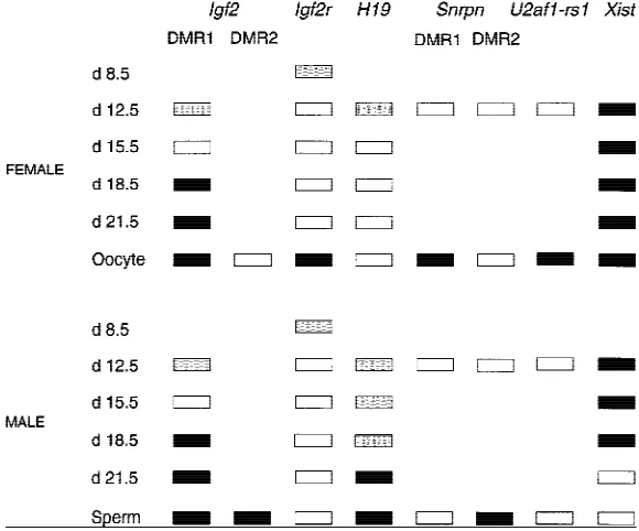
The global demethylation occurring at early stages of germ cell development also includes imprinted genes (Brandeis et al. 1993). Hence, Igf2r, p57Kip2, Peg1, Peg3, Snrpn, U2afrs1, and Nnat are all demethylated by E12.5 (Fig. 2; Brandeis
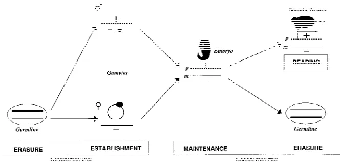
[image:2.612.78.410.70.228.2]et al. 1993; Tada et al. 1998). H19 and Igf2 are not completely demethylated and slightly higher levels of methylation are present in male compared to fe-male PGCs (Tada et al. 1998; H. Sasaki, pers. comm.). This is based on direct examination of PGCs or inferred from the use of embryonic germ (EG) cell lines. Little is known about stages before E12.5 except for one study of E8.5 EG cells that found that the maternal methylation of Igf2r region 2 was still present in some cell lines, suggesting that demethylation takes place between E8.5 and E12.5 (Labosky et al. 1994). This timing is partly sup-ported by functional studies. Although chimeras made with E8.5 EG cells develop normally (Labosky et al. 1994), those made with E12.5 EG cells (from both sexes) show enhanced fetal growth, lethality, and skeletal malformations (some of these pheno-types are characteristic of androgenetic chimeras) (Labosky et al. 1994; Tada et al. 1998). In these chi-meras, hypomethylation was observed in the nor-mally maternally methylated genes, and Igf2r was repressed (which together with expected biallelic expression of paternal genes would contribute to the androgenetic-like phenotype). H19 and Igf2 seemed to be 50% methylated in chimeras, suggest-ing that these imprints were retained at E12.5 when Figure 1 Key stages of genomic imprinting during development. (Erasure) The
‘‘imprint’’ (inherited from the previous generation, generation one) is erased on both parental chromosomes during germ cell development (note that this figure shows complete erasure of all imprints in the germ line, which is one of two models discussed in the text). (Establishment) A new imprint is established (+ or
the EG cells were derived (Tada et al. 1998). Whether the paternal copy of H19 becomes de-methylated (erased imprint) in male germ cells at any stage is not clear (see below). However, Igf2,
H19, Igf2r, and Snrpn are all biallelically expressed in
both germ lines beginning from E11.5 (Szabo and Mann 1995; Villar et al. 1995), suggesting that im-prints are indeed largely erased (or not recognized) at these stages.
Fusion experiments between EG cells and thy-mic lymphocytes show that EG cells have a domi-nant demethylating activity that acts on imprinted as well as nonimprinted genes and repeat sequences (Tada et al. 1997). The only sequences so far de-tected that might escape this global demethylation in early germ cells seem to be the 58region of Xist, which remains methylated in both sexes (Fig. 2; Ariel et al. 1995; Razin and Shemer 1995) and the paternal copy of H19 in male germ cells as men-tioned above (H. Sasaki, pers. comm.).
Establishment
The timing of methylation establishment in im-printed genes is quite clear for the female, but less clear for the male germ line (Fig. 2). In the female, oocytes in dictyate stage arrest (from E13.5) are ap-parently not methylated until after birth, when
methylation occurs during oocyte growth. This has been established for re-peat sequences (Howlett and Reik 1991),
Igf2r (Brandeis et al. 1993; Sto¨ger et al. 1993), imprinted transgenes (Chaillet et al. 1991; Ueda et al. 1992), and inferred for Peg1, Peg3, and Snrpn from functional studies (see below). These methylation events are coincident with the presence of high levels of DNA methyltransferase (Dnmt1) in the nucleus of the growing oocyte (Mertineit et al. 1998). Whether other imprinted genes that remain un-methylated in the oocyte (e.g., H19) are specifically protected from de novo methylation at these stages is not known. That such protection may be necessary is indicated by the fact that the p57Kip2
gene, which is normally paternally meth-ylated, and is demethylated in EG cells, became de novo methylated in EG cell chimeras (Tada et al. 1998). As pointed out above, the establishment of im-printed gene methylation in the male germ line is less clearly defined (Fig. 2). However, H19 and Igf2 methylation is ap-parently established around the time of birth (Brandeis et al. 1993; H. Sasaki, pers. comm.). This is again coincident with high levels of Dnmt1 protein in nuclei of spermatogonia (Mertineit et al. 1998). Whether the grandpaternal copy of H19 becomes completely demethylated before this de novo meth-ylation occurs is not clear (H. Sasaki, pers. comm.). It has been proposed that H19 is targeted specifically for de novo methylation in male germ cells, rather than being protected from it in female ones (Surani 1998).
Two sets of functional studies have examined germ cells by nuclear transplantation at presumed stages of erasure and before establishment. Gynoge-netic embryos have been prepared with the second maternal genome originating from ungrown oo-cytes (Kono et al. 1996; Obata et al. 1998). These embryos show improved development when com-pared to gynogenetic ones, and express the (pre-sumably unmethylated) Peg1, Peg3, and Snrpn genes. In contrast, Igf2r and p57Kip2were repressed in the
genome originating from the immature oocyte, showing again that they need to be methylated for activity. The nucleus from male germ cells from E15.5 to E16.5 was also transplanted, and chimeras were obtained in which the transplanted nucleus participated in embryonic and extraembryonic de-velopment to E10.5 (Kato and Tsunoda 1995). Inas-Figure 2 Methylation patterns in imprinted genes in the germ line.
[image:3.612.73.363.72.313.2]much as androgenetic cells are not expected to con-tribute to embryonic development in chimeras (they contribute to extraembryonic tissues), this may indicate that these germ cells are still at a stage before acquiring the paternal imprints.
Genetic observations of ‘‘imprinting muta-tions’’ in the Prader-Willi/Angelman syndromes (PWS/AS) have indicated previously that such mu-tations may interfere with the required switch in epigenotype in the germ lines (Horsthemke et al. 1997). It has been proposed that such mutations prevent the chromosome of the opposite parental sex switching epigenotype, but do not affect the epi-genotype of the same sex chromosome (Horsthe-mke et al. 1997). This would appear to lend support to the ‘‘same sex, no reprogramming’’ hypothesis. However, as argued previously, these observations are not conclusive and do not currently allow the distinction between the models (Kelsey and Reik 1997; Reik and Walter 1998).
The Embryo
Ontogeny of Allelic Methylation
The ontogeny of DNA methylation at imprinted loci after fertilization, as in the germ line, has to be seen in the context of the genome as a whole and the global changes in methylation that occur during early embryonic development. Much of the meth-ylation arriving with the gametes, including the gross difference between the maternal and paternal genomes, is erased rapidly during preimplantation development. The higher methylation levels that characterize adult somatic tissues begin to be laid down after implantation, with the onset of general de novo methylation (Monk et al. 1987; Sanford et al. 1987; Howlett and Reik 1991; Kafri et al. 1992; Yoder et al. 1997a). The essential role of methyl-ation in development is indicated by the mid-gestational failure of mouse embryos that lack the product of the DNA methyltransferase gene Dnmt1 (Li et al. 1992; Lei et al. 1996); abnormalities include the deregulation of imprinted gene expression (Li et al. 1993).
The enzymology of the embryonic demethyl-ation and remethyldemethyl-ation events is beginning to be understood. The principal DNA methyltransferase is Dnmt1, in addition to its activity as a maintenance methylase, through its action on hemimethylated DNA at replication, Dnmt1 has also been suggested to be the predominant de novo methylation activity in mouse embryos (Yoder et al. 1997b). Although present in abundance as a maternal gene product in
early embryos, the protein is cytoplasmic during the first cleavage stages, except for a brief appearance in the nucleus at the eight-cell stage, and becomes lo-calized in nuclei only at implantation (Carlson et al. 1992; Trasler et al. 1996; Mertineit et al. 1998). Therefore, generalized demethylation could come about by a passive dilution of methyl groups at each cell division (Howlett and Reik 1991). In addition, demethylation in early embryos may occur by an active process (Kafri et al. 1993). A demethylating activity has been characterized partially in vitro, in extracts of myoblast and embryonic carcinoma (EC) cells, and is mediated by an RNA component, whose activity, sequence specificity, or cell-type specificity may be modulated by protein factors (Weiss et al. 1996). De novo methylase activity, as measured by methylation of newly integrated retroviral DNA, is readily detected in mouse embryonic stem (ES) cells (which derive from the inner cell mass of the blas-tocyst) and in undifferentiated EC cells, but is down-regulated in differentiated EC cells and postimplantation embryos (Ja¨hner and Jaenisch 1984; Lei et al. 1996). Undifferentiated ES cells in which the Dnmt1 gene has been homozygously de-leted retain a low level of genomic methylation and the ability to methylate retroviral DNA (Lei et al. 1996), indicating the existence of methyltransferase activities in addition to Dnmt1.
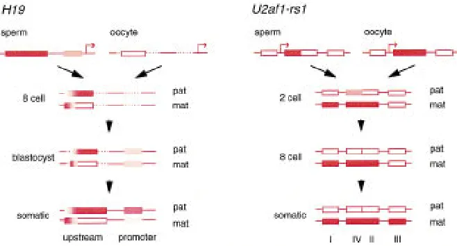
and later development. In contrast, closer to the promoter, differential methylation is less pro-nounced. Both parental alleles are relatively hypo-methylated in the blastocyst, although methylation was high in sperm. The higher level of methylation of the paternal allele is attained in the embryo postimplantation (Tremblay et al. 1997).
Rapid postzygotic changes in methylation have also been described in some detail at the mouse
U2af1-rs1 gene, although without the resolution of
the bisulfite approach (Shibata et al. 1997). A ga-metic difference exists in the tandem repeat con-taining 58UTR (region II, Fig. 3). Exclusive maternal allele methylation is retained throughout preim-plantation development and into somatic tissues. A flanking part of the 58UTR (region IV) is methylated in both gametes and in one-cell embryos, but meth-ylation is lost from the paternal allele from the two-cell stage. In contrast, the promoter (region I) is un-methylated biallelically to the one-cell stage, and methylation specifically on the maternal allele is de-tected first at the two-cell stage. Further
methyl-ation of the maternal allele occurs after implantation, ex-emplified by region III within the gene.
These two examples re-veal a very dynamic pattern of methylation at imprinted genes during preimplantation development. More limited PCR analysis has shown the existence of gametic imprints at the Igf2r, Snrpn, and Grf1/
Cdc25Mm genes (Brandeis et
al. 1993; Sto¨ger et al. 1993; Shemer et al. 1996, 1997; Shi-bata et al. 1998), with some indication of similar remodel-ing. There are several implica-tions of such dynamic behav-ior. First, a ‘‘core’’ of differen-tial methylation exists, which is retained throughout devel-opment (described hence-forth as a core differentially m e t h y l a t e d r e g i o n , c o r e DMR). Remodeling of methyl-ation occurs at sites flanking the core DMR from very early stages, which ultimately stabi-lize to produce the allelic methylation of somatic tis-sues. Second, the two alleles may be undergoing simultaneously opposing modi-fication processes. In this, differential methylation runs counter to two trends; methylation on one al-lele is protected from the general demethylation oc-curring over the rest of the genome, and hypometh-ylation of the other must resist the subsequent glo-bal remethylation. It is possible that an unusually high density of Me
CpG at a CpG island-like se-quence (nonimprinted CpG islands in contrast re-main hypomethylated) is sufficient to resist de-methylation (Howell et al. 1998). In addition, the protection of CpG islands from de novo methyl-ation afforded by the transcription factor Sp1 offers some precedent for the resistance of the unmethyl-ated allele to modification (Brandeis et al. 1994; Macleod et al. 1994). If methylation is lost from a core DMR, as occurs in Dnmt11/1ES cells, returning
[image:5.612.80.409.72.249.2]methyltransferase activity cannot restore it, without passage through the germ line (Tucker et al. 1996). This might indicate that once methylation has been erased from the normally methylated allele, reme-thylation is prevented in somatic tissues by the same Figure 3 The ontogeny of allelic methylation at the H19 and U2af1-rs1 genes.
factors that protect against methylation of the nor-mally unmethylated allele.
Somatic Maintenance of Allelic Methylation
As implied above, gametic imprints represent only a part of the differential methylation at imprinted loci in somatic tissues. At the H19 gene, for ex-ample, methylation of the paternal, repressed allele extends to the promoter and encompasses the body of the gene during postimplantation development (Bartolomei et al. 1993; Ferguson-Smith et al. 1993; Sasaki et al. 1995). Likewise, at the Igf2r gene, the repressed promoter of the paternal allele becomes methylated only late in gestation (Sto¨ ger et al. 1993). Such differential methylation may thus be regarded as a secondary effect, possibly in response to transcriptional inactivity, as it arises after mono-allelic expression has become established.
Differential methylation need not involve the whole gene or the entirety of a region containing a cluster of imprinted genes. For the small intronless
U2af1-rs1 gene, methylation does extend over the
entire length of the repressed maternal allele, with the paternal copy being hypomethylated (Shibata et al. 1996; Feil et al. 1997). Generally, however, pa-rental allele-specific methylation appears to be re-stricted to discrete elements. At Igf2r, a gene span-ning∼90 kb, DMRs occur at the promoter and the gametic imprint in intron 2, elements separated by ∼30 kb (Sto¨ger et al. 1993). At the Igf2/H19 cluster, differential methylation is confined to the H19 gene (Bartolomei et al. 1993; Ferguson-Smith et al. 1993) and three elements in Igf2: DMR0 and DMR1 in up-stream introns and DMR2 around the final intron (Feil et al. 1994; Moore et al. 1997). The CpG island at Igf2 promoter P2 is unmethylated biallelically (Sasaki et al. 1992) and the 70-kb region between
H19 and Igf2 has equal levels of methylation on
both chromosomes (Koide et al. 1994). However, differential methylation of these elements does de-pend on the integrity of the presumed imprinting signals associated with core DMRs. Deletion of the
H19 gene, including the upstream region, leads to
altered methylation at the three Igf2 DMRs on the same parental allele (Forne´ et al. 1997; Moore et al. 1997). In the Prader-Willi syndrome (PWS), im-printing mutations including deletion of the DMR at the ‘‘imprinting center’’ at the SNRPN CpG island result in altered methylation in cis of loci over a region of at least 1 Mb (Horsthemke 1997). One model foresees that such ‘‘secondary’’ DMRs may represent staging posts in the spreading of the im-printed epigenotype across a cluster of imim-printed
genes outward from an imprinting center repre-sented by the core DMR (Reik and Walter 1998).
In general, methylation effects have shown little tissue-specific variation. Differential methyl-ation of the H19 gene occurs in liver and brain, de-spite expression in the former and not in the latter, and is not altered after the postnatal repression of the gene (Bartolomei et al. 1993). Other imprinted genes that show pronounced tissue-specific varia-tion in expression levels generally have constitutive differential methylation. At the Igf2 gene, in con-trast, tissue-specific DMRs have been described. DMR0 is methylated on the repressed maternal al-lele specifically in placenta (Moore et al. 1997); methylation at DMR2 is restricted to the paternal allele in liver and other endoderm-derived tissues (Feil et al. 1994). It is possible that such methylation reflects tissue-specific transcription effects; DMR0, for example, is located close to noncoding upstream exons restricted to placental transcripts. It remains to be seen whether genes that are widely expressed but subject to highly tissue-specific imprinting, such as Gnas and Ube3a (Williamson et al. 1996; Albrecht et al. 1997), will show equally tissue-specific differential methylation, or whether they also contain core DMRs that could serve as a memory of parental origin in all tissues.
Allelic methylation differences are generally re-garded to be stable in somatic tissues once estab-lished in the embryonic phase. Cultured fibroblasts are reluctant to express the maternal Igf2 allele, u n l e s s c h a l l e n g e d w i t h a g e n t s s u c h a s 5 -azadeoxycytidine (an inhibitor of methyltransfer-ase) or sustained growth arrest (Eversole-Cire et al. 1993; Ungaro et al. 1997). Changes in epigenotype leading to relaxation of imprinting are also encoun-tered in pathological situations, where they might be seen to offer a selective advantage on cellular phenotype. Biallelic methylation of the H19 pro-moter occurs frequently in Wilms’ tumor of the kid-ney. The silencing of the normally active maternal
H19 promoter may lead to derepression of the
ma-ternal IGF2 allele, through competition with the shared enhancers (Moulton et al. 1994; Steenman et al. 1994), resulting in a growth advantage from en-hanced IGF2 expression. ‘‘Relaxation’’ of IGF2 and
H19 imprinting is a frequent occurrence in many
tumor types.
Cis-Acting Signals
Tandem Repeats
sequences involved in the establishment or mainte-nance of imprinting? H19 transgenes having dele-tions of the core DMR fail to show correct imprint-ing, but other interventions within H19 transgenes also impair imprinting (Pfeifer et al. 1996; Elson and Bartolomei 1997). When assayed in Drosophila (whose genome is unmethylated) this region func-tions as a cis-acting silencer (Lyko et al. 1997). De-letion of the core DMR (region 2) of the Igf2r gene in the context of transgenic yeast artificial chromo-somes (YACs) also eliminates imprinting of the transgene (Wutz et al. 1997). The PWS ‘‘imprinting mutations’’ are small deletions including the DMR at the CpG island of the SNRPN locus, which may lead to incorrect resetting of the imprint in the pa-ternal germ line (Horsthemke 1997). A similar dele-tion at the mouse Snrpn gene has now also been shown to block the switch from maternal to pater-nal epigenotype (Yang et al. 1998). Does this knowl-edge help to identify key cis-acting sequences? One provocative finding has been the association of short tandem repeats with DMRs.
Neumann et al. (1995) first proposed that direct tandem repeats, embedded in CG-rich sequences and associated with regions of differential methyl-ation, may constitute a feature common and possi-bly unique to imprinted genes. This hypothesis was based on the concept that the imprinting mecha-nism may have evolved from the host defense func-tion of DNA methylafunc-tion (Barlow 1993). For ex-ample, an insertion of a retrovirus-like intracisternal A-particle (IAP) upstream of the mouse agouti locus can induce imprinting. The resulting ectopic expres-sion of agouti is silenced specifically with paternal transmission, correlating with hypermethylation of the IAP long terminal repeat sequence (Michaud et al. 1994). Therefore, sequences with properties of ‘‘foreign DNA’’ may have been recognized by com-ponents of the methylation system to attract allele-specific modification and given rise to imprinting (Barlow 1993).
Many, if not all, imprinted genes contain tan-dem direct repeats. However, comparison of such direct repeats reveals few obvious common features. No sequence homology can be found between re-peats in different genes (the consensus sequences tend, however, to be G-rich); the repeat units can be of different lengths, and the number of times they are repeated is variable; their location with respect to the gene also differs (upstream, 58UTR, intronic; some being transcribed, others not) as does their relationship with CpG islands and allele-specific methylation patterns (they are found within, or at variable distances from CpG islands or DMRs). A
comparison between mouse and human regarding a possible role for the repeats is not conclusive. The
IGF2 gene in humans lacks a repeat in the
homolo-gous region to the mouse and yet is imprinted (Moore et al. 1997). A block of repeats may, how-ever, exist further upstream of IGF2, as reported for the H19 gene (Jinno et al. 1996). On the other hand, the tandem repeat region and differential methyl-ation in the U2af1-rs1 58UTR is specific to this gene; related U2af1-rs loci in the mouse and human ge-nomes lack this sequence feature and are bialleli-cally expressed (Shibata et al. 1997). Finally, the
IGF2R gene is polymorphically imprinted in
hu-mans (Xu et al. 1993) but contains numerous large direct repeats and is differentially methylated (Smr-zka et al. 1995). What experimental evidence sup-ports a role for these repeats in the establishment or maintenance of imprinting, given this diversity of properties?
Imprinting of the RSVIgmyc mouse transgene, which expresses the c-myc oncogene derived from a translocation, seems to require a cis-acting signal that is principally derived from the tandemly re-peated sequences that make up the 38portion of the murine immunoglobulin a (IgA) heavy-chain switch region (Chaillet et al. 1995). Sequence con-text also seems to be important for imprinting, as the sequence elements of RSVIgmyc are not im-printed in their normal endogenous location. A re-cent analysis of additional mutations of the
RSVIg-myc transgene, however, indicates that the IgA
re-peat sequences are not absolutely required for transgene imprinting (Howell et al. 1998). Similar results have also been observed with H19 trans-genes; deletion of the G-rich repeat upstream of H19 eliminates transgene imprinting (M. Bartolomei, pers. comm.). However, a larger transgene carrying this deletion did, in fact, show imprinted expression (M. Bartolomei, pers. comm.). These observations suggest that the repeats, although potentially im-portant for attracting allele-specific methylation and expression, are not sufficient and must interact with other sequence features or context. Further-more, although deletion of the repeat-containing region from an Igf2r YAC abolishes imprinting, shorter transgenes that contain the repeats fail to attract parental-specific methylation (Wutz et al. 1997).
spread out in regions of the gene. Ultimately, it would be their organization or context (rather than sequence) that confers imprinting potential. These elements may constitute a network of responsive elements, under the hierarchical control of imprint-ing centers.
Although the role of direct repeats needs to be better defined by more precise genetic experiments, their involvement can be envisaged in a number of ways. Because organizational conservation rather than sequence homology is observed, secondary DNA structure may play an important role. Repeats may form alternative secondary structures that at-tract de novo methylation either in the gametes or soon after fertilization. Alternatively, the repeats could nucleate specific chromatin structures and the observed methylation patterns would be a con-sequence of this. Either way, germ-line-specific fac-tors that protect one allele from these modifications need to be involved (Fig. 4); for instance, methyl-ation at Alu repeats is blocked in sperm by an Alu-binding protein (SABP)
(Chesno-kov and Schmid 1995).
We propose a model in which the allele-specific methyl-ation patterns at imprinted loci are established in regions where opposing de novo methylation signals (emanating from repeti-tive elements) and demethyl-ation signals (induced by CpG-rich environment) interact (Fig. 4). Trans-acting factors dictate the allele-specific patterns by ei-ther blocking the de novo meth-ylation or demethmeth-ylation path-ways (Fig. 4). Although core DMRs are methylated (or not) in the germ line and these states are then inherited after fertilization, it should be pointed out that re-peats could have a role in either establishment (in the germ line) or in maintenance of allelic methylation (in embryos), or in both processes.
There is precedent for the role of repeat sequences in gene silencing and possibly in induc-i n g r e g induc-i o n a l m e t h y l a t induc-i o n . Hence, repeat-induced gene si-lencing has been observed in or-ganisms as diverse as fungi, sects, plants, and mammals,
[image:8.612.237.554.348.572.2]in-cluding those in which there is no methylation (Trends Genet. 1997). A de novo methylation center has been identified in the mouse adenine phospho-ribosyltransferase gene (aprt) that imparts a methyl-ation signal to upstream and downstream HpaII sites (Mummaneni et al. 1993). The bulk of the methylation center signal seems to emanate from tandem B1 repetitive elements (Turker and Bestor 1997) and it has been postulated that Sp1-binding sites in the promoter region are necessary to stop the spreading of the de novo methylation (Mum-maneni et al. 1995). Hypothetically, repeat ele-ments may also function as recognition ‘‘nuclei’’ by protein complexes such as the methyltransferase, PCNA–p21–methyltransferase complexes, or even protein complexes involved in chromosomal repres-sion, as in telomeric silencing mediated by RAP1– SIR protein complexes in yeast (Moretti et al. 1994). Some DMRs acquire their allelic methylation af-ter fertilization. The local spacing and cooperativity between these DMRs and core DMRs (i.e., somatic
and germ-line methylation imprints) may be impor-tant for the propagation of the imprinting signal and stabilization of specific epigenotypes in im-printing clusters (Reik and Walter 1998). The dis-ruption of any of these elements could result in fail-ure to maintain or establish a parental epigenotype.
RNAs, Competition, and Regional Control of Epigenotype
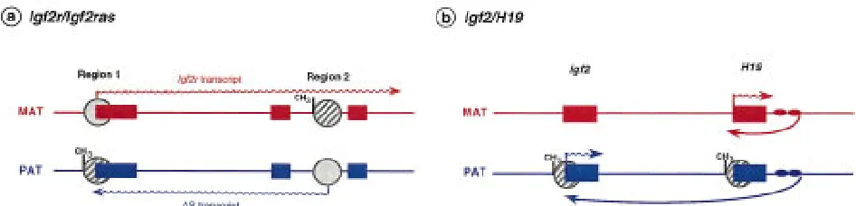
Transcripts originating from the noncoding or an-tisense strand, in a parental allele-specific manner, have been found in three imprinted genes so far. An antisense transcript (with no apparent ORF) is ex-pressed abundantly from the paternal allele of Igf2r, initiating within region 2. This antisense is thought to be responsible for monoallelic expression of Igf2r by repressing specifically the paternal allele of Igf2r in cis (Wutz et al. 1997), an example of ‘‘expression competition’’ (Fig. 5a) in which the antisense is per-ceived as an ‘‘imprintor’’ and the Igf2r gene as its imprinted target (Barlow 1997). Deletion of the an-tisense promoter or enhancer leads to nonim-printed expression of Igf2r. Although no develop-mental details have yet been reported, it is conceiv-able that antisense expression coincides with the onset of monoallelic expression of Igf2r at E5–E6 (Lerchner and Barlow 1997). This would explain the lack of correlation between the onset of repression and promoter methylation (region 1) on the pater-nal allele (Sto¨ger et al. 1993). Transcription could be repressed first by the antisense and then
methyl-ation would ‘‘lock’’ the repressed state later in de-velopment. The antisense may repress the paternal
Igf2r allele in cis in several ways, such as physically
coating the paternal allele (in a similar way to Xist; see below), occlusion of the Igf2r promoter by tran-scribing through it in the opposite direction, or competition for transcription factors or enhancers (Reik and Constaˆncia 1997).
An imprinted antisense RNA has also been found to overlap the maternally expressed UBE3A gene and is thought to regulate tissue specificity of
UBE3A imprinting (Rougeulle et al. 1998). The
an-tisense RNA is expressed exclusively in the brain from the paternal chromosome, thus apparently limiting expression of the UBE3A gene to the ma-ternal chromosome in this tissue. In other tissues, where the antisense RNA is not detectable, UBE3A is expressed biallelicaly (Rougeulle et al. 1998).
Several imprinted antisense transcripts (Igf2as-a, b, and c) have been described in the upstream region of the Igf2 gene (Moore et al. 1997), but in contrast to Igf2r they are expressed from the same allele as Igf2 and at low levels. The role of these antisense transcripts is unclear, but overlapping an-tisense transcription may influence the activity of promoters 0 and 1, because of their proximity to these two regions, and regulate Igf2 levels in a tissue-specific fashion.
H19 is an example of an imprinted, untrans-lated RNA involved, through enhancer competi-tion, in regulating the closely linked but
recipro-Figure 5 Expression competition mechanisms at the Igf2r/Igf2ras and Igf2/H19 loci. (a) Exons of Igf2r are depicted as solid boxes, with arrows indicating the transcribed alleles; regions 1 and 2 are sites of differential methylation (hatched circles with CH3: methylation) arising during embryonic development and in the egg, respectively. When
[image:9.612.82.555.468.582.2]cally imprinted genes Igf2 and Ins2 (Leighton et al. 1995a). The mechanism appears to be competition for shared enhancer elements located downstream of H19 (Bartolomei et al. 1993; Leighton et al. 1995b) (Fig. 5b). When the maternal H19 promoter is activated by the downstream enhancer, the Igf2 gene is not expressed from the maternal allele. Pa-ternal expression of the Igf2 gene is observed when the enhancer is unable to interact with the H19 pa-ternal promoter because it is methylated (Fig. 5). Proper imprinting of Igf2 and H19 on the maternal chromosome seems to be dependent on the posi-tion of the enhancers relative to the two genes. Mice carrying an extra set of the H19 endoderm-specific enhancers between Igf2 and H19 show expression of the normally silent Igf2 gene in liver, consistent with relief from competition (Webber et al. 1998). When a single set of enhancers is located equidis-tantly from Igf2 and H19 on the maternal chromo-some, Igf2 is expressed instead of H19, suggesting that the strength of the H19 promoter is not the main determinant in the competition for enhancers (Webber et al. 1998). The core DMR associated with the H19 promoter may determine methylation of the H19 promoter but, in addition, may also play a role in allowing the 38enhancers access to the Igf2 promoters.
Unusual RNAs are also found in the upstream region of the SNRPN gene and may be involved in imprinting control for the entire PWS/AS region. Deletions and a splice mutation affecting these up-stream transcripts are associated with AS, as they lead to an apparent failure to switch the paternal epigenotype in the maternal germ line (Dittrich et al. 1996). In contrast, deletion of the SNRPN CpG island region and promoter leads to an apparent inability to switch the maternal regional epigeno-type in the paternal germ line, and hence to PWS. However, these observations could also be ex-plained by extending the expression or enhancer competition model to the PWS/AS situation (Barlow 1997; Tilghman et al. 1998). Promoter competition between the SNRPN and UBE3A genes for a neuronal enhancer has been proposed recently (Tilghman et al. 1998). Methylation inactivating the SNRPN pro-moter on the maternal chromosome would lead to activation of the UBE3A gene.
A common feature of the enhancer competition model is that deletion of DMRs, promoter, or en-hancers would imbalance the competition system. However, this model does not explain how regional methylation can be altered by DMR or promoter deletions (Sutcliffe et al. 1994; Buiting et al. 1995; Forne´ et al. 1997; Moore et al. 1997; Wutz et al.
1997). It has been suggested that cis-acting RNAs that interact with DMRs could be involved in this regional spreading (Reik and Walter 1998).
A precedent for the role of an untranslated RNA in regional silencing in cis is the Xist gene. The Xist RNA is expressed from the inactive X chromosome in female mammals and is essential for inactivation of genes along the X chromosome (Penny et al. 1996; Marahrens et al. 1997). The accumulation of
Xist transcripts along the length of the inactive
chromosome (Clemson et al. 1996; Lee and Jaenisch 1997) is thought to be required for the nucleation and spread of heterochromatin from the X inacti-vation center (Xic) (Panning et al. 1997). Inactiva-tion of the paternal X chromosome occurs in extra-embryonic tissues and consistent with this the pa-ternal Xist copy is expressed preferentially in female preimplantation embryos (Kay et al. 1993). How-ever, in embryonic lineages, in which X inactiva-tion is random, the imprinted expression needs to be reprogrammed. This may involve expression of unstable Xist transcripts from both X chromosomes (Panning et al. 1997; Sheardown et al. 1997), which could be brought about by using alternative pro-moters (Sheardown et al. 1997). The future inactive X chromosome then stabilizes Xist RNA by an un-known mechanism (possibly involving a promoter switch), whereas the active X chromosome fails to accumulate Xist. This initial decision is ‘‘locked’’ by subsequent silencing of Xist, at the transcriptional level, on the active X chromosome by DNA meth-ylation (Panning and Jaenisch 1996; Panning et al. 1997).
Reading the Imprint
The different methylation states of the two alleles have to be translated into monoallelic expression. This is termed the reading of the imprint. The read-ing mechanism requires the involvement of various
trans-acting factors, some of which can detect the
throughout the genome (Yoder et al. 1997a). Hence, unmethylated regions are relatively rare and usually encountered in the form of CpG islands associated with the promoters of expressed housekeeping genes (Bird 1995). If such regions become errone-ously methylated (for example, in tumors) then the associated gene is repressed. This is the case for the CpG islands of tumor suppressors VHL, p16, and Rb (Laird and Jaenisch 1996). Certain transgenes also show a tight inverse correlation between methyl-ation levels and expression (Allen et al. 1990; Chail-let et al. 1991).
Is it possible to treat monoallelic expression through imprinting as just a special case of methyl-ation silencing? It could be postulated that the ‘‘hard work’’ of distinguishing the two parental al-leles has already been accomplished by the imposi-tion of the differential methylaimposi-tion pattern. The next step is the ‘‘blind’’ transcriptional response to the methylation pattern, which only requires the factors responsible for the initiation or inhibition of transcription to associate with the appropriate al-lele. This would be determined not only by the availability of the appropriate transcription factors for the enhancer/promoter elements, but also how these transcription factors respond to the methyl-ation patterns that may exist in those regions.
Reading Factors
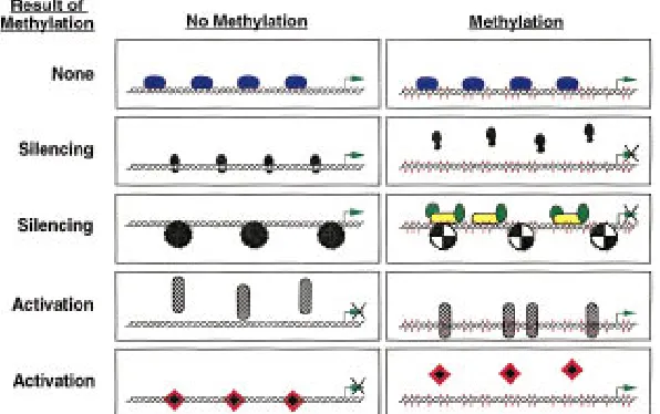
DNA-binding factors can respond in a number of ways to the methylation of their target sites. Some seem relatively unaffected by the methylation status of their binding sites (Tate and Bird 1993). These include Sp1, MTF-1, Krox-20, CTF/NF1, and TCR-ATF. Table 1 lists those factors whose binding affini-ties are altered by the methylation of their target sequences and Figure 6 illustrates the potential ef-fects of methylation on trans-acting factor binding and subsequent transcriptional control.
The silencing of transcription brought about by methylation has been divided into two categories: (1) that resulting from the inability of methylation-sensitive transcription factors to bind to their target sequences and contribute to gene activation, and (2) that resulting from the binding of methylation-dependent silencer proteins that may act by nucle-ating chromatin structures in the gene region. The
Xist gene possesses a transcription-promoting
ele-ment that binds factors belonging to both of these categories (Huntriss et al. 1997). When unmethyl-ated, a factor binds and may activate expression of the gene. However, with methylation, the activator is no longer able to bind and is replaced by a
meth-ylation-dependent binding protein that may act to silence gene expression.
Two proteins of the second category, MeCP1 and MeCP2, appear to be crucial in general methyl-ation-dependent silencing. MeCP2 is an abundant protein that possesses domains that bind methyl-ated DNA and repress transcription (probably indi-rectly). Immunofluorescence studies show that the MeCP2 protein is tightly associated with chromo-somes, especially in heterochromatic regions (Nan et al. 1997). Interestingly, MeCP2 may carry out part of its inhibitory role by becoming a key part of the chromatin complex itself as it has been shown that MeCP2 can replace the linker histone H1 in the compacted DNA of silenced regions. MeCP1 is a complex of several proteins including one, PCM1, which has a methyl-binding domain homologous to MeCP2 and a cysteine-rich domain homologous to those found in the DNMT1 and HRX proteins (Cross et al. 1997). The complex is widely expressed at a low level and requires a greater density of meth-ylated cytosines than does MeCP2 before binding occurs. Binding of MeCPs to methylated DNA re-sults in reduced accessibility of chromatin to endo-nucleases (DNase I, restriction enzymes). Consistent with an involvement of such proteins in allelic si-lencing, some imprinted genes show markedly re-duced DNase accessibility in regions of extensive methylation (Bartolomei et al. 1993; Ferguson-Smith et al. 1993; Feil and Kelsey 1997; Feil et al. 1997). However, the link between such proteins and the establishment of silencing through the forma-tion of specific chromatin structures is still un-known (Kass et al. 1997). A key process may be the balancing act between the acetylation and deacety-lation of histones within nucleosomes (Pazin and Kadonaga 1997). Deacetylation of these proteins, carried out by ‘‘histone deacetylases’’ (in mammals there are two, HD1 and HD2) in conjunction with another protein, Sin3, is associated with the silenc-ing of genes such as Xenopus TRbA (Wong et al.
1998). It has been suggested that the specificity of the deacetylation process may be dictated by DNA-bound factors that serve to anchor the activity in appropriate areas. Indeed, it has been shown re-cently that MeCP-dependent silencing acts through deacetylation of histones (Jones et al. 1998; Nan et al. 1998).
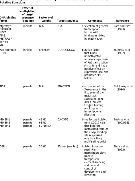
Table 1. Methylation-Sensitive and -Dependent DNA-Binding Proteins and their Putative Functions
DNA-binding factor
Effect of methylation
of target sequence (binding)
Factor mol.
weight Target sequence Comments Reference
Myc/Myn CREB/ATF NFKB AP-2 MLTF/USF EBP-80 VBP1
inhibits N.A. N.A. a selection of general
transcription factors with binding inhibited by methylation
Tate and Bird (1993)
Xist promoter BP1
inhibits unknown GCGCCGCGG putative factor that binds unmethylated sequence upstream of Xist transcription start site and has a positive effect on expression (see Xist promoter BP2 below)
Huntriss et al. (1997)
AP-1 permits N.A. TGACTCG methylation of the
X-sequence in the first exon of the metastasis-associated gene mts-1 induces Fos/Jun binding resulting in transcriptional silencing
Tulchinsky et al. (1996)
MMBP-1 MMBP-2 MMBP-3
permits permits permits
42 kD 63 kD 50–60 kD
CACGTG three factors isolated from C2C12 cells that bind the methylated form of the c-Myc binding site (expressed only in actively proliferating cells)
Suetake et al. (1993/95)
DBPm permits 50 kD 35-mer (see Ref.) isolated from pea seed. Plant methylation plays roles in
transposable element silencing and general control of development and flowering
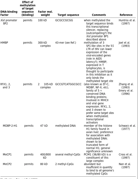
Table 1. (Continued)
DNA-binding Factor
Effect of methylation
of target sequence (binding)
Factor mol.
weight Target sequence Comments Reference
Xist promoter BP2
permits 100 kD GCGCCGCGG when methylated the target sequence binds this transcriptional silencer, replacing (outcompeting?) the Xist promoter BP1 described above
Huntriss et al. (1997)
HMBP permits 300-kD
complex
43-mer (see Ref.) methylation of 3 SP1-like sites in the 58 LTR of HIV can lower expression of the viral-encoded genes (role in AIDS latency?); HMBP, expressed in T lymphocytes, is thought to participate in this inhibition as it only binds the methylated target site
Joel et al. (1993)
RFX1, 2, and 3
permits 22105-kD complex
GCCGTCATGGCGCC (also known as EF-C, MDBP, NF-X, etc), family of 5 + conserved DNA binding proteins. Involved in MHCII and viral gene expression. RFX1, 2, and 3 shown to prefer some target sites when methylated; transcriptional activators
Zhang et al. (1993) Emery et al.
(1996)
MDBP-2-H1 permits 47 kD methylated DNA member of the histone H1 family found in avian liver; preference for association with methylated DNA; shown to be truncated form of normal H1; general significance unknown
Schwarz et al. (1977)
MeCP1 permits 400/800
kD
several methyl-CpGs PCM1 (36/80 kD) is a constituent of this large complex
Cross et al. (1997)
MeCP2 permits 80 kD 2 methyl-CpGs abundant but
insufficient in quantity to bind to all genome’s methylated CpGs
Nan et al. (1997)
amounts of biallelic H19 expression in early em-bryos. The drug treatment slowed the rate of pater-nal allele silencing, which resulted in increased numbers of patches of biallelic H19 expression be-yond the normal stages. From these results we can conclude that the presence of biallelic expression from imprinted genes early in development may be the result of the immaturity of the silencing com-plex as well as the unrefined nature of the methyl-ation pattern. Other routes to heterochromatizmethyl-ation exist that do not require changes in histone acety-lation. The polycomb group of genes has been shown to play important chromatin-mediated si-lencing effects in Drosophila (position effect variega-tion) and the regulation of mammalian genes such as the Hox clusters (Pirrotta 1997). The full reper-toire of chromatin or chromatin-organizing genes potentially involved in the control of monoallelic expression remains to be cataloged.
The two categories of silencing described above are a useful guide but cannot explain all observa-tions. For example, the actively transcribed paternal allele of the Igf2 gene is the allele that shows higher methylation levels (at DMR1 and DMR2). Assuming that these regions play an important role in the
con-trol of monoallelic expression, it be-comes necessary to explain this appar-ent reversal in the effect of methylation on transcription. It could be proposed that either a methylation-dependent activator protein is binding to the pa-ternal DMRs (acting in the same way as
RFX1/RFX2/RFX3), or that a
methyl-ation-sensitive silencer protein is associ-ated with the repressed maternal allele.
Reading Disorders
This review has already mentioned dis-eases arising from the loss of the correct imprint or from the failure to switch the imprint to reflect the parental ori-gin. Is it possible that there exist defects in the reading process that have patho-logical consequences? Targeted disrup-tion of the methyl-cytosine-binding protein gene MeCP2 has an early em-bryonic lethal phenotype leading to speculation that this is a critical com-ponent in the conversion of methyl-ation into silencing that cannot be re-placed by compensatory or parallel pathways (Tate et al. 1996). It will be interesting to determine whether ex-pression of imprinted genes is altered in these knock-out mice. Are there less severe phenotypes arising from other reading machinery defects? There are several instances when a seemingly allele-specific methylation pattern is not translated into genuine monoallelic expression. This is the case for the human Igf2r gene where only some individuals (genotypes) show monoallelic expression. A pos-sible cause of this variability could be that functional polymorphisms are present in the human genes re-sponsible for the reading of the imprint as opposed to functional alterations in the imprint itself.
Conclusions
[image:14.612.77.375.75.262.2]Major reprogramming of the imprints occurs in the germ lines of developing embryos. Both sex germ cells undergo global demethylation including im-printed and nonimim-printed genes. The bulk of this demethylation probably occurs between E8.5 and E12.5. Functional studies on germ cells from this stage in chimeras or nuclear transplantations show that this demethylation is indeed associated with substantial reprogramming of imprinted genes. It is not yet clear whether all imprinted genes undergo Figure 6 Four mechanisms by which methylation can alter
complete reprogramming. New methylation im-prints are established in the female germ line during oocyte growth after birth, and in males probably around the time of birth. In addition to germ-line-specific de novo methylation, protection from methylation may also be required.
Regions of differential methylation have been identified at all imprinted genes. Their developmen-tal kinetics appear to be diverse and complex, some being stable and potentially associated with gametic imprinting signals, others arising later in develop-ment and more likely to be involved in mainte-nance of imprinting or spreading of an imprinted epigenotype in a cluster of imprinted genes. Ge-nome-wide alterations in methylation occur in the preimplantation and early postimplantation em-bryo. However, methylation of core DMR regions of imprinted genes remains unaffected. This requires protective mechanisms that not only prevent the methylated allele from becoming demethylated but also prevent the demethylated allele from de novo methylation. High methylation densities may play a role by causing resistance to demethylation. Mul-tiple cis-acting sequences are probably required for the creation and maintenance of the methylation state. The precise methylation patterns are elabo-rated during postimplantation development.
Diverse sequence and structural motifs are asso-ciated with imprinted genes. DMRs are CpG-rich and associated with direct repeats; although this as-sociation with imprinted loci is strong, evidence for a functional role is, at present, inconclusive. One model for control of methylation at imprinted loci may involve the interplay between de novo meth-ylation caused by repeats and demethmeth-ylation in-duced by the CpG-rich environment. Other regula-tion mechanisms such as antisense transcripregula-tion and enhancer competition play an important role in the imprinting process, probably both in indi-vidual genes and in imprinting clusters.
The mechanisms underlying the translation of the imprint into monoallelic gene expression are largely unknown. The methylated allele of most im-printed genes is the inactive one. Repression could be brought about by the inability of transcription factors to bind or alternatively, by the binding of MeCPs and associated silencing factors. Expression from the methylated allele is rare but may involve the displacement of repressors or the binding of methylation-dependent activating factors.
ACKNOWLEDGMENTS
We apologize to authors whose work could not be cited
be-cause of space limitations. We thank M. Bartolomei, T. Bestor, J. Chaillet, H. Sasaki, A. Surani, and T. Kono for communicat-ing unpublished results, and members of the laboratories of W. Reik and G. Kelsey for helpful discussions. Work in our laboratory is supported by Biotechnology and Biological Sci-ences Research Council, European Union, Medical Research Council, and Cancer Research Campaign. M.C. acknowledges support by a JNICT/PRAXIS XXI Scholarship (Portugal).
REFERENCES
Albrecht, U., J.S. Sutcliffe, B.M. Cattanach, C.V. Beechey, D. Armstrong, G. Eichele, and A.L. Beaudet. 1997. Imprinted expression of the murine Angelman syndrome gene Ube3a in hippocampal and Purkinje neurons. Nature Genet.
17: 75–78.
Allen, N.D., M.L. Norris, and M.A. Surani. 1990. Epigenetic control of transgene expression and imprinting by genotype specific modifiers. Cell 61: 853–861.
Ariel, M., E. Robinson, J.R. McCarrey, and H. Cedar. 1995. Gamete-specific methylation correlates with imprinting of the murine Xist gene. Nature Genet. 9: 312–315.
Barlow, D.P. 1993. Methylation and imprinting: From host defense to gene regulation? Science 260: 309–310.
———. 1997. Competition—a common motif for the imprinting mechanism? EMBO J. 16: 6899–6905.
Bartolomei, M.S., A.L. Webber, M.E. Brunkow, and S.M. Tilghman. 1993. Epigenetic mechanisms underlying the imprinting of the mouse H19 gene. Genes & Dev.
7: 1663–1673.
Bird, A.P. 1995. Gene number, noise reduction and biological complexity. Trends Genet. 11: 94–100.
Brandeis, M., T. Kafri, M. Ariel, J.R. Chaillet, J. McCarrey, A. Razin, and H. Cedar. 1993. The ontogeny of allele-specific methylation associated with imprinted genes in the mouse.
EMBO J. 12: 3669–3677.
Brandeis, M., D. Frank, I. Keshet, Z. Siegfried, M.
Mendelsohn, A. Nemes, V. Temper, A. Razin, and H. Cedar. 1994. Sp1 elements protect a CpG island from de novo methylation. Nature 371: 435–438.
Buehr, M. 1997. The primordial germ cells of mammals: Some current perspectives. Exp. Cell Res. 232: 194–207.
Buiting, K., S. Saitoh, S. Gross, B. Dittrich, S. Schwartz, R.D. Nicholls, and B. Horsthemke. 1995. Inherited
microdeletions in the Angelman and Prader-Willi syndromes define an imprinting centre on human chromosome 15. Nature Genet. 9: 395–400.
Carlson, L.L., A.W. Page, and T.H. Bestor. 1992. Properties and localization of DNA methyltransferase in
Chaillet, J.R., T.F. Vogt, D.R. Beier, and P. Leder. 1991. Parental-specific methylation of an imprinted transgene is established during gametogenesis and progressively changes during embryogenesis. Cell 66: 77–83.
Chaillet, J.R., D.S. Bader, and P. Leder. 1995. Regulation of genomic imprinting by gametic and embryonic processes.
Genes & Dev. 9: 1177–1187.
Chesnokov, I.N. and C.W. Schmid. 1995. Specific Alu binding protein from human sperm chromatin prevents DNA methylation. J. Biol. Chem. 270: 18539–18542.
Clemson, C.M., J.A. McNeil, H.F. Willard, and J.B. Lawrence. 1996. Xist RNA paints the inactive X chromosome at interphase: Evidence for a novel RNA involved in nuclear/chromosome structure. J. Cell. Biol.
132: 259–275.
Cross, S.H., R.H. Meehan, X. Nan, and A. Bird. 1997. A component of the transcriptional repressor MeCP1 shares a motif with DNA methyltransferase and HRX proteins.
Nature Genet. 16: 256–259.
Dittrich, B., K. Buiting, B. Korn, S. Rickard, J. Buxton, S. Saitoh, R.D. Nicholls, A. Poustka, A. Winterpacht, B. Zabel, and B. Horsthemke. 1996. Imprint switching on human chromosome 15 may involve alternative transcripts of the
SNRPN gene. Nature Genet. 14: 163–170.
Ehrlich, K.C. 1993. Partial purification of a pea seed DNA-binding protein that specifically recognizes 5-methylcytosine. Prep. Biochem. 23: 423–438.
Elson, D.A. and M. Bartolomei. 1997. A 58differentially methylated sequence and the 38-flanking region are necessary for H19 transgene imprinting. Mol. Cell. Biol.
17: 309–317.
Emery, P., B. Durand, B. Mach, and W. Reith. 1996. RFX proteins, a novel family of DNA binding proteins conserved in the eukaryotic kingdom. Nucleic Acids Res. 24: 803–807.
Eversole-Cire, P., A.C. Ferguson-Smith, H. Sasaki, K.D. Brown, B.M. Cattanach, F.A. Gonzales, M.A. Surani, and P.A. Jones. 1993. Activation of an imprinted Igf2 gene in mouse somatic cell cultures. Mol. Cell. Biol. 13: 4928–4938.
Feil, R. and G. Kelsey. 1997. Genomic imprinting: A chromatin connection. Am. J. Hum. Genet. 61: 1213–1219.
Feil, R., J. Walter, N.D. Allen, and W. Reik. 1994. Developmental control of allelic methylation in the imprinted mouse Igf2 and H19 genes. Development
120: 2933–2943.
Feil, R., M.D. Boyano, N.D. Allen, and G. Kelsey. 1997. Parental chromosome-specific chromatin conformation in the imprinted U2af1-rs1 gene in the mouse. J. Biol. Chem.
272: 20893–20900.
Ferguson-Smith, A.C., H. Sasaki, B.M. Cattanach, and M.A. Surani. 1993. Parental-origin-specific epigenetic
modification of the mouse H19 gene. Nature 362: 751–755.
Forne´, T., J. Oswald, W. Dean, J.R. Saam, B. Bailleul, L. Dandolo, S.M. Tilghman, J. Walter, and W. Reik. 1997. Loss of the maternal H19 gene induces changes in Igf2
methylation in both cis and trans. Proc. Natl. Acad. Sci.
94: 10243–10248.
Horsthemke, B. 1997. Imprinting in the
Prader-Willi/Angelman syndrome region on human chromosome 15. In Frontiers in molecular biology (series ed. B.D. Hames and D.M. Glover); Genomic imprinting (ed. W. Reik and A. Surani), pp. 177–190. IRL Press, Oxford, UK.
Horsthemke, B., B. Dittrich, and K. Buiting. 1997. Imprinting mutations on human chromosome 15. Hum.
Mutat. 10: 329–337.
Howell, C.Y., A.L. Steptoe, M.W. Miller, and J.R. Chaillet. 1998. Cis-acting signal for inheritance of imprinted DNA methylation patterns in the preimplantation mouse embryo. Mol. Cell. Biol. 18: 4147–4156.
Howlett, S.K. and W. Reik. 1991. Methylation levels of maternal and paternal genomes during preimplantation development. Development 113: 119–127.
Huntriss, J., R. Lorenzi, A. Purewal, and M. Monk. 1997. A methylation-dependent DNA-binding activity recognising the methylated promoter region of the mouse Xist gene.
Biochem. Biophys. Res. Commun. 235: 730–738.
Ja¨hner, D. and R. Jaenisch. 1984. DNA methylation in early mammalian development. In DNA methylation: Biochemistry
and biological significance (ed. A. Razin, H. Cedar, and A.D.
Riggs), pp. 189–219. Springer-Verlag, New York, NY.
Jinno, Y., K. Sengoku, M. Nakao, K. Tamate, T. Miyamoto, T. Matsuzaka, J.S. Sutcliffe, T. Anan, N. Takuma, K. Nishiwaki, Y. Ikeda, T. Ishimaru, M. Ishikawa, and N. Niikawa. 1996. Mouse/human sequence divergence in a region with a paternal-specific methylation imprint at the human H19 locus. Hum. Mol. Genet. 5: 1155–1161.
Joel, P., W. Shao, and K. Pratt. 1993. A nuclear protein with enhanced binding to methylated Sp1 sites in the AIDS virus promoter. Nucleic Acids Res. 21: 5786–5793.
Jones, P.L., G.J.C. Veenstra, P.A. Wade, D. Vermaak, S.U. Kass, N. Landsberger, J. Strouboulis, and A.P. Wolffe. 1998. Methylated DNA and MeCP2 recruit histone deacetylase to repress transcription. Nature Genet. 19: 187–191.
Kafri, T., M. Ariel, M. Brandeis, R. Shemer, L. Urven, J. McCarrey, H. Cedar, and A. Razin. 1992. Developmental pattern of gene-specific DNA methylation in the mouse embryo and germ line. Genes & Dev. 6: 705–714.
Kafri, T., X. Gao, and A. Razin. 1993. Mechanistic aspects of genome-wide demethylation in the preimplantation mouse embryos. Proc. Natl. Acad. Sci. 90: 10558–10562.
Kass, S.U., D. Pruss, and A.P. Wolffe. 1997. How does DNA methylation repress transcription? Trends Genet.
Kato, Y. and Y. Tsunoda. 1995. Germ-cell nuclei of male fetal mice can support development of chimeras to mid-gestation following serial transplantation. Development
121: 779–783.
Kay, G.F., G.D. Penny, D. Patel, A. Ashworth, N. Brockdorff, and S. Rastan. 1993. Expression of Xist during mouse development suggests a role in the initiation of X-chromosome inactivation. Cell 72: 171–182.
Kelsey, G. and W. Reik. 1997. Imprint switch mechanism indicated by mutations in Prader-Willi and Angelman syndromes. Bioessays 19: 361–365.
Koide, T., J. Ainscough, M. Wijgerde, and M.A. Surani. 1994. Comparative analysis of Igf-2/H19 imprinted domain: Identification of a highly conserved intergenic DNaseI hypersensitive region. Genomics 24: 1–8.
Kono, T., Y. Obata, T. Yoshimzu, T. Nakahara, and J. Carroll. 1996. Epigenetic modifications during oocyte growth correlate with extended parthenogenetic development in the mouse. Nature Genet. 13: 91–94.
Labosky, P.A., D.P. Barlow, and B.L.M. Hogan. 1994. Mouse embryonic germ (EG) cell-lines—transmission through the germline and differences in the methylation imprint of insulin-like growth-factor 2 receptor (IGF2R) gene compared with embryonic stem (ES) cell lines. Development
120: 3197–3204.
Laird, P.W. and R. Jaenisch. 1996. The role of DNA methylation in cancer genetics and epigenetics. Annu. Rev.
Gen. 30: 441–464.
Lee, J.T. and R. Jaenisch. 1997. Long-range cis effects of ectopic X-inactivation centres on a mouse autosome. Nature
386: 275–279.
Lei, H., S.P. Oh, M. Okano, R. Juttermann, K.A. Goss, R. Jaenisch, and E. Li. 1996. De novo DNA cytosine
methyltransferase activities in mouse embryonic stem cells.
Development 122: 3195–3205.
Leighton, P.A., R.S. Ingram, J. Eggenschwiler, A. Efstratiadis, and S.M. Tilghman. 1995a. Disruption of imprinting caused by deletion of the H19 gene region in mice. Nature
375: 34–39.
Leighton, P.A., J.R. Saam, R.S. Ingram, C.L. Stewart, and S.M. Tilghman. 1995b. An enhancer deletion affects both
H19 and Igf2 expression. Genes & Dev. 9: 2079–2089.
Lerchner, W. and D.P. Barlow. 1997. Paternal repression of the imprinted mouse Igf2r locus occurs during implantation and is stable in all tissues of the post-implantation mouse embryo. Mech. Dev. 61: 141–149.
Li, E., T.H. Bestor, and R. Jaenisch. 1992. Targeted mutation of the DNA methyltransferase gene results in embryonic lethality. Cell 69: 915–926.
Li, E., C. Beard, and R. Jaenisch. 1993. Role of DNA methylation in genomic imprinting. Nature 366: 362–365.
Lyko, F., J.D. Brenton, M.A. Surani, and R. Paro. 1997. An imprinting element from the mouse H19 locus functions as a silencer in Drosophila. Nature Genet. 16: 171–173.
Macleod, D., J. Charlton, J. Mullins, and A.P. Bird. 1994. Sp1 sites in the mouse aprt gene promoter are required to prevent methylation of the CpG island. Genes & Dev.
8: 2282–2292.
Marahrens, Y., B. Panning, J. Dausman, W. Strauss, and R. Jaenisch. 1997. Xist-deficient mice are defective in dosage compensation but not spermatogenesis. Genes & Dev.
11: 156–166.
Mertineit, C., J.A. Yoder, T. Taketo, D.W. Laird, J.M. Trasler, and T.H. Bestor. 1998. Sex-specific exons control DNA methyltransferase in mammalian germ cells. Development
125: 889–897.
Michaud, E.J., M.J. van Vugt, S.J. Bultman, H.O. Sweet, M.T. Davisson, and R.P. Woychik. 1994. Differential expression of a new dominant agouti allele (Aiapy
) is correlated with methylation state and is influenced by parental lineage.
Genes & Dev. 8: 1463–1472.
Monk, M., M. Boubelik, and S. Lehnert. 1987. Temporal and regional changes in DNA methylation in the embryonic, extraembryonic and germ cell lineages during mouse embryo development. Development 99: 371–382.
Moore, T., M. Constaˆncia, M. Zubair, B. Bailleul, R. Feil, H. Sasaki, and W. Reik. 1997. Multiple imprinted sense and antisense transcripts, differential methylation and tandem repeats in a putative imprinting control region upstream of mouse Igf2. Proc. Natl. Acad. Sci. 94: 12509–12514.
Moretti, P., K. Freeman, L. Coodly, and D. Shore. 1994. Evidence that a complex of SIR proteins interacts with the silencer and telomere-binding protein RAP1. Genes & Dev.
8: 2257–2269.
Moulton, T., T. Crenshaw, H. Yao, J. Moosihasuwan, N. Lin, F. Dembitzer, T. Hensle, L. Weiss, L. McMorrow, T. Loew, W. Kraus, W. Gerald, and B. Tycko. 1994. Epigenetic lesions at the H19 locus in Wilms’ tumour patients. Nature Genet.
7: 440–447.
Mummaneni, P., P.L. Bishop, and M.S. Turker. 1993. A
cis-acting element accounts for a conserved methylation
pattern upstream of the mouse adenine phosphoribosyltransferase gene. J. Biol. Chem.
268: 552–558.
Mummaneni, P., K.A. Walker, P.L. Bishop, and M.S. Turker. 1995. Epigenetic gene inactivation induced by a cis-acting methylation center. J. Biol. Chem. 270: 788–792.
Nan, X., J. Campoy, and A. Bird. 1997. MeCP2 is a transcriptional repressor with abundant binding sites in genomic chromatin. Cell 88: 471–481.
Nan, X., H.-H. Ng, C.A. Johnson, C.D. Laherty, B.M. Turner, R.N. Eisenman, and A. Bird. 1998. Transcriptional
involves a histone deacetylase complex. Nature
393: 386–389.
Neumann, B., P. Kubicka, and D.P. Barlow. 1995.
Characteristics of imprinted genes. Nature Genet. 9: 12–13.
Obata, Y., T. Kaneko-Ishino, T. Koide, Y. Takai, T. Ueda, I. Domeki, T. Shiroishi, F. Ishino, and T. Kono. 1998. Disruption of primary imprinting during oocyte growth leads to the modified expression of imprinted genes during embryogenesis. Development 125: 1553–1560.
Olek, A. and J. Walter. 1997. The pre-implantation ontogeny of the H19 methylation imprint. Nature Genet.
17: 275–276.
Panning, B. and R. Jaenisch. 1996. DNA hypomethylation can activate Xist expression and silence X-linked genes.
Genes & Dev. 10: 1991–2002.
Panning, B., J. Dausman, and R. Jaenisch. 1997. X chromosome inactivation is mediated by Xist RNA stabilization. Cell 90: 907–916.
Pazin, M.J. and J.T. Kadonaga. 1997. What’s up and down with histone deacetylation and transcription? Cell
89: 325–328.
Penny, G.D., G.F. Kay, S.A. Sheardown, S. Rastan, and N. Brockdorff. 1996. Requirement for Xist in X chromosome inactivation. Nature 379: 131–137.
Pfeifer, K., P.A. Leighton, and S.M. Tilghman. 1996. The structural H19 gene is required for transgene imprinting.
Proc. Natl. Acad. Sci. 93: 13876–13883.
Pirrotta, V. 1997. PcG complexes and chromatin silencing.
Curr. Opin. Genet. Dev. 7: 249–258.
Razin, A. and R. Shemer. 1995. DNA methylation in early development. Hum. Mol. Genet. 4: 1751–1755.
Reik, W. and M. Constaˆncia. 1997. Genomic imprinting: Making sense or antisense? Nature 389: 669–671.
Reik, W. and J. Walter. 1998. Imprinting mechanisms in mammals. Curr. Opin. Genet. Dev. 8: 154–164.
Ro¨mer, I., W. Reik, W. Dean, and J. Klose. 1997. Epigenetic inheritance in the mouse. Curr. Biol. 7: 277–280.
Rossant, J. 1993. Immortal germ cells? Curr. Biol. 3: 47–49.
Rougeulle, C., C. Cardoso, M. Fontes, L. Colleaux, and M. Lalande. 1998. An imprinted antisense RNA overlaps UBE3A and a second maternally expressed transcript. Nature Genet.
19: 15–16.
Sanford, J.P., H.J. Clark, V.M. Chapman, and J. Rossant. 1987. Differences in DNA methylation during oogenesis and spermatogenesis and their persistence during early
embryogenesis in the mouse. Genes & Dev. 1: 1039–1046.
Sapienza, C., J. Paquette, T.H. Tran, and A. Peterson. 1989.
Epigenetic and genetic factors affect transgene methylation imprinting. Development 107: 155–168.
Sasaki, H., A.C. Ferguson-Smith, A.S.W. Shum, S.C. Barton, and M.A. Surani. 1995. Temporal and spatial regulation of
H19 imprinting in normal and uniparental mouse embryos. Development 121: 4195–4202.
Sasaki, H., P.A. Jones, J.R. Chaillet, A.C. Ferguson-Smith, S.C. Barton, W. Reik, and M.A. Surani. 1992. Parental imprinting: Potentially active chromatin of the repressed maternal allele of the mouse insulin-like growth factor II (Igf2) gene. Genes & Dev. 6: 1843–1856.
Schwarz, S., D. Hess, and J.-P. Jost. 1997. The methylated DNA binding protein-2-H1 (MDBP-2-H1) consists of histone H1 subtypes which are truncated at the C-terminus. Nucleic
Acids Res. 25: 5052–5056.
Sheardown, S.A., S.M. Duthie, C.M. Johnston, A.E.T. Newall, E.J. Formstone, R.M. Arkell, T.B. Nesterova, G.-C. Alghisi, S. Rastan, and N. Brockdorff. 1997. Stabilization of Xist RNA mediates initiation of X chromosome inactivation. Cell
91: 99–107.
Shemer, R., Y. Birger, W.L. Dean, W. Reik, A.D. Riggs, and A. Razin. 1996. Dynamic methylation adjustment and counting as part of imprinting mechanisms. Proc. Natl.
Acad. Sci. 93: 6371–6376.
Shemer, R., Y. Birger, A.D. Riggs, and A. Razin. 1997. Structure of the imprinted mouse Snrpn gene and establishment of its parental-specific methylation pattern.
Proc. Natl. Acad. Sci. 94: 10267–10272.
Shibata, H., K. Yoshino, S. Sunahara, Y. Gondo, M. Katsuki, T. Ueda, M. Kamiya, M. Muramatsu, Y. Murakami, I. Kalcheva, C. Plass, V.M. Chapman, and Y. Hayashizaki. 1996. Inactive allele-specific methylation and chromatin structure of the imprinted gene U2af1-rs1 on chromosome 11. Genomics 35: 248–252.
Shibata, H., T. Ueda, M. Kamiya, A. Yoshiki, M. Kusakabe, C. Plass, W.A. Held, S. Sunahara, M. Katsuki, M.
Muramatsu, and Y. Hayashizaki. 1997. An oocyte-specific methylation imprint center in the mouse
U2afbp-rs/U2af1-rs1 gene marks the establishment of
allele-specific methylation during preimplantation development. Genomics 44: 171–178.
Shibata, H., Y. Yoda, R. Kato, T. Ueda, M. Kamiya, N. Hiraiwa, A. Yoshiki, C. Plass, R.S. Pearsall, W.A. Held, M. Muramatsu, H. Sasaki, M. Kusakabe, and Y. Hayashizaki. 1998. A methylation imprint mark in mouse imprinted gene Grf1/Cdc25Mm locus shares a common feature with
U2afbp-rs gene: An association with a short tandem repeat
and a hypermethylated region. Genomics 49: 30–37.
Smrzka, O.W., I. Fae´, R. Sto¨ger, R. Kurzbauer, G.F. Fisher, T. Henn, A. Weith, and D.P. Barlow. 1995. Conservation of a maternal-specific methylation signal at the human IGF2R locus. Hum. Mol. Genet. 4: 1945–1952.