T E C H N I C A L N O T E
Open Access
Construction and use of a prokaryotic expression
system for
Helicobacter pylori
AhpC
Khalid Mehmood
1,2,3*and Fariha Hasan
1Abstract
Background:Helicobacter pyloriis an important pathogen responsible for human gastric problems like
inflammation, ulcers and cancer. It is widely prevalent in developing countries with low socioeconomic status. Since the infection remains asymptomatic in most individuals, efforts for efficient diagnostic markers to identify high risk patients are warranted. In this study, we constructed an expression vector that overexpresses theH. pyloriAhpC protein as a glutathione S-transferase fusion protein. We furthermore examined whether this recombinant fusion protein retained immunogenicity and thus would be useful as a diagnostic marker.
Findings:The full-lengthtsaA gene fromH. pyloristrain G27, which encodes AhpC, was cloned in plasmid vector pGEX-6P-2 to create the recombinant plasmid vectorpGEX-tsaA. The nucleotide sequence of the clone showed 100% homology with corresponding published sequence of original gene. Over-expression of the target protein GST-AhpC was achieved inE. coliBL21 (DE3) cells by induction with isopropyl-beta-D-thiogalactoside (IPTG). GST-AhpC was extracted and identified using SDS-PAGE as a 52 kDa protein. Western blotting results using commercial antibodies against whole cellH. pylorishowed that the fusion protein retained immunogenecity. Conclusion:A recombinant prokaryotic expression system was successfully established with high expression efficiency for target fusion genepGEX-tsaA. The expressed GST-AhpC protein showed immunoreactivity against commercial anti-H. pyloriantibodies. This recombinant fusion protein can be developed as a diagnostic marker for screening patients with chronicH. pyloriinfections.
Keywords:Prokaryotic expression system,H. pylori,tsaA, Alkyl hydroperoxide reductase, Recombinant fusion protein, AhpC
Findings Background
Helicobacter pylorihas been recognized as a human gas-tric pathogen able to colonize in the stomachs of around half of the world’s population [1]. Most infected indivi-duals remain asymptomatic, however, the infection may cause acute and chronic gastritis or peptic ulceration, besides being a risk factor for development of gastric adenocarcinoma, mucosa-associated lymphoid tissue (MALT) lymphoma and primary gastric non-Hodgkin’s lymphoma [2-5].
H. pylori infection is acquired in early childhood. Like all developing countries, the prevalence of H. pylori in-fection in Pakistan is very high in children. Results of urea breath test in infants from suburbs of Karachi revealed that 80% were positive forH. pylori[6].H. pyl-oriis able to colonize human stomach for life, if not era-dicated. Persistent colonization requires H. pylori to avoid damage from by-products of oxygen metabolism and oxidative host responses.H. pylorihas an impressive array of antioxidant proteins. The bacterium protects it-self against such oxidative damage by expressing enzymes like superoxide dismutase SodB [7], catalase KatA [8] and KatA-associated protein KapA [9]. The ac-tivities of alkyl hydroperoxide reductase AhpC [10], thiol peroxidases Bcp and Tpx [11] have also been reported to protect H. pylori against organic peroxides. NADPH quinone reductase MdaB [12] and the iron-binding
* Correspondence:[email protected]
1
Department of Microbiology, Quaid-i-Azam University, Islamabad 45320, Pakistan
2
Microbiology and Environmental Toxicology Department, University of California Santa Cruz, Santa Cruz, CA 95064, USA
Full list of author information is available at the end of the article
protein NapA [13] were also found involved in resistance to oxygen stress.
H. pylori AhpC is a thioredoxin (Trx)-dependent AhpC and a member of the 2-Cys peroxiredoxin family (2-Cys Prxs). AhpC is one of the major proteins for anti-oxidant defense inH. pyloriand plays an important role in gastric colonization by the microbe [10]. The tsaA gene was originally annotated as HP1563 in H. pylori 26695 [14] and HP1471 in H. pylori J99 [15], however Chalker et al. (2001) annotated the gene as ahpC, be-cause this nomenclature is more usual for bacterial alkyl hydroperoxide reductase enzymes [16]. AhpC has been reported in many bacteria, like Escherichia coliand Sal-monella typhimurium[17]. H. pyloriAhpC was reported much near to eukaryotic Prxs unlike reductases found in many other bacterial species and indeed, could act like a molecular chaperone similar to Prxs present in yeast and human [18]. Recently, AhpC was found to be con-sistently expressed in higher amounts inH. pyloristrains isolated from gastric cancer patients than in patients presenting gastritis only [19]. The 26-kDa protein first reported as an antigenically conserved H. pylori species specific protein, is now being predicted to be a useful diagnostic marker for detection of H. pylori infection [20]. It was also found associated with a specific anti-body response in patients with adenocarcinoma [21].
In the present study, a recombinant expression plas-mid containing wholetsaAgene fromH. pyloriG27 was constructed. The plasmid was cloned in E. coli BL21 cells and recombinant fusion protein was expressed, extracted, identified and analyzed for immunoreactivity with commercial antiH. pyloriantibodies. The results of this initial work provide a basis for future studies using this fusion protein to develop a specific diagnostic mar-ker for detection of advanced stage diseases like peptic ulcer, gastric cancer and adenocarcinoma due to chronic H. pyloriinfection in Pakistani population.
Results
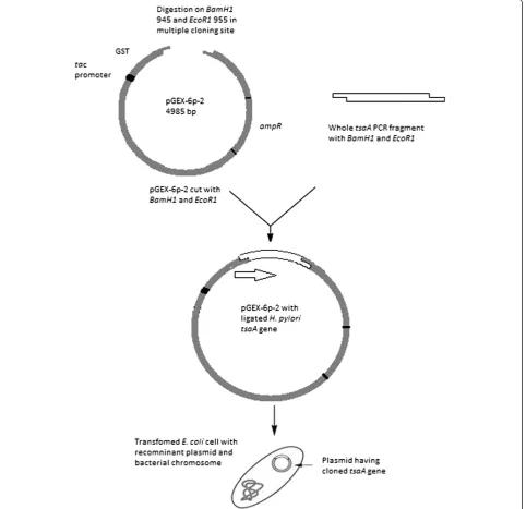
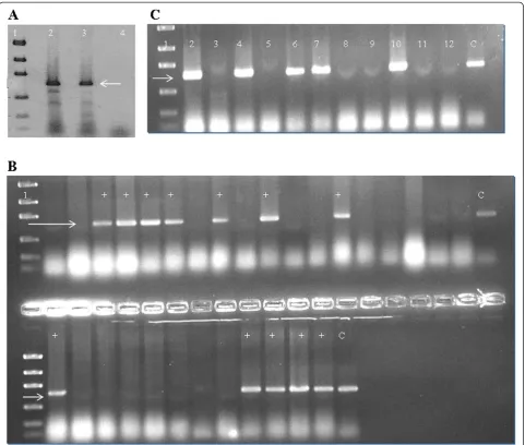
Polymerase chain reaction, cloning and transformation The full-length tsaAgene was amplified as described in the methods, digested with restriction enzymes and ligated withpGEX-6p-2that had been cut with the same enzymes to generate pGEX-tsaA. This plasmid places the GST coding sequences N-terminal to the TsaA/AhpC coding sequences and thus should generate rGST-AhpC fusion protein (Figure 1). pGEX-tsaA was used to trans-formE. coliDH10B to ampicillin resistance, and the cor-rect nature of pGEX-tsaA was verified using PCR and DNA sequencing (Figure 2).
Expression of target fusion protein
We next examined whether pGEX-tsaA would overex-press rGST-AhpC. For this experiment we transformed
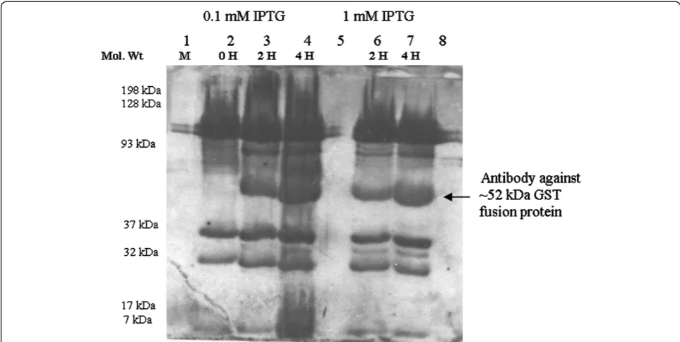
pGEX-tsaAinto theE. colistrain BL21 (DE3) that encodes a chromosomal T7 RNA Polymerase under the control of atacpromoter. Under IPTG induction, thetacpromoter gets activated that drives expression of the rGST-AhpC. We thus added IPTG at concentrations of 0.1 and 1 mmol/L to mid-log phase cultures of pGEX-tsa- BL21 (DE3) grown at 37 °C, and collected whole cell proteins for SDS-PAGE gel analysis. We found that pGEX-tsaA expressed robust amounts of rGST-AhpC that migrated at the expected size of 52 kDa (Figure 3). Further analysis demonstrated that this rGST-AhpC was mainly present as inclusion bodies, based on the observation that the protein was not soluble after cell lysis (Data not shown).
Immunoreactivity of GST-AhpC
Although it is well known that antibodies against AhpC are generated during H. pylori infection, we did not know if such antiserum would recognize a rGST-AhpC fusion protein. We thus tested whether a commercial rabbit antibody that had been generated against whole cell H. pylori would recognize our recombinant rGST-AhpC. We obtained robust recognition, confirming that this recombinant protein still displays critical antigenic determinants (Figure 4).
Purification of fusion protein
Target fusion protein rGST-AhpC was extracted from large volume LB-Amp culture (1 L) using repeat sonic-ation and washing followed by centrifugsonic-ation after each step. The samples were checked on 10-12% SDS-PAGE gel for protein quality. Pellet after third sonication and centrifugation step (Pellet-III) showed best results with a good intense band and little degradation/cleavage pro-ducts. Pellet-III sample was dialyzed and the super-natant, after dialysis was concentrated to approx. 3 ml. Right sized band for fusion protein was observed in case of both dialyzed and concentrated protein samples with no apparent degradation product as compared to pellet-III sample without dialysis and concentration (Figure 5).
Discussion
The study of virulence factors and important antigens of H. pylori is important to understand bacterial mechan-isms involved in persistent colonization and inflamma-tion. Prokaryotic expression systems for several important H. pylorivirulence factors have been success-fully constructed and recombinant fusion proteins or their purified proteins used in seroprevalence studies or screened as H. pylori vaccine candidates [22-24], how-ever, little attention has been paid towards protein pro-ducts of housekeeping genes liketsaA (ahpC).
Wang et al. (2005) reported that mutant H. pylori strains defective in AhpC were more sensitive to oxidative stress conditions compared to wild-type cells [25]. Also,
Olczak et al., (2003) reported that ahpC/tsaA mutant strains were unable to colonize mouse stomach [26]. An important feature of AhpC is that it seems to promote the activity of other proteins. Specifically, loss of AhpC resulted in partial inactivation of catalase, suggesting that AhpC is required for proper catalase activity [27].
At least two studies reported that antibodies against AhpC were found only in H. pylori infections that resulted in disease. Wang et al.,2004 found antibodies against AhpC in sera of peptic ulcer, cancer and
[image:3.595.59.542.91.558.2]adenocarcinoma patients infected with H. pylori[21]. In a recent report, Haunget al.,2011 examined protein ex-pression levels of AhpC inH. pylori from different clin-ical manifestations and observed 5-fold increase in AhpC protein expression in strains isolated from gastric cancer patients than those from gastritis [19]. This describes the potential of AhpC as an efficient biomarker to identify and risk patient population presenting differ-ent stages of dseases associated with chronic H. pylori infections.
Figure 1Schematic diagram of cloning and transformation oftsaAgene.WholetsaAgene havingBamH1andEcoR1sites was ligated with
Another important H. pylori antigen associated with disease is CagA. Patients infected with cagA+ H. pylori have higher risk of developing gastric cancers than those infected with cagA-strains [28,29]. Various studies have reported that 60 to 70% ofH. pyloristrains isolated from European and North American populations carried the cagAgene [30-32], whereas over 90% of the isolates from Asia-Pacific populations were found to becagA+[33-35]. The fact that nearly all strains of H. pylori from coun-tries like Taiwan and China as well as a high proportion in western countries arecagA positive makes it difficult to use cagA only as a marker for screening high-risk patients in these populations.
[image:4.595.58.540.88.496.2]ThetsaAgene fromH pyloristrain G27, cloned in this study, showed 100% nucleotide homology with published corresponding sequences. In the present study, SDS-PAGE demonstrated that the constructed expression sys-tems pGEX-6p-2-tsaA-BL21(DE3) efficiently produced the target recombinant GST fusion protein, however mainly presented in the form of inclusion body. How-ever, we successfully obtained free of inclusion body fu-sion protein using repeat sonication and centrifugation protocol which can be extended to subsequent refolding and cleavage from GST tag. The commercial rabbit anti-serum against the whole cell ofH. pylorirecognized and combined with rGST-AhpC as confirmed by Western Figure 2A. Target amplification fragments oftsaAgene.Lane 1: Low mass ladder (New England Biolab); Lanes 2 & 3: Target amplification fragments oftsaAgene (~600 bp) form G27 and positive control 26695; Lane 4: Blank control.B. PCR showingE. coliBL21 colonies after cloning, positive fortsaAgene by direct colony PCR method. Lane 1 is low mass ladder. Lanes with + sign show successful transformants.C. PCR fortsaA
using plasmid extracts of transformedE. coliBL21 colonies. Lane 1 is low mass ladder. Lanes with bands at target site (~600 bp) show successful transformants.
blot, indicating that the fusion protein had high immu-noreactivity. However, we could not explain presence of lower intensity bands other than that of the right sized band, both in SDS-PAGE (Figure 3B) and their subse-quent recognition in Western Blot (Figure 4). Perhaps these are E. coli BL21 (DE3) proteins which cross-reacted with the commercial rabbit antiserum against the whole cell of H. pylori. Several other approaches have been used for construction and use of prokaryotic expression system for potential H. pylori vaccine candidates likeflaA, flab, ureBandcagAby other groups [22-24].
Limitations of present study include use of GST fusion protein itself and not its cleaved purified product, and not testing the sera of potential patient groups to dem-onstrate the effectiveness of this approach due to some constraints.
Conclusions
[image:5.595.59.538.88.520.2]Immunoreactivity against rGST-AhpC was tested against commercial anti-H. pylori antibodies raised in rabbit serum. The fusion protein was then processed to get free from inclusion body. This recombinant fusion protein can serve as a potential diagnostic marker for screening advanced clinical manifestations due to chronicH. pylori infections. Such strategies are need of the time to iden-tify high risk patients keeping in view the genetic diver-sity of H. pylori regarding a well accepted biomarker CagA across the globe.
Methods Bacterial strains
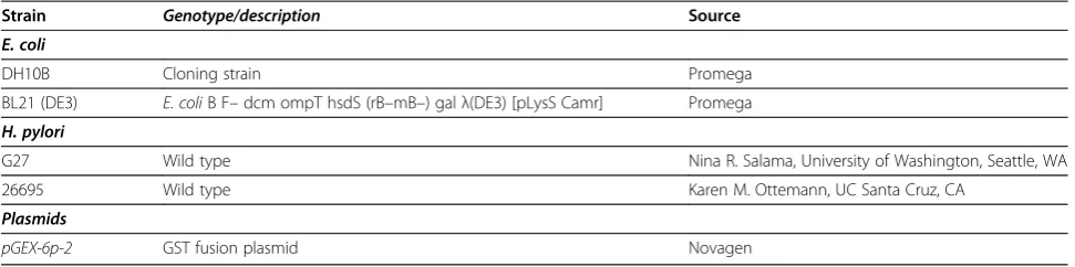
Bacterial strains used are given in Table 1. E. colistrain DH10B was used for cloning and BL21 (DE3) used for protein expression.
Growth media and chemicals
For solid-medium culture, H. pylori was grown on Columbia blood agar (Becton Dickinson) plates with 5% defibrinated horse blood (Hemostat Labs, Davis, CA), 5 μg of trimethoprim/ml, 8 μg of amphotericin B/ml, 10 μg of vancomycin/ml, 50 μg of cycloheximide/ml, 5 μg of cefsulodin/ml, 2.5 U of polymyxin B/ml, 0.2% (wt/vol) β-cyclodextrin (Sigma) (CHBA) at 37°C under conditions of 10% O2, 10% CO2 and 80% N2. All
anti-biotics were from Sigma or ISC Bioexpress. E. coli strains were grown on LB Agar or broth with Ampicillin and incubated on 37°C for overnight (LB-Amp). For long-term storage ofH. pyloristrains, 3–5 day growth of H. pylori was transferred to brucella broth having 10% FBS (BB10), 1% β-cyclodextrin, 5% dimethyl sulfoxide and 25% glycerol. The cells were frozen at −70°C after pipetting and vortexing.
DNA extraction
[image:6.595.56.538.89.331.2]Genomic DNA from 2–3 days culture ofH. pyloristrain G27 was extracted using Wizard genomic kit (Promega) according to manufacturer’s instructions.
Figure 4Western Blot showing detection of target ~52 kDa GST fusion protein by Rabbit antiH. pyloriantibodies as primary antibody. Lane 1: Kaleidoscope colour marker; Lanes 2–4: Protein samples induced inE. coliBL21 cells with 0.1 mM IPTG at 0, 2 and 4 hours respectively; Lanes 6–7: Protein samples induced with 1 mM IPTG; Lanes 5 & 8: Blank controls.
Figure 5SDS-PAGE showing different purification steps for fusion protein from pellet after sonication step III (P-III).Lane M: Kaleidoscope colour marker; Lane 1: Dialyzed pellet before
lyophilization; Lane 2: Pellet-III after lyophilization; Lane 3: Pellet-III before lyophilization: Lane 4: Dialyzed pellet after lyophilization; Lane 5: Dialyzed supernatant before lyophilization; Lane 6: Filtrate after concentration and Lane 7: Concentrated fusion protein.
[image:6.595.57.292.512.655.2]Primers
Primers were designed from genome sequence ofH. pyl-ori 26695 (HP1563) to amplify the full-length sequence of HP1563/tsaAusing the U.C. Santa Cruz archeal gen-ome browser (http://archaea.ucsc.edu). Forward primer
5′-CTCTGGATCCATGTTAGTTACAAAACTTGCC-3′
having restriction endonuclease site of BamH1 and re-verse primer 5′ -CTCTGAATTCTTAAAGCTTAATG-GAATTTTC-3′ with EcoR1 site were used to amplify full lengthtsaAgene fromH. pyloriG27 genomic DNA. Primers were synthesized by Eurofins MWG Operon, (Huntsville, USA).
Polymerase chain reaction
PCR reaction mixture contained 100 ng ofH. pyloriG27 genomic DNA, 2.5μl of dNTPs, 2.5 μl of 30 mM MgCl2
PCR buffer, 200 ng of each primer and 1μl of Taq poly-merase. Final volume was made to 25 μl with sterile water. Genomic DNA of H. pylori 26695 in same con-centration was used as positive control. PCR conditions were: Initial melting at 94°C for 3 minutes, then 30 cycles of repeat melting at 94°C for 40 sec, annealing at 52°C for 40 seconds and initial extension at 72°C for 30 seconds, then final extension at 72°C for 3 minutes and storage at 4°C until run on the gel. The results of PCR were observed under UV light after electrophoresis on 1.5% agarose gel stained with ethidium bromide. The expected size of tsaA amplification fragment was ~600 bp.
Sequencing
Sequencing of PCR products was done by the DNA se-quencing facility, UC Berkeley, CA. A sample containing 100 ng gel purified DNA and 0.8 pg of forward or re-verse primer was adjusted to a total volume of ~13μl in dH2O. Sequences obtained were subjected to nucleotide
BLAST feature of National Center for Biotechnology In-formation (NCBI) website (http://blast.ncbi.nlm.nih.gov/ Blast.cgi) for homology analyses with published tsaA sequence of G27.
Cloning of tsaA gene into plasmid vector
Gel purified tsaA whole gene was cloned into plasmid vectorpGEX-6p-2.Both thetsaAPCR product and plas-mid were digested with restriction endonucleases BamHI and EcoRI (New England Biolabs) at 37°C over-night. Digested DNA and plasmid were gel purified and concentration was determined by Nanodrop. The plas-mid sample was treated with Shrimp alkaline phosphat-ase (New England BioLabs). Ligation was done with 0.5
μl T4 ligase enzyme (New England Biolabs) by incubat-ing the ligation mix at 16°C for overnight. This plasmid was transformed into E. coli DH10B using electropor-ation; ampicillin-resistant colonies were screened using direct colony PCR using the original tsaA primers. One colony containing the correct plasmid was used to purify plasmid for sequencing.
Expression and identification of target recombinant protein
pGEX-Recombinant plasmid was transformed through electroporation intoE. coli BL21 (DE3) (Promega). One BL21 (DE3) colony of successful transformant contain-ing pGEX-tsaA was cultured in LB-Amp broth at 37°C until OD600of 0.5, after which IPTG was added at
con-centrations of 0.1 and 1.0 and mmol/L. Cultures were incubated for the times indicated in Figure 3A. Cells were then collected by centrifugation at 6000 X g force. The bacterial pellet was resuspended in 200–300 μl of 2x Laemmli sample buffer (1.52 g Tris-Base, 2 g SDS, 20 ml 100% glycerol, 2 mlβ-merceptoethanol and 1 mg bromophenol blue in dH2O to make 100 ml), and
re-suspended pellets were boiled for 5 minutes on water bath for denaturing proteins. SDS-PAGE (10%) was used to measure the molecular mass and output of the target recombinant fusion protein (rGST-AhpC).
Immunoreactivity of recombinant fusion protein
Immunoreactivity of the fusion protein was then deter-mined. Briefly, PVDF membrane (BioRad) cut to the size of the gel was pre-wet in 100% Methanol. Gel, sponge pads, 4 pieces of Whatman filter paper and MeOH-Table 1 Strains and plasmid used in the study
Strain Genotype/description Source
E. coli
DH10B Cloning strain Promega
BL21 (DE3) E. coliB F–dcm ompT hsdS (rB–mB–) galλ(DE3) [pLysS Camr] Promega
H. pylori
G27 Wild type Nina R. Salama, University of Washington, Seattle, WA
26695 Wild type Karen M. Ottemann, UC Santa Cruz, CA
Plasmids
[image:7.595.56.542.100.221.2]soaked membrane were soaked in the transfer buffer (1X SDS-PAGE Running Buffer, 20% MeOH) for 10 minutes. The cassettes were closed and placed in the module and contents of the gel transferred to membrane at 250 mA for 75 minutes. The commercial rabbit antiserum against whole-cellH pylori(Santa Cruz Biotech. USA) in 1:1000 dilution and HRP-labeling sheep antiserum against rabbit IgG in 1:7500 dilution (Santa Cruz Biotech, USA) were used as the first and second antibodies to deter-mine the immunoreactivity of rGST-AhpC by Western blotting, respectively. Results were visualized using lumi-nol and Biomax Light Film (Kodak).
Isolation of fusion protein from inclusion bodies
The fusion protein was isolated from inclusion bodies by using an online protocol (http://www.its.caltech.edu/ ~bjorker/Protocols) with some modifications. Briefly, culture from single colony of successful transformant grown in 1 L LB-Amp broth was centrifuged (Sorvall RC5C Plus, Thermo Fischer Scientific, Rockford, USA) at 7000 Xgfor 20 minutes at 4°C. Resultant supernatant was carefully decanted and pellet was resuspended in 13 ml of solution buffer (50 mM Tris–HCl, 25% sucrose, 1 mM NaEDTA, 0.1% Sodium azide and 10 mM DTT) on ice and sonicated (Sonic Dismembrator, Fischer Sci-entific, Rockford, USA) at 50% amplitude for 4–5 times on ice. Then 100μl Lysozyme, 250μl DNase I and 50μl MgCl2 were mixed with 12.5 ml of lysis buffer (50 mM
Tris- HCl, 1% Triton X-100, 1% Na deoxycholate, 100 mM NaCl, 0.1% Sodium azide and 10 mM DTT) and sample was incubated for 45 minutes at room temperature. In next step, 350μl of NaEDTA was added to the sample and freezed in liquid nitrogen. The sample was then thawed at 37°C for 30 minutes. 200 μl MgCl2
solution was added to the solution and placed at room temperature for an hour. Then 350 ml NaEDTA was added again to the solution and placed on ice. This solu-tion was centrifuged at 11000 Xgat 4°C for 20 minutes and supernatant was discarded in a tube. The pellet was washed with 10 ml washing buffer (50 mM Tris–HCl, 0.5% Triton X-100, 100 mM NaCl, 1 mM NaEDTA, 0.1% Sodium azide and 1 mM DTT) and sonicated on ice. Solution was spun again at 11000 Xgfor 20 minutes at 4°C and supernatant was discarded in a tube. Pellet was again resuspended in 10 ml washing buffer but this time without Triton X-100 and sonicated. The same procedure was repeated for third time. Samples from both supernatant and pellet after each centrifugation step were retained for checking protein quality. In the last step, pellet was dissolved in 5 ml of 8 M Guanidi-nium solution (pH 8.0) in the presence of 4 mM DTT and shaken at room temperature till complete dissol-ution. The dissolved sample was stored at−80°C.
Dialysis and concentration
Solution was diluted to 50 ml with minimal protein buf-fer (150 mM NaCl and 50 mM Tris–HCl at pH 8.0) and dialyzed against 1 L of MPB using SnakeSkin dialysis tubing (Thermo Fischer Scientific, Rockford, USA) with gentle shaking at 4 °C. After 2 hours of shaking, the buf-fer was changed and then dialyzed overnight. Solution was pelleted at 3000 X g for 20 minutes at 4 °C. The re-sultant supernatant was concentrated to about 3 ml vol-ume using Amicon Ultra-15 concentrator (Millipore, USA) following manufacturer’s instructions. Both con-centrated and dialyzed pellets were analyzed for proteins on SDS-PAGE. Both the pellet and concentrate were lyo-philized (FreeZone 2.5 Freeze Dry Systems, Labconco, Kansas, USA) for long term storage and further use.
Competing interests
The authors declare no competing interests.
Authors’contributions
KM conceived the study, carried out the experimental work and prepared the initial draft of the manuscript. FH helped in result interpretation and preparation of final draft. All authors read and approved the final manuscript.
Acknowledgment
We are thankful to Pakistan Higher Education Commission (HEC) for funding this research work, and Karen M. Ottemann (UC Santa Cruz, CA) for providing lab space and technical advice.
Author details
1Department of Microbiology, Quaid-i-Azam University, Islamabad 45320, Pakistan.2Microbiology and Environmental Toxicology Department, University of California Santa Cruz, Santa Cruz, CA 95064, USA.3Department of Pharmacy, Hazara University, Havelian, Abbottabad, Pakistan.
Received: 29 February 2012 Accepted: 21 June 2012 Published: 25 June 2012
References
1. Brown LM:Helicobacter pylori: epidemiology and routes of transmission.
Epidemiol Rev2000,22(2):283–297.
2. Nakamura S, Matsumoto T, Suekane H, Takeshita M, Hizawa K, Kawasaki M, Yao T, Tsuneyoshi M, Iida M, Fujishima M:Predictive value of endoscopic ultrasonography for regression of gastric low grade and high grade MALT lymphomas after eradication of Helicobacter pylori.
Gut2001,48(4):454–460.
3. Uemura N, Okamoto S, Yamamoto S, Matsumura N, Yamaguchi S, Yamakido M, Taniyama K, Sasaki N, Schlemper RJ:Helicobacter pylori infection and the development of gastric cancer.N Engl J Med2001,345(11):784–789. 4. Morgner A, Bayerdorffer E, Neubauer A, Stolte M:Gastric MALT lymphoma
and its relationship to Helicobacter pylori infection: management and pathogenesis of the disease.Microsc Res Tech2000,48(6):349–356. 5. Kate V, Ananthakrishnan N, Badrinath S:Effect of Helicobacter pylori
eradication on the ulcer recurrence rate after simple closure of perforated duodenal ulcer: retrospective and prospective randomized controlled studies.Br J Surg2001,88(8):1054–1058.
6. Nizami SQ, Bhutta ZA, Weaver L, Preston T:Helicobacter pylori colonization in infants in a peri-urban community in Karachi, Pakistan.
J Pediatr Gastroenterol Nutr2005,41(2):191–194.
7. Seyler RW Jr, Olson JW, Maier RJ:Superoxide dismutase-deficient mutants of Helicobacter pylori are hypersensitive to oxidative stress and defective in host colonization.Infect Immun2001,69(6):4034–4040. 8. Basu M, Czinn SJ, Blanchard TG:Absence of catalase reduces long-term
survival of Helicobacter pylori in macrophage phagosomes.Helicobacter
2004,9(3):211–216.
and functional analysis of a novel gene product designated‘ KatA-associated protein’, KapA (HP0874).Microbiology2002,148(Pt 12):3813–3825. 10. Baker LM, Raudonikiene A, Hoffman PS, Poole LB:Essential
thioredoxin-dependent peroxiredoxin system from Helicobacter pylori: genetic and kinetic characterization.J Bacteriol2001,183(6):1961–1973.
11. Wang G, Olczak AA, Walton JP, Maier RJ:Contribution of the Helicobacter pylori thiol peroxidase bacterioferritin comigratory protein to oxidative stress resistance and host colonization.Infect Immun2005,73(1):378–384. 12. Wang G, Maier RJ:An NADPH quinone reductase of Helicobacter pylori
plays an important role in oxidative stress resistance and host colonization.Infect Immun2004,72(3):1391–1396.
13. Evans DJ Jr, Evans DG, Takemura T, Nakano H, Lampert HC, Graham DY, Granger DN, Kvietys PR:Characterization of a Helicobacter pylori neutrophil-activating protein.Infect Immun1995,63(6):2213–2220. 14. Tomb JF, White O, Kerlavage AR, Clayton RA, Sutton GG, Fleischmann RD,
Ketchum KA, Klenk HP, Gill S, Dougherty BA,et al:The complete genome sequence of the gastric pathogen Helicobacter pylori.Nature
1997,388(6642):539–547.
15. Alm RA, Ling LS, Moir DT, King BL, Brown ED, Doig PC, Smith DR, Noonan B, Guild BC, deJonge BL,et al:Genomic-sequence comparison of two unrelated isolates of the human gastric pathogen Helicobacter pylori.
Nature1999,397(6715):176–180.
16. Chalker AF, Minehart HW, Hughes NJ, Koretke KK, Lonetto MA, Brinkman KK, Warren PV, Lupas A, Stanhope MJ, Brown JR,et al:Systematic identification of selective essential genes in Helicobacter pylori by genome prioritization and allelic replacement mutagenesis.J Bacteriol
2001,183(4):1259–1268.
17. Storz G, Jacobson FS, Tartaglia LA, Morgan RW, Silveira LA, Ames BN:An alkyl hydroperoxide reductase induced by oxidative stress in Salmonella typhimurium and Escherichia coli: genetic characterization and cloning of ahp.J Bacteriol1989,171(4):2049–2055.
18. Chuang MH, Wu MS, Lo WL, Lin JT, Wong CH, Chiou SH:The antioxidant protein alkylhydroperoxide reductase of Helicobacter pylori switches from a peroxide reductase to a molecular chaperone function.Proc Natl Acad Sci U S A2006,103(8):2552–2557.
19. Huang CH, Chuang MH, Lo WL, Wu MS, Wu YH, Wu DC, Chiou SH:
Alkylhydroperoxide reductase of Helicobacter pylori as a biomarker for gastric patients with different pathological manifestations.Biochimie
2011,93(7):1115–1123.
20. O’Toole PW, Logan SM, Kostrzynska M, Wadstrom T, Trust TJ:Isolation and biochemical and molecular analyses of a species-specific protein antigen from the gastric pathogen Helicobacter pylori.J Bacteriol1991,
173(2):505–513.
21. Wang JT, Chang CS, Lee CZ, Yang JC, Lin JT, Wang TH:Antibody to a Helicobacter pylori species specific antigen in patients with adenocarcinoma of the stomach.Biochem Biophys Res Commun1998,
244(2):360–363.
22. Yan J, Liang SH, Mao YF, Li LW, Li SP:Construction of expression systems for flaA and flaB genes of Helicobacter pylori and determination of immunoreactivity and antigenicity of recombinant proteins.World J Gastroenterol2003,9(10):2240–2250.
23. Yan J, Wang Y, Shao SH, Mao YF, Li HW, Luo YH:Construction of prokaryotic expression system of ltB-ureB fusion gene and identification of the recombinant protein immunity and adjuvanticity.World J Gastroenterol2004,10(18):2675–2679.
24. Yan J, Wang Y, Shao SH, Mao YF, Li HW, Luo YH:Construction of prokaryotic expression system of 2 148-bp fragment from cagA gene and detection of cagA gene, CagA protein in Helicobacter pylori isolates and its antibody in sera of patients.World J Gastroenterol2004,
10(8):1183–1190.
25. Wang G, Conover RC, Olczak AA, Alamuri P, Johnson MK, Maier RJ:
Oxidative stress defense mechanisms to counter iron-promoted DNA damage in Helicobacter pylori.Free Radic Res2005,39(11):1183–1191. 26. Olczak AA, Seyler RW Jr, Olson JW, Maier RJ:Association of Helicobacter
pylori antioxidant activities with host colonization proficiency.Infect Immun2003,71(1):580–583.
27. Wang G, Conover RC, Benoit S, Olczak AA, Olson JW, Johnson MK, Maier RJ:
Role of a bacterial organic hydroperoxide detoxification system in preventing catalase inactivation.J Biol Chem2004,279(50):51908–51914. 28. Blaser MJ, Perez-Perez GI, Kleanthous H, Cover TL, Peek RM, Chyou PH,
Stemmermann GN, Nomura A:Infection with Helicobacter pylori strains
possessing cagA is associated with an increased risk of developing adenocarcinoma of the stomach.Cancer Res1995,55(10):2111–2115. 29. Parsonnet J, Friedman GD, Orentreich N, Vogelman H:Risk for gastric cancer in people with CagA positive or CagA negative Helicobacter pylori infection.Gut1997,40(3):297–301.
30. Tummuru MK, Cover TL, Blaser MJ:Cloning and expression of a high-molecular-mass major antigen of Helicobacter pylori: evidence of linkage to cytotoxin production.Infect Immun1993,61(5):1799–1809. 31. Covacci A, Censini S, Bugnoli M, Petracca R, Burroni D, Macchia G, Massone A,
Papini E, Xiang Z, Figura N,et al:Molecular characterization of the 128-kDa immunodominant antigen of Helicobacter pylori associated with cytotoxicity and duodenal ulcer.Proc Natl Acad Sci U S A1993,90(12):5791–5795. 32. Figueiredo C, Van Doorn LJ, Nogueira C, Soares JM, Pinho C, Figueira P,
Quint WG, Carneiro F:Helicobacter pylori genotypes are associated with clinical outcome in Portuguese patients and show a high prevalence of infections with multiple strains.Scand J Gastroenterol2001,36(2):128–135. 33. Ito Y, Azuma T, Ito S, Miyaji H, Hirai M, Yamazaki Y, Sato F, Kato T, Kohli Y,
Kuriyama M:Analysis and typing of the vacA gene from cagA-positive strains of Helicobacter pylori isolated in Japan.J Clin Microbiol1997,35(7):1710–1714. 34. Park SM, Park J, Kim JG, Yoo BC:Relevance of vacA genotypes of
Helicobacter pylori to cagA status and its clinical outcome.Korean J Intern Med2001,16(1):8–13.
35. Yang H, Wu SV, Pichuantes S, Song M, Wang J, Zhou D, Xu Z, Quan S, Polito A, Walsh JH:High prevalence of cagA-positive strains in Helicobacter pylori-infected, healthy, young Chinese adults.J Gastroenterol Hepatol
1999,14(5):476–480. doi:10.1186/1756-0500-5-328
Cite this article as:Mehmood and Hasan:Construction and use of a prokaryotic expression system forHelicobacter pyloriAhpC.BMC Research
Notes20125:328.
Submit your next manuscript to BioMed Central and take full advantage of:
• Convenient online submission
• Thorough peer review
• No space constraints or color figure charges
• Immediate publication on acceptance
• Inclusion in PubMed, CAS, Scopus and Google Scholar
• Research which is freely available for redistribution