R E S E A R C H A R T I C L E
Open Access
Parity predisposes breasts to the oncogenic
action of PAPP-A and activation of the
collagen receptor DDR2
Elizabeth Slocum
1,3, Amanda Craig
2,3, Augusto Villanueva
2,3and Doris Germain
1,3*Abstract
Background:Women who had children at a young age (less than 25) show a reduced overall risk of breast cancer. However, epidemiological studies showed that for all other women, pregnancy increases the risk of breast cancer and the risk remains higher for decades. Further, even in women who had children at a young age, there is a transient increase risk that peaks 6 years after pregnancy. Women diagnosed with breast cancer following pregnancy show a higher rate of metastasis. Yet, the factors that increase the predisposition of post-partum breasts to more aggressive cancers remain unknown. Pregnancy-associated plasma protein A (PAPP-A) is a secreted protease that is overexpressed in more than 70% of breast cancers. However, PAPP-A is a collagen-dependent oncogene. We initiated this study to test the effect of PAPP-A on the predisposition of post-partum breasts.
Methods:We used PAPP-A mouse models for the analysis of its effect on virgin, involuting, or post-partum mammary glands. We performed second-harmonic generation microscopy for the analysis of collagen, defined tumor-associated collagen signature (TACS), the rate of mammary tumors, and the status of the collagen-DDR2-Snail axis of metastasis. We knockdown DDR2 by CRISPR and performed invasion assays. A transcriptomic approach was used to define a PAPP-A and parity-dependent genetic signature and assess its correlation with breast cancer recurrence in humans. Results:We confirmed that post-partum mammary glands have a higher level of collagen than virgin glands and that this collagen is characterized by an anti-proliferative architecture. However, PAPP-A converts the anti-proliferative post-partum collagen into pro-tumorigenic collagen. We show that PAPP-A activates the collagen receptor DDR2 and metastasis. Further, deletion of DDR2 by CRISPR abolished the effect of PAPP-A on invasion. We defined a PAPP-A-driven genetic signature that identifies patients at higher risk of metastasis.
Conclusions: These results support the notion that information about pregnancy may be critical in the prognosis of breast cancer as passage through a single pregnancy predisposes to the oncogenic action of PAPP-A. Our data indicate that history of pregnancy combined with the expression of PAPP-A-driven genetic signature may be useful to identify patients at higher risk of metastatic disease.
Keywords: Pregnancy-associated breast cancer, DDR2, Parity, Pregnancy-associated plasma protein A, Involution, Insulin-like growth factor (IGF) signaling, Collagen, TACS, LARP6
© The Author(s). 2019Open AccessThis article is distributed under the terms of the Creative Commons Attribution 4.0 International License (http://creativecommons.org/licenses/by/4.0/), which permits unrestricted use, distribution, and reproduction in any medium, provided you give appropriate credit to the original author(s) and the source, provide a link to the Creative Commons license, and indicate if changes were made. The Creative Commons Public Domain Dedication waiver (http://creativecommons.org/publicdomain/zero/1.0/) applies to the data made available in this article, unless otherwise stated. * Correspondence:doris.germain@mssm.edu
1
Department of Medicine, Division of Hematology/Oncology, Tisch Cancer Institute, Icahn School of Medicine at Mount Sinai, New York, NY, USA 3Graduate School of Biomedical Sciences, Icahn School of Medicine at Mount
Sinai, New York, NY, USA
Background
Diverse factors contribute to the etiology of breast can-cer, including parity and breastfeeding [1–5]. Pregnancy however has a dual effect on breast cancer risk. On one hand, at a young age (before the age of 25), pregnancy confers a lifelong protective effect [1]. On the other hand, pregnancy is associated with a transient risk of de-veloping breast cancer in all women, and this risk peaks 6 years after birth [1–4]. In fact, a recent meta-analysis combining the results of 15 studies revealed that women remain at higher risk of breast cancer following preg-nancy for more than 20 years, suggesting that healthcare providers should consider parity as a risk factor [6]. Fur-thermore, the relative risk directly correlates with the age of the mother. Notably, women above the age of 35 at the time of their first pregnancy remain at higher risk of developing breast cancer compared to nulliparous women for over three decades after pregnancy [1, 2].
Therefore, contrary to the current definition of
pregnancy-associated breast cancer (PABC), which is empirically restricted to breast cancers diagnosed within the first 2 years post-partum, the diagnosis of PABC vs. non-PABC is very challenging [1–4].
As women in developed countries are steadily delaying childbearing to a later age, PABC incidence is expected to drastically increase, yet the link between pregnancy and breast cancer remains a poorly understood area of breast cancer research [3, 4, 7]. This is particularly sig-nificant as PABC are classically more aggressive than non-PABC. PABC patients typically exhibit higher rates of recurrence, metastasis, and poorer survival rates com-pared to age-, stage-, and hormone-matched nulliparous controls [1, 8, 9]. These observations highlight the need for reliable biomarkers and a deeper understanding of the molecular underpinnings of PABC.
Recent breakthroughs have identified involution as a pro-tumorigenic phase of breast development [8–10]. Involution is a transient event that is initiated by the cessation of lactation and which resets the mammary tis-sue architecture to a non-pregnant, post-partum state. Involution involves a drastic remodeling of the breast through a complex coordination of selected epithelial cell apoptosis, extracellular matrix remodeling, and adi-pogenesis. The signaling pathways involved share similar characteristics to wound healing and tumor-promoting microenvironments [10,11].
One key feature contributing to the pro-tumorigenic ef-fect of involution is the increased deposition of collagen [8,9,12]. High collagen abundance and stromal stiffening can disrupt the homeostasis of the extracellular matrix and are associated with an increased risk for breast cancer, cell invasion, and metastasis [13–20]. Importantly, the col-lagen of an involuting breast transiently adopts an altered architecture, referred to as tumor-associated collagen
signature 3 (TACS3) [8,13]. In contrast to normal colla-gen, TACS3 is characterized as fibrillar, linearized collagen fibers perpendicular to the tumor or ductal border [8,13]. TACS can be further classified as TACS1, 2, or 3 depend-ing on the angle of the collagen fiber [13]. TACS3 is asso-ciated with worst prognosis and most advanced stage of breast cancer [13,17].
A post-partum breast, defined as a breast after the completion of involution and therefore does not have any of the wound healing characteristics of the invo-luting breast, retains the high collagen content ob-served in involution and is therefore different from breasts that have never gone through pregnancy [8,
14]. Unlike involution-collagen, however, post-partum collagen adopts a TACS1 architecture. Unlike TACS3, TACS1 collagen is anti-proliferative and has been postulated to contribute to the protective effect of pregnancy [13]. These observations raise the
possibil-ity that oncogenic events able to convert the
post-partum collagen into an involution-like collagen may predispose post-partum breasts to the develop-ment of breast cancer.
Pregnancy-associated plasma protein A (PAPP-A) or pappalysin is a secreted protease that is widely overex-pressed in breast cancer, and therefore, we seek to define
its role in breast cancer [21]. We reported that
patients at higher risk of metastasis and is characterized by a profound remodeling of the extracellular matrix.
Methods
Animal procedures
Mice overexpressing PAPP-A in the mammary gland (MMTV-PAPP-A) in FVB/n background were generated as previously described (Takabatake et al). The animals were sacrificed, and the tissues were stored at −80 °C for bio-chemical analyses. For the post-partum time course experi-ments, 2-month-old nulliparous FVB/n non-transgenic and PAPP-A transgenic females (n= 20 mice per type, 5 mice per group) were bred and subsequently housed separately post-fertilization. All females were kept with their litter for either a 2-day- or 2-week-long lactation period. Involution mice were sacrificed 12 days post-weaning, and late post-partum mice were sacrificed 2 months post-weaning. The mammary glands were harvested for histological ana-lyses or as RNA and protein samples at−80 °C.
Histology and immunohistochemistry
Histological slides were prepared as 4μm formalin-fixed sections embedded in paraffin and stained with H&E by the Oncological Sciences Department Histology core facil-ity at Icahn School of Medicine at Mount Sinai. For im-munostaining of paraffin-embedded sections, the samples were deparaffinized in xylene and rehydrated in water. Antigen retrieval was performed using citrate buffer pH 6 for 30 min at 95 °C. Peroxidase blocking was completed using endogenous peroxidase solution (Dako Dual En-dogenous Enzyme Block #S20003) at room temperature for 15 min. Antibody Diluent (MP Biomedicals Normal Antibody Diluent #980631) was used for serum blocking for 30 min at room temperature. All buffer washes be-tween incubations were completed with TBS. Primary antibody (1:100 Snail, Cell Signaling Technology L70G2 #3895) incubation was performed overnight at 4 °C in an enclosed moist chamber. Biotinylated mouse antibody (Jackson ImmunoResearch #115-065-205) diluted 1:200 in TBS for 1 h at room temperature was used for the second-ary antibody incubation. Washes between antibody
incu-bations were done with TBS with .04% Tween.
Streptavidin peroxidase (Vector Labs #SA-5004) was incu-bated for 30 min at room temperature, followed by a TBS with .04% Tween and TBS wash. The slides were devel-oped using AEC (Vector Labs #SK-4200) for 15 min at room temperature and mounted using VectaMount (Vec-tor Labs #H-5501). Images were acquired using a Zeiss AX10 light microscope and scored according to the pa-rameters shown.
Second-harmonic generation microscopy
All mouse mammary gland second-harmonic generation (SHG) images were captured on an Olympus FV1000MPE
Fluoview multiphoton microscope (Tokyo, Japan) in the Microscopy CoRE at the Icahn School of Medicine at Mount Sinai, supported with funding from NIH Shared Instrumentation Grant (1S10RR026639-01). Image acqui-sition was done using an Olympus XLPlanN 25×/1.05 nu-merical aperture water immersion lens with the excitation wavelength set at 900 nm. Imaging specs included Coher-ent Chameleon Vision II Ti:S laser (Santa Clara, CA) with a 680- to 1080-nm tuning range, dispersion compensation, and 140 fs pulse width. Signal collection was performed by backward imaging with a 420- to 460-nm band-pass filter, a 485 dichroic mirror (GR/XR filter cube, Olympus), and an external detector. Images were acquired at a 512 × 512 pixel resolution with consistent power levels. Protocol for collagen intensity vs. distance analysis was adapted from Lyons et al. and performed in six replicate ducts per mouse in five mice per group (n= 30 ducts per group). SHG images were opened in ImageJ (version 1.46r), and a rectangular selection of 50 × 100 pixels was made border-ing the ductal edge in four compass directions. Each re-gion of interest (ROI) was analyzed using the profile plot function, and intensity of the SHG signal over a 40-μM distance was averaged as the signal for a single duct.
Tumor-associated collagen signature analysis
SHG images of the mammary ducts and tumors (acquired as described in the“Second-harmonic generation micros-copy” section) were imported into the curvelet-based alignment analysis software CurveAlign (version 2.2 R2012a), available through the Laboratory of Optical and Computational Instrumentation at the University of Wisconsin-Madison. The borders of the mammary ducts and tumors were manually indicated, and the CurveAlign software measured the angle and quantity of collagen fi-bers with respect to the designated borders within a preset pixel distance. The individual curvelet angles were re-ported as an output value between 0 and 90. Based on prior literature, angles between 60 and 90 were character-ized as a positive TACS3 region, angles between 30 and 60 as TACS2, and angles 0–30 as TACS1. The number of in-dividual collagen fibers having an angle of interaction within each of the three signatures was counted and nor-malized to the total number of collagen fibers assessed within an ROI by the software.
Masson’s trichrome collagen stain and analysis
stain was converted to black using Adobe Photoshop CS5’s (version 12.0 × 64) magic wand tool. The images were imported into ImageJ (version 1.46r), and the black selections were quantified as percent area positive for collagen. The images were normalized against the per-iphery of the duct epithelia in which the final ratio rep-resents collagen/epithelia.
Cell culture and treatment conditions
MCF-7PAPP-A and MCF-7PAPP-AE483Q cells were
gen-erated as previously described (Takabatake et al).
MCF-7, MCF-7PAPP-A, MCF-7PAPP-A/DDR2 KO, and
MCF-7PAPP-AE483Q cells were grown in Dulbecco’s
modified Eagle’s medium (DMEM) supplemented
with 10% fetal bovine serum and 1% penicillin/
streptomycin at 37 °C/5% CO2. rIGF-1 (Preprotech,
#100-11) treatments were done in serum-free media at 10 nM for indicated time points. Imatinib (R&D Systems, #59-061-00) and PQ401 (Enzo Life Sciences ALX-270-459-M005) treatments were performed at indicated concentrations in complete media for 48 h, and untreated samples were treated with vehicle control (DMSO) for 48 h. PAPP-A- and PAPP-A
E483Q-conditioned media treatments were
per-formed at 10 ng/mL in complete media for 24 h. Cells treated with conditioned media were seeded on collagen I-coated plates 24 h prior to treatment. Preparation of collagen-coated plates was performed
as follows: type I rat tail collagen (Corning,
#354236) was diluted at 50μg/mL in 0.1 nM HCl
and incubated on plates at room temperature for 1 h followed by two PBS washes.
Media collection and PAPP-A ELISA
MCF-7PAPP-A and MCF-7PAPP-AE483Qcells were grown to 80% confluence in complete media. Media were then switched to serum-free and collected 24 h later, and PAPP-A protein was concentrated from media using 50 kDa cutoff Amicon Ultra-15 Centrifugal Filter Units (EMD Millipore, #UFC905024) spun at 5000g for 20 min at 4 °C. Concentrated condition media were then stored at
−20 °C, used for rIGFBP-5 protease assay, or quantified by ELISA. PAPP-A concentration was measured using Quantikine PAPP-A ELISA (R&D Systems, # DPPA00) following the manufacturer’s protocol adapted with a pri-mary incubation for 18 h at 4 °C and PAPP-A conjugate incubation for 6 h at room temperature.
IGFBP-5 protease assay
Cell-free protease assays were performed using PAPP-A protein secreted in 24 h serum-free culture media from MCF-7 PA and MCF-7 PA E483Q cells. Culture media from parental MCF-7 were used as a negative control for PAPP-A protein in cell media. Media were collected
and concentrated as described above. Fifty nanograms of rIGFBP-5 (Abcam, #ab49835) was co-incubated with 15μL of concentrated media and 15μL of DMEM for 3 h at 37 °C. The reaction was inactivated by the addition of 1× Laemmli sample buffer to 40μL final volume and boiling for 5 min. Degradation of IGFBP-5 in samples was then detected by western blot.
Immunoblot
Cell lysates were prepared in RIPA lysis buffer (25 mM Tris HCl, pH 7.6, 150 mM NaCl, 1% NP-40, 0.1% SDS, 1% sodium deoxycholate) supplemented with protease and phosphatase inhibitors. Thirty mi-crograms total protein in 1× Laemmli buffer per sample was separated on a 10% SDS-glycine poly-acrylamide gels ran at 80 V for 30 min and 200 V for 45 min. Proteins were transferred to nitrocellulose membranes (GE Healthcare) for 1.5 h at 85 V. Mem-branes were blocked in 5% milk in TBS-T and incubated on a rotator overnight at 4 °C in the fol-lowing primary antibodies: DDR2 1:250 (Cell Signal-ing Technology #12133S), p-DDR2 Y740 1:600 (R&D Systems, #MAB25382), Snail 1:1000 (Cell Signaling Technology L70G2 #3895), Larp6 1:600 (Abnova, #H00055323-B01P), IGFBP-5 1:1000 (EMD Millipore, #06-110), PAPP-A 1:200 (Santa Cruz Biotechnology, # sc-50518), phospho-Akt (Ser473) 1:1000 (Cell Sig-naling Technology 587F11 #4051), Akt 1:10,000 (Cell Signaling Technology #9272), and actin 1:10,000 (EMB Millipore #MAB1501R). After three washes in TBS-T, and 10-min 10% milk/TBS-T incubation, the membranes were incubated in secondary anti-rabbit or anti-mouse antibodies conjugated to horseradish peroxidase (Thermo Fisher Scientific or KwikQuant) in 10% milk/TBS-T for 1 h at room temperature.
Sig-nal was detected using ECL (GE Healthcare,
#RPN2106 or KwikQuant) following the
manufac-turer’s protocols. TBS-T was prepared with 0.1%
Tween.
RNA isolation and RT-qPCR
Transwell in vitro invasion assay
Cell permeable membrane inserts (Falcon, #08-771-21)
were coated with 200μL of diluted 1:100 growth
factor-reduced Matrigel basement membrane matrix (Corning, #356230) in cold PBS. Inserts were placed in 24-well plates (Falcon), and artificial extracellular matrix was allowed to polymerize for 2 h at room temperature. Following polymerization incubation, excess Matrigel so-lution was removed by pipette. Fifty thousand cells in 500μL of serum-free DMEM were seeded atop the poly-merized Matrigel in the inserts. Five hundred microliters of complete DMEM containing 10% FBS was added to each lower chamber. Invasion assays were incubated at 37 °C/5% CO2. After 48 h, invaded cells were fixed and
stained using the Hema 3 Manual Staining Stat Pack (Thermo Fisher Scientific, #23-123-869) according to the manufacturer’s protocol and mounted using Per-mount toluene solution (Thermo Fisher Scientific). All experiments were done in technical triplicates.
Xenografts using MCF-7, MCF-7PAPP-A, and MCF-7PAPP-A
DDR2 KO
cells
A week prior to cell injection, a 60-day release 0.5 mg
β-estradiol 17-acetate pellet (Innovative Research of America, #SE-271) was inserted in the cervical region of virgin female athymic nude mice aged 2 months. Using a 25-G needle, 2.5 million cells (MCF-7 parental or MCF-)
in 100μL of Matrigel basement membrane matrix
(Corning, #354234) were injected in the left inguinal mammary fat pads and 2.5 million cells in 100μL of 1:1 Matrigel/type 1 collagen mixture (collagen: Corning, #354236) were injected in the right inguinal mammary fat pads (n= 5 mice, 10 tumors total). Palpable tumors were measured by caliper 3 times per week for 3 weeks beginning 14 days post-injection. Final tumor volumes were recorded by weight, and tumors were harvested for histological analyses or as RNA and protein samples at
−80 °C. The lungs were collected for histological analysis following perfusions with sterile PBS and 10% formalin.
MTT assay
MCF-7 and MCF-7PAPP-Acells were seeded at 50% con-fluence and treated with imatinib and PQ401 at the indi-cated time points in triplicate samples for 48 h. Following treatment, complete media with MTT at 0.5
mg/mL was added to each well and incubated at 37 °C for 3 h protected from light. After incubation, and DMSO was used as MTT solvent and each will be mixed thoroughly to homogenize the color and subsequently read at absorbance OD = 570 nm. The samples were av-eraged and normalized to the vehicle control sample.
Met1 xenograft and conditioned media injections
Two-month-old virgin female FvBN mice were bred and then housed separately post-fertilization. Pregnant mice were checked daily for litter drop and then underwent a lactation period of 2 days. Mice were injected with met1
cells at a time point of post-partum 2 months
post-lactation. Met1 cells were kindly provided by Dr. Emily Gallagher at Mount Sinai. Two hundred fifty
thousand met1 cells in 100μL of control MCF-7- or
PAPP-A-conditioned media at 0.1 ng/μL were injected into the upper right and left inguinal mammary fat pads (n = 5 mice, 6 tumors per group). The lower left and right inguinal mammary fat pads received injections of MCF-7-concentrated media or concentrated PAPP-A from MCF-7PAPP-Amedia at 0.1 ng/μL alone (n= 5 mice, 6 mammary glands per group, PAPP-A concentrated as described above). Subsequently, every 2 days for 3 weeks following the initial injection, 100μL of control media or 0.1 ng/μL PAPP-A containing media was injected into the upper and lower left and right inguinal mammary fat pads or Met1 xenografts. All injections were done using a 25-G needle. Final tumor volume was recorded by weight, and all tumors and mammary glands were har-vested for histological analysis. The lungs were collected for histological analysis following perfusions with sterile PBS and 10% formalin. Clusters of cancer cells were de-tected by H&E and counted as micrometastases.
Gene set analysis
Publicly available primary breast cancer gene expression and phenotype data published by Kao et al. (GSE20685)
was downloaded from Gene Expression Omnibus [27].
PAPP-A/SNAI1/COL1A1 gene signature was composed of the probes for each gene with the highest average ex-pression across all samples. The probes used for each
gene are as follows: PAPP-A (201981_at), SNAI1
(219480_at), andCOL1A1 (202310_s_at).The secondary validation dataset in Additional file4: Figure S4 was col-lected from Chanrion et al. (GSE9893), and the probes
used werePAPP-A: H200006224,COL1A1: H200008097,
and SNAI1: H200004861 [28]. Normalized expressions of PAPP-A/SNAI1/COL1A1 were averaged to calculate an expression score for each patient (n= 327). The top 50% of patients with the highest expression scores were
designated as PAPP-A/SNAI1/COL1A1 high (n= 163),
and the bottom 50% were designated asPAPP-A/SNAI1/
COL1A1 low (n= 164). Differential gene expression
Gene Forward Reverse
Human DDR2 5′-TCA CCC AGA CCC ATG AAT AC-3′
5′-GGG AAG GAA ATG GCA TTA GG-3′
Human Col1a1 5′-TCT GCG ACA ACG GCA AGG TG-3′
5′-GAC GCC GGT GGT TTC TTG GT-3′
Humanβ-actin 5′-ATC CTC ACC CTG AAG TAC CC-3′
between the two groups of patients was calculated using by LIMMA [29]. Genes with a false discovery rate (FDR) less than 0.05 were ranked by Log2fold change and used
for pre-ranked gene set enrichment analysis (GSEA) using GenePattern [30]. Enrichment scores were deter-mined as a running sum statistic at maximum deviation from zero.
Survival analysis
For the outcome analysis, we constructed Kaplan-Meier curves for time to distant metastasis as the endpoint. This was defined as the time from primary disease diag-nosis to the detection of distant metastasis. Differences between patients with a high and low score of our
pre-defined PAPP-A/SNAI1/COL1A1 signature were
evalu-ated using the log-rank test. Patients who died without metastasis were censored at the time of death. The ana-lysis was performed using R Packages Survival, Survmi-ner, and RMS [31].
CRISPR-Cas9 genome editing
For lentiviral vector production, HEK293T cells were used in standard cell culture conditions with DMEM supplemented with 10% FBS. Cells at 80% confluence were co-transfected using Lipofectamine 2000 (Invitro-gen #11668-019) following the manufacturer’s protocol with 12μg of lentiviral vector, 9μg of psPAX2, and 3μg of pMD2.G. Fresh media (DMEM supplemented with 10% FBS and penicillin/streptomycin) were exchanged 16 h post-transfection, and supernatants were collected 48 h post-transfection and sterile filtered (.45 μm). Tar-get cells (MCF-7 and MCF-7PAPP-A) were infected with the lentiCRISPRv2 (Addgene #52961) plasmid lentiviral supernatant described above and polybrene (8 μg/mL) for 24 h. Cells were selected with puromycin for 2 weeks prior to clonal isolation and expansion. gRNAs (see oligo sequence below) targeting DDR2 were designed using the publicly available MIT CRISPR Design program.
Results
PAPP-A transgenic mice sustain involution-like collagen in post-partum mammary glands in vivo
To test whether the expression of PAPP-A affects the amount, distribution, or orientation of collagen in post-partum mammary glands, we harvested the
mam-mary glands from parous PAPP-A transgenic or
non-transgenic mice during involution (12 days
post-weaning) or post-partum (2 months post-weaning). We previously shown that due to the use of a truncated MMTV promoter, where the hormonal responsive
element is deleted, the expression of PAPP-A in our mouse model is constitutive but is not enhanced by pregnancy [22]. We showed that PAPP-A levels are simi-lar in virgin, pregnant, lactating, involuting, and post-partum mammary glands [22].
First, the amount of collagen in each group was ana-lyzed using Masson’s trichrome staining. As internal con-trols, we confirmed that the amount of collagen in the non-transgenic females is increased during involution (Fig. 1a, b) and is maintained in the post-partum mam-mary glands (Fig.1a, b) [8]. Further, in the PAPP-A trans-genic mice, we confirmed that while the level of collagen in virgin females is not significantly different from that in non-transgenic virgin females, collagen during involution is significantly higher in PAPP-A transgenic mice com-pared to non-transgenic mice (Fig. 1a, b) [22]. We then analyzed the level of collagen in the post-partum glands in the PAPP-A transgenic mice. We found that the level of collagen remains higher in the post-partum mammary glands in PAPP-A transgenic mice (Fig.1a, b).
Second, we then performed a second-harmonic gener-ation to determine the distribution and orientgener-ation of collagen (Fig. 1c). Again as internal controls, we con-firmed that in the non-transgenic mice, the collagen dur-ing involution shows a wide distribution as indicated by the presence of collagen across longer distances from the ducts and that in contrast, collagen is closer to the mammary duct borders, which is a characteristic of
TACS1, in the post-partum mammary glands (Fig. 1d)
[8]. We also confirmed that in the PAPP-A transgenic mice, collagen spreads over a wider range of distances from the ducts during involution compared to the non-transgenic mice (Fig.1e) [22]. We then analyzed the distribution of collagen in the post-partum glands in the PAPP-A transgenic mice. We found that in sharp con-trast to the non-transgenic mice, the same wide distribu-tion was maintained in the post-partum glands in transgenic mice (Fig.1e).
Third, we scored the angle of collagen fiber orienta-tion. TACS3 regions are quantified as percent number of individual collagen fibers (curvelets) at an angle of 60– 90° relative to the ductal border [13]. The total curvelets were analyzed using the CurveAlign software, and re-gions of interest showing an angle of collagen at more than 60° relative to the duct border were scored as TACS3. We found that in the non-transgenic mice, the levels of TACS3 increases during involution but are de-creased in the post-partum glands (Fig. 1f ). In contrast, in the PAPP-A transgenic mice, TACS3 collagen was in-creased during involution and was maintained at similar levels in the post-partum glands (Fig.1g).
We concluded that the constitutive expression of PAPP-A in the mammary gland maintains the elevation in the amount, distribution, and orientation of collagen
DDR2 gRNA 1 5′-CAC CGT TCA TGG AGT GGT CGG TGA C-3′
in the post-partum mammary gland. These alterations result in the persistence of involution-like collagen into the post-partum mammary glands. Considering that the collagen of involution is pro-tumorigenic, this result raises the possibility that the expression of PAPP-A, after involution is completed, in a post-partum mammary gland may contribute to the development of PABC.
Injections of PAPP-A in post-partum mammary glands of non-transgenic mice convert post-partum collagen into involution-like collagen and promote PABC
In contrast to the PAPP-A transgenic mice, in human breast cancers, rather than being constitutive, the ex-pression of PAPP-A is sporadic and can occur at any time before or after pregnancy. Notably, we show that
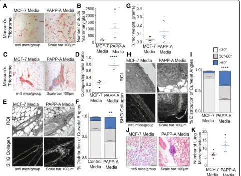
Fig. 1PAPP-A sustains an altered collagen phenotype in post-partum mammary glands.aRepresentative images of Masson’s trichrome collagen
stain (blue) on virgin, involuting, or late post-partum mammary glands from non-transgenic and PAPP-A transgenic mice.n= 5 mice per group.
Scale bar 100μm.bQuantification of collagen per epithelial region by Masson’s trichrome stain on non-transgenic and PAPP-A transgenic virgin,
involuting, or late post-partum mammary glands froma.n= 5 mice per group, each point represents the average of ten ducts per mouse per
time point. Mean ± SEM, unpairedttest with Welch’s correction: **p< 0.005, ***p< 0.0005.cRepresentative second-harmonic generation (SHG)
imaging of collagen on magnified ducts of histological sections of non-transgenic or PAPP-A transgenic virgin, involuting, or late post-partum mammary glands (lower panels). The yellow dotted lines indicate mammary ducts borders; collagen is seen in white. Corresponding regions of
interest (ROI) are shown in top panels.n= 5 mice per group. Scale bar 100μm.dGraph of collagen intensity relative to the distance from the
mammary duct borders of SHG images of non-transgenic mammary glands inc. Mean ± SEM.n= 5 mice per group, unpairedttest with Welch’s
correction: *p< 0.05.eGraph of collagen intensity relative to the distance from the mammary duct borders of SHG images of PAPP-A transgenic
mammary glands inc. Mean ± SEM.n= 5 mice per group.fQuantification of TACS3 per total curvelets analyzed in virgin, involuting, or late
post-partum mammary glands of non-transgenic mice using the CurveAlign software. TACS3 characterized as curvelet angles 60–90 relative to the
ductal border.n= 5 mice per group. Mean + SEM, unpairedttest with Welch’s correction (comparisons between the TACS3 group only). *p< .05,
**p< 0.005.gQuantification of TACS3 per total curvelets analyzed in virgin, involuting, or late post-partum mammary glands of PAPP-A
transgenic mice using the CurveAlign software. TACS3 characterized as curvelet angles 60–90 relative to ductal border.n= 5 mice per group.
[image:7.595.57.541.87.463.2]PAPP-A is a transcriptional target of mutant p53 sug-gesting that mutations in p53 represent one mechanism by which PAPP-A is abnormally overexpressed in breast cancer [32–34]. Having established that constitutive ex-pression of PAPP-A in the PAPP-A transgenic mice maintains an involution-like collagen in the post-partum glands (Fig. 1), we next investigated whether sporadic expression of PAPP-A in an established post-partum mammary gland of non-transgenic mice is able to con-vert the post-partum collagen architecture into an involution-like collagen architecture. To test this possi-bility, we established a cohort of non-transgenic post-partum mice (2 months post-weaning). In parallel, we collected media from MCF-7 cells overexpressing PAPP-A (MCF-7PAPP-A), which we previously character-ized and shown to secrete proteolytically active PAPP-A [22]. We then concentrated the PAPP-A from the media and injected PAPP-A (PAPP-A media) or control media concentrated from MCF-7 cells into the post-partum mammary glands of the non-transgenic mice every 2 days for 3 weeks.
We found that injections of PAPP-A in the
post-partum mammary glands of non-transgenic mice induce a remarkable increase in proliferation, indicated by the significantly higher number of ducts per mam-mary glands (Fig.2a, b). Further, the post-partum mam-mary glands injected with PAPP-A had significantly higher collagen abundance (Fig. 2c, d). SHG imaging of collagen orientation and CurveAlign analysis revealed that post-partum mice injected with PAPP-A adopted a TACS3 phenotype, compared to the mammary glands injected with control media, which exhibited predomin-antly TACS1 collagen (Fig. 2e, f ). As a control, we also injected PAPP-A media into the mammary glands of vir-gin non-transgenic mice. However, injections of PAPP-A had no significant effect on the proliferation or collagen in the mammary glands of virgin mice (Additional file1: Figure S1A, B). These results suggest that the sporadic presence of PAPP-A is sufficient to convert post-partum collagen into a high TACS3 involution-like collagen architecture.
Considering that involution-collagen is pro-tumorigenic, we then tested whether the effect of sporadic expression of PAPP-A affects tumor growth. To address this possibility, we used Met1 breast epithelial cancer cells, however, as these cells are known to form aggressive metastatic tumors in mice, we injected only 250,000 cells in order to increase the probability to detect differences between the groups. Met1 cells were injected into the post-partum mammary glands of non-transgenic mice along with control media or media containing PAPP-A followed by injections of the re-spective media every 2 days for 3 weeks. We found that tu-mors injected with PAPP-A are on average larger compared to the control group, but this difference did not reach
statistical significance (Fig. 2g). In addition, CurveAlign analysis of the collagen orientation on these tumors re-vealed that the mammary tumors injected with PAPP-A had a significantly higher frequency of TACS3 regions com-pared to the control group (Fig.2h, i).
As TACS3 is associated with increased metastasis [13], we analyzed these mice for lung metastases. Consistent with the higher TACS3 phenotype, mice injected with PAPP-A had a substantial increase in the frequency of micro-metastases to the lungs compared to the control group within this short period of time (Fig.2j, k).
Taken together, these results indicate that sporadic ex-pression of PAPP-A between pregnancies is able to con-vert the elevated levels of collagen of a post-partum gland to provide a pro-tumorigenic microenvironment. These results therefore imply that the abnormal gain of the overexpression of PAPP-A at any time after a first pregnancy may be sufficient to promote the develop-ment of PABC. Further, they indicate that PAPP-A also enhances the metastatic potential of cancer cells.
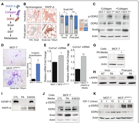
PAPP-A promotes a DDR2/Snail signaling axis of invasion through collagen mRNA stabilization and IGF signaling in vitro
In addition to a structural role of collagen in metastasis [13, 35], collagen activates the discoidin domain
recep-tors DDR1 and DDR2 [36–38]. DDR1 and 2 are
fre-quently overexpressed in breast cancers and have recently been implicated in breast cancer metastasis [36,
39–41]. DDR2 has been linked with the invasive TACS3 phenotype, while the loss of DDR2 is associated with a non-invasive collagen signature, TACS2 [40,41].
A recent report described a novel signaling role for
DDR2 in breast cancer metastasis through the
stabilization of the epithelial to mesenchymal transition (EMT) transcription factor Snail (Fig. 3a) [39]. In addition to a critical role in activating EMT, Snail may also promote cancer stem-like subpopulations, drug re-sistance, and tumor cell invasion [42, 43]. Since we found that PAPP-A leads to an increase in collagen [22] (Fig. 1a, b) and promotes metastasis (Fig.2j), we sought to address whether the DDR2/Snail axis of metastasis is active in PAPP-A-driven PABC (Fig.3a).
Fig. 2PAPP-A converts post-partum collagen into an altered, pro-tumorigeneic involution-like collagen phenotype.aRepresentative images of
the mammary ducts stained with Masson’s trichrome collagen (blue) on non-transgenic late post-partum mammary glands treated with control
or PAPP-A injections.n= 5 mice per group. Scale bar 100μm (images were taken at × 5).bQuantification of the number of ducts per mammary
gland from non-transgenic post-partum mice treated with control or PAPP-A media injections.n= 5 mice per group, each point represents the
average of each mouse per group. Mean ± SEM, unpairedttest: *p< 0.05).cRepresentative images of Masson’s trichrome collagen stain (blue) on
non-transgenic post-partum mammary glands treated with control or PAPP-A media injections.n= 5 mice per group. Scale bar 100μm.d
Quantification of collagen per epithelial region by Masson’s trichrome stain on non-transgenic post-partum mammary glands treated with control
or PAPP-A media injections.n= 5 mice per group, each point represents the average of ten ducts per mouse per group. Mean ± SEM, unpaired
ttest with Welch’s correction. ****p< 0.0001.eRepresentative second-harmonic generation (SHG) imaging of collagen on magnified ducts of
histological sections of non-transgenic late post-partum mammary glands treated with control or PAPP-A media injections (lower panels). Yellow
dotted lines indicate the mammary duct borders. Corresponding regions of interest (ROI) are shown in the top panels.n= 5 mice per group.
Scale bar 100μm.fQuantification of TACS3 per total curvelets analyzed in non-transgenic post-partum mammary glands treated with control or
PAPP-A media injections using the CurveAlign software. TACS3 characterized as curvelet angles 60–90 relative to the ductal border.n= 5 mice
per group. Mean + SEM, unpairedttest with Welch’s correction (comparisons between the TACS3 group only). **p< 0.005.gTumor weights at
time of harvesting Met1 xenograft tumors from non-transgenic post-partum mice treated with control or PAPP-A media injections.n= 5 mice
per group, two xenografts injected per mouse. In the MCF-7 media group, 5/10 tumors took while 8/10 tumors grew in the PAPP-A media group.
Each point represents a tumor weight. Mean ± SEM.hRepresentative second-harmonic generation (SHG) imaging of collagen on magnified
histological sections of Met1 xenograft tumors in non-transgenic late post-partum mice treated with control or PAPP-A media injections (lower
panels). Yellow dotted lines indicate the mammary duct borders. Corresponding regions of interest (ROI) are shown in the top panels.n= 5 mice
per group. Scale bar 100μm.iQuantification of TACS3 per total curvelets analyzed in Met1 xenograft tumors in non-transgenic late post-partum
mice treated with control or PAPP-A media injections using the CurveAlign software. TACS3 characterized as curvelet angles 60–90 relative to the
ductal border.n= 5 mice per group. Mean + SEM, unpairedt test with Welch’s correction (comparisons between the TACS3 group only).
****p< 0.0001.jRepresentative images of lung micrometastases in post-partum non-transgenic mice xenografted with Met1 cells and
treated with control or PAPP-A media injections.n= 5 mice per group. Scale bar 100μm.kQuantification of lung micrometastases in
post-partum non-transgenic mice xenografted with Met1 cells and treated with control or PAPP-A media injections.n= 5 mice per group, each point
[image:9.595.58.541.88.441.2]post-partum mammary glands. We found a significant ele-vation in Snail in the MCF-7PAPP-A cells (Fig.3c). While the levels of phosphorylated DDR2 (p-DDR2) could not
be determined by immunohistochemistry due to the lack of anti-p-DDR2 antibody that recognizes mouse DDR2, we next aimed at determining the level of p-DDR2 in
Fig. 3PAPP-A promotes DDR2 activation through collagen mRNA stabilization and IGF signaling in vitro.aSchematic of the proposed mechanism of
activation of DDR2/Snail signaling axis of invasion [39].bRepresentative images of Snail IHC (Snail: red; nuclei: blue) of virgin, involuting, or
post-partum mammary glands from non-transgenic or PAPP-A transgenic mice.n= 5 mice per group. Quantification of Snail IHC is shown,n= 5 mice per
group. Mean ± SEM, unpairedttest with Welch’s correction (comparisons between the score 3 group only). *p< 0.05, **p< 0.005.cImmunoblot of
indicated markers in MCF-7 or MCF-7PAPP-Acells co-cultured with and without collagen. Scale bar 100μm.dRepresentative images of 48 h Transwell in
vitro invasion assays of MCF-7 and MCF-7PAPP-Acells. Experiments repeated in technical triplicate. Scale bar 100μm. Quantification is shown, unpairedt
test with Welch’s correction. *p< 0.05.eRelative fold change of col1a1 mRNA transcript in MCF-7PAPP-Aover MCF-7 cells measured by real-time qPCR
and normalized to actin. Mean + SEM, triplicate experiment. Unpairedttest with Welch’s correction. *p< 0.05.fRelative fold change of col1a1 mRNA
transcript in MCF-7 cells treated in vitro with or without PAPP-A media for 24 h. PAPP-A media concentration is at 10 ng/mL of PAPP-A protein
(quantified by ELISA). Measured by real-time qPCR and normalized to actin. Mean + SEM, triplicate experiment. Unpairedttest with Welch’s correction.
**p< 0.005.gImmunoblot of LARP6 in MCF-7 cells treated in vitro with or without PAPP-A media for 24 h. PAPP-A media concentration is at 10 ng/mL
of PAPP-A protein (quantified by ELISA).hRepresentative immunoblot of LARP6 in the mammary glands from non-transgenic or PAPP-A transgenic
mice during virgin, involution, or late post-partum.n= 5 mice per group.iImmunoblot of rIGFBP-5 following a 3-h incubation in culture media from
MCF-7 (CTL), MCF-7PAPP-A(PA), or MCF-7 overexpressing proteolytic-dead mutant PAPP-A E483Q (E483Q). Immunoblot of PAPP-A secreted in culture
media from MCF-7, MCF-7PAPP-A, and MCF-7PAPP-A/E483Qcells.jImmunoblot of 7 cells treated in vitro with culture media from 7 (CTL),
[image:10.595.60.541.86.511.2]MCF-7 and MCF-7PAPPAcells. MCF-7PAPPAcells showed higher phosphorylated DDR2 (p-DDR2) and Snail expres-sion co-cultured with and without collagen I (Fig. 3c). Therefore, these observations confirm that the DDR2/ Snail axis is activated following the overexpression of PAPP-A. Since Snail is associated with invasion, we also measured the invasion capacity of MCF-7 and MCF-7PAPP
A
cells using Transwell invasion assay. We found that MCF-7PAPPAcells have a significantly higher cell invasive capacity (Fig.3d).
Since we found that collagen is required in vivo for the activation of PAPP-A [17], we were surprised by the observation that in MCF-7PAPP-Acells, DDR2 is activated in the absence of extraneous collagen. We hypothesized that this observation may be due to the ability of PAPP-A to promote the production of collagen in these cells. To test this possibility, we measured the level of
collagen 1 mRNA in MCF-7 and MCF-7PAPPAcells. We
found an elevation in collagen mRNA expression MCF-7PAPPAcells (Fig. 3e). Further, since we found that injecting PAPP-A promotes collagen deposition in vivo (Fig. 2c), we also measured collagen mRNA levels in MCF-7 cells treated with media containing PAPP-A. We found higher mRNA transcripts of col1a1 under these conditions (Fig.3f ).
La ribonucleoprotein domain family member 6
(LARP6) is a post-transcriptional modifier of col1a1 that enhances col1a1 transcripts’ translation by binding to a specific 5′UTR stem-loop of col1a1 thereby both stabil-izing col1a1 transcripts and aiding in their transport from the nucleus to the cytoplasm [46, 47]. IGF signal-ing has been previously reported to increase collagen production specifically by upregulating LARP6 [48, 49]. Therefore, since PAPP-A increases IGF signaling, we an-alyzed the level of LARP6 in MCF-7 cells treated with PAPP-A. We found that MCF-7 cells treated with PAPP-A resulted in an upregulation of LARP6 protein (Fig. 3g). To verify if this observation holds true in PAPP-A transgenic mice, we analyzed the level of LARP6 in non-transgenic and PAPP-A transgenic mam-mary glands during involution and in post-partum glands. We found that LARP6 protein is upregulated in
vivo during involution but more significantly in
post-partum glands (Fig.3h). These results indicate that in addition to its known role in promoting IGF signaling, PAPP-A also promotes metastasis through the activation of DDR2 and promotes the stabilization of collagen through activation of LARP6.
PAPP-A is a large protease with multiple uncharacter-ized domains. Therefore, it remains possible that the ac-tion of PAPP-A on DDR2 is independent of its proteolytic function. To test this possibility, we treated MCF-7 cells with media containing wild-type PAPP-A and media containing a proteoloytic-dead mutant form
of PAPP-A (E483Q). We confirmed that the E483Q mu-tant is inactive and unable to cleave recombinant IGFBP-5 in vitro (Fig. 3i). We found that wild-type but not inactive PAPP-A activates DDR2 and Snail in MCF-7 cells in vitro (Fig. 3j). Since PAPP-A activates IGF signaling, we then asked if IGF-1 downstream of PAPP-A is able to activate DDR2. To investigate this possibility, we tested the effect of exogenous IGF-1 on DDR2 in vitro. We found that IGF-1 activates DDR2 in MCF-7 cells, but this effect was markedly increased in MCF-7PAPP-A cells (Fig. 3k). Further, MCF-7PAPP-Acells expressing inactive PAPP-A also show some activation although the effect was markedly decreased compared to MCF-7PAPP-Acells (Additional file2: Figure S2).
These results suggest that through its ability to in-crease IGF signaling and the elevation in collagen, PAPP-A leads to the increased activation of DDR2 signaling.
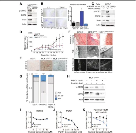
Loss of DDR2 abolishes the ability of PAPP-A to regulate cell invasion and Snail expression in vitro and in vivo
To further validate the role of DDR2 in the increased in-vasion of cells overexpressing PAPP-A, we used CRISPR
to knock out DDR2 in MCF-7 and MCF-7PAPP-A cells.
First, we confirmed that DDR2 expression is abolished in the MCF-7PAPPA-DDR2KO cells (Fig. 4a). Inhibition of DDR2 resulted in undetectable levels of Snail by immu-noblot (Fig.4a). Further, the decrease in Snail correlated with a marked decrease in cell invasive capacity (Fig.4b). We confirmed that inhibition of DDR2 abolishes the ac-tivation of Snail by PAPP-A using MCF-7 cells and MCF-7 cells where DDR2 was inhibited by CRISPR (MCF-7DDR2KO) despite treatment with PAPP-A contain-ing media (Fig.4c).
Therefore, these results suggest that in addition to in-creasing IGF signaling and collagen production, PAPP-A leads to the activation of a distinct collagen signaling path-way mediated by DDR2 which promotes invasion.
To further explore the potential clinical relevance of DDR2 signaling in PAPP-A-driven PABC, we sought to in-vestigate the effect of DDR2 in vivo by comparing the growth of MCF-7, MCF-7PAPP-A, and MCF-7PAPP-A/DDR2KO xenografts in the mammary fat pads of nude mice.
Add-itionally, we previously found that using a 1:1
Matrigel-collagen reduces the growth of MCF-7 xenografts compared to Matrigel alone [22]. This observation suggests that Matrigel-collagen mixture mimics the collagen-rich and anti-proliferative environment of a post-partum mam-mary gland [14, 22]. We therefore performed these xeno-grafts using a 1:1 Matrigel-collagen mixture.
Fig. 4Loss of DDR2 abolishes cell invasion, Snail expression, and tumor growth by PAPP-A.aImmunoblot of indicated markers in MCF-7PAPP-A
CTL and MCF-7PAPP-ADDR2−/−cells.bRepresentative images of 48 h Transwell in vitro invasion assays of MCF-7 CTL, MCF-7 DDR2−/−, MCF-7PAPP-A
CTL, and MCF-7PAPP-ADDR2−/−cells. Experiments repeated in technical triplicate. Scale bar 100μm. Quantification is shown, unpairedttest with
Welch’s correction. *p< 0.05.cImmunoblot of indicated markers in MCF-7 CTL or MCF-7 DDR2−/−cells treated with or without PAPP-A media for
24 h.dRelative tumor growth of MCF-7, MCF-7PAPP-A, and MCF-7PAPP-ADDR2−/−xenografts in a Matrigel-collagen mixture.n= 5 mice per group,
ten tumors each. Each point represents the average of five mice, mean ± SEM. Unpairedttest at end point (MCF-7PAPP-Avs. MCF-7PAPP-A
DDR2−/−). *p< 0.05eRepresentative images of Snail IHC on MCF-7, MCF-7PAPP-A, and MCF-7PAPP-ADDR2−/−xenograft tumors in Matrigel-collagen
mixture.n= 3 mice, six tumors total per group. Scale bar 100μm.fRepresentative images of Masson’s trichrome collagen stain (blue) and
second-harmonic generation (SHG) imaging of collagen (lower panels). Yellow dotted lines indicate the mammary tumor borders. Corresponding
regions of interest (ROI) on magnified histological sections of MCF-7, MCF-7PAPP-A, or MCF-7PAPP-ADDR2−/−xenograft tumors in a
Matrigel-collagen mixture.n= 3 mice per group. Scale bar 100μm.gQuantification of TACS3 per total curvelets analyzed in MCF-7, MCF-7PAPP-A, or
MCF-7PAPP-A DDR2−/−xenograft tumors in a Matrigel-collagen mixture using the CurveAlign software. TACS3 characterized as curvelet angles 60–90
relative to the ductal border.n= 3 mice, six tumors total per group. Mean + SEM, unpairedttest with Welch’s correction (comparisons between
TACS3 group only, MCF-7PAPP-Avs. MCF-7PAPP-ADDR2−/−). *p< 0.05.hImmunoblot of indicated markers in MCF-7PAPP-Acells treated for 48 h at
the indicated concentrations of PQ401 and imatinib.iProliferation assay of MCF-7 and MCF-7PAPP-Acells treated with imatinib at the indicated
concentrations for 48 h.jProliferation assay of MCF-7 and MCF-7PAPP-Acells treated with PQ401 at the indicated concentrations for 48 h.k
[image:12.595.59.538.87.552.2](Fig. 4d). Therefore, we conclude that DDR2 is a signifi-cant mediator of the accelerated growth of cells overex-pressing PAPP-A in vivo.
Further, in agreement with the results obtained in vitro, we found that MCF-7PAPP-A xenografts have the
highest Snail protein expression by IHC, while
MCF-7PAPP-A/DDR2KO xenografts exhibit undetectable
Snail expression (Fig.4e).
We next analyzed the orientation of collagen in these xenografts. We found that MCF-7 xenografts exhibit
ele-vated TACS1 but low TACS3, while MCF-7PAPP-A
xeno-grafts show elevated TACS3 orientation (Fig. 4f, g). Strikingly, MCF-7PAPP-A/DDR2KO xenografts had signifi-cantly less TACS3 regions of collagen and distribution of curvelet angles comparable to that seen in MCF-7 tu-mors (Fig.4f, g). These results suggest a critical role for DDR2 in the ability of PAPP-A to promote and maintain collagen into a TACS3 orientation.
Imatinib has been shown to inhibit the activity of DDR2 [50, 51]. Since PAPP-A activates both IGF and DDR2 signaling, we tested the ability of the IGF inhibi-tor PQ401 alone or in combination with imatinib on the growth of MCF-7PAPP-A cells. First, we confirmed that imatinib reduces the activation of DDR2 in these cells (Fig. 4h). However, the reduction was enhanced in cells treated with imatinib in combination with PQ410 (Fig. 4h). We found that while imatinib has no effect on
the growth of either MCF-7 or MCF-7PAPP-A cells
(Fig. 4i) and PQ401 inhibited cell growth in both cell lines equally (Fig. 4j), the combination selectively inhib-ited the growth of the MCF-7PAPP-A cells (Fig. 4k). Mechanistically, we found that MCF-7PAPP-A cells have higher p-Akt levels than MCF-7 cells (Additional file 3: Figure S3). However, a decrease in p-Akt levels was only observed in MCF-7PAPP-A cells when treated with the
combination treatment of PQ401 and imatinib
(Additional file3: Figure S3). This observation is consist-ent with the observation that MCF-7PAPP-A cells have higher DDR2 activation in response to treatment with IGF (Fig.3k). Further, a recent study reported a positive crosstalk between DDR1 and IGF signaling, therefore raising the possibility that a similar crosstalk exists be-tween DDR2 and IGF signaling [52].
These results indicate that complete inhibition
achieved by either deletion of DDR2 by CRISPR or treat-ment with pharmacological combination is necessary to
abolish the growth of the MCF-7PAPP-A cells, while
DDR2 inhibition by imatinib alone does not.
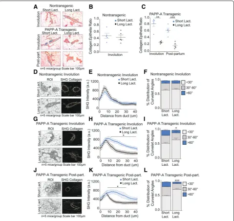
Long lactation inhibits PAPP-A-driven TACS3 formation in post-partum mammary gland
We previously reported that a long lactation of 2 weeks or more prior to involution inhibits the formation of
PABC in the PAPP-A transgenic mice [22]. As long
lactation represents an alternative method to inhibit the action of PAPP-A upstream of DDR2, we tested the ef-fect of long lactation on the level of collagen deposition in post-partum glands. We harvested mammary glands from parous PAPP-A transgenic mice during involution
(12 days post-weaning) or post-partum (2 months
post-weaning) as described in Fig. 1 with the exception that mothers were kept with their litters for 2 weeks prior to the initiation of involution. These mammary glands were compared to the mammary glands from in-voluting and post-partum PAPP-A transgenic mice following a short lactation described in Fig. 1. In non-transgenic mice, the length of lactation had no ef-fect as expected (Fig. 5a, b) and was therefore not pur-sued further. In PAPP-A transgenic mice, however, we found that long lactation significantly decreased collagen abundance in both involuting and post-partum mam-mary glands (Fig.5a, b).
We next analyzed the orientation of collagen. Consist-ent with the lack of effect of long lactation on collagen abundance, we found that long lactation had no effect on collagen orientation in involuting mammary glands from non-transgenic mice (Fig. 5d–f ). However, long lactation resulted in a distribution of collagen that is closer to the ductal border and decreased TACS3 in in-voluting mammary glands of PAPP-A transgenic mice (Fig. 5g–i). Importantly, the distribution of collagen and the frequency of TACS3 in PAPP-A transgenic involut-ing mammary glands after a long lactation mimic those observed in the non-transgenic involuting mammary glands (Fig.5e, f ).
In post-partum mammary glands from PAPP-A trans-genic mice, we found that a long lactation also signifi-cantly altered collagen orientation, as indicated by a decreased presence of collagen dispersed from the ductal border and decreased TACS3 (Fig.5j–l). Furthermore, the decrease in TACS3 in PAPP-A transgenic post-partum mammary glands following a long lactation was even more significant than that observed in involuting mam-mary glands after a long lactation (Fig. 5f, i, l). These re-sults highlight that a longer lactation ablates the ability of PAPP-A to maintain an involution-like collagen in post-partum mammary glands.
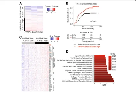
PAPP-A-driven signature correlates with distant metastasis in human breast cancers
does not act as an oncogene in the absence of collagen. Therefore, for PAPP-A to be useful as a diagnostic bio-marker, it must be analyzed in the context of high colla-gen content. However, another difficulty arises from the fact that information regarding breastfeeding is either sparse or entirely missing from medical charts. There-fore, the use of combined expression of PAPP-A and col-lagen as a biomarker performs poorly as the impact of high expression of both genes can be ablated by long breastfeeding. Having identified the involvement of a DDR2/Snail axis in PAPP-A-driven PABC, we reasoned that Snail in combination with PAPP-A and collagen might represent a more reliable readout for monitoring the activation of the pathway. To test this hypothesis, we investigated the correlation between a three-gene signa-ture of PAPP-A, COL1A1, and SNAI1 and clinical out-comes in breast cancer patients. First, we generated a score based on the average gene expression of PAPP-A/
SNAI1/COL1A1from a publicly available dataset of 327
patients with primary breast cancer (GSE20685) [27]. We next stratified patients based on this score into
PAPP-A/SNAI1/COL1A1 high and low (Fig. 6a). We
found that the PAPP-A/SNAI1/COL1A1-high patient
population develops distant metastases at a significantly higher rate than those patients with low values of the
PAPP-A/SNAI1/COL1A1 expression score (P= 0.042, Fig.6b). This finding was validated in an additional
data-set (GSE9893) with the PAPP-A/SNAI1/COL1A1-high
patients developing distant metastases at a significantly higher rate (p= .0045, Additional file4: Figure S4) [28].
To unravel the major molecular differences between
patients with high and low values for our PAPP-A/
SNAI1/COL1A1 expression score, we next conducted differential gene expression between these patients [29]. We found 78 significantly upregulated genes of interest in the PAPP-A/SNAI1/COL1A1-high patient population (Fig. 6c, Additional file5: Table S1). Of these 78 genes, highlighted in red is PAPP-A, COL1A1, and SNAI1 as
(See figure on previous page.)
Fig. 5Long lactation inhibits PAPP-A-driven collagen deposition and TACS3 formation in post-partum mammary glands.aRepresentative images
of Masson’s trichrome collagen stain (blue) on involuting or post-partum mammary glands from non-transgenic and PAPP-A transgenic mice
following a short (2 days) or long (14 days) lactation period.n= 5 mice per group. All images from mice following short lactation are from the
same experiment shown in Fig.1a. Scale bar 100μm.bQuantification of collagen per epithelial region by Masson’s trichrome stain on
non-transgenic involuting mammary glands following a short (2 days) or long (14 days) lactation period froma.n= 5 mice, each point represents the
average of ten ducts per mouse per time point. Mean ± SEM, unpairedttest with Welch’s correction. Analysis on short lactation samples is from
the same analysis performed in Fig.1b.cQuantification of collagen per epithelial region by Masson’s trichrome stain on PAPP-A transgenic
involuting or post-partum mammary glands following a short (2 days) or long (14 days) lactation period froma.n= 5 mice, each point represents
the average of ten ducts per mouse per time point. Mean ± SEM, unpairedttest with Welch’s correction: **p< 0.005, ***p< 0.0005. Analysis on
short lactation samples is from the same analysis performed in Fig.1c.dRepresentative second-harmonic generation (SHG) imaging of collagen
on magnified ducts of histological sections of non-transgenic involuting mammary glands following a short (2 days) or long (14 days) lactation
period (right panels) and corresponding regions of interest (ROI) (left panels) (n= 5 mice per group). All images of short lactation samples are
from the same experiment shown in Fig.1d. Scale bar 100μm.eGraph of collagen intensity relative to the distance from the mammary duct
borders of SHG images of non-transgenic involuting mammary glands following a short (2 days) or long (14 days) lactation period ind.
Mean ± SEM.n= 5 mice per group, unpairedttest with Welch’s correction. Analysis on short lactation samples is from the same analysis
performed in Fig.1e.fQuantification of TACS3 per total curvelets analyzed in non-transgenic involuting mammary glands following a short (2
days) or long (14 days) lactation period using the CurveAlign software. TACS3 characterized as curvelet angles 60–90 relative to the ductal border.
n= 5 mice per group. Mean + SEM, unpairedttest with Welch’s correction (comparisons between the TACS3 group only). Analysis on short
lactation samples is from the same analysis performed in Fig.1g.gRepresentative second-harmonic generation (SHG) imaging of collagen on
magnified ducts of histological sections of PAPP-A transgenic involuting mammary glands following a short (2 days) or long (14 days) lactation
period (right panels) and corresponding regions of interest (ROI) (left panels) (n= 5 mice per group). All images of short lactation samples are
from the same experiment shown in Fig.1d. Scale bar 100μm.hGraph of collagen intensity relative to the distance from the mammary duct
borders of SHG images of PAPP-A transgenic involuting mammary glands following a short (2 days) or long (14 days) lactation period ine.
Mean ± SEM.n= 5 mice per group, unpairedttest with Welch’s correction. Analysis on short lactation samples is from the same analysis
performed in Fig.1f.iQuantification of TACS3 per total curvelets analyzed in PAPP-A transgenic involuting mammary glands following a short (2
days) or long (14 days) lactation period using the CurveAlign software. TACS3 characterized as curvelet angles 60–90 relative to the ductal border.
n= 5 mice per group. Mean + SEM, unpairedttest with Welch’s correction (comparisons between the TACS3 group only). *p< 0.05. Analysis on
short lactation samples is from the same analysis performed in Fig.1g.jRepresentative second-harmonic generation (SHG) imaging of collagen
on magnified ducts of histological sections of PAPP-A transgenic post-partum mammary glands following a short (2 days) or long (14 days)
lactation period (right panels) and corresponding regions of interest (ROI) (left panels) (n= 5 mice per group). All images of short lactation
samples are from the same experiment shown in Fig.1d.kGraph of collagen intensity relative to the distance from the mammary duct edge of
SHG images of PAPP-A transgenic post-partum mammary glands following a short (2 days) or long (14 days) lactation period inf. Mean ± SEM.n
= 5 mice per group, unpairedttest with Welch’s correction: *p< 0.05. Analysis on short lactation samples is from the same analysis performed in
Fig.1f.lQuantification of TACS3 per total curvelets analyzed in PAPP-A transgenic post-partum mammary glands following a short (2 days) or
long (14 days) lactation period using the CurveAlign software. TACS3 characterized as curvelet angles 60–90 relative to the ductal border.n= 5
mice per group. Mean + SEM, unpairedttest with Welch’s correction (comparisons between the TACS3 group only). *p< 0.05. Analysis on short
expected in addition to DDR2 and LARP6 (Fig. 6c). These 78 genes are involved in extracellular matrix com-ponents, collagen, collagen processing and remodeling, EMT, IGF signaling, Wnt signaling, and cell invasion and migration (Fig.6c).
All significantly upregulated genes from the differential gene expression data were used for gene set expression analysis (GSEA) to identify significantly activated path-ways in thePAPP-A/SNAI1/COL1A1-high patient popula-tion [30]. We found 13 statistically significant and relevant pathways active in the PAPP-A/SNAI1/COL1A1-high pa-tient group (Fig.6d, Additional file6: Table S2). Of note, EMT, extracellular matrix organization, and collagen for-mation were the top 3 highly activated pathways in the
PAPP-A/SNAI1/COL1A1-high patient group (Fig. 6d). This is consistent with the increased frequency of TACS3 collagen and cell invasion observed in our PAPP-A over-expressing cells in vitro and in vivo.
Discussion
The existence of PABC as a distinct breast cancer subtype remains controversial. The major difficulties in the study of PABC are the lack of reliable bio-markers and the time frame after pregnancy of
diag-nosis of PABC. Currently, PABC is empirically
defined as breast cancers arising 1–2 years after
preg-nancy despite the epidemiological findings that
women who had children remain at higher risk of breast cancer for years compared to women who never had children [1]. Clearly distinguishing a PABC from a sporadic cancer that is unrelated to pregnancy in women diagnosed several years after their last pregnancy is a challenge.
Data presented in the current study support the pro-vocative notion that the passage through a single preg-nancy is sufficient to predispose a breast to the oncogenic action of PAPP-A. Our data imply that the acquisition of a
Fig. 6PAPP-A/SNAI1/COL1A1gene signature correlates with distant metastasis in breast cancer patients.aHeatmap of patients clustered based on
their score for thePAPP-A/SNAI1/COL1A1signature (categorized according to the mean score value). Each row represents one patient,n= 327.b
Kaplan-Meier curve for time to distant metastasis according to thePAPP-A/SNAI1/COL1A1score as defined ina. Number of patients at risk at each
time point over a 150-month period is recorded below in table.cHeatmap representing the relative expression of a selected panel of 78 genes
(FDR < .05) in thePAPP-A/SNAI1/COL1A1-low andPAPP-A/SNAI1/COL1A1-high patient populations. Genes are organized by pathways, as labeled
adjacent to the gene names. The red text highlights relevant genes investigated in this study (PAPP-A,DDR2,SNAI1,COL1A1, andLARP6). Each
column represents a patient, and each row represents a gene.dChart of GSEA pathways significantly enriched in thePAPP-A/SNAI1/COL1A1-high
[image:16.595.60.538.88.412.2]mutation leading to PAPP-A overexpression at any point after pregnancy, even years, can hijack the high collagen abundance of a post-partum breast and convert this colla-gen into a TACS3 pro-tumoricolla-genic collacolla-gen.
Considering that PAPP-A has been reported to be overexpressed in nearly all breast cancers [21] and that the majority of women worldwide have at least one pregnancy, the implication of our findings is that PAPP-A-driven PABC may be very frequent. If so, targeting PAPP-A for therapy may be an attractive therapeutic target. This possibility is supported by the fact that antibody against PAPP-A has been developed and shown to be effective in ovarian cancer [53]. Fur-ther, as genetic ablation of the PAPP-A gene leads to a reduction in several age-related pathologies and in-creases longevity by 30% [54], the prediction is that anti-PAPP-A therapy, in marked contrast to current therapies, would have limited side effects and may ac-tually improve general health.
Further, considering the elevated rate of PAPP-A overexpression in breast cancer, targeting PAPP-A in the prevention setting also appears as a potential av-enue of future research. In this regard, extended breastfeeding has been shown to have a protective ef-fect against breast cancer [55]. Our results suggest that the inhibition of PAPP-A by its inhibitors stan-niocalcins 1 and 2 (STC1, STC2) during lactation may play an important role in the protective effect of breastfeeding. Mechanistically, we reported that the protective effect of lactation is due to the production of STC1 and STC2 by the ovaries during lactation [22, 56, 57]. Our results proposed a model whereby long lactation allows for the saturation of PAPP-A by STC1 and 2 such that upon initiation of involution, PAPP-A remains inactive and cannot cleave IGFBP-5 [22]. As a result, the pro-tumorigenic cascade initi-ated by PAPP-A is prevented [22].
Our findings that collagen is an important co-factor that contributes to the oncogenic activity of PAPP-A also raises the question of the potential link between PAPP-A and breast density. Mammographic density is one of the best predictors of breast cancer risk [58]. One prediction is that women with dense breasts, which have a higher content of collagen, maybe more prone to PAPP-A-driven PABC. In addition, a recent GWAS have shown that low IGFBP-5 is linked to increased risk of breast cancer [26]. This observation is also consistent with an important role of PAPP-A in breast cancer, since breasts with low endogenous level of IGFBP-5 are likely to be more sensitive to the oncogenic action of PAPP-A since the high PAPP-A/IGFBP-5 ratio would favor a more effective elimination of IGFBP-5. Therefore, the re-sults of our study suggest that in the context of PAPP-A-driven PABC, breast density, SNPs in IGFBP-5,
and history of breastfeeding must all be considered (Fig.7).
Collectively, therefore, combining breast density, SNPs that reduce expression of IGFBP-5, pregnancy, and breastfeeding history holds the potential for the develop-ment of a predictive test for risk of breast cancer. Cur-rently, genetic testing for the risk of breast cancer is limited to the sequencing of BRCA1 gene for women with a history of breast cancer. This possibility will be tested in the future.
In addition to offering a potentially important link between several known factors of breast cancer risk, our current study also identifies novel functions of PAPP-A, namely the elevation of collagen through LARP6 [46, 47] and the activation of the collagen re-ceptor DDR2. We propose a model whereby both the crosstalk between the high IGF signaling by proteo-lytic cleavage of IGFBP-5 and increasing collagen de-position synergizes to activate DDR2 [22, 52, 59]. Further, we show that the resulting activation of DDR2 leads to an increase in Snail and cell invasive capacity. The effects of PAPP-A on invasion are sig-nificant as PABC is typically a more aggressive form of breast cancer, characterized by higher rates of re-currence, metastasis, and poorer patient survival [1, 4,
60, 61]. This finding is also consistent with the
obser-vation that PABC exhibit an increased rate of
triple-negative breast cancer, which have the highest rate of mutation in p53 and we have previously shown mutant p53 activates the transcription of PAPP-A [1, 33, 62, 63]. We show here that PAPP-A promotes these physiological effects through TACS3 maintenance and DDR2/Snail signaling axis of inva-sion. How TACS3 is formed remains a mystery, but it has been reported that inhibition of DDR2 abolishes its formation [39].
We show that inhibition of DDR2 by CRISPR abol-ishes the ability of PAPPA to promote invasion. The role of DDR2 in PAPP-A-driven PABC adds to the growing evidence of the importance of DDR2 in breast cancer metastasis and that it is active in both tumor and stro-mal cells [40]. Our findings suggest the possibility of DDR2 as a novel therapeutic target in PAPP-A-driven PABC. A summary of how LARP6 and DDR2 add to our current understanding of PAPP-A-driven PABC is shown in Fig.7.
Conclusions
Despite extensive research in breast cancer and the fact that pregnancy and breastfeeding are the funda-mental roles of breasts, history of pregnancy and breastfeeding is not included in standard clinical charts nor considered in diagnosed patients. In
empirically defined as those arising within 2 years after pregnancy and therefore misconceived as being rare. In fact, a recent meta-analysis combining the results of 15 studies confirmed that women remain at higher risk of breast cancer following pregnancy for more than 20 years, suggesting that healthcare providers should consider parity as a risk factor [6]. The results presented here challenge the current def-inition of PABC and support the conclusions of this recent study. Our data suggest that parity imposes a long-term predisposition to PAPP-A-driven breast cancers, a notion that much better reflects the epidemio-logical findings [1–3,6]. Further, the data presented in the current study strongly argue that information regarding pregnancy and lactation history should be mandatory in medical charts of breast cancer patients. Our results sug-gest that this information, combined with the analysis of collagen and thePAPP-Agenetic signature, holds the po-tential to identify patients at higher risk of recurrence. Further, the current study highlights the potential thera-peutic interventions. Therefore, the translational impact spans from the need for basic information on medical charts to the development of a diagnostic signature and targeted intervention.
Additional files
Additional file 1:Figure S1.PAPP-A media injections do not affect
collagen abundance in virgin mammary glands. a) Representative images
of Masson’s trichrome collagen stain (blue) on non-transgenic virgin
mammary glands treated with control or PAPP-A injections.n= 3 mice per
group. Scale bar 100μm. b) Quantification of collagen per epithelial region
by Masson’s trichrome stain on non-transgenic virgin mammary glands
treated with control or PAPP-A injections.n= 3 mice per group, each point
represents the average of ten ducts per mouse per group. Mean ± SEM,
unpairedttest with Welch’s correction. (PDF 232 kb)
Additional file 2:Figure S2.Mutant PAPP-A decreases DDR2 activation
by IGF-1: a) immunoblot of DDR2 and phospho-DDR2 in MCF-7PAPP-Aand
MCF-7PAPP-A-E483Qcells treated with recombinant 10 nM IGF-1 at indicated
time points. (PDF 139 kb)
Additional file 3:Figure S3.Combination of PQ401 and imatinib
treatment blocks p-Akt in vitro: a) immunoblot of the indicated markers
in MCF-7 and MCF-7PAPP-Acells treated at the indicated concentrations of
PQ401 and imatinib. (PDF 157 kb)
Additional file 4:Figure S4.Secondary validation of time to distant metastasis: a) Kaplan-Meier curve for time to distant metastasis according
to thePAPP-A/SNAI1/COL1A1score. Number of patients at risk at each
time point over a 160-month period is recorded below in table. (PDF 162 kb)
Additional file 5:Table S1.LIMMA differential gene expression analysis
PAPP-A/SNAI1/COL1A1gene signature: complete data on differential gene
expression analysis onPAPP-A/SNAI1/COL1A1-high vs.PAPP-A/SNAI1/
COL1A1-low patient groups. Each row is one gene (indicated by each probe used), and each column is as labeled. (XLS 9760 kb)
[image:18.595.56.539.86.316.2]