Oral presentations
O1
Digital breast tomosynthesis: a comparison of the
accuracy of digital breast tomosynthesis,
two-dimensional digital mammography and
two-dimensional screening mammography
(film-screen)
MJ Michell1, RK Wasan1, P Whelehan1, A Iqbal1, CP Lawinski2, AN Donaldson3, DR Evans1, C Peacock1, ARM Wilson1
1Breast Radiology Department, King’s College Hospital NHS
Foundation Trust, London, UK, 2KCARE, Medical Engineering and
Physics, King’s College Hospital NHS Foundation Trust, London, UK,
3Department of Medical Statistics, King’s College London, London, UK
Breast Cancer Research2009, 11(Suppl 2):O1 (doi: 10.1186/bcr2365) Introduction Digital breast tomosynthesis (DBT) may improve the accuracy of mammography by enabling visual separation of overlapping tissues (Andersson et al. 2008, Poplack et al. 2007).
Methods Following local research ethics approval, all women attending the assessment clinic for evaluation of a mammographic abnormality found on routine screening (film-screen) were invited to take part in the study subject to informed consent. Participants underwent bilateral view dimensional (2D) digital mammography and bilateral two-view DBT. Mammography scores using the RCR Breast Group classifi-cation were sequentially obtained for the screening mammogram, 2D digital and DBT, and these were each compared with the final assessment outcome.
Results Ninety-one percent of eligible women participated. Results from the first 300 participants are presented in Table 1 below. Kappa coefficients for agreement of each imaging method with final assessment outcome were calculated. Screening mammograms had the lowest agreement with the final outcome (Kappa = 0.02; P= 0.22), 2D digital mammography was better (Kappa = 0.26; P= 0.0000) and DBT had the highest score (Kappa = 0.37; P= 0.0000).
Conclusion The preliminary results of this ongoing study show that DBT increases the diagnostic confidence of the radiologist. This supports the need for a larger multicentre study.
O2
Optimal mammography reading volumes: evidence
from real life
E Cornford1, J Reed2, A Murphy2, A Evans1, R Bennett3
1Nottingham Breast Institute, Nottingham, UK, 2East Midlands QA
Reference Centre, Nottingham, UK, 3Cancer Screening Evaluation
Unit, Institute of Cancer Research, London, UK
Breast Cancer Research2009, 11(Suppl 2):O2 (doi: 10.1186/bcr2366) IntroductionThe aim of the study was to assess real life reader perfor-mance as a function of volume of mammograms read in a large multicentre cohort.
MethodsThirty-seven film readers within the East Midlands Screening Programme had 3 years of consecutive screen reading results available for comparison.
Markers of screen reading performance (overall and first reader cancer detection rates, recall rates, positive predictive value of recall and missed cancers) were compared with volume of films read. Readers were categorised into four groups, according to film reading volume over the 3-year period: <15,000 (that is, on average less than the recommended 5,000/year); 15,000 to <20,000; 20,000 to <25,000; and ≥25,000. Statistical analysis was undertaken using SPSS for Windows version 13.
ResultsThe recall rate in low volume readers (<5,000/year) was 6.9% and was significantly higher than in the other groups combined (4.8%;
P≤0.001). These readers also had a lower positive predictive value than higher volume readers (11.7% versus 15.7%, P ≤ 0.001).The cancer detection rate at first read was significantly lower in the higher volume readers (≥25,000) in comparison to the other groups combined (6.6 per 1,000 versus 8.2 per 1000, P≤0.001).
Conclusion These data support the recommendation that readers should read a minimum of 5,000 mammograms/year. They also suggest that there is an upper limit above which reader performance deteriorates (in terms of cancer detection). With the imminent programme expansion this has implications for service quality. Consideration should be given to the introduction of an upper limit of mammographic reads.
Breast Cancer Research
Volume 11 Supplement 2, November 2009
Meeting abstracts
Royal College of Radiologists Breast Group Annual Scientific
Meeting
Belfast, Northern Ireland
1–3 November 2009
Published: 26 October 2009
© 2009 BioMed Central Ltd
Table 1 (abstract O1)
Imaging score Screening mammogram 2D digital mammogram DBT
Assessment outcome = normal or benign (n=238) M1 or M2 44 (18.4%) 130 (54.6%) 174 (73.1%)
Assessment outcome = malignant (n=62) M5 22 (35.4%) 25 (40.3%) 33 (53.2%)
O3
Pathological features and patterns of metastatic
disease in locally advanced inflammatory and
non-inflammatory breast cancer
MZ Mvere1, SJ Tennant1, AJ Evans1, I Ellis2, J James1, M Shehat2, S Chan2
1Nottingham Breast Institute, Nottingham, UK, 2Nottingham University
Hospitals NHS Trust, Nottingham, UK
Breast Cancer Research2009, 11(Suppl 2):O3 (doi: 10.1186/bcr2367) IntroductionLocally advanced primary breast cancer (LAPC) is asso-ciated with a high risk of metastatic disease in spite of multimodality treatment. The aim of this study was to describe and compare the patterns of metastatic disease of the non-inflammatory (nIBC) and inflam-matory (IBC) breast cancer subtypes as detected by staging computed tomography scan of the chest, abdomen and pelvis (CTCAP).
Methods Over a 29 month period, 97 patients underwent staging CTCAP for LAPC or IBC and were identified from the hospital’s computerised radiology system. The formal staging CTCAP scan and initial core biopsy pathology reports were reviewed. Statistical analyses were carried out using Fisher’s exact test or chi square test via an online analysis package (‘SISA-Binomial’).
Results Thirty-eight patients (39%) presented with IBC and 59 patients (61%) with nIBC. Out of 97 patients, 16 (16.5%) had metastatic disease at staging CTCAP. Of these, 10 of 38 patients (26%) of the IBC group had metastases compared to 6 of 59 patients (10%) of the nIBC group (P= 0.034). Metastatic disease was present within the lungs, pleura and lymph nodes, with no liver or bone metastases. However, compared to nIBC, IBC patients were more likely to present with pleural metastases (4 of 38 (10.5%) versus 1 of 59 (1.7%), P= 0.05), the majority presenting with bilateral pleural based nodules.
ConclusionA distinct pattern of pleural metastatic disease has been shown in patients with IBC.
O4
Surgical management of women with
screen-detected breast cancer: preoperative factors
indicating risk of multiple operations versus a single
operation in women undergoing surgery for screen
detected breast cancer
RJ Currie1, EAM O’Flynn2, MJ Michell2
1Breast Care Unit, Royal Devon and Exeter Foundation Trust, Exeter,
UK, 2National Breast Screening Training Centre, King’s College
Hospital NHS Foundation Trust, London, UK
Breast Cancer Research2009, 11(Suppl 2):O4 (doi: 10.1186/bcr2368) Introduction Seventeen percent of women undergoing surgery for screen-detected breast cancer undergo more than one operation (BASO audit 2008). The aim of this study is to identify pre-operative data items that predict which cases will require more than one application.
Methods Over an 11 year period (April 1996 to April 2007) 1,193 women presented with screen-detected breast cancer through the South East London Breast Screening Programme. For each case, imaging and biopsy data were reviewed in order to predict which women undergoing wide local excision (WLE) would require further operations. Age, invasive cancer size, ductal carcinoma in situ(DCIS) size, histology, cancer grade, nodal status, mammographic sign and level of suspicion of malignancy (RCR Breast Group 1-5 scale) were compared.
Results Of the 1,193 women, 916 (77%) had a WLE or excision biopsy. These were split into two groups: group 1 (520 women (57%)) had a single operation; and group 2 (396 women (43%)) went on to have further operations, including repeat WLE to clear margins 238 (60%), mastectomy 133 (34%) and axillary dissection 25 (6%). Twenty-eight women went on to have a third operation. There were significant associations with increasing size of DCIS (P< 0.001),
increasing tumour grade (P = 0.005), radiologists suspicion of 3/4 (P= 0.002) and mammographic sign (spiculated mass plus micro-calcifications and asymmetric density (ASD), P= 0.003) for repeat operations. There was no statistical difference in patient’s age, invasive cancer size, histology or nodal status.
Conclusion The multidisciplinary team should pay particular attention to DCIS extent and mammographic features of the tumour when planning surgical management in order to minimise the number of women requiring repeat operations.
O5
Multistatic radar: first trials of a new breast imaging
modality
M Shere1, A Preece2, I Craddock3, J Leendertz2, M Klemm3
1Frenchay Hospital, Bristol, UK, 2Medical Physics Department,
University of Bristol, Bristol, UK, 3Engineering Department, University
of Bristol, Bristol, UK
Breast Cancer Research2009, 11(Suppl 2):O5 (doi: 10.1186/bcr2369) None of the current breast imaging modalities are perfect. Mammography has quite a low sensitivity, uses ionising radiation and is uncomfortable. Ultrasound has better sensitivity, is cheap, but is user dependent and not very good for screening. Magnetic resonance imaging has good sensitivity but is expensive. Positron emission tomography is extremely expensive. Other modalities have been tested, such as thermal imaging, red light imaging and elastography. We present a new modality - microwave RADAR imaging.
This uses electromagnetic radiation in the gigahertz range (similar to a mobile phone, but much lower power). Research has been going on for many years using single emitters and antennae, which has proven the concept but was unable to produce high definition images. The breakthrough came with using technology developed for land mine radar detection - multistatic arrays.
The third generation imaging apparatus uses 31 microwave emitters arranged in a hemisphere in which the breast sits in a prone table system. This gives 435 different pathways through the breast, which go into a network analyser and then are translated into a three-dimensional image for viewing and manipulation.
We have now done the initial trials with first use in humans; 160 women with lesions have been scanned and are being compared with mammography and ultrasound. The results are encouraging; most lesions are visible and it has a good ability to distinguish between benign and malignant lesions with a modality that is quicker, cheaper, safer and more comfortable.
Pictures of the apparatus and images produced will be shown.
O6
Interval cancer review in the Leicestershire
symptomatic breast service
SL Tennant, H Daintith, M Al-Attar, E Denton, L Grosvenor, D Lister, H Khan
University Hospitals Leicester, Leicester, UK
Breast Cancer Research2009, 11(Suppl 2):O6 (doi: 10.1186/bcr2370) Introduction Interval cancer review is an important part of quality assurance within the NHSBSP. No equivalent assessment is per-formed within the symptomatic service. We attempted to apply a similar review to the symptomatic work performed at our centre.
Methods All patients referred to our centre with a breast symptom are seen in ‘one-stop’ triple assessment clinics. We identified, and reviewed the imaging of, patients diagnosed with in situ or invasive carcinoma at our unit during 2007 who had undergone breast imaging via the symptomatic service within 3 years prior to diagnosis.
Fourteen cancers were felt to be true intervals, six mammographically occult, and five suspicious. Nine were unclassifiable. Of the unclassi-fiable cases, one had missing imaging and eight did not have mammo-graphy at prior attendance. Only four of these were symptomatic in the same area as their cancer was later diagnosed.
Conclusion ‘Interval cancers’ within the symptomatic population are a small but heterogeneous group. We discuss the details and present a pictorial review of the interesting cases.
Poster presentations
P1
Is it safe practice not to biopsy fibroadenomas in
women under 30?
K Taylor, P Britton, L Sonoda, M Wallis, R Sinnatamby
Cambridge Breast Unit, Addenbrooke’s Hospital, Cambridge, UK Breast Cancer Research2009, 11(Suppl 2):P1 (doi: 10.1186/bcr2371) Introduction Fibroadenomas (FAs) present as common breast lesions in young women, often necessitating core biopsy/fine needle aspira-tion. Our unit protocol has been to biopsy suspected FAs in women aged 20 years and over. Literature suggests there is a case for safe non-biopsy in the under 25s. We wanted to establish whether it would be safe practice to stop biopsying FAs in women <30 years of age. Methods A theoretical incidence of a benign presentation of breast cancer in our unit was established using national statistics and Stavros criteria [1]. Using this, an imaging criteria-based protocol for non-biopsy of FAs was devised, which we retrospectively tested against our departmental practice over the period 2000 to 2008 in women <30 years of age.
Results Between 2000 and 2008, 9 cancers were diagnosed in this age group and 490 fibroepithelial lesions were core biopsied, resulting in 479 histologically B2 FAs and 11 B3 lesions. Of the 11 B3 lesions, subsequent surgical histology downgraded 9 to FAs, the remaining 2 being phyllodes tumours. Case review of both the phyllodes and the cancers, applying retrospective non-biopsy criteria, demonstrated that none of these lesions (B3 and above) would have been missed. Conclusion Our new protocol provides a sound evidence base for non-biopsy of FAs in women <30 years of age. This is safe practice provided there is rigid adherence to the protocol and this is currently being audited.
Reference
Stavros T, Thickman D, Rapp C, Dennis M, Parker S, Sisney G: Solid breast nodules: use of sonography to distinguish between benign and malignant lesions. Radiology 1995, 196:123-134.
P2
“Might be something, might not” - what happens
when indeterminate grade screening mamograms are
arbitrated?
J Prynn2, N Barr1, C Boggis1, E Hurley1, M Wilson1
1University Hospital of South Manchester Foundation Trust,
Manchester, UK, 2Manchester University, Manchester, UK
Breast Cancer Research2009, 11(Suppl 2):P2 (doi: 10.1186/bcr2372) Introduction Arbitration of screening mammograms following independent double reading with or without consensus has been shown to reduce recall rates and maintain cancer detection rates. This study examined the effect of arbitration of indeterminate (score 3) mammograms only following consensus decision to recall.
Methods This was a retrospective audit. Data were gathered from two sets of women undergoing analogue screening at a large unit in Northwest England, before and after introduction of arbitration. Data were collected from film reading records and the National Breast Screening Service. The results were analysed by the authors. Chi
squared tests were used to examine for any statistical significance in differences between the two data sets.
Results Overall recall rate was significantly reduced after introducing arbitration (2.5% (142 of 5,639) versus 4.9% (313 of 6,397),
P< 0.0001). The recall rate was lower even without arbitration (3.4%). However, arbitration of recalled score 3 mammograms reduced the recall rate by a further 27%. There was no statistically significant difference in the proportion of films graded 2, 3, 4 or 5 individually or overall after arbitration was introduced. The Positive Predictive Value (PPV) at assessment for recall of mammograms scored 3 by both readers was 26% (5 of 19) compared to 1% (1 of 97) where scores were discordant.
Conclusion Arbitration of indeterminate grade mammograms can further reduce recall rates following consensus and does not influence reader scores. However, further study should be done to determine the positive predictive value of concordant score 3 mammograms prior to arbitration.
P3
Does breast magnetic resonance imaging
measurement correlate with pathology in assessment
of primary breast cancer?
AN Khan, M Hoosein, H Khan, L Grosvenor, M Al-Attar
University Hospitals of Leicester, Leicester, UK
Breast Cancer Research2009, 11(Suppl 2):P3 (doi: 10.1186/bcr2373) Introduction Magnetic resonance imaging (MRI) has been increasingly used for diagnosis and staging of breast tumours in selected clinical scenarios. The aim of our study was to compare MRI and histological measurement of primary breast lesions.
MethodsRetrospectively, patients with breast cancer who underwent MRI of the breast between 2006 and 2009 were identified from the Radiology Information System database. The maximum dimension of breast lesion on MRI was recorded by two breast radiologists and all lesions were ‘T’ staged. Correlation between MRI and histological measurement was performed using SPSS version 14.0. Measurement agreement by MRI and histology was analysed using the Bland and Altman (B&A) plot.
ResultsThis study included 98 patients. The mean size on MRI was 27 mm and that on histology was 28.87 mm. On the basis of MRI, 43 cases were classified as T1, 48 as T2 and 7 as T3. There was significant correlation between the MRI and histological measurements, with a correlation coefficient of 0.770 (P= 0.0001) and R2value of
0.594. On B&A analysis the arithmetic mean difference (AMD) between MRI and histological measurements was 1.86 mm but the limits of agreement (LOA) were -24.5 to 28 mm. T stage comparison on B&A plot showed an AMD of -4.6 mm (LOA -28 to 18.9 mm) for T1, 1.25 mm (LOA -22.9 to 25.4 mm) for T2 and -6.42 mm (LOA -53 to 40 mm)for T3.
ConclusionA high degree of correlation exists between breast MRI and histological measurement using correlation coefficients. However, on B&A plot there is significant over and underestimation of T stage by MRI.
P4
Does magnetic resonance imaging alter the clinical
management of patients with breast cancer? A three
year single centre experience
H Khan, MM Hoosein, AN Khan, M Al-Attar
University Hospitals of Leicester, Leicester, UK
Breast Cancer Research2009, 11(Suppl 2):P4 (doi: 10.1186/bcr2374) IntroductionMagnetic resonance imaging (MRI) has been used as an adjunct to breast imaging for over 20 years. It has gained increasing popularity for the diagnosis and staging of selected cases of breast cancer, which cannot be obtained with clinical examination and conventional imaging alone. The aim of this study was to identify cases where clinical management was altered by MRI findings.
excluded. The final number of patients in the study was 152. Mean age at presentation was 55 years; age range 19 to 89 years. The indications for imaging were: lobular carcinoma (n = 29, 19.1%); dense breasts (n = 16, 10.5%); axillary lymph node metastases with no breast primary (n = 13, 8.6%); Paget’s disease (n = 2, 1.3%) and problem solving, including clinical/imaging discrepancy (n = 92, 60.5%). Results: The use of MRI for breast imaging changed the clinical management or intended surgical procedure in 74 (48.7%) patients. Of these, 52 (70.2%) had an alternative surgical procedure and 22 (29.7%) avoided an invasive biopsy. Four (2.6%) patients underwent an unnecessary invasive procedure due to MRI. MRI was most useful in the categories of problem solving (aiding diagnosis and staging of breast diseases) and in Paget’s disease of the nipple (57 of 92 and 2 of 2 patients, respectively).
Conclusion The use of MRI positively impacts on the clinical management of patients with breast cancer, and adds further evidence to support the use of such a modality in selected patients.
P5
Breast cancers missed by the arbitration process
CM Lee1, L McLean1, A O’Brien21Breast Screening Unit, Royal Victoria Infirmary, Newcastle Upon Tyne,
UK, 2Breast Screening Unit, Queen Elizabeth Hospital, Gateshead, UK
Breast Cancer Research2009, 11(Suppl 2):P5 (doi: 10.1186/bcr2375) IntroductionIn our institution double reading of screening mammo-grams with arbitration by consensus was introduced in January 2000. Discordant double reading opinions are discussed at a weekly arbitration meeting that all screen readers attend and a decision is made as to whether the patient should be recalled to assessment or returned to routine screening. To date, there is little known about the outcome of arbitrated cases that are returned to routine screening. We aim to identify the number of breast cancer cases that subsequently developed at the arbitrated site that were returned to routine screening at the arbitration meeting and to determine the mammographic and pathological features of these cancers.
MethodsFrom KC-62 records all screen-detected and interval cancer patients were identified between January 2001 and March 2006. From this, we identified all cases that had undergone arbitration but were not recalled at the screen prior to the cancers being diagnosed. Mammograms and histology were reviewed.
Results Eight patients subsequently developed cancer at the previously arbitrated site. Six were detected at the next screening round and two presented as interval cases; within 1 year, three out of eight of these cases were better demonstrated on the cranio-caudal view. Six out of the eight cases were mammographically subtle densi-ties that developed into masses, one was architectural distortion and two were microcalcification. All cases but one had a good Nottingham Prognostic Index.
ConclusionCancers that developed following non-recall of arbitrated cases were mostly subtle masses, better demonstrated on cranio-caudal view and mainly low grade tumours.
P6
R3 breast lesions - are we categorising and
managing them correctly?
N Tahir, N Sharma, BJG Dall
Leeds Teaching Hospitals, Leeds, UK
Breast Cancer Research2009, 11(Suppl 2):P6 (doi: 10.1186/bcr2376) Introduction The Royal College of Radiologists Breast Group has recently brought out a breast imaging classification system to ensure clear communication regarding the likelihood of malignancy within breast lesions and the need for further investigation to establish a definite diagnosis. The R3 category is used for indeterminate/probably benign findings, where there is a small risk of malignancy and further investigation in the form of further imaging and usually biopsy is required. Our aim was to audit symptomatic imaging in a large teaching hospital to ensure: lesions classified as R3 were appropriately
managed; and lesions diagnosed as malignant on histopathology were not inappropriately categorised as R3 on imaging.
Methods A retrospective 6-month audit was carried out reviewing all cases coded as R3. Further investigation and outcome of these lesions was then assessed using radiology, pathology and multi-disciplinary meeting records. All cases that proved to be malignant on histology had their imaging reviewed by three independent radiologists to see if they had been correctly coded as R3.
Results This study included 140 lesions. Of these, 28 (20%) were downgraded to benign on further imaging and 111 (79%) were biopsied. One patient refused biopsy. Nineteen (14%) lesions were malignant. On imaging review of malignant cases, the consensus was R3 in all. Conclusion Review of our practice shows that we are adequately managing R3 breast lesions in 100% of cases. It is important that all R3 lesions are biopsied as a significant proportion will be malignant. In our study 14% were malignant.
P7
How important is mammographic image manipulation
when examining digital screening cases?
Y Chen1, J James2, A Gale1
1Loughborough University, Loughborough, UK, 2Nottingham Breast
Institute, Nottingham, UK
Breast Cancer Research2009, 11(Suppl 2):P7 (doi: 10.1186/bcr2377) Introduction A group of screeners was presented with recent digital screening cases on a mammographic workstation and asked to examine these images either with or without using any image manipulation functions. Their performance and visual search behaviour was measured to determine how using these functions affected their case reading behaviour and performance.
Methods Two sets of 20 cases were matched for abnormality presence and mammographic appearances as closely as possible. Seven radiologists and advanced practitioners then examined these cases on a GE digital mammography workstation whilst their eye movements were recorded using a head-mounted eye tracker. For 20 cases they were not allowed to manipulate the images and for the other 20 they could manipulate the images (that is, pan, zoom and adjust contrast and window level) if they wanted to. Case viewing order was randomised. For each case they rated their confidence in abnormality presence, its location, case density and their screening decisions. Their performance and search behaviour were also compared to those of an experienced radiologist who was very familiar with the case set. Results The data demonstrated that participants were as able to identify abnormalities without the need of using image enhancement manipulations as they were with them (P> 0.5). However, using these tools increased their rated confidence in their case decisions as well as resulted in overall slower examination times compared to the experienced radiologist.
Conclusion Whilst image post-processing manipulations are not necessary for reporting screening cases appropriately, they do affect reporting confidence and mammographic case visual examination.
P8
Incident round cancers - imaging characteristics at
diagnosis and on the previous screening round
EAM O’Flynn1, R Currie1, J Gonzalez2, L Meacock1, MJ Michell11Department of Breast Radiology, King’s College Hospital NHS
Foundation Trust, London, UK, 2Department of Clinical Research
Statistics, King’s College Hospital NHS Foundation Trust, London, UK Breast Cancer Research2009, 11(Suppl 2):P8 (doi: 10.1186/bcr2378) Introduction The incident round cancer detection rate is increasing. We have reviewed the imaging characteristics of these cancers at diagnosis and, if present, on the previous screening round to document their imaging progression.
screening round. Age, mammographic and histological size, mammographic sign, tumour type, grade and nodal status at diagnosis were documented. When visible previously, mammographic size and sign, position in breast and interpretation were noted.
Results Twenty-six percent (216 of 844) of incident round cancers were potentially detectable on the previous screening mammograms (group 1). Of these, 69% were interpreted as subtle/uncertain and 29% as suspicious with the majority in the ‘milky way’ (55%). Seventy-four percent (628 of 844) were not visible previously (group 2). The most frequent mammographic sign at diagnosis was a spiculated mass (group 1, 56%; group 2, 48%; P= 0.0025). If present previously (group 1), the most likely signs were a mass (57%) (P = 0.001), micro-calcification (13%) or an asymmetric density (10%). There was a significant difference in mammographic size between the cancers at diagnosis (mean 17 mm) and on the previous round (mean 10 mm) (P= 0.01). Most tumours were grade 2 at diagnosis (group 1, 46%; group 2, 45%). In group 1 there were significantly more grade 1 tumours (43%) and nearly half the amount of grade 3 tumours (11%) (P= 0.0001). Conclusion If visible on previous mammograms, incident round cancers are likely to be small, of low grade and appear as a mass, asymmetric density or focus of microcalcification.
P9
Role of large volume ‘mammotome’ biopsy in the
management of screen-detected radial scars
S Rajan, K Mankad, A-M Wason, P CarderBradford Teaching Hospitals NHS Foundation Trust, Bradford, UK Breast Cancer Research2009, 11(Suppl 2):P9 (doi: 10.1186/bcr2379) Introduction Conventionally, radial scars (RSs) are surgically excised due to the risk of associated malignancy. However, in the absence of atypia on needle core biopsy (NCB), this risk is low. Mammotome biopsy allows further sampling such that a benign diagnosis may be accepted more confidently. We aimed to review its role as an alternative to surgery in the screening population.
Methods Cases of screen-detected RS on NCB diagnosed between July 2004 and September 2008 were identified from pathology computer records. From January 2006, the mammotome device was used to further sample RS with no atypia on NCB. Prior to this, such patients underwent diagnostic surgery. Radiology and subsequent histology from both groups were analysed.
Results Twenty-five RSs were included in the study. Three had atypia on NCB and proceeded straight to surgery. Of the remaining 22 patients without atypia, 14 were planned for mammotome and 8 for surgical biopsy. Of the 14 mammotome patients, 11 proved benign. One case demonstrated atypia (lobular in situneoplasia) on mammo-tome biopsy, prompting surgery at this point. Two patients proceeded to surgery due to failure of the mammotome to localise and adequately sample the lesion. Of the eight surgical biopsy patients, six were benign, one proved malignant (ductal carcinoma in situ) and one patient declined the operation.
Conclusion Introduction of the mammotome device successfully avoided surgery in 78% (11 of 14) of eligible patients. Selective use of the mammotome in RS without atypia may significantly reduce the need for diagnostic surgery in the screening population.
P10
Ultrasound-guided, large-bore, vacuum-assisted
needle excision of fibroadenomata of the breast:
a patient satisfaction survey
R Gordon2, M Shere1
1Frenchay Hospital, Bristol, UK, 2University of Bristol Medical School,
Bristol, UK
Breast Cancer Research2009, 11(Suppl 2):P10 (doi: 10.1186/bcr2380) Introduction Vacuum-assisted large-bore needle excision of fibro-adenomata of the breast is an established procedure approved by the National Institute for Health and Clinical Excellence. However, despite its perceived advantages over formal surgical excision to both patient
and clinician of speed, cost, convenience and cosmesis, it is not commonly used in the UK. As fibroadenomata are diagnosed by triple assessment and do not usually need to be excised for clinical reasons, it becomes a cosmetic procedure. We therefore did a survey of patients to determine their satisfaction with the procedure.
Methods We looked at the case records of 198 patients who had had 211 fibroadenomata removed in our unit between 1999 and 2006. We sent these patients a questionnaire about satisfaction and cosmetic outcome.
Results The mean age of patients was 30.1 years (range 15 to 64). The mean size of fibroadenoma excised was 17.9 mm (range 6.6 to 36.5). 102 questionnaires were returned (51.5%). The mean score (5 point scale) for anxiety surrounding the procedure was 3.2, the mean pain score was 2.5. Only 21.6% could see a visible scar and of these only two said that it bothered them; 16.7% said that they could still feel a lump; and 83.9% of those who had previously had a surgical excision said that they preferred the mammotome excision.
Conclusion This is a well tolerated procedure and has advantages for the patient, the surgeon and the health care provider. As it has become a cosmetic procedure, the measure of outcome is patient satisfaction. We feel that this procedure should be the standard of care.
P11
In the NHS breast screening programme does
radiographer-reported clinical history add value to
mammography?
RJ Currie, GJ Porter, JR Steel
Primrose Breast Care Unit, Derriford Hospital, Plymouth, Devon, UK Breast Cancer Research2009, 11(Suppl 2):P11 (doi: 10.1186/bcr2381) Introduction Currently, all women attending breast screening are asked by radiographers about breast symptoms. Recording and assessing these responses takes time. We have studied the usefulness of this practice. No group that we are aware of have validated this since the routine use of two-view mammography.
Methods In our region, 40,204 women were screened over a 22-month period. All responses from the 703 (1.7%) women reporting breast symptoms were analysed and films reviewed from all cancers detected in this group.
Results The commonest complaints were pain 386 (55%) or a lump 163 (23%). Less frequent symptoms included skin and nipple changes. Of the symptomatic women, 157 (22%) were recalled for assessment. More women complaining of a lump were recalled (58%) than those reporting skin changes (39%) or pain (7%).
From those recalled, 23 cancers were diagnosed, of which 14 were in women reporting a lump. Out of the 23 cancers, 21 had M4/M5 mammographic abnormalities (91%). Two women with cancer had normal screening mammograms but were recalled due to their clinical history. The first patient reported a lump. The second clinical recall was due to an asymptomatic skin dimple noticed by the radiographer. Both clinical abnormalities corresponded to invasive breast cancers. Conclusion Of the 40,204 women screened, 2 (<0.0001%) had cancer diagnosed following a clinical history recall with normal screening mammograms. This is a low yield and questions how useful the clinical history is, given the extra time and resources required for assessment.
P12
Comparison of ultrasound localisation techniques for
impalpable breast cancer
D Johnston, A Juette, M Shaw, S Pain, P Malcolm
Norfolk and Norwich University Hospital, Norwich, UK
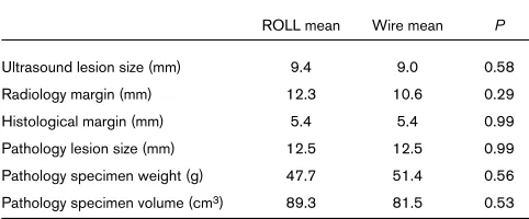
lesion localisation (ROLL) at the beginning of 2008 for ultrasound visible lesions.
Methods All ultrasound guided localisations from January 2006 to December 2008 were reviewed. Wire localisation had been used between January 2006 and December 2007, being superseded by ROLL for a 1-year period leading up to December 2008. For all wire and ROLL localisations, lesion size on ultrasound, radiological margin on specimen radiography, histological margin, lesion size and both specimen volume and weight were recorded. Benign lesions and skin marking only were excluded.
Results See Table 1. During the study period there were 69 ROLL procedures (30 exclusions) and 56 Wire localisations (23 exclusions).
Table 1 (abstract P12)
ROLL mean Wire mean P
Ultrasound lesion size (mm) 9.4 9.0 0.58
Radiology margin (mm) 12.3 10.6 0.29
Histological margin (mm) 5.4 5.4 0.99
Pathology lesion size (mm) 12.5 12.5 0.99
Pathology specimen weight (g) 47.7 51.4 0.56
Pathology specimen volume (cm3) 89.3 81.5 0.53
Conclusion ROLL achieved marginally superior results in terms of specimen weight although no significant differences were demon-strated in the measured indices. Our department is very happy with the technique as early experience suggests ROLL is better tolerated by patients and is surgically less restrictive with potential for a better cosmetic result.
P13
Breast screening unit size and performance on
self-assessment (PERFORMS)
HJ Scott, AG Gale
Loughborough University, Loughborough, UK
Breast Cancer Research2009, 11(Suppl 2):P13 (doi: 10.1186/bcr2383) Introduction The UK Breast Screening Programme (UKBSP) is comprised of approximately 100 individual Breast Screening Units (BSUs) that vary in size (measured by number of women screened). Previous research using UKBSP real life data (Blanks, Bennett, Wallis and Moss, 2002), attributed differences in performance, related to BSU size, to smaller units’ slightly lowered cancer detection rates and positive predictive value scores (compared to large/medium units). All BSUs on the UKBSP annually take part in the PERFORMS scheme as way of self-assessing their film-reading skills. We looked at the performance of all film-readers who had completed the last PERFORMS round (SA08) by BSU size in order to explore any group differences mediated by unit capacity.
Methods Each BSU’s size was approximated by ranking each unit by number of readers who had completed the last PERFORMS round. Subsequently, these BSUs were allocated into three main groups approximating their unit’s size: small = 1 to 4 readers, 30% (n= 157); medium = 5 to 7 readers, 34.5% (n= 181); and large = 8 or more readers), 35% (n = 186). Several performance measures were compared, including ‘percentage correct recall’ and ‘malignancies detected’ (measures of sensitivity), ‘percentage correct return to screen’ (a measure of specificity), and negative and positive predictive value scores.
Results Analysis of variance (one-way) did not produce any significant findings (P = not significant) for any of the measures, indicating equivocal performance. Descriptive statistics showed smaller units scored less than 1% below medium/large BSUs for malignancies detected, correct recall and negative predictive value only.
Conclusion Unlike real-life screening, smaller units perform at a similar level to all others on self-assessment.
P14
A survey of UK breast surgeons and radiologists to
determine current and aspired mammography
surveillance practice after treatment for primary
breast cancer
C Robertson1, R Thomas1, S Heys1, A Maxwell2, F Gilbert1, and the Mammographic Surveillance Health Technology
Assessment Group1
1University of Aberdeen, Aberdeen, UK, 2Royal Bolton Hospital,
Bolton, UK
Breast Cancer Research2009, 11(Suppl 2):P14 (doi: 10.1186/bcr2384) Introduction There is considerable debate about the optimal organisation of a surveillance mammography service following breast cancer treatment in the UK. The optimal frequency and duration of surveillance mammography is unclear, leading to variation in follow-up protocols. The aim of our survey was to describe the variation in current mammography surveillance practice.
Methods A web-based, anonymous survey of members of the Association of Breast Surgery (ABS) at the British Association of Surgical Oncology (569) and Royal College of Radiology (RCR) Breast Group (479). Participants were invited to complete the survey via an email-based web-link sent by membership administrators. Results The survey was sent to 1,048 members from 106 NHS trusts and 185 (18%) responded: 64 (35%) radiologists, 119 (64%) surgeons, 2 (1%) other. The majority of respondents (158, 85%) initiated surveillance mammography (SM) 12 months after completion of surgery; 140 (76%) conduct SM annually following breast conserving surgery. Following mastectomy most conduct SM annually (103, 56%), or biennially (48, 26%). Most discharge from clinical follow-up at 5 (85, 46%) or 10 years (29, 16%) and from SM follow-up at 5 (45, 24%) or 10 (66, 36%) years. Forty-three percent of respondents followed one of two patterns of surveillance: initiate SM at 12 months, annual SM, with discharge at 5 years (34 of185); or initiate at 12 months, annual SM, with discharge at 10 years (46 of 185). Respondents varied greatly in the combinations of start, frequency, duration and discharge from SM. Conclusion Whilst common patterns in surveillance mammography practice exist, there is considerable variation in the way surveillance is organised.
P15
Performance in digital mammography with and
without film prior mammograms
S Taylor-Phillips1, MG Wallis3, A Duncan2, AG Gale1
1Loughborough University, Loughborough, UK, 2University Hospital
(Coventry), Coventry, UK, 3Addenbrooke’s Hospital, Cambridge, UK
Breast Cancer Research2009, 11(Suppl 2):P15 (doi: 10.1186/bcr2385) Introduction In the transition to digital mammography the prior mammograms are in film format. There are difficulties making comparisons between digital current and film prior mammograms due to differences in image appearance and display brightness. This study investigates cancer detection performance in digital mammography with and without film prior mammograms.
MethodsTwo radiologists and two radiography advanced practitioners read a set of 160 (41% malignant) difficult digital mammography cases twice, once with film prior mammograms and once without. Participants noted whether they would recall each case in the NHS breast screening programme.
ResultsThe number of false negative cases (that is, missed cancers) did not differ between conditions. There was a trend towards a larger number of false positive cases (that is, normal cases that were recalled) when prior mammograms were not available (24% increase,
[image:6.612.56.297.223.323.2]ConclusionInitial results indicate that film prior mammograms may be beneficial to cancer detection performance in digital mammography, and therefore should be displayed. Completion of the study with four extra participants will allow firmer conclusions to be drawn.
P16
Is mammogram indicated in patients presenting with
breast pain alone in the presence of a normal clinical
examination?
H Winter, M Dilworth, K Darvall, M Sintler
Sandwell General Hospital, West Bromwich, UK
Breast Cancer Research2009, 11(Suppl 2):P16 (doi: 10.1186/bcr2386) IntroductionPatients with breast pain are commonly seen in one-stop breast clinics. Breast pain alone, however, is rarely associated with an underlying malignancy. The Royal College of Radiologists do not recommend routine mammography or ultrasound when examination is normal. We analysed the outcomes of referrals to breast clinic over 1 year. Methods A retrospective analysis of 1,673 patients attending the breast clinic over a 12-month period. Patients were subcategorised into those presenting with breast pain alone, and in whom mammo-grams were performed. The results of clinical and radiological examination were correlated.
ResultsThere were 359 (21.5%) patients who presented with breast pain alone. Of these, 251 patients were aged ≥35 years with normal examinations; 230 of these patients (91.6%) had mammograms, 220 (96%) of which were normal. One out of 251 (0.4%) patients who attended with breast pain had microcalcifications on mammography, which was later confirmed as ductal carcinoma in situ. This patient was aged 52 years. She had not previously attended for screening mammography and was classed as low risk.
ConclusionThe incidence of breast cancer at age 50 to 52 years in the NHS Breast Screening Programme is 4 per 1,000 women screened. This is far less in younger age groups. Our study demon-strates that the pick-up rate of breast cancer from mammograms in breast pain alone is low. The case we have identified is likely to be an incidental finding. We continue to screen women with breast pain over the age of 35 years. Should we continue to perform opportunistic screening or should we tailor our screening with age/risk?
P17
Screen-detected breast cancer: does presence of
minimal signs on previous mammograms predict
staging/grading of cancer?
GJ Bansal1, K Gower-Thomas2
1University Hospital of Wales, Cardiff, UK, 2Breast Test Wales, Cardiff, UK
Breast Cancer Research2009, 11(Suppl 2):P17 (doi: 10.1186/bcr2387) Methods The previous mammograms of 148 patients with screen-detected breast cancer were examined at Breast Test Wales, Cardiff. Women who showed ‘minimal signs’ on previous screen mammograms formed the study group. Age, average size of tumour, tumour characteristic, grade and lymph node status were compared to a control group, which had normal previous mammograms. Student t-test and chi-square test were used, with P-value <0.05 being taken as significant. Images were interpreted first by a senior registrar and then by a breast radiologist who was blinded to the results of current mammograms.
ResultsThe average age of all the patients was 68.9 years and the mean interval between the two screening mammograms was 44.75 months; 17.56% showed minimal signs at the site of the tumour on previous screen mammograms and formed the study group. There was no statistically significant difference between the two groups with respect to age, average size of tumour, grade or lymph node status, with P-values being 0.609, 0.781, 0.938 and 0.444, respectively. The only statistically significant difference was more microcalcifications seen in the study group (P= 0.003). Five patients in the study group showed positive lymph node or were greater than 2 cm and, therefore, may have had possible gain from earlier diagnosis.
ConclusionThis study did not demonstrate a statistical difference in grading/staging between cancers with minimal signs on previous mammograms and those with normal previous mammograms.
P18
Haematoma-directed ultrasound guidewire
localisation of breast lesions
CM Lee, A Redman
Breast Screening Unit, Queen Elizabeth Hospital, Gateshead, UK Breast Cancer Research2009, 11(Suppl 2):P18 (doi: 10.1186/bcr2388) Introduction The standard technique for surgical excision of mammographically detected, ultrasound invisible, non-palpable breast lesions is by pre-operative stereotactic guidewire localization (SGL). Disadvantages of SGL include patient discomfort, ionizing radiation, the requirement for more staff and longer procedure time. Ultrasound visible clips are used for localisation after vacuum-assisted core biopsies (VACB) but clip migration and visibility are problems. Post-VACB, the biopsy cavity fills with haematoma, which is ultrasonographi-cally visible and can be used as a ‘marker’ for guidewire localisation. Centres in America have successfully used ultrasound intraoperatively to identify the post-biopsy haematoma and guide surgical excision; but no centres have attempted to use ultrasound pre-operatively to locate the post-biopsy haematoma and direct guidewire placement. We aim to describe this new technique of haematoma-directed ultrasound guidewire localisation (HUGL) and compare its accuracy with SGL. MethodsBetween September 2007 and June 2009, 15 patients with mammographically detected, non-palpable, ultrasound invisible breast lesions had VACB followed by HUGL. We compared this technique with 15 consecutive patients who underwent SGL.
Results Both techniques located all lesions successfully. The mean skin to hook distance and wire overthrow for HUGL were 47.5 mm and 10.4 mm, respectively; the corresponding values for SGL were 67.5 mm and 15.3 mm, respectively. Histology of the final surgical specimen confirmed the presence of targeted lesions in all cases. Conclusion This study demonstrates the effectiveness of HUGL of breast lesions. As this technique is potentially more comfortable, technically easier, faster and cheaper than SGL, consideration should be given to routinely employing this as a first line technique.
P19
Breast imaging and biopsy in women after
therapeutic mammoplasty: comparison with women
after wide local excision
S Taneja, S Doyle, S McCulley, D Macmillan, A Evans
Nottingham Breast Institute, Nottingham, UK
Breast Cancer Research2009, 11(Suppl 2):P19 (doi: 10.1186/bcr2389) Introduction Therapeutic mammoplasty (TM) is being increasingly performed to allow breast conservation in women with breasts suitable for some form of breast reduction.
This study was performed to compare the ipsilateral post-operative mammography findings, frequency of ultrasound and image-guided biopsy post-TM with a group of women who had undergone wide local excision (WLE).
Methods Ninety-one women post-TM were compared with 86 women post-WLE. All women had intact breast irradiation and were of similar age (mean age 57 years and 56 years, respectively), had had at least one post-operative mammogram and the same surgeon was present at all operations (DM).
The presence and type of mammographic calcification, and focal and generalised reaction were noted as were ultrasound (US) examination and image guided biopsies. The chi-square test and Fisher’s exact test were used for statistical analysis.
of US and image-guided biopsy were also similar: 26 of 86 (30%) versus 23 of 91 (25%) and 10 of 98 (12%) versus 9 of 91 (10%), respectively. Having corrected for length of follow-up, no statistically significant difference in the frequency of features analysed was observed. Conclusion TM is not associated with greater post-operative mammo-graphic calcification, US examinations or image-guided biopsy.
P20
Negative axillary ultrasound in primary breast cancer:
how reassured should we really be?
SJ Hall, SE Brown, GJR Porter, J Steel, K Paisley, RM Watkins, C Holgate
Plymouth Hospitals NHS Trust, Plymouth, UK
Breast Cancer Research2009, 11(Suppl 2):P20 (doi: 10.1186/bcr2390) IntroductionAxillary ultrasound has become an important adjunct in the staging of breast cancer in recent years. We retrospectively studied a sample of 155 women with invasive breast cancer and normal axillary ultrasound to investigate whether the characteristics of the primary tumour could predict the likelihood of false negative axillary ultrasound.
Methods Screening and symptomatic patients were identified from pathology records and information collated from pathology and imaging records.
ResultsOf the 155 normal ultrasounds, 45 (29%) were positive at axillary surgery.
True and false negative groups were compared in terms of the following: tumour size, pathological type and grade, lymphovascular invasion and oestrogen receptor (ER) status.
Breast tumour size was significantly different, with the average size in the true negative group 21 mm and in the false negative group 30 mm (P<0.002).
There was no significant difference in tumour grade or ER status. However, the histological type varied significantly between the groups, with excess lobular carcinomas in the false negative group (6 of 110 versus 6 of 45, P< 0.001).
The false negative group was more likely to show lymphovascular invasion in the breast (31% versus 5%, P< 0.001).
ConclusionThere are significant differences in tumour characteristics between women with true negative and those with false negative axillary ultrasound in terms of size, primary tumour histological type and presence of lymphovascular invasion. In particular, axillary assessment in primary lobular carcinoma may be more difficult and a negative result should be interpreted with caution.
P21
Axillary ultrasound in staging breast cancer:
diagnostic accuracy and effect on subsequent axillary
surgery - the Plymouth experience
SJ Hall, SE Brown, GJR Porter, J Steel, K Paisley, RM Watkins, C Holgate
Plymouth Hospitals NHS Trust, Plymouth, UK
Breast Cancer Research2009, 11(Suppl 2):P21 (doi: 10.1186/bcr2391) Introduction Routine axillary ultrasound and sampling of abnormal nodes began in Plymouth in 2005 for the staging of primary breast cancer.
MethodsWe collected imaging and pathological data on 385 women with invasive breast cancer from before and after the introduction of axillary ultrasound.
Results Without axillary ultrasound, 136 of 208 (65%) women underwent axillary node sampling (ANS) based on mammographic and clinical findings. The remaining 72 (35%) women had axillary clearance surgery (ANC). Following ANS, 29 (21%) were histologically positive, 27 having subsequent ANC. Of those who had initial ANC, 37 of 72 (51%) were positive.
In our study 177 axillary ultrasounds were performed, and 112 were normal. Of these, 32 (29%) had ANC, 14 (44%) being histologically
positive. Following abnormal axillary ultrasound, 51 of 65 (78%) under-went ANC with 45 (88%) being positive.
There was no significant difference in primary tumour size or mean patient age between the groups before and after the introduction of ultrasound.
Statistical analysis shows significant reduction (P < 0.02) in the proportion of ANC in the ultrasound group and, importantly, a reduction in histologically negative ANCs (P< 0.01).
In our study, axillary ultrasound had a sensitivity of 54%, a specificity of 83%, a positive predictive value of 71% and a negative predictive value of 71%.
Conclusion Introduction of axillary ultrasound was associated with less frequent ANC. Importantly, there has been significant reduction in histologically negative clearances. Our study supports the use of axillary ultrasound in guiding axillary surgical management.
P22
Screen-detected ductal carcinoma in situ - fine
needle aspiration versus core biopsy
CM Lee, B Kaye, L McLean
Breast Screening Unit, Royal Victoria Infirmary, Newcastle Upon Tyne, UK
Breast Cancer Research2009, 11(Suppl 2):P22 (doi: 10.1186/bcr2392) Introduction Our current practice in the investigation of micro-calcification is to perform fine needle aspiration (FNA), with immediate results, and vacuum-assisted core biopsy (VACB), which was introduced in April 2007. The British Association Of Surgical Oncology 2006/2007 audit reported fewer centres using cytology alone for diagnosis of screen-detected cancer. We assessed the contribution of FNA to non-operative diagnosis of microcalcification, and the benefit to women in having immediate FNA results. We also investigated the effect of introducing VACB, rather than core biopsy for sampling microcalcification, on the number of repeat cores, and on the non-operative diagnosis of microcalcification.
MethodsWe reviewed the accuracy of FNA versus core biopsy of 150 patients with microcalcification between April 2008 and March 2009 by reference to imaging and histology findings and comparison with results from the previous 2 years.
ResultsIn 2008/2009, 49% cases were malignant microcalcification. The non-operative diagnostic FNA and core biopsy results for 2008/2009 were 42% and 90%, respectively. The corresponding values for 2007/2008 were 28% and 88%, respectively, and for 2006/2007 39% and 63%, respectively. Introduction of VACB has resulted in higher non-operative diagnostic malignant cores - 90% in 2008/2009 and 88% in 2007/2008 versus 63% in 2006/2007 - and fewer repeat cores - 11% in 2008/2009 and 16% in 2007/2008 versus 33% in 2006/2007.
Conclusion The continued use of FNA in the investigation of micro-calcification will be reviewed in terms of its role in giving a same-day diagnosis in clinics, particularly when VACB is used for first-line diagnosis.
P23
Can we predict the likelihood of malignancy in
mammographically indeterminate microcalcification?
AN Khan, M Hoosein, N Hartley, S Tennant, H Daintith, E Denton, M Al-AttarUniversity Hospitals of Leicester, Leicester, UK
Results The total number of patients with M3 microcalcification was 242. On final histology, 99 (41%) patients had breast cancer and 143 (59%) had benign breast disease. In the cancer group, the mean age was 57 years (95% confidence interval (CI) 55.7 to 58.4 years), the mean cluster size was 18.62 mm (95% CI 14.86 to 22.38), and 84% of patients had a new microcalcification and16% had an increase in size of microcalcification. Of the malignant M3 microcalcifications, 65% were in the outer quardrant, 26% were in the medial quardrant and 9% were central. In the benign group, the mean age was 54.5 years (95% CI 53.9 to 55.8), the mean size of microcalcification was 18.62 mm (95% CI 14.86 to 22.38), 76% had a new microcalcification and 22% had an increase in size of microcalcification. Of the benign M3 microcalcifications, 51% were in the outer quadrant, 35% were in the medial quadrant and 14% were central. Except for age, there was no statistically significant difference between the studied variables in the two groups. On the KruskalWallis test, patients with cancer were older compared to patients with benign disease (P= 0.01).
Conclusion Increasing age is the only strong predictor of malignancy in M3 microcalcification. Size, site, new microcalcification and increasing microcalcification size were not related with the probability of cancer.
P24
Abstract withdrawn
P25
Preoperative ultrasound assessment of axillary
lymph nodes in breast cancer: histopathological
correlation in 100 cases
A O’Connor
University Hospitals Aintree NHS Foundation Trust, Liverpool, UK Breast Cancer Research2009, 11(Suppl 2):P25 (doi: 10.1186/bcr2395) Introduction With the widespread adoption of sentinel lymph node biopsy, it is important to identify involved nodes preoperatively so that surgery can be planned appropriately.
Methods One hundred consecutive cases of breast cancer were selected where the patient had axillary sampling or clearance. Ultrasound appearances of ipsilateral lymph nodes were scored U1 to U5 according to length/width ratio, cortical depth, size, and absence of fatty hilum. Fine needle aspiration cytology was performed if the nodes scored U3 to U5. Ultrasound and cytology findings were correlated with the resection specimens.
ResultsOf the 100 cases, 44 had positive lymph nodes at surgery. Fourteen of these had a preoperative C5 result. All 14 cases coded U4 or U5 had positive nodes, although 6 of these had cytology of C1 to C4. U3 with C3 to C5 occurred in seven cases, which were all positive. U3 with C1 to C2 occurred in 22 cases, of which 8 were positive. Fifty-seven cases were graded U1 or U2, of which 16 were positive. C1 occurred in six cases, of which four were positive. All 21 cases with C3 to C5 and/or U4 to U5 were positive. C2 falsely reassured in 7 of 19 cases where the nodes were subsequently shown to be involved. Sensitivity and specificity of a U3 to U5 result for positive nodes were 63% and 73%, respectively.
Conclusion About one-third of positive patients avoided a second operation. Ways of improving the results are considered, including repeating all C1 cases, repeating C2 cases especially if U4 or U5, and fine needle aspiration even when nodes appear normal or benign.
P26
A 5-year case review of granulomatous mastitis
A O’Connor, J LeadbetterRoyal Liverpool and Broadgreen University Hospitals NHS Trust, Liverpool, UK
Breast Cancer Research2009, 11(Suppl 2):P26 (doi: 10.1186/bcr2396) Introduction Granulomatous mastitis is a rare inflammatory condition said to predominate in women of reproductive age, often presenting as a lump that may mimic malignancy.
Methods The histopathology database was searched for ‘granuloma-tous mastitis’ over 5 years. Case notes, imaging, and pathology were reviewed.
ResultsSeven cases were identified. The age range was 34 to 72 years, and mean age was 54 years. Presentation was a lump in two cases, chronic breast infection in three, indeterminate mass on screening mammogram in one, and a suspicious mass and large axillary node on screening mammogram in one. Two cases had imaging features suggesting malignancy; one was a lump at the site of previous breast cancer excision and the other was a screen-detected mass in a patient with a chronic skin condition. Fine needle aspiration cytology was C2 in six cases and C1 in one case. Five of the C2 results gave a diagnosis of granulomatous mastitis, the sixth was abscess. Core biopsy was carried out in three cases: a suspicious screening case with C2; for exclusion of malignancy in a probable abscess; and multiple suspicious masses with C1 cytology. In all three cases core biopsy showed B2 granulomatous mastitis. Only one case went for operation: multifocal suspicious masses in a patient with previous ipsilateral breast cancer, with histopathology confirming granulomatous mastitis.
Conclusion This series confirms that granulomatous mastitis is uncommon. Only two patients were premenopausal. In three patients there was clinically apparent chronic inflammation. In only two cases was malignancy considered the most likely diagnosis.
P27
Axillary lymph node ultrasound and fine needle
aspiration in pre-operative breast cancer staging
AAM Leaver, L McLeanRoyal Victoria Infirmary, Newcastle-upon-Tyne, UK
We present an audit of these results that prompted a change in local criteria for FNA.
Methods Patients who had entered the treatment pathway and undergone axillary surgery between October 2008 and May 2009 were identified from multidisciplinary team (MDT) meeting records. We investigated whether axillary ultrasound was always performed, and if criteria for FNA yielded sensitivities and specificities that compared favourably with the literature.
ResultsRecords were available for 133 female patients with invasive breast cancer, 89% (118 patients: 67 screening, 51 symptomatic) of whom underwent axillary ultrasound. Axillary ultrasound sensitivity was 49% (23 of 47: symptomatic 58% (18 of 31), screening 31% (5 of 16)), and specificity was 93% (66 of 71: symptomatic 80% (16 of 20), screening 98% (50 of 51)). Axillary FNA sensitivity was 88% (14 of 16: symptomatic 85% (11 of 13), screening 100% (3 of 3), and specificity was 100% (2 of 2).
Combination of ultrasound and FNA had an overall sensitivity of 45% (21 of 47), and a specificity of 96% (68 of 71).
Ultrasound had a relatively high false negative rate (22%, 26 of 118) and therefore led to low rate of subsequent FNA. FNA had a low false negative rate (11%, 2 of 18).
Conclusion These results, with literature review, have been used to alter local ultrasound guidelines for FNA. Further audit to validate these new guidelines is ongoing.
P28
Correlation of age and hormone replacement therapy
with breast density as assessed by Quantra™
PL Skippage1, LS Wilkinson1, SD Allen2, N Roche2, M Dowsett3, R A’Hern3
1South West London Breast Screening Unit, Tooting, London, UK, 2Royal Marsden NHS Foundation Trust, Sutton, Surrey, UK, 3Institute
of Cancer Research, Fulham, London, UK
Breast Cancer Research2009, 11(Suppl 2):P28 (doi: 10.1186/bcr2398) Breast density is a significant predictor in the risk of developing breast cancer. Several methods are available for assessing breast density but all are subject to intra-observer variability and are unable to assess the breast as a three-dimensional structure. Using Quantra™ to quantify breast density, we have correlated this with risk factors to determine what impact these variables have on breast density.
Women attending for full-field digital mammography at the South West London Breast Screening Unit between December 2008 and March 2009 were invited to participate in the study by questionnaire. Consenting women returned the questionnaire, allowing further data collection, including demographics, menopausal status and hormone replacement therapy use. Data were correlated against breast density measurements to determine the degree of association. Mammograms were assessed on a Hologic™ workstation and breast density calculated using Quantra™. Quantra™ is an automated algorithm for the volumetric assessment of breast tissue composition from digital mammograms. We invited 683 women to participate (those with implants or mastectomy were excluded) and 321 completed returned questionnaires were assessed. The mean age of participants was 59 years (range 49 to 81). Mean density was 19.4% (range 8.5 to 49.0%). There was a decrease in density with age (Spearman rank correlation coefficient -0.21). Correlation between density and hormone replacement therapy use showed a significant positive result.
Quantra™ has shown to be an accurate, reproducible tool for quantifying breast density, demonstrated by its correlation with lifestyle and demographic data. Given its ease of acquisition, this may be the future of breast density quantification in the digital age.
P29
The feasibility of vision-supported computer-based
training in digital mammography
Y Chen1, AG Gale1, A Evans2
1Loughborough University, Loughborough, UK, 2Nottingham Breast
Institute, Nottingham, UK
Breast Cancer Research2009, 11(Suppl 2):P29 (doi: 10.1186/bcr2399) Introduction Full-field digital mammography (FFDM) screening necessitates training more users to interpret digital images, as well as facilitating computer-based training employing a range of display devices. The feasibility of training naïve observers to examine mammographic images using different forms of vision-supported training on a PC monitor was examined.
Methods A set of recent screening cases were first examined by an experienced radiologist and both his visual search behaviour and verbal commentary recorded. Twenty naive observers were then familiarised with abnormal mammographic appearance, concentrating on masses and calcifications. They were then split into four different training groups (examining mammographic images with: overlay of the radiologist’s visual search; playback of the radiologist’s commentary; mammographic regions of interest highlighted; or only regions of interest presented) using 20 two-view cases and a control group. Before and after training, each participant was tested on a set of 21 cases and required to identify whether an abnormality was present. Participants’ eye-movements were recorded and a 21” LCD monitor was used throughout to view the images.
Results Examination of visual search and performance data, pre- and post-training, indicated that only 14% of responses identified the correct features and their locations. Errors were due to search (>60%), detection (<20%) and interpretation (<18%) factors. Approaches that emphasised the region of interest around an abnormality caused observers to fixate these areas for longer periods and produced fewer errors.
Conclusion The introduction of FFDM allows a variety of displays and computer-based approaches to be used for training purposes.
P30
Breast cancer screening - how do we communicate
with women of South Asian origin?
AK Jain, J Serevitch
The Nightingale Centre and Genesis Prevention Centre, Manchester, UK Breast Cancer Research2009, 11(Suppl 2):P30 (doi: 10.1186/bcr2400) Introduction The aim of this study was to investigate how various Breast Screening Units (BSUs) communicate with women of South Asian origin (SAOW).
Methods Structured questionnaires and letters setting the study objectives were sent to the Directors/Office Managers of 99 BSUs in the UK. Further reminders were sent via the RCR Breast Group and directly to BSUs with larger SAOW populations.
Results To date, 60 responses have been received, with 59 completed questionnaires. The BSU size varies considerably, with 3 BSUs inviting less than 10,000 women and 6 inviting over 50,000 annually. The percentage of invited SAOW also varies, from <5% to 25 to 30%. Only one BSU sends the first invitation/reminder in South Asian languages.
Sixteen BSUs (27%) record the patient’s language and 20 (34%) give leaflets in their language when they attend for mammography. Four BSUs give them normal recall letters in their language. Three BSUs send their assessment recall letter and nine give them biopsy leaflets in their language. Three BSUs send them a normal assessment results letter in their language.
Seventy-six percent of the BSU think it would be useful to record a patient’s language for improving services.
No BSU has separate funding for targeting SAOW for breast screening; only one has a dedicated team to do so.