RESEARCH NOTE
Immunohistochemical over expression
of p53 in head and neck Squamous cell
carcinoma: clinical and prognostic significance
Atif Ali Hashmi
1, Zubaida Fida Hussain
1, Shumaila Kanwal Hashmi
2, Muhammad Irfan
1, Erum Yousuf Khan
1,
Naveen Faridi
1, Amir Khan
3*and Muhammad Muzzammil Edhi
4Abstract
Objective: Immunohistochemical over expression of p53 is considered as a marker of poor prognosis in many cancers. Therefore, we aimed to evaluate immunohistochemical overexpression of p53 in 121 cases of head and neck Squamous cell carcinoma and its association with various clinicopathologic features and survival.
Results: Total 66.1% (80 cases) expressed positive p53 expression, 34% (29 cases) revealed no p53 expression, while focal positive p53 expression was noted in 9.9% (12 Cases). Moreover, high p53 expression (> 70%) was noted in 26.4% (32 cases), while 19% (23 cases) showed 51–70% p53 expression. On the basis of intensity of p53 staining; strong p53 expression was noted in 39.7% (48 cases), while 24.8% (30 cases) and 10.7% (13 cases) revealed intermediate and weak p53 expression respectively. Significant association of p53 intensity of expression with extranodal extension and higher tumor grade (grades II and III) was noted. p53 is useful prognostic biomarker in head and neck Squamous cell carcinoma and therefore we suggest that more large scale studies are needed to evaluate its prognostic significance in our population.
Keywords: Head and Squamous cell carcinoma, Oropharyngeal Squamous cell carcinoma, p53, Areca nut, Gutka
© The Author(s) 2018. This article is distributed under the terms of the Creative Commons Attribution 4.0 International License (http://creat iveco mmons .org/licen ses/by/4.0/), which permits unrestricted use, distribution, and reproduction in any medium, provided you give appropriate credit to the original author(s) and the source, provide a link to the Creative Commons license, and indicate if changes were made. The Creative Commons Public Domain Dedication waiver (http://creat iveco mmons .org/ publi cdoma in/zero/1.0/) applies to the data made available in this article, unless otherwise stated.
Introduction
Despite advancements in cancer treatment, head and neck Squamous cell carcinoma (HNSCC) remains sig-nificant cause of morbidity and mortality worldwide, and hence 5 years survival rate is still below 50% [1, 2]. Owing to the widespread use of chewing tobacco and areca nut (pan/gutka) in South-Asia, oropharyngeal Squamous cell carcinoma (OSCC) is on a rise in this part of the world [3]. Oral carcinogenesis is a multistep process involving multiple proto-oncogenes and tumor suppressor genes including p16, cyclin D1, p53 and EGFR. TP53 is a tumor suppressor gene located on chromosome 17p. Mutations of TP53 gene is one of the most common event in human carcinogenesis. As mutated protein is not easily digesti-ble, therefore it accumulates inside the cancer cell leading
to immunohistochemical overexpression. p53 overex-pression in OSCC is considered a marker of poor prog-nosis [4]. Moreover, p53 overexpression may result in decreased sensitivity of tumor cells to chemotherapeutic drugs [5]. Therefore, we aimed to evaluate immunohisto-chemical overexpression of p53 in our cases of HNSCC and its association with various clinicopathologic fea-tures and survival so that therapeutic protocols could be devised for loco-regional population.
Main text
Patients and methods
This was a retrospective study in which, 144 cases of HNSCC excision specimens were identified from previ-ous records of pathology department. All patients had elective surgeries at Liaquat National hospital, Kara-chi from January 2008 till December 2013 over a period of 7 years. The approval of the study was taken from research and ethical review committee of the institution.
Open Access
*Correspondence: dramirkhan04@gmail.com 3 Kandahar University, Kandahar 3802, Afghanistan
Informed written consent was taken from all of the patients. Hematoxylin and eosin stained slides of all cases and paraffin blocks of 77 cases were recruited and new sections were cut when felt necessary. Slides of all cases were evaluated by two senior histopathologists indepen-dently and pathologic characteristics like tumor type, grade, tumor-stage, nodal-stage, lymphovascular invasion and perineural were interpreted. Clinical records of 57 patients were available and are thus reviewed from insti-tutional records to evaluated patients age, smoking, alco-hol and gutka/pan use history, history of radiation and chemotherapy. Moreover, representative tissue blocks of 121 cases were available for p53 immunohistochemistry.
Immunohistochemistry
p53 IHC was performed using DAKO EnVision method using DAKO anti-human p53 protein, clone DO-7 according to manufacturers protocol. Nuclear staining for p53 was both quantitatively and qualitatively evalu-ated. Intensity of staining was grouped into no stain-ing (0), weak (1+), intermediate (2+), strong (3+) while percentage of positively stained cells were calculated as a continuous variable. Intermediate to strong staining in more than 10% cancer cells was taken as positive while weak to intermediate staining in less than 10% tumor cells was interpreted as focal positive (Additional file 1: Figure S1). No staining in cancer cells was taken as nega-tive. Moreover, p53 immunostaining was also categorized according to percentage of staining cells into different groups.
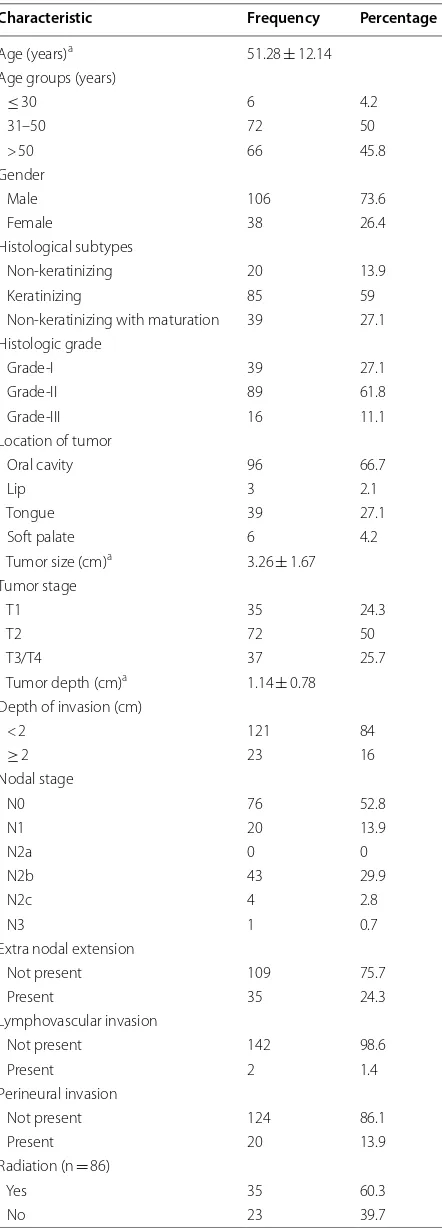
Table 1 Clinicopathologic features of Squamous cell carcinoma head and neck (n = 144)
Characteristic Frequency Percentage (%)
Age (years)a 51.28 ± 12.14
Age groups (years)
≤ 30 6 4.2
31–50 72 50
> 50 66 45.8
Gender
Male 106 73.6
Female 38 26.4
Histological subtypes
Non-keratinizing 20 13.9 Keratinizing 85 59 Non-keratinizing with maturation 39 27.1 Histologic grade
Grade-I 39 27.1
Grade-II 89 61.8 Grade-III 16 11.1 Location of tumor
Oral cavity 96 66.7
Lip 3 2.1
Tongue 39 27.1
Soft palate 6 4.2 Tumor size (cm)a 3.26 ± 1.67
Tumor stage
T1 35 24.3
T2 72 50
T3/T4 37 25.7
Tumor depth (cm)a 1.14 ± 0.78
Depth of invasion (cm)
< 2 121 84
≥ 2 23 16
Nodal stage
N0 76 52.8
N1 20 13.9
N2a 0 0
N2b 43 29.9
N2c 4 2.8
N3 1 0.7
Extra nodal extension
Not present 109 75.7
Present 35 24.3
Lymphovascular invasion
Not present 142 98.6
Present 2 1.4
Perineural invasion
Not present 124 86.1
Present 20 13.9
Radiation (n = 86)
Yes 35 60.3
No 23 39.7
Table 1 (continued)
Characteristic Frequency Percentage (%)
Chemotherapy (n = 86)
Yes 34 58.6
No 24 41.4
Recurrence (n = 86)
Yes 33 56.9
No 25 43.1
History of pan (n = 57)
Yes 34 59.6
No 23 40.4
History of smoking (n = 57)
Yes 4 7
No 53 93
History of alcohol (n = 57)
Yes 1 1.8
No 56 98.2
[image:2.595.302.539.100.309.2] [image:2.595.58.279.111.725.2]Statistical analysis
Statistical package for social sciences (SPSS 21) was used for data compilation and analysis. Mean and standard deviation were evaluated for quantitative variables. For qualitative variables, frequency and percentage were cal-culated. Chi square was applied to determine association. P value of ≤ 0.05 was considered as significant. Statistical power of each variable was calculated by using software PASS version 11. Power of each tested variable is men-tioned in Tables 2 and 3.
Demographic profile of patients
Mean age of the patients included in our study was
found to be 51.28 ± 12.14. Most common age group
was between 31 and 50 years. Male gender was more
common. Oral cavity was the most common origin of SCC found in our study (96 cases). High tumor stage (T3/T4) was noted in 25% (37 cases). Mean tumor size was 3.26 ± 1.67 cm while mean tumor depth was found to be 1.14 ± 0.78 cm. High nodal stage (N2/N3) was seen in 34.4% cases. 85 cases revealed keratinizing phenotype, while 11.1% cases were of high grade (grade III). Perineu-ral and lymphovascular invasion was seen in 13.9 and 1.4% cases respectively as represented in Table 1.
Immunohistochemical expression of p53 in head and neck Squamous cell carcinoma
Total 66.1% (80 cases) expressed positive p53 expression, 34% (29 cases) revealed no p53 expression, while focal positive p53 expression was noted in 9.9% (12 Cases).
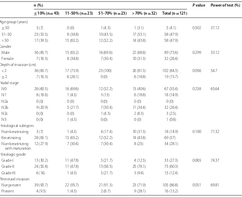
Table 2 Association of p53 over expression categories with clinic pathologic parameters of head and neck Squamous cell carcinoma
Chi square test was applied
P-value ≤ 0.05 considered as significant
n (%) P value Power of test (%)
≤ 10% (n = 43) 11–50% (n = 23) 51–70% (n = 23) > 70% (n = 32) Total (n = 121)
Age group (years)
≤ 30 3 (7) 0 (0) 1 (4.3) 1 (3.1) 5 (4.1) 0.502 37.12 31–50 23 (53.5) 8 (34.8) 10 (43.5) 17 (53.1) 58 (47.9)
> 50 17 (39.5) 15 (65.2) 12 (52.2) 14 (43.8) 58 (47.9) Gender
Male 36 (83.7) 15 (65.2) 16 (69.6) 22 (68.8) 89 (73.6) 0.299 33.12 Female 7 (16.3) 8 (34.8) 7 (30.4) 10 (31.3) 32 (26.4)
Depth of invasion (cm)
< 2 36 (83.7) 17 (73.9) 23 (100) 26 (81.3) 102 (84.3) 0.056 54.7
≥ 2 7 (16.3) 6 (26.1) 0 (0) 6 (18.8) 19 (15.7) Nodal stage
N0 26 (60.5) 16 (69.6) 12 (52.2) 13 (40.6) 67 (55.4) 0.258 65.64 N1 8 (18.6) 1 (4.3) 3 (13) 6 (18.8) 18 (14.9)
N2a 0 (0) 0 (0) 0 (0) 0 (0) 0 (0) N2b 9 (20.9) 5 (21.7) 7 (30.4) 11 (34.4) 32 (26.4) N2c 0 (0) 0 (0) 1 (4.3) 2 (6.3) 3 (2.5) N3 0 (0) 1 (4.3) 0 (0) 0 (0) 1 (0.8) Histological subtypes
Non-keratinizing 3 (7) 1 (4.3) 4 (17.4) 10 (31.3) 18 (14.9) 0.100 71.32 Keratinizing 28 (65.1) 15 (65.2) 12 (52.2) 14 (43.8) 69 (57)
Non-keratinizing
with maturation 12 (27.9) 7 (30.4) 7 (30.4) 8 (25) 34 (28.1) Histologic grade
Grade-I 13 (30.2) 11 (47.8) 5 (21.7) 4 (12.5) 33 (27.3) 0.065 74.37 Grade-II 24 (55.8) 11 (47.8) 13 (56.5) 25 (78.1) 73 (60.3)
Grade-III 6 (14) 1 (4.3) 5 (21.7) 3 (9.4) 15 (12.4) Perineural invasion
[image:3.595.58.538.307.698.2]Moreover, high p53 expression (> 70%) was noted in 26.4% (32 cases), while 19% (23 cases) showed 51–70% p53 expression. 11–50 and < 10%/no expression was noted in 19% (23 cases) and 35.5% (43 cases) respectively. On the basis of intensity of p53 staining; strong p53 expression was noted in 39.7% (48 cases), while 24.8% (30 cases) and 10.7% (13 cases) revealed intermediate and weak p53 expression respectively. Tables 2 and 3 repre-sent association of p53 expression with various clinico-pathologic parameters in HNSCC. Significant association of p53 intensity of expression with extranodal extension and higher tumor grade (grades II and III) was noted. Association with those clinicopathological parameters with statistical power of less than 50 were not included in Tables 2 and 3.
Discussion
In the present study, we found that 66.1% of HNCC revealed p53 overexpression; moreover significant asso-ciation of p53 was noted with extranodal extension and tumor grade, which are key prognostic factors of
HNSCC, thus proving the prognostic significance of this biomarker.
[image:4.595.56.538.113.414.2]p53 overexpression in HNSCC varies in different parts of the world, owing to divergent risk factors and patho-genesis of the disease. Kerdpon et al. reported a positive association of alcohol use with p53 over expression and negative association with betal nut and tobacco [6]. P53 expression in HNSCC ranges from 25 to 90%. Dragomir et al. reported 31% p53 expression [7], whereas, Gonza-lez-Moles revealed 57.7% expression of p53 [8]. On the other hand, 85.6% p53 expression was reported in a study from India [5], 63.3% expression was noted in a research conducted in Brazilian population [9], while as low as 28.5% expression was noted in a study conducted in Iran [10]. Varied expression of p53 in HNSCC may be due to different use of techniques, methods of interpretation or due to difference in ethnicity and risk factors involved in HNSCC pathogenesis. Previous literature also showed association of p53 expression with tumor grade and other histologic features. Dave et al. in a study involv-ing 40 cases of HNCC found a significant association of p53 expression with tumor grade and other histologic Table 3 Association of p53 expression intensity with clinicopathologic parameters of head and neck Squamous cell carcinoma
Chi square test was applied
P-value ≤ 0.05 considered as significant
n (%) P-value Power of test (%)
No intensity
(n = 30) Weak (n = 13) Intermediate (n = 30) Strong (n = 48) Total (n = 121)
Age group (years)
≤ 30 3 (10) 0 (0) 1 (3.3) 1 (2.1) 5 (4.1) 0.445 43.47 31–50 15 (50) 8 (61.5) 11 (36.7) 24 (50) 58 (47.9)
> 50 12 (40) 5 (38.5) 18 (60) 23 (47.9) 58 (47.9) Gender
Male 25 (83.3) 11 (84.6) 22 (73.3) 31 (64.6) 89 (73.6) 0.269 38.17 Female 5 (16.7) 2 (15.4) 8 (26.7) 17 (35.4) 32 (26.4)
Nodal stage
N0 17 (56.7) 9 (69.2) 22 (73.3) 19 (39.6) 67 (55.4) 0.053 82.44 N1 8 (26.7) 0 (0) 2 (6.7) 8 (16.7) 18 (14.9)
N2a 0 (0) 0 (0) 0 (0) 0 (0) 0 (0) N2b 5 (16.7) 4 (30.8) 6 (20) 17 (35.4) 32 (26.4) N2c 0 (0) 0 (0) 0 (0) 3 (6.3) 3 (2.5) N3 0 (0) 0 (0) 0 (0) 1 (2.1) 1 (0.8) Extranodal extension
Not present 22 (73.3) 11 (84.6) 28 (93.3) 29 (60.4) 90 (74.4) 0.008 81.53 Present 8 (26.7) 2 (15.4) 2 (6.7) 19 (39.6) 31 (25.6)
Histologic grade
Grade-I 6 (20) 7 (53.8) 12 (40) 8 (16.7) 33 (27.3) 0.046 74.91 Grade-II 20 (66.7) 4 (30.8) 14 (46.7) 35 (72.9) 73 (60.3)
parameters like degree of keratinization [11]. Although we found significant association of p53 expression with tumor grade, however association with other histologic parameters was not noted. Although no significant asso-ciation was noted between p53 expression and other clin-icopathological parameters in our study, however as the statistical power for these associations was less than 50% therefore, no conclusion could be derived.
Many studies have proved the prognostic significance of p53 in HNCC, owing to association of p53 overexpres-sion with overall survival, recurrence, high tumor grade and T and N stage [5, 8, 12]. Conversely, a meta-analysis involving 174 studies revealed no association of p53 over-expression as a marker of poor prognosis [13]. We found a significant association of intensity of p53 expression with extranodal extension and tumor grade, with a large sample size and significant statistical power the asso-ciations are important; therefore we suggest more large scale studies to evaluate prognostic significance of p53 expression in HNSCC and its association with disease free survival in loco-regional population.
Limitation
Major limitation of the study was that, this was a single center data, however as it’s a major tertiary care hospi-tal of the province therefore the results may have major clinical implications. Furthermore, recurrence status of patients was not available to evaluate association p53 expression with disease free survival.
Abbreviations
HNSCC: head and neck Squamous cell carcinoma; OSCC: oropharyngeal Squamous cell carcinoma.
Authors’ contributions
AAH and ZFH: main author of manuscript, have made substantial contribu-tions to conception and design of study. SKH, MI, EYK, NF, AK and MME: have been involved in requisition and analysis of the data and revision of the manuscript. All authors read and approved the final manuscript.
Author details
1 Liaquat National Hospital and Medical College, Karachi, Pakistan. 2 CMH Multan Institute of Medical Sciences, Multan, Pakistan. 3 Kandahar University, Kandahar 3802, Afghanistan. 4 Brown University, Providence, RI, USA.
Acknowledgements
We gratefully acknowledge all staff members of Pathology, Liaquat National Hospital, Karachi, Pakistan for their help and cooperation.
Competing interests
The authors declare that they have no competing interests. Additional file
Additional file 1: Figure S1. p53 expression in oral Squamous cell carcinoma.
Availability of data and materials
Please contact author, Atif Ali Hashmi (doc_atif2005@yahoo.com) for data requests.
Consent to publish
Not applicable.
Ethics approval and consent to participate
Ethics committee of Liaquat National Hospital, Karachi, Pakistan approved the study. Written informed consent was obtained from the patients for the participation.
Funding
There was no funding available for this manuscript.
Publisher’s Note
Springer Nature remains neutral with regard to jurisdictional claims in pub-lished maps and institutional affiliations.
Received: 2 April 2018 Accepted: 27 June 2018
References
1. Ferlay J, Shin HR, Bray F, Forman D, Mathers C, Parkin DM. GLOBOCAN 2008, Cancer Incidence and Mortality Worldwide: IARC CancerBase No. 10. Lyon: International Agency for Research on Cancer; 2010. 2. Warnakulasuriya S. Global epidemiology of oral and oropharyngeal
cancer. Oral Oncol. 2009;45:309–16.
3. Gupta N, Gupta R, Acharya AK, Patthi B, Goud V, Reddy S, Garg A, Singla A. Changing trends in oral cancer—a global scenario. Nepal J Epidemiol. 2017;6(4):613–9.
4. Abbas NF, Labib El-Sharkawy S, Abbas EA, El-Shaer AM. Immunohisto-chemical study of p53 and angiogenesis in benign and preneoplastic oral lesions and oral squamous cell carcinoma. Oral Surg Oral Med Oral Pathol Oral Radiol Endod. 2007;103:385–90.
5. Khan H, Gupta S, Husain N, Misra S, Mps N, Jamal N, Ghatak A. Correlation between expressions of cyclin-D1, EGFR and p53 with chemoradiation response in patients of locally advanced oral squamous cell carcinoma. BBA Clin. 2014;21(3):11–7.
6. Kerdpon D, Sriplung H, Kietthubthew S. Expression of p53 in oral squamous cell carcinoma and its association with risk habits in southern Thailand. Oral Oncol. 2001;37(7):553–7.
7. Dragomir LP, Simionescu C, Mărgăritescu C, Stepan A, Dragomir IM, Popescu MR. P53, p16 and Ki67 immuno expression in oral squamous carcinomas. Rom J Morphol Embryol. 2012;53(1):89–93.
8. Gonzalez-Moles MA, Galindo P, Gutierrez-Fernandez J, Sanchez-Fernan-dez E, Rodriguez-Archilla A, Ruiz-Avila I, Bravo M. P53 protein expres-sion in oral squamous cell carcinoma survival analysis. Anticancer Res. 2001;21(4B):2889–94.
9. Abrahao AC, Bonelli BV, Nunes FD, Dias EP, Cabral MG. Immuno-histochemical expression of p53, p16 and hTERT in oral squamous cell carcinoma and potentially malignant disorders. Braz Oral Res. 2011;25(1):34–41.
10. Etemad-Moghadam S, Keyhani A, Yazdani K, Alaeddini M. Status of p53 and p27 (KIP1) in Iranian patients with oral squamous cell carcinoma. Iran Red Crescent Med J. 2015;17(10):e19359.
11. Dave KV, Chalishazar M, Dave VR, Panja P, Singh M, Modi TG. Immunohis-tochemical expression of p53 and its clinicopathological correlation with modified Anneroth’s histological grading system. J Oral Maxillofac Pathol. 2016;20(1):29–35.
12. Monteiro LS, Diniz-Freitas M, Garcia-Caballero T, Warnakulasuriya S, Forteza J, Fraga M. Combined cytoplasmic and membranous EGFR and p53 overexpression is a poor prognostic marker in early stage oral squa-mous cell carcinoma. J Oral Pathol Med. 2012;41(7):559–67.