City, University of London Institutional Repository
Citation:
Delvecchio, G., Dima, D. & Frangou, S. (2015). The effect of ANK3 bipolar-risk polymorphisms on the working memory circuitry differs between loci and according to risk-status for bipolar disorder. American Journal of Medical Genetics Part B: Neuropsychiatric Genetics, 168B(3), pp. 188-196. doi: 10.1002/ajmg.b.32294This is the accepted version of the paper.
This version of the publication may differ from the final published
version.
Permanent repository link:
http://openaccess.city.ac.uk/15084/Link to published version:
http://dx.doi.org/10.1002/ajmg.b.32294Copyright and reuse: City Research Online aims to make research
outputs of City, University of London available to a wider audience.
Copyright and Moral Rights remain with the author(s) and/or copyright
holders. URLs from City Research Online may be freely distributed and
linked to.
City Research Online: http://openaccess.city.ac.uk/ [email protected]
1
Title: The effect of ANK3 bipolar-risk polymorphisms on the working memory circuitry
differs between loci and according to risk-status for bipolar disorder
Authors: Giuseppe Delvecchio MSc, Danai Dima PhD, Sophia Frangou MD, PhD
Affiliations: Mr Delvecchio and Dr Dima are with the Social Genetic and Developmental
Psychiatry Center, Institute of Psychiatry, King’s College London, UK; Dr Dima is also with the
Psychosis Research Program, Department of Psychiatry, Icahn School of Medicine at Mount
Sinai, USA; Dr Frangou is with the Psychosis Research Program, Department of Psychiatry,
Icahn School of Medicine at Mount Sinai.
Cite: Delvecchio, G, Dima, D & Frangou, S (2015). The effect of ANK3 bipolar-risk
polymorphisms on the working memory circuitry differs between loci and according to
risk-status for bipolar disorder. Am J Med Genet B Neuropsychiatr Genet 168B(3)
Keywords: bipolar disorder, relatives, high-risk, working memory, genetic, polymorphism,
2
Abstract
Polymorphisms at the rs10994336 and rs9804190 loci of the Ankyrin 3 (ANK3) gene have been strongly associated with increased risk for bipolar disorder (BD). However, their
potential pathogenetic effect on BD-relevant neural circuits remains unknown. We
examined the effect of BD-risk polymorphisms at rs10994336 and rs9804190 on the working
memory (WM) circuit using functional magnetic resonance imaging (fMRI) data obtained
from euthymic patients with BD (n=41), their psychiatrically healthy first-degree relatives
(n=25) and unrelated individuals without personal or family history of psychiatric disorders
(n=46) while performing the N-back task. In unrelated healthy individuals, the
rs10994336-risk-allele was associated with reduced activation of the ventral visual cortical components
of the WM circuit while the rs9804190-risk-allele was associated with inefficient
engagement of the prefrontal cortical components of the WM. In patients and their healthy
relatives, risk alleles at either loci were associated with hyperactivation in the ventral
anterior cingulate cortex. Additionally, Rs9804190-risk-allele carriers with BD evidenced
abnormal activation within the posterior cingulate cortex. This study provides new insights
on the neurogenetic correlates of allelic variation at different genome-wide supported
3
Introduction
Allelic variation in the Ankyrin3 (ANK3) gene located on chromosome 10q21.2 has been most convincingly associated with increased risk for bipolar disorder (BD). The first report
concerned a genome-wide association between BD and a single nucleotide polymorphism
(SNP) at rs9804190 identified in two independent samples from the US and Germany (Baum
et al., 2008). This association signal within a 70 kilobase region at the 3’ end of the gene was
later confirmed in a larger study by the Psychiatric GWAS Consortium Bipolar Disorder
Working Group (Sklar et al., 2011). Three linked susceptibility loci at rs10994336 (Ferreira et
al., 2008; Lett et al., 2011; Tesli et al., 2011), rs10994397 (Sklar et al., 2011) and rs1938526
(Takata et al., 2011; Lee et al., 2011; Dedman et al., 2012) have also been identified within a
250 kilobase region at the 5’ end of the gene. The association signals within the 3’ and 5’
regions do not overlap and there is no evidence of linkage disequilibrium or other
interaction between the corresponding SNPs (Schulze et al., 2009). These two regions are
therefore considered as two independent genetic risk factors for BD.
The biological mechanisms linking allelic variation in the ANK3 gene to increased risk for BD have yet to be clearly defined. The ANK3 gene encodes for multiple protein isoforms of Ankyrin-G (AnkG) (Kordeli et al., 1995), a multi-functional protein with several distinct
domains including spectrin- and trans-membrane binding domains. Brain-specific isoforms
of AnkG are localized in the nodes of Ranvier and at axonal initial segments (AIS) (Kordeli et
al., 1995). AnkG is involved in maintenance of neuronal polarity (Rasband, 2010) and in the
clustering of ion gated channels required for action potential generation and propagation
(Rasband, 2010; Zhou et al., 1998). Alterations in AnkG sequence or intracellular levels could
disrupt these mechanisms and affect the function of neural circuits involved in mood and
cognition. Congruent with this hypothesis, reduced ANK3 expression of brain-specific transcripts in mouse models affects AIS throughout the brain (Leussis et al., 2013). These
mice also exhibit a number of traits considered relevant to BD, specifically increased risk
taking behaviour (decreased latency in the elevated plus maze and light-dark transition),
greater reward salience (decreased latency to approach food in the novelty-suppressed
feeding and increased sucrose preference), and increased reactivity to chronic stress
(increased forced swim test immobility and elevated baseline and reactive corticosterone
4 In human post-mortem samples, the BD-risk-alleles have been associated with reduced
neuronal ANK3 expression in multiple brain regions (Roussos et al., 2012; Rueckert et al.,
2013). However, in healthy individuals there are significant differences in the phenotypic
traits associated with allelic variation at the 5’ compared to the 3’ ANK3 region. Behaviourally, 5’risk-allele carriers (rs10994336) show increased anxiety-related
temperamental traits (Roussos et al., 2011) while 3’ risk-allele carriers (rs9804190) show
abnormalities in psychosis-related traits (Roussos et al., 2012). White matter connectivity is
reduced in 5’risk-allele carriers (rs10994336) but not in 3’ risk-allele carriers (rs9804190)
(Linke et al., 2012). In terms of cognitive function, 5’risk-allele carriers (rs10994336), but not
3’ risk-allele carriers (rs9804190), underperform in tasks of sustained attention and set
shifting (Linke et al., 2012; Ruberto et al., 2011; Hatzimanolis et al., 2012; Zhang et al.,
2013). The one phenotypic trait shared by risk-alleles in both 3’ and 5’ ANK3 regions is working memory disruption (Roussos et al., 2012; Ruberto et al., 2011) which is also a
documented feature of BD.
Disruption in working memory (WM) circuitry in BD has been associated both with disease
expression (Adler et al., 2004; Lagopoulos et al., 2007; Frangou et al., 2008; Townsend et al.,
2010; Jogia et al., 2012; Pomarol-Clotet et al., 2012; Fernández-Corcuera et al., 2013) and
familial risk (Drapier et al., 2008; Thermenos et al., 2010; Thermenos et al., 2011). Disease
expression is associated with diminished function in dorsolateral frontoparietal regions
involved in information encoding and maintenance (Adler et al., 2004; Lagopoulos et al.,
2007; Frangou et al., 2008; Townsend et al., 2010; Jogia et al., 2012; Pomarol-Clotet et al.,
2012; Fernández-Corcuera et al., 2013) and with failure to deactivate the default mode
network (DMN) as evidenced by aberrant activation within medial prefrontal cortex and the
anterior cingulate cortex (ACC) (Jogia et al., 2012; Pomarol-Clotet et al., 2012;
Fernández-Corcuera et al., 2013). The failure to suppress ACC activation during the N-back has also
been reported in unaffected first-degree relatives of patients and is likely to represent a
genetically mediated vulnerability trait for BD (Drapier et al., 2008; Thermenos et al., 2010).
The current study examined the effect of SNP rs10994336 and rs9804190 on the neural
circuitry subserving WM in a sample of 112 individuals comprising euthymic patients with
BD (n=41), their psychiatrically healthy first-degree relatives (n=25) and unrelated healthy
5 increased risk for BD conferred by the two independent loci. We tested whether the
pathogenetic effect of the risk-alleles at rs10994336 and rs9804190 independently
contribute to failure to suppress DMC activation during the n-back task in patients and their
healthy relatives, and whether a similar effect would be observed in unrelated individuals
without a personal or family history of psychiatric disorders.
Subjects and methods
Participants
All participants were selected from the VIBES study cohort which comprises 75 families
identified through a proband with BD type I and screened to exclude pedigrees with
schizophrenia or schizophrenia spectrum disorders. Details of the VIBES rationale and design
have been reported previously (Frangou, 2009). The sample considered in the present study
comprised 41 euthymic patients with BD, 25 of their psychiatrically healthy first-degree
relatives, and 46 healthy unrelated individuals, all of white British ancestry (Table 5-1). The
study received institutional ethical approval. All individuals provided written informed
consent prior to participation.
All participants were assessed by trained psychiatrists with patient or non-patient versions
of the Structured Clinical Interview for Interview (SCID) (First et al., 2002a,b), the Hamilton
Depression Rating Scale (HDRS) (Hamilton, 1960), the Young Mania Rating Scale (YMRS)
(Young et al., 1978), the expanded Brief Psychiatric Rating Scale (BPRS) (Lukoff et al., 1986)
and the Wechsler Adult Intelligence Scale-Revised (WAIS-R) (Wechsler, 1981). Patients
fulfilled criteria for BD type I based on the Diagnostic and Statistical Manual of Mental
Disorders, 4th edition, revised (American Psychiatric Association, 1994). We included only
psychiatrically healthy relatives of BD probands based on the absence of a personal lifetime
history of any psychiatric disorder. Unrelated healthy individuals without a personal or
family history of psychiatric disorders were selected to match patients and relatives on age,
sex, and IQ.
Exclusion criteria for all participants were current and hereditary neurological disorders,
DSM-IV lifetime drug or alcohol dependence or drug or alcohol abuse in the preceding six
6 patients were required to have been in remission, defined as scoring below 7 in HDRS and
YMRS, for a minimum of one month based on prospective weekly assessments, and to have
remained on the same medication type and dose for at least six months. There was a
significant effect of group on all symptom rating scales (F(2,112)>9.82, p<0.001) with patients
having higher scores than healthy relatives and unrelated healthy individuals; there was no
difference between the latter two groups (Table 5-1). The HDRS, YMRS and BPRS were
highly correlated with each other (all r=0.73, p<0.0001). As only the BPRS is suited for non-patient populations (healthy relatives and unrelated healthy individuals) this scale was
chosen to control for psychopathology in subsequent analyses.
Thirty BD patients were on psychotropic medication; 12 on antipsychotics (7 on atypical, 2
on typical and 3 on both), 21 on mood stabilisers (lithium =15, sodium valproate=6), and 13
on selective serotonin reuptake inhibitors. None received anticholinergics or
benzodiazepines. Medicated and unmedicated BD patients did not differ in age of onset,
illness duration, IQ, HDRS, YMRS and BPRS total scores (all p> 0.31).
DNA extraction and genotyping
DNA was obtained from buccal swabs using conventional procedures. The ANK3 rs10994336 (risk-allele T) as well as the ANK3 rs9804190 (risk-allele C) genotype were determined by the TaqMan allelic discrimination assay (Applied Biosystems, Assay ID C_31344821_10).
Endpoint analysis was performed using the Applied Biosystems 7900HT Fast Real-Time PCR
System. Genotypes were called with the SDS 2.3 software and the output was checked
visually to ensure genotypes fell into distinct clusters. Call rate was 100% as buccal swabs
were repeated for 7 individuals for whom initial genotyping was undetermined. Accuracy
was assessed by duplicating 15% of the sample. Reproducibility was 100%.
Within each group (patients, healthy relatives, unrelated healthy individuals) homozygote
and heterozygote risk-allele carriers for each SNP were considered as detailed in
Supplemental Tables 5-S1 and 5-S2. There was no effect of genotype or group-by-genotype
7
Neuroimaging
Experimental Paradigm: The n-back task was employed in a block design incorporating alternating experimental and sensorimotor control conditions. A series of letters in yellow
font were displayed on a blue screen for two seconds each. Participants were instructed to
indicate by a button press whether the letter currently displayed matched the letter from
the preceding n trials. In the sensorimotor control (0-back) condition, the letter “X” was the
designated target. In the experimental conditions (1, 2, 3-back) the target letter was defined
as any letter that was identical to the one presented in the preceding one, two, or three
trials. There were 18 epochs in all, each lasting 30 seconds, comprising 14 letters with a ratio
of target to non-target letters ranging from 2:12 to 4:10 per epoch. The entire experiment
lasted 9 minutes and included a total of 49 target and 203 non-target stimuli. To avoid any
systematic order effects the conditions were pseudo-randomised. Performance was
evaluated in terms of reaction time to target letters and accuracy (% correct responses).
Group differences in accuracy were examined using analysis of variance followed by
pairwise comparisons with Bonferroni correction.
Acquisition Parameters: Gradient echo planar magnetic resonance (MR) images were acquired using a 1.5-Tesla GE Neuro-optimised Signa MR system (General Electric,
Milwaukee, WI, USA) fitted with 40 mT/m highspeed gradients, at the Maudsley Hospital,
London. Foam padding and a forehead strap were used to limit head motion. A quadrature
birdcage head coil was used for radio frequency (RF) transmission and reception. A total of
180 T2*-weighted MR brain volumes depicting blood-oxygenation level-dependent (BOLD)
contrast were acquired at each of 36 near-axial planes parallel to the inter-commissural
(AC-PC) plane; repetition time (TR) = 3000ms, echo time (TE) = 40ms, slice thickness = 3mm,
voxel dimensions = 3.75 x 3.75 x 3.30mm, interslice gap = 0.3mm, matrix size = 64 * 64, flip
angle=90°. Prior to each acquisition sequence, four dummy data acquisition scans were
performed to allow the scanner to reach a steady state in T1 contrast. During the same
session, a high-resolution T1-weighted structural image was acquired in the axial plane for
subsequent co-registration (inversion recovery prepared, spoiled gradient-echo sequence;
TR = 18ms, TE = 5.1 ms, TI = 450 ms, slice thickness = 1.5 mm, voxel dimensions = 0.9375 ×
0.9375 x 1.5 mm, matrix size 256 * 192, field of view = 240 x 180 mm, flip angle = 20°,
8
Neuroimaging Data Analysis: All analyses were implemented using Statistical Parametric Mapping (SPM8) (www.fil.ion.ucl.ac.uk/spm/software/spm8/). The BOLD images were
realigned to the fifth volume and corrected for interscan movements by means of a rigid
body transformation with three rotation and three translation parameters. Subsequently,
the 180 fMRI images were spatially normalized to the standard template of the Montreal
Neurological Institute (MNI) and re-sampled to a voxel size of 2x2x2mm. Finally, the images
were smoothed using an 8 mm full-width-half-maximum Gaussian kernel.
The smoothed single-subject images were analyzed via multiple regression using a standard
linear convolution model, with vectors of onset representing the 1, 2, 3-back and the 0-back
condition as the sensorimotor control. Serial correlations were removed using an AR(1)
model. A high pass filter (128s) was applied to remove low-frequency noise.
As the effect of any single SNP on neural networks is expected to be subtle, all subsequent
analyses were restricted to the 3-back condition because (a) individual differences in
cognitive and neural efficiency are more apparent at high WM load (Gevins and Smith,
2000), and (b) the effect of diagnosis in patients with BD and their relatives is also most
consistently seen at high WM load (Jogia et al., 2012; Palaniyappan and Liddle, 2014).
Images representing the 3-back vs. 0-back contrast from each subject were entered in
second level random-effects.
First, we investigated the main effect of each risk-SNP (rs10994336 and rs9804190) and
their interaction on the WM circuitry in healthy unrelated individuals. This analysis allowed
us to relate our findings to the literature that has examined the effect of ANK3 only in unrelated healthy individuals. Second, full factorial ANCOVA was used to the effect of each
SNP and their interactions in patients, healthy relatives and unrelated healthy individuals
with BPRS and accuracy as covariates. Suprathreshold clusters were identified using Family
Wise Error (FWE) correction of P<0.05. Stereotactic coordinates of the peak maxima of the
suprathreshold clusters were converted (www.mrc-cbu.cam.ac.uk/Imaging/mnispace.html)
from the Montreal Neurological Institute spatial array (www.mni.mcgill.ca) to that of
Talairach and Tournoux (Talairach and Tournoux, 1988). Mean signal change from
suprathreshold clusters was extracted using the MarsBaR toolbox
9 effect of age of onset, duration of illness, number of episodes, and medication dose at the
time of scanning (lithium and antipsychotic). Threshold for statistical significance was set at
p< 0.005 following Bonferroni correction.
Results
Effect of ANK3 allelic variation on clinical features
Rs10994336 or rs9804190 risk associated patients had significantly higher HDRS, YMRS and
BPRS scores compared to all other groups (F(2, 112)>6.5, p<0.02) (Supplemental Tables 5-S1
and 5-S2). There was no effect of genotype on patients’ age of onset, duration of illness and
number of mood episodes (t39<1.2, p>0.2).
Effect of ANK3 allelic variation on cognitive task performance
There was no effect of group, genotype or group by genotype interaction for either SNP on
general intellectual ability or response time (p>0.1)(Table 5-1, Supplemental Tables 5-S1 and 5-S2). In contrast, there was a significant effect of group on accuracy for the 3-back
condition only, where relatives were significantly better than both other groups
(F(2,112)>24.31, p<0.003) (Table 5-1). Within the relatives group, non-risk associated relatives
for either rs10994336 or rs9804190 had significantly higher accuracy (p<0.01) (Supplemental Tables 5-S1 and 5-S2).
Effect of ANK3 allelic variation on WM-related activation in unrelated healthy individuals
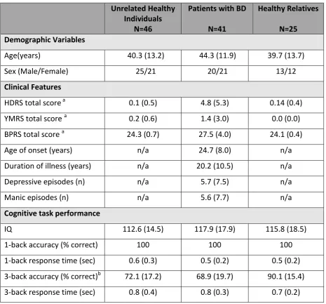
Healthy carriers of the rs10994336 risk-allele showed significantly decreased lateral
temporal cortical activation within the middle (BA 21) and inferior (BA 20) temporal gyrus
(Fig. 5-1). In contrast, healthy homozygotes of the rs9804190 risk-allele showed increased
activation in the lateral prefrontal cortex within the inferior (BA 47/11) and middle (BA 46)
frontal gyrus (Fig. 5-1). The coordinates of the peak height voxel of the corresponding
suprathreshold clusters are presented in Table 5-2.
Effect of ANK3 allelic variation on WM-related activation in BD patients and their healthy relatives
An effect of group (patients, healthy relatives, healthy unrelated individuals) was observed
10 and in the ventral ACC (BA 24/32). When compared to unrelated healthy individuals, brain
activation in patients was significantly (a) reduced in the left (BA 9) and right middle frontal
gyri (BA 10) and, (b) increased in the superior and middle temporal gyri (BA 21/22) on the
right and in the ACC bilaterally (BA 24/32). In comparison to patients, healthy relatives had
greater activation in the middle frontal gyrus bilaterally. No differences were observed
between unrelated healthy individuals and healthy relatives. The coordinates of the peak
activations of the suprathreshold clusters are shown in Supplemental Table 5-S3.
In patients, there were no significant correlations between mean signal change in
suprathreshold clusters and age of onset, duration of illness, mood episodes or medication
dose (p> 0.1).
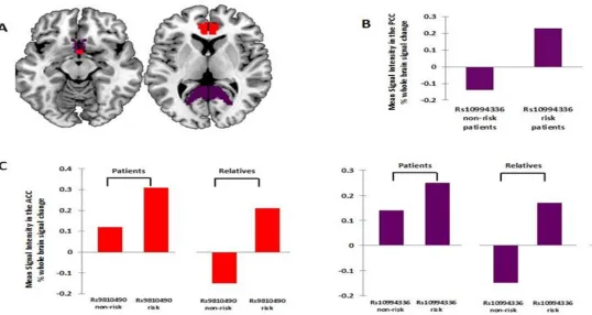
For the ANK3rs10994336, we found a significant group by genotype interaction in the right ventral ACC (x=4, y=19, z=-3, cluster size=35, z-value=3.67) and left ventral posterior
cingulate cortex (PCC; x=-28, y=-64, z=16, cluster size=166, z-value=4.10). In the right ACC,
the risk T-allele was associated with increased activation in BD patients and their healthy
relatives compared to unrelated healthy individuals. In the left PCC, the risk T-allele was
related with increased activation in BD patients compared to their healthy relatives and to
unrelated healthy individuals (Fig. 5-2).
For the ANK3 rs9804190, a significant group by genotype interaction was found in the right ACC (x=4, y=17, z=-4; cluster size= 58; z-value=3.95) in which patients and healthy relatives
who were risk C-allele homozygotes showed increased activation compared to unrelated
healthy individuals (Fig. 5-2).
Discussion
There are two key findings from this study. First, in healthy individuals without personal or
family history of psychiatric disorders, the rs10994336 and rs9804190 BD-risk alleles had
different effects on the working memory (WM) network, although neither affected task
performance. Second, both BD-risk alleles were associated with failure to deactivate the
default mode network (DMN) in patients and in their healthy relatives; accuracy was
11 Unrelated healthy carriers of the rs10994336 risk-allele showed reduced engagement of the ventral visual cortex within the middle and inferior temporal gyri (Table 5-2). This accords
with previous reports from two independent samples which found that the largest effect
size of the rs10994336 risk-allele was on reduced sensitivity in target detection and
increased errors of commission during the degraded symbol continuous performance task
(Ruberto et al., 2011; Hatzimanolis et al., 2012). Although the ventral visual cortex is an
integral part of the WM circuitry, the core WM network involves the frontoparietal cortices
(Owen et al., 2005; Leech et al., 2011; Rottschy et al., 2012). These regions are also core
components of the superordinate cognitive control network that supports a broad range of
executive function tasks (Niendam et al., 2012). Unrelated healthy rs9804190-risk allele
homozygotes evidenced greater activation within the prefrontal components of the WM
network although their task performance was comparable to that of the non-risk associated
unrelated individuals (Table 5-2). This pattern is typically interpreted as evidence of cortical
inefficiency, and is consistent with behavioural data from an independent sample that also
found that healthy rs9804190-risk-allele homozygotes underperform in a wide array of
executive function tasks (Roussos et al., 2012).
These findings suggest that in the absence of increased background genetic risk for BD or
other psychiatric disorders the two ANK3 BD-risk loci affect different regions of the WM circuitry. The reason for these regional differences is unclear. Available data suggest that 3’
risk-alleles (rs9804190) are associated with reduced transcript levels of brain-specific AnkG
isoforms. To date, the region most commonly implicated is the cerebellum where ANK3
expression is generally highest (Rueckert et al., 2013). Information about other brain regions
is incomplete because the available post-mortem studies have limited statistical power due
to the small number of donors and provide incomplete brain coverage (Roussos et al., 2012;
Rueckert et al., 2013). With regards to rs10994336, the effect of the risk-allele on ANK3
expression in the brain is unknown. The rs10994336 polymorphism is located in an intronic
region (Tesli et al., 2011) but could affect gene expression through cis- or trans-regulatory
mechanisms (Quinn et al., 2010). Alternatively, rs10994336 may be in strong linkage
disequilibrium with other, yet unidentified, genetic loci that drive the effects observed here.
Rs10994336 or rs9804190 risk-associated patients and relatives showed hyperactivity within
12 processing and generation (Critchley et al., 2003) and a key component of the anterior DMN
(Raichle et al., 2001; Buckner et al., 2008). Activation within the ventral ACC is increased
during the processing of arousing stimuli or during mental stress (Critchley et al., 2003). The
n-back task is quite challenging and may engender mild mental stress but it is not expected
to result in ventral ACC hyperactivation. In fact, deactivation of the ventral ACC is normally
observed during the n-back task within the context of anticorrelated activity between the
DMN and the frontoparietal cognitive control network (Leech et al., 2011; Esposito et al.,
2006).
Accordingly, healthy unrelated individuals in this study showed deactivation of the ventral
ACC during the n-back task regardless of genotype. As expected, hyperactivation within the
ventral ACC was observed in the patients regardless of genotype (Jogia et al., 2012;
Pomarol-Clotet et al., 2012; Fernández-Corcuera et al., 2013) but it was more pronounced in
rs10994336 or rs9804190 risk associated individuals. Amongst the healthy relatives, ventral
ACC hyperactivity was only present in risk associated individuals for either risk-allele.
Additionally, BD carriers of the rs10994336 risk-allele also showed hyperactivity within the
PCC, centred on the ventral and extending to dorsal regions (Vogt et al., 2006). The PCC has
dense anatomical connections with multiple cortical and subcortical regions (Hagmann et
al., 2008) and is a core component of the posterior DMN (Raichle et al., 2001; Buckner et al.,
2008). Healthy individuals performing the n-back task show deactivation in both dorsal and
ventral PCC (Leech et al., 2011; Esposito et al., 2006) so the persistent PCC activation seen in
patients suggests that the rs10994336 risk-allele compromises the ability to deactivate this
brain region.
Taken together, these findings suggest that aberrant hyperactivation within the ventral ACC
is a key mechanism mediating the risk-conferring effects of rs10994336 and rs9804190 in
connection to the WM circuitry. It is noteworthy that this effect appeared to require the
concomitant presence of additional risk factors for BD as it was not observed in unrelated
individuals who had no personal or family history of such risk factors. This is consistent with
the multifactorial pathogenetic model of BD that involves interaction between multiple
genetic and non-genetic risk factors (Sullivan et al., 2012). The effect of any individual factor
depends on the relative prevalence of other risk factors that are part of the same
13 reported here, several neuroimaging studies have shown differential effects of various
susceptibility polymorphisms (e.g. DISC1, NRG1, COMT) on brain structure and function in patients, high-risk groups and unrelated healthy individuals (Addington et al., 2007; Mechelli
et al., 2008; Prata et al., 2008; Tsuchimine et al., 2013; Narr et al., 2009; Whalley et al.,
2012).
In conclusion, our results point to a differential effect of BD-risk associated polymorphisms
at ANK3 rs10994336 and rs9804190 modulated by risk-status for the disorder. This suggests
that the BD-risk conferring mechanisms associated with these genetic variants are
influenced by other genetic and possibly non-genetic factors that contribute to risk status.
Inability to suppress key nodes of the DMN emerged as a common final pathway through
which either risk-allele may contribute to the pathogenesis of BD. Mood stabilizing
medications such as Lamotrigine interact with the ANK3 system through ion channels bound
by AnkG to the axonal initial segment. Our results therefore lend further support to our
previous study on patients with BD treated with Lamotrigine that showed “normalization”
of the WM circuitry (Lang et al., 1993; Haldane et al., 2008) and suggest that ANK3-related
molecular pathways may be a fruitful ground for the identification of new drug targets for
14
References
Addington AM, Gornick MC, Shaw P, Seal J, Gogtay N, Greenstein D, Clasen L, Coffey M,
Gochman P, Long R, Rapoport JL. Neuregulin 1 (8p12) and childhood-onset schizophrenia:
Susceptibility haplotypes for diagnosis and brain developmental trajectories. Mol
Psychiatry2007;12: 195-205.
Adler CM, Holland SK, Schmithorst V, Tuchfarber MJ, Strakowski SM. Changes in neuronal
activation in patients with bipolar disorder during performance of a working memory task.
Bipolar Disord 2004; 6: 540-549.
American Psychiatric Association. Diagnostic and Statistical Manual of Mental Disorders, 4th
ed. Washington, DC: American Psychiatric Press 1994.
Baum AE, Akula N, Cabanero M, Cardona I, Corona W, Klemens B, Schulze TG, Cichon S,
Rietschel M, Nöthen MM, Georgi A, Schumacher J, Schwarz M, Abou Jamra R, Höfels S,
Propping P, Satagopan J, Detera-Wadleigh SD, Hardy J, McMahon FJ. A genome-wide
association study implicates diacylglycerol kinase eta (DGKH) and several other genes in the
etiology of bipolar disorder. Mol Psychiatry 2008; 13: 197-207.
Buckner RL, Andrews-Hanna JR, Schacter DL. The brain's default network: anatomy,
function, and relevance to disease. Ann N Y Acad Sci 2008; 1124: 1-38.
Critchley HD, Mathias CJ, Josephs O, O'Doherty J, Zanini S, Dewar BK, Cipolotti L, Shallice T,
Dolan RJ. Human cingulate cortex and autonomic control: converging neuroimaging and
clinical evidence. Brain 2003; 126: 2139-2152.
Dedman A, McQuillin A, Kandaswamy R, Sharp S, Anjorin A, Gurling H. Sequencing of the
ANKYRIN 3 gene (ANK3) encoding ankyrin G in bipolar disorder reveals a non-conservative
amino acid change in a short isoform of ankyrin G. Am J Med Genet B Neuropsychiatr Genet
15 Drapier D, Surguladze S, Marshall N, Schulze K, Fern A, Hall MH, Walshe M, Murray RM,
McDonald C. Genetic liability for bipolar disorder is characterized by excess frontal
activation in response to a working memory task. Biol Psychiatry 2008; 64: 513-520.
Esposito F, Bertolino A, Scarabino T, Latorre V, Blasi G, Popolizio T, Tedeschi G, Cirillo S,
Goebel R, Di Salle F. Independent component model of the default-mode brain function:
Assessing the impact of active thinking. Brain Res Bull 2006; 70: 263-269.
Fernández-Corcuera P, Salvador R, Monté GC, Salvador Sarró S, Goikolea JM, Amann B,
Moro N, Sans-Sansa B, Ortiz-Gil J, Vieta E, Maristany T, McKenna PJ, Pomarol-Clotet E.
Bipolar depressed patients show both failure to activate and failure to de-activate during
performance of a working memory task. J Affect Disord 2013; 148: 170-178.
Ferreira MA, O'Donovan MC, Meng YA, Jones IR, Ruderfer DM, Jones L, Fan J, Kirov G, Perlis
RH, Green EK, Smoller JW, Grozeva D, Stone J, Nikolov I, Chambert K, Hamshere ML,
Nimgaonkar VL, Moskvina V, Thase ME, Caesar S, Sachs GS, Franklin J, Gordon-Smith K,
Ardlie KG, Gabriel SB, Fraser C, Blumenstiel B, Defelice M, Breen G, Gill M, Morris DW, Elkin
A, Muir WJ, McGhee KA, Williamson R, MacIntyre DJ, MacLean AW, St CD, Robinson M, Van
Beck M, Pereira AC, Kandaswamy R, McQuillin A, Collier DA, Bass NJ, Young AH, Lawrence J,
Ferrier IN, Anjorin A, Farmer A, Curtis D, Scolnick EM, McGuffin P, Daly MJ, Corvin AP,
Holmans PA, Blackwood DH, Gurling HM, Owen MJ, Purcell SM, Sklar P, Craddock N;
Wellcome Trust Case Control Consortium. Collaborative genome-wide association analysis
supports a role for ANK3 and CACNA1C in bipolar disorder. Nat Genet 2008; 40: 1056-1058.
First MB, Spitzer RL, Gibbon M, Williams JBW. Structured Clinical Interview for DSM-IV-TR
Axis I Disorders, Research Version, Patient Edition. (SCID-I/P) New York, NY: Biometrics
Research, New York State Psychiatric Institute 2002a.
First MB, Spitzer RL, Gibbon M, Williams JBW. Structured Clinical Interview for DSM-IV-TR
Axis I Disorders, Research Version, Non-patient Edition. (SCID-I/NP) New York, NY:
16 Frangou S, Kington J, Raymont V, Shergill SS. Examining ventral and dorsal prefrontal
function in bipolar disorder: a functional magnetic resonance imaging study. Eur Psychiatry
2008 23: 300-308.
Frangou S. Risk and resilience in bipolar disorder: rationale and design of the Vulnerability to
Bipolar Disorders Study (VIBES). Biochem Soc Trans 2009; 37: 1085-1089.
Gevins A, Smith ME. Neurophysiological measures of working memory and individual
differences in cognitive ability and cognitive style. Cereb Cortex 2000; 10: 829-839.
Hagmann P, Cammoun L, Gigandet X, Meuli R, Honey CJ, Wedeen VJ, Sporns O. Mapping the
structural core of human cerebral cortex. PLoS Biol 2008; 6:e159.
Haldane M, Jogia J, Cobb A, Kozuch E, Kumari V, Frangou S. Changes in brain activation
during working memory and facial recognition tasks in patients with bipolar disorder with
Lamotrigine monotherapy. Eur Neuropsychopharmacol 2008; 18: 48-54.
Hamilton M. A rating scale for depression. J Neurol Neurosurg Psychiatry 1960; 23: 56-62.
Hatzimanolis A, Smyrnis N, Avramopoulos D, Stefanis CN, Evdokimidis I, Stefanis NC. Bipolar
disorder ANK3 risk variant effect on sustained attention is replicated in a large healthy
population. Psychiatr Genet 2012; 22: 210-213.
Jogia J, Dima D, Kumari V, Frangou S. Frontopolar cortical inefficiency may underpin reward
and working memory dysfunction in bipolar disorder. World J Biol Psychiatry 2012; 13:
605-615.
Kordeli E, Lambert S, Bennett V. AnkyrinG. A new ankyrin gene with neural-specific isoforms
localized at the axonal initial segment and node of Ranvier. J Biol Chem 1995; 270:
17 Lagopoulos J, Ivanovski B, Malhi GS. An event-related functional MRI study of working
memory in euthymic bipolar disorder. J Psychiatry Neurosci 2007; 32: 174-184.
Lang DG, Wang CM, Cooper BR. Lamotrigine, phenytoin and carbamazepine interactions on
the sodium current present in N4TG1 mouse neuroblastoma cells. J Pharmacol Exp Ther
1993; 266: 829-835.
Lee MT, Chen CH, Lee CS, Chen CC, Chong MY, Ouyang WC, Lee MT, Chen CH, Lee CS, Chen
CC, Chong MY, Ouyang WC, Chiu NY, Chuo LJ, Chen CY, Tan HK, Lane HY, Chang TJ, Lin CH,
Jou SH, Hou YM, Feng J, Lai TJ, Tung CL, Chen TJ, Chang CJ, Lung FW, Chen CK, Shiah IS, Liu
CY, Teng PR, Chen KH, Shen LJ, Cheng CS, Chang TP, Li CF, Chou CH, Chen CY, Wang KH, Fann
CS, Wu JY, Chen YT, Cheng AT. Genome-wide association study of bipolar I disorder in the
Han Chinese population. Mol Psychiatry 2011; 16: 548-556.
Leech R, Kamourieh S, Beckmann CF, Sharp DJ. Fractionating the default mode network:
distinct contributions of the ventral and dorsal posterior cingulate cortex to cognitive
control. J Neurosci 2011; 31: 3217-3224.
Lett TA, Zai CC, Tiwari AK, Shaikh SA, Likhodi O, Kennedy JL, Müller DJ. ANK3, CACNA1C and
ZNF804A gene variants in bipolar disorders and psychosis subphenotype. World J Biol
Psychiatry 2011; 12: 392-397.
Leussis MP, Berry-Scott EM, Saito M, Jhuang H, de Haan G, Alkan O, Luce CJ, Madison JM,
Sklar P, Serre T, Root DE, Petryshen TL. The ANK3 bipolar disorder gene regulates
psychiatric-related behaviors that are modulated by lithium and stress. Biol Psychiatry 2013;
73: 683-690.
Linke J, Witt SH, King AV, Nieratschker V, Poupon C, Gass A, Hennerici MG, Rietschel M,
Wessa M. Genome-wide supported risk variant for bipolar disorder alters anatomical
18 Lukoff D, Nuechterlien K, Ventura J. Manual for the expanded Brief Psychiatric Rating Scale.
Schizophr Bull 1986; 12: 594-608.
Mechelli A, Prata DP, Fu CH, Picchioni M, Kane F, Kalidindi S, McDonald C, Demjaha A,
Kravariti E, Toulopoulou T, Murray R, Collier DA, McGuire PK. The effects of neuregulin1 on
brain function in controls and patients with schizophrenia and bipolar disorder. Neuroimage
2008; 42: 817-826.
Narr KL, Szeszko PR, Lencz T, Woods RP, Hamilton LS, Phillips O, Robinson D, Burdick KE,
DeRosse P, Kucherlapati R, Thompson PM, Toga AW, Malhotra AK, Bilder RM. DTNBP1 is
associated with imaging phenotypes in schizophrenia. Hum Brain Mapp 2009; 30:
3783-3794.
Niendam TA, Laird AR, Ray KL, Dean YM, Glahn DC, Carter CS. Meta-analytic evidence for a
superordinate cognitive control network subserving diverse executive functions. Cogn Affect
Behav Neurosci 2012; 12: 241-268.
Owen AM, McMillan KM, Laird AR, Bullmore ET. N-back working memory paradigm: a
meta-analysis of normative functional neuroimaging studies. Hum Brain Mapp 2005; 25: 46-59.
Palaniyappan L, Liddle PF. Diagnostic Discontinuity in Psychosis: A Combined Study of
Cortical Gyrification and Functional Connectivity. Schizophr Bull 2014; 40: 675-684.
Pomarol-Clotet E, Moro N, Sarró S, Goikolea JM, Vieta E, Amann B, Fernandez-Corcuera P,
Sans-Sansa B, Monté GC, Capdevila A, McKenna PJ, Salvador R. Failure of de-activation in
the medial frontal cortex in mania: evidence for default mode network dysfunction in the
disorder. World J Biol Psychiatry 2012; 13: 616-626.
Prata DP, Mechelli A, Fu CHY, Picchioni M, Kane F, Kalidini S, McDonald C, Howes O, Kravariti
E, Demjaha A, Toulopoulou T, Diforti M, Murray RM, Collier DA, McGuire PK. Opposite
effects of catechol-O-methyltransferase Val158Met on cortical function in healthy subjects
19 Quinn EM, Hill M, Anney R, Gill M, Corvin AP, Morris DW. Evidence for cis-acting regulation
of ANK3 and CACNA1C gene expression. Bipolar Disord 2010; 12: 440-445.
Raichle ME, MacLeod AM, Snyder AZ, Powers WJ, Gusnard DA, Shulman GL. A default mode
of brain function. Proc Natl Acad Sci USA 2001; 98: 676-682.
Rasband MN. The axon initial segment and the maintenance of neuronal polarity. Nat Rev
Neurosci 2010; 11: 552-562.
Rottschy C, Langner R, Dogan I, Reetz K, Laird AR, Schulz JB, Fox PT, Eickhoff SB. Modelling
neural correlates of working memory: a coordinate-based meta-analysis. Neuroimage 2012;
60: 830-846.
Roussos P, Giakoumaki SG, Georgakopoulos A, Robakis NK, Bitsios P. The CACNA1C and
ANK3 risk alleles impact on affective personality traits and startle reactivity but not on
cognition or gating in healthy males. Bipolar Disord 2011; 13: 250-259.
Roussos P, Katsel P, Davis KL, Bitsios P, Giakoumaki SG, Jogia J, Rozsnyai K, Collier D, Frangou
S, Siever LJ, Haroutunian V. Molecular and genetic evidence for abnormalities in the nodes
of Ranvier in schizophrenia. Arch Gen Psychiatry 2012; 69: 7-15.
Ruberto G, Vassos E, Lewis CM, Tatarelli R, Girardi P, Collier D, Frangou S. The cognitive
impact of the ANK3 risk variant for bipolar disorder: initial evidence of selectivity to signal
detection during sustained attention. PLoS One 2011; 6: e16671.
Rueckert EH, Barker D, Ruderfer D, Bergen SE, O'Dushlaine C, Luce CJ, Sheridan SD,
Theriault KM, Chambert K, Moran J, Purcell SM, Madison JM, Haggarty SJ, Sklar P. Cis-acting
regulation of brain-specific ANK3 gene expression by a genetic variant associated with
20 Schulze TG, Detera-Wadleigh SD, Akula N, Gupta A, Kassem L, Steele J, Pearl J, Strohmaier J,
Breuer R, Schwarz M, Propping P, Nöthen MM, Cichon S, Schumacher J; NIMH Genetics
Initiative Bipolar Disorder Consortium, Rietschel M, McMahon FJ. Two variants in Ankyrin 3
(ANK3) are independent genetic risk factors for bipolar disorder. Mol Psychiatry 2009; 14:
487-491.
Sklar P, Ripke S, Scott LJ, Andreassen OA, Cichon S, Craddock N; Psychiatric GWAS
Consortium Bipolar Disorder Working Group. Large-scale genome-wide association analysis
of bipolar disorder identifies a new susceptibility locus near ODZ4. Nat Genet 2011; 43:
977-983.
Sullivan PF, Daly MJ, O'Donovan M. Genetic architectures of psychiatric disorders: the
emerging picture and its implications. Nat Rev Genet 2012; 13: 537-551.
Takata A, Kim SH, Ozaki N, Iwata N, Kunugi H, Inada T, Ujike H, Nakamura K, Mori N, Ahn
YM, Joo EJ, Song JY, Kanba S, Yoshikawa T, Kim YS, Kato T. Association of ANK3 with bipolar
disorder confirmed in East Asia. Am J Med Genet B Neuropsychiatr Genet 2011; 156B:
312-315.
Talairach J, Tournoux P. Co-planar stereotaxic atlas of the human brain. New York, NY:
Thieme 1988.
Tesli M, Koefoed P, Athanasiu L, Mattingsdal M, Gustafsson O, Agartz I, Rimol LM, Brown A,
Wirgenes KV, Smorr LL, Kähler AK, Werge T, Mors O, Mellerup E, Jönsson EG, Melle I,
Morken G, Djurovic S, Andreassen OA. Association analysis of ANK3 gene variants in nordic
bipolar disorder and schizophrenia case-control samples. Am J Med Genet B Neuropsychiatr
Genet 2011; 156B: 969-974.
Thermenos HW, Goldstein JM, Milanovic SM, Whitfield-Gabrieli S, Makris N, Laviolette P,
Koch JK, Faraone SV, Tsuang MT, Buka SL, Seidman LJ. An fMRI study of working memory in
persons with bipolar disorder or at genetic risk for bipolar disorder. Am J Med Genet B
21 Thermenos HW, Makris N, Whitfield-Gabrieli S, Brown AB, Giuliano AJ, Lee EH, Faraone SV,
Tsuang MT, Seidman LJ. A functional MRI study of working memory in adolescents and
young adults at genetic risk for bipolar disorder: preliminary findings. Bipolar Disord 2011;
13: 272-286.
Townsend J, Bookheimer SY, Foland-Ross LC, Sugar CA, Altshuler LL. fMRI abnormalities in
dorsolateral prefrontal cortex during a working memory task in manic, euthymic and
depressed bipolar subjects. Psychiatry Res 2010; 182: 22-29.
Tsuchimine S, Yasui-Furukori N, Kaneda A, Kaneko S. Differential effects of the
catechol-O-methyltransferase Val158Met genotype on the cognitive function of schizophrenia patients
and healthy Japanese individuals. PLoS One 2013; 8: e76763.
Vogt BA, Vogt L, Laureys S. Cytology and functionally correlated circuits of human posterior
cingulate areas. Neuroimage 2006; 29: 452-466.
Wechsler D. Wechsler Adult Intelligence Scale-Revised. San Antonio. TX: The Psychological
Corporation 1981.
Whalley HC, Sussmann JE, Johnstone M, Romaniuk L, Redpath H, Chakirova G, Mukherjee P,
Hall J, Johnstone EC, Lawrie SM, McIntosh AM. Effects of a mis-sense DISC1 variant on brain
activation in two cohorts at high risk of bipolar disorder or schizophrenia. Am J Med Genet B
Neuropsychiatr Genet 2012; 159B: 343-353.
Young RC, Biggs JT, Ziegler VE, Meyer DA. A rating scale for mania: reliability, validity and
sensitivity. Br J Psychiatry 1978; 133: 429-435.
Zhang C, Cai J, Zhang J, Li Z, Guo Z, Zhang X, Lu W, Zhang Y, Yuan A, Yu S, Fang Y. Genetic
modulation of working memory deficits by ankyrin 3 gene in schizophrenia. Prog
22 Zhou D, Lambert S, Malen PL, Carpenter S, Boland LM, Bennett V. AnkyrinG is required for
clustering of voltage-gated Na channels at axon initial segments and for normal action
23
Table 5-1 Study Sample
Unrelated Healthy Individuals
N=46
Patients with BD
N=41
Healthy Relatives
N=25 Demographic Variables
Age(years) 40.3 (13.2) 44.3 (11.9) 39.7 (13.7)
Sex (Male/Female) 25/21 20/21 13/12
Clinical Features
HDRS total score a 0.1 (0.5) 4.8 (5.3) 0.14 (0.4)
YMRS total score a 0.2 (0.6) 1.4 (3.0) 0.0 (0.0)
BPRS total score a 24.3 (0.7) 27.5 (4.0) 24.1 (0.4)
Age of onset (years) n/a 24.7 (8.0) n/a
Duration of illness (years) n/a 20.2 (10.5) n/a
Depressive episodes (n) n/a 5.7 (7.5) n/a
Manic episodes (n) n/a 5.6 (7.7) n/a
Cognitive task performance
IQ 112.6 (14.5) 117.9 (17.9) 115.8 (18.5)
1-back accuracy (% correct) 100 100 100
1-back response time (sec) 0.6 (0.3) 0.5 (0.2) 0.5 (0.2)
3-back accuracy (% correct)b 72.1 (17.2) 68.9 (19.7) 90.1 (15.4)
3-back response time (sec) 0.8 (0.4) 0.8 (0.3) 0.7 (0.2)
Except for sex, all data are presented as mean (standard deviation); BD= Bipolar Disorder;
24
Table 5-2 Brain regions showing significant effects of allelic variation at 10994336 and rs9810490 in the 3-back vs 0-back contrast in unrelated healthy individuals
Region Gyrus Laterality Brodmann Area
Talairach and Tournoux Coordinates
Cluster size z-value
X y z
ANK3 rs10994336: Risk-Allele Homozygotes < Non-Risk Allele Carriers
Temporal Middle Temporal
Left 21 -40 12 -28 120 3.41
Right 48 -5 -15 90 3.76
59 -14 -4 38 3.60
Inferior Temporal
Left 20 -42 -12 -24 56 3.43
Right 44 -11 -20 90 3.80
ANK3 rs9810490: Risk Allele Carriers > Non-Risk Allele Homozygotes
Frontal Middle Frontal
Left 46 -40 38 20 46 3.51
Inferior Frontal
Left 47/10 -46 48 -4 51 3.48
25
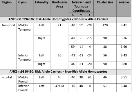
Figure 5-1 Effect of genotype on regional brain activation in the 3-back vs 0-back contrast in unrelated healthy individuals.
26
Figure 5-2 Effect of genotype on regional brain activation in the 3-back vs 0-back contrast in patients with bipolar disorder and their
27
Supplemental Material
Supplemental Table 5-S1 Effect of ANK3 rs10994336 genotype (risk-allele T)
Effect of Genotype Effect of Group by Genotype
Risk Associated TT+CT No-risk Associated CC Risk Associated TT+CT No-risk Associated CC
N = 40 N = 72
Unrelated Healthy Individuals
N = 14
BD patients
N = 16
Healthy Relatives N=10
Unrelated Healthy Individuals
N = 32
BD patients
N = 25
Healthy Relatives
n=15
Demographic Variables
Age(years) 39.9 (13.1) 42.8 (12.28) 40.6 (12.2) 42.0 (10.7) 40.8 (8.3) 39.3 (12.3) 43.3 (12.3) 38.3 (13.7)
Sex
(Male/Female)
21/19 34/38 7/7 9/7 4/6 18/14 11/14 7/8
Clinical Features
HDRS total score a 2.1 (4.3) 0.5 (0.81) 0.4 (0.9) 5.3 (4.6) 0.3 (0.6) 0.1 (0.4) 1.5 (0.9) 0.1 (0.5)
YMRS total score a 0.7 (2.1) 0.1 (0.3) 0.2 (0.4) 1.6 (2.9) 0.0 (0.0) 0.2 (0.6) 0.7 (1.4) 0.0 (0.0)
BPRS total score a 25.6 (3.2) 24.9 (1.0) 24.8 (1.1) 27.3 (4.3) 24.7 (1.1) 24.2 (0.6) 25.9 (1.9) 24.1 (0.3)
Cognitive Performance
IQ 117.2 (17.6) 116.9 (16.1) 110.7 (12.9) 112.3 (16.2) 112.0 (20.4) 116.7 (14.5) 121.7 (16.3) 118.3 (18.5)
3-back accuracy (%) b, c
78.7 (23.1) 66.9 (29.8) 70.5 (19.8) 73.3 (36.5) 83.8 (22.8) 75.1 (24.5) 67.2 (23.5) 90.78 (14.9)
3-back response time (sec)
0.84 (0.38) 0.77 (0.33) 0.99 (0.37) 0.68 (0.23) 0.54 (0.21) 0.86 (0.46) 0.94 (0.36) 0.75 (0.21)
Except for sex, all data are presented as mean (standard deviation); BD= Bipolar Disorder; BPRS= Brief Psychiatric Rating Scale; HDRS=Hamilton Depression Rating Scale;
YMRS=Young Mania Rating Scale. a BD carriers of the risk-allele > unrelated healthy individuals, relatives, P<0.02; b relatives> unrelated healthy individuals, P=0.003;
28
Supplemental Table 5-S2 Effect of ANK3 rs9810490 genotype (risk-allele C)
Effect of Genotype Effect of Group by Genotype
Risk Associated CC No-risk Associated TT+CT Risk Associated CC No-risk Associated TT+TC
N = 63 N = 49
Unrelated Healthy Individuals
N = 28
BD Patients
N = 21
Healthy Relatives
N = 14
Unrelated Healthy Controls
N = 18
BD Patients
N = 20
Healthy Relatives
N = 11 Demographic Variables
Age(years) 38.8 (13.6) 43.9 (12.1) 40.1 (13.26) 43.5 (12.51) 40.2 (14.3) 40.1 (11.9) 44.8 (9.5) 39.2 (13.8)
Sex (Male/Female) 29/34 29/20 14/14 8/13 7/7 11/8 12/9 6/5
Clinical Features
HDRS total score a 1.7 (3.9) 2.1 (4.1) 0.1 (0.4) 5.5 (5.6) 0.1 (0.1) 1.2 (3.3) 4.5 (5.4) 0.07 (0.3)
YMRS total score a 0.7 (2.1) 0.5 (1.8) 0.2 (0.5) 2.0 (3.5) 0.0 (0.0) 0.06 (0.25) 1.2 (2.7) 0.0 (0.0)
BPRS total score a 25.8 (1.5) 25.1 (1.9) 24.3 (0.6) 29.3 (5.0) 24.2 (0.6) 24.7 (1.0) 26.3 (2.7) 24.1 (0.3)
Cognitive Performance
IQ 111.8 (15.6) 120.4 (18.7) 117.6 (16.3) 114.1 (13.9) 108.5 (18.1) 119.4 (16.5) 118.3 (21.5) 122.9 (18.1)
3-back accuracy (%) b, c
72.02 (25.9) 83.01 (23.1) 67.1 (25.7) 73.5 (30.5) 85.6 (17.0) 85.8 (26.1) 62.5 (23.2) 92.5 (14.2)
3-back response time ( sec)
0.76 (0.48) 0.58 (0.42) 0.85 (0.50) 0.61 (0.49) 0.70 (0.37) 0.81 (0.51) 0.41 (0.44) 0.60 (0.29)
Except for sex, all data are presented as mean (standard deviation); BD= Bipolar Disorder; BPRS= Brief Psychiatric Rating Scale; HDRS=Hamilton Depression Rating Scale;
YMRS=Young Mania Rating Scale. a BD carriers of the risk-allele > unrelated healthy individuals, relatives, P<0.02, relatives> unrelated healthy individuals, P = 0.003; relatives>
29
Supplemental Table 5-S3 Brain regions showing significant effect of group in the 3-back vs 0-back contrast
Gyrus Laterality Brodmann Area Talairach and Tournoux Coordinates z-value
x y Z
Patients with BD > Healthy unrelated controls
Anterior Cingulate
Left 24/32 -14 46 6 3.56
Right 10 26 -6 3.49
Superior Temporal
Right 22 54 1 -4 3.93
Middle Temporal
Right 21 62 -8 -4 4.25
Patients with BD < Healthy unrelated controls
Middle Frontal Left 9 -34 14 40 3.70
Right 10 38 56 -8 3.26
Patients with BD < Healthy relatives
Middle Frontal Left 9 -42 20 34 4.46
Right 9 40 32 34 4.01
30
Supplemental Figure 5-S1 Box plot with the mean signal change in the posterior Cingulate
Cortex in subjects carriying the ANK3 rs10994336.
Supplemental Figure 5-S2 Box plot with the mean signal change in the Anterior Cingulate
31
Supplemental Figure 5-S3 Box plot with the mean signal change in the posterior Cingulate