JOURNAL OF VIROLOGY, Oct. 1977, p. 363-377 CopyrightC1977 AmericanSociety for Microbiology
Vol. 24,No. 1 Printed in U.S.A.
Morphogenesis of Bacteriophage
029
of
Bacillus
subtilis:
Mapping
and Funetional Analysis
of the
Head
Fiber Gene
BERNARD E. REILLY,* RODGER A. NELSON, AND DWIGHT L. ANDERSON
Department ofMicrobiologyand SchoolofDentistry, University ofMinnesota, Minneapolis, Minnesota 55455 Received for publication 18 May 1977
A
set of mutants
of Bacillus subtilis bacteriophage
429 unable
to
synthesize
the head fiber protein has been
identified
by sodium
dodecyl
sulfate-polyacryl-amide
gel electrophoresis
and
autoradiography. Infectious phage
are
produced
during restrictive infection. We
have focused
on mutant
sus8.5(900)
because
the
mutation is
suppressible by both the
su+3
and
su+44
hosts, and
it can
be
mapped
by three- and four-factor crosses. After restrictive
infection
with mutant
sus8.5(900),
afragment
about 70% of the size of the normal fiber is
produced
aswell as particles
that
are
fast-sedimenting
in sucrose
gradients
relative to 429+.
These particles have the buoyant density of particles
with
the fibers removed
and have the absolute
plating
efficiency of 4)29+. Fiber protein
is
absent
from
prohead
as
well
as virion. A
second
set
of mutants
produces fiber
protein with a
slightly altered electrophoretic
mobility. This
type
of fiber
protein
is
either
present or
absent on both
prohead and
virion. A
third class of mutants,
typified
by
914,
produces
a
"normal" fiber,
but a major head protein of
altered
electropho-retic
mobility. After infection
by this
mutant,
the fiber is absent from both
prohead and virion, and the biological and physical properties of the
914-particle
are
similar to those of
particles produced
after
infection
of the su- host
by
sus8.5(900).
These observations suggest that the head fiber is
notan
essential
component
of the
prohead
or
virion
and that
the assembly
process is
efficient in
the absence of fiber protein. Three- and four-factor genetic crosses
have
estab-lished the order sus8(769)-8(914)-sus8.5(900)-sus9(756)
and indicate that cistrons
8
and
8.5
code for the
major
head
protein
and head
fiber protein, respectively.
The
virion
of
bacteriophage
429
has protein
fibers attached
at
both ends
of its
prolate head
(2, 28). Fiberless particles produced by
di-methyl sulfoxide treatment retain their
infec-tivity (27).
Three
polypeptides have been
as-signed
tothe
matureviral
head (18), and there
is
general agreement that the major head
pro-tein
and fiber are the major head constituents
(10, 12, 22).
Point mutants
incistron 8 were
unable
to
synthesize head
protein or
fiber
dur-ing restrictive
infection (5, 9, 15), and tryptic
peptide analysis of these
proteins
indicated that
they
were
distinct
proteins (27).
Pulse-chase
experiments
demonstrated
that fiber was a
ma-jor component of the viral
prohead, a particle
that may be the first intermediate on the 429
assembly pathway
(20).
We
have isolated
a series ofmutants that
produce fiber protein with altered
electropho-retic
mobility or do not synthesize the fiber
protein. After infection of the nonpermissive
host, certain of these mutants produce both
proheads
and
infectious particles that lack
fi-bers. The absolute
plating
efficiency
of
the
fi-berless
particles is the
same aswild-type
phage. Another mutant
produces
an
altered
major
head protein and an apparently normal
fiber,
but the fiber is excluded from both
pro-head and virion.
Genetic studies
suggest
that
these
mutations map
between
the mutants
sus8(769)
and sus9(756) of cistrons
8and 9.
MATERIALS AND METHODS
The
429
cistronsdefined by quantitativecomple-mentation have been numberedsequentially from left toright(1to17),accordingtotheir relative map position. A protein product of cistron n has been referred to as pn. Forexample,theproductof cistron 11,the neck lower collar protein, isnownamedp11 (16). Although we have focused on thehead fiber protein as astructural component, wehave found thephenotypes of some ofour new mutants to be quitecomplicated. For thisreason wehave referred tothesemutantscollectivelyasthe"900series," and
particularmutantshaveatrivialdesignation,e.g., mutant912,that doesnotimply that the912
pheno-typenecessarily reflectsapoint mutation.
Phage and bacteria. Phage
429k
and the mutant susl4(1241), a mutant havingthedelayed lysis phe-363on November 10, 2019 by guest
http://jvi.asm.org/
364 REILLY, NELSON, AND ANDERSON notype, were employed (19, 24). The mutants used to constructthe recombinants sus7(614)-sus8(769) and sus8(769)-sus9(756), andsuslO(302), have been de-scribed (26).
The properties of Bacillus subtilis SpoA12 and B. subtilis L15, the nonpermissive (su-) and permis-sive bacterial hosts, respectively, have been de-scribed (26). To study suppression, we have em-ployed B. subtilis MO-101-P thr- [met-]+ spoA- su+44 (17) and a prototroph derived from B. subtilis L5, strain L6.Inthe text these mutantswill be referred to assu+44and su+3,respectively.
Lysate production andbacteriophage assay. Our methods and media for lysate preparation have been
described (3, 24), and standard phage techniques
were used (1). The phage forgenetic experiments were stored inDifco antibiotic medium 3. Viruses and lysatestobe examined by electrophoresis and
autoradiographywerepreparedinthe minimal me-dium M40 (23) supplemented with 50
/ig
of trypto-phan per ml (M40t medium), and input phage for these experiments were in TMS buffer (0.05 M Tris-hydrochloride [pH7.8]-0.1 MNaCl-0.01 MMgCl2).Growth conditions, UV irradiation, and infec-tion. Protocols described previously (12, 29) were
generallyfollowed. Bacteria were grown to2 x 108 cells/ml in M40t medium, concentratedto 2 x 10⁹
cells/ml by centrifugation, UVirradiated for 10 min (50ergs/mm2pers) in M40twhenindicated,infected withamultiplicity of infection (MOI) of 20 or 50, and then diluted to 2 x 10⁸ cells/ml with prewarmed (37°C) M40t medium after adsorption for 2.5 or 5 min. The MOI as well as the time and temperature ofadsorption isspecified in the figure legendsfor each type of experiment. The M40t medium was sometimessupplemented with 200 ,ug of Casamino Acids(Difco)per ml (M40tamedium).
Radioactive labeling of proteins. UV-irradiated cells at 2 x 10⁹/ml in M40t medium at 37°C were infected with phage at an MOIof 50 (zero time).The culture was diluted to 2 x 10⁸ cells/ml with pre-warmed M40tmediumand incubated withshaking
at 37°C. '4C-labeled amino acids (5 uCi/ml) were added at 10 min afterinfection, andproteins were
continuouslylabeled until 110 min afterinfection,at which timelabelingwasterminated (12). Cells were collected by centrifugation, and both the concen-trated cells andthe culture supernatant were proc-essed for sodiumdodecylsulfate (SDS)-gel electro-phoresis asdescribedpreviously(12).Any modifica-tions inthe times oflabeling,etc., are indicated in theappropriate figurelegend.
Isolation of viral structures. For the isolation of
fast-sedimenting mature phage, concentrated cells (2 x 10⁹cells/ml) in M40ta medium were infected at an MOI of20. After a 5-min adsorption period at 23°C, the cells were diluted 10-fold withprewarmed
(37°C) M40ta medium and incubated at37°C with shaking. Proteins were continuously labeled with
'4C-aminoacids(10,Ci/ml)from35 to 110minafter infection. Labeling was terminated (12), and in-fected cellswerecollectedbycentrifugationand
sus-pendedin avolume of TMS 1/50 that of the
original
culture;theconcentrated cellswerelysedand proc-essed for sucrosegradient analysisasdescribed
pre-viously (11, 29). Samples were centrifuged in
gra-dients of5 to 20% sucrose in TMS buffer inSW50.1
rotor at35,000 rpm (120,000xg) for 25min at 23°C. Thegradients were formed and fractionated as de-scribed previously (29). For the isolation of pro-heads, UV-irradiated B. subtilis SpoA12 in M40t
medium were infected and processed as described previously (20). Labeling in this case wasfrom 6 to 100 min after infection ("4C-labeled amino acids, 5 ,Ci/ml), and the lysate preparation was as de-scribed above. Conditions of centrifugation for the isolation of proheads are described in the appropri-atefigure legend.
Electron microscopy. Phage purified by density gradient centrifugation in cesium chloride and
di-alyzedagainstTMSbufferwerefixedby adding 8%
glutaraldehyde(Polysciences, Warrington, Pa.) to a final concentration of approximately 0.2%. Grids
with carbon-coated Formvar films werefloated on
the sample dropsfor 5 to 10min,andthespecimens were then negatively stained with uranylacetate.
Todetermine absolute plating efficiency, phage ina sample were counted by procedures described previously (4). Purified phage in water and 0.087-,umpolystyrenelatexparticles (Dow Chemical Co.,
Midland,Mich.) were mixed and sprayed on carbon-coatedFormvarfilmsbyuseof an EFFA sprayer (E. F.Fullam, Inc., Schenectady, N.Y.). The specimens wereshadowed with platinum-iridium at an angle of
approximately20°. Micrographs ofdropletpatterns containingphageand reference latexparticleswere made at an instrumental magnification of x4,500 andenlargedapproximatelythreetofour times for
counting. Micrographs were taken with a Philips EM301 electron microscope, using an accelerating
potential of80kV.
Electrophoresis. Proteins were separated by SDS-polyacrylamide gel electrophoresisasdescribed previously (5, 6).Acrylamideconcentrationsare in-dicated in theappropriatefigure legends.Gelswere
successivelyfixed in 45% methanol-10% aceticacid
and 30%methanol-10%aceticacidpriortodrying.
The driedgelswereprocessedforautoradiography
asdescribedpreviously (5).
Mutagenesisandgeneticmethods. Alysateof the mutant with the delayed lysis phenotype,
susl4(1241), replicatedinthepresenceof bromode-oxyuridine,wasthesourceofmutants(25).Putative mutants were processed for examination by
SDS-polyacrylamide gel electrophoresis and
autoradiog-raphy, and the conditions for infection and isotopic labeling have been reported (5, 29). If the protein profile indicated that the major head proteinorthe head fiber were missing or exhibited an altered elec-trophoretic mobility, then the mutants wereplaced
inthe "900 series."
The details of two- and three-factorcrosses aswell as recombinant virus construction have been de-scribed (25, 26).
Themethods employed to position a member of the 900 serieswere asfollows. With eachmutantof the 900 series, anattempt was made to construct three recombinants (see Table 3). Forexample,the recombinant sus9(756)-900 was isolated from the crosssus8(769)-sus9(756) X 900.Theserecombinants J. VIROL.
on November 10, 2019 by guest
http://jvi.asm.org/
MORPHOGENESIS OF BACTERIOPHAGE 429 365 werecloned three times and then examined for the
presence of fibers by electron microscopy, and the
observationswerethen confirmed by electrophoretic
analysis. All recombinants used for genetic analysis had the wild-type allele of cistron 14 and did not exhibit delayed lysis.
In the three- and four-factor crosses, recombi-nants able to replicate in the sue host were selected at random and cloned twice on the sue host, and thenlysateswereproduced. Ananalysisof the pro-teinprofile by electrophoresis and autoradiography enabled us toidentify theallele of the cistroncoding
for the head protein or fiber protein that was pres-ent.
Chemicals and isotopes. The serylprotease inhib-itor phenylmethylsulfonylfluoride (Sigma Chemical Co., St. Louis, Mo.) was dissolved in 95%ethanol to give a stock solution of 6 mg/ml and used at a final concentrationof 300
jg/ml.
Egg-white lysozyme (Agrade),DNase I (B grade), and RNase (bovine pan-creatic) werefromCalbiochem, La Jolla, Calif.
The
'4C-labeled
amino acid mixture (NewEng-land Nuclear Corp., Boston, Mass.; NEC-445; 100
jiCi/ml,
about 200 mCi/mmol) was adjusted to neu-trality by the addition of 0.1 volume of 1 N NaOH immediately before use. [3H]thymidine was from New England Nuclear Corp. (NET-027Z; 1 mCi/ml, 50Ci/mmol).RESULTS
Isolation of mutants and
preliminary
char-acterization.
A
series
of
putative
sus mutantswere
isolated
after the
replication
of the
de-layed lysis mutant
of
4)29,
susl4(1241), in
the
presence
of
bromodeoxyuridine
(19,
25).
The
pattern
of
429-specific protein synthesis
after
infection of the nonpermissive host
(su-)
was
examined
by SDS-polyacrylamide gel
electro-phoresis and autoradiography (5). In 13 of 437
429-specific
protein
profiles examined, the head
fiber
protein was missing or
exhibited
a
slightly
altered
electrophoretic mobility (Fig.
1and
Ta-ble 1). For
example, the fiber protein is missing
after
907 or908
su-infection
(Fig.
lh
and
1).
A
fragment apparently derived from the
fiber
pro-tein
and
having about
70%
of the mass
of the
fiber protein
is
present in
the
900-profile
slightly
below the
position
of the protein A2
(Fig.
id,
arrow).
The
profile
representing the
909 su-
infection
(Fig. lp) illustrates increased
relative
mobility of the fiber protein as
com-pared
with its normal
position
when obtained
from the
purified
virion
(Fig.
leand n)
and
shown
inthe
susl4(1241) profile (Fig. la),
whereas the
profile of 902 su infection (Fig. lg)
shows decreased
fiber
mobility.
Other mutant
infections
yielding fibers with slightly altered
electrophoretic
mobilities include
904, 906, 905,
and 901, as illustrated
inFig.
if,
j, k, and o,
respectively.
All
mutantsof
the
900series were
able to
replicate in the su- host (Table 1), which
sug-gests that the fiber
protein is nonessential.
To
further characterize the mutants,
lysates
were
prepared with either
the
permissive host
su+3
orsu+44
(Table
1and Fig. 2).
Of the
mu-tants
tested, apparently only 900 can be
sup-pressed because both
the fragment and
apro-tein
that migrates to
the
position
of the fiber
protein are
visible in the
electrophoretic profile
(Fig. 2e).
Partial characterization of the 900
parti-cles. We have
arbitrarily
focused
onthe
proper-ties
of
the 900 particle
from
the su
host
be-cause
these
properties
aretypical
of the
fiber-less particle
and
are
frequently
distinctive
when
compared
with
4)29+.
When
900
particles
were
examined
by
veloc-ity
sedimentation in 5 to 20% sucrose
gradients,
they
were
fast sedimenting
relative to
14-con-trol
phage (Fig.
3C and
A, respectively).
Elec-tron microscopy
and SDS-gel electrophoresis
of
samples of the fractions
containing
fast-sedi-menting
particles showed
no
visible
fiber and
no
fragment of the head fiber protein (data not
shown). When
the
fast-sedimenting particles
were
further
purified by
equilibrium density
gradient centrifugation
inCsCl
and
examined
by electron
microscopy,
the
900particles lacked
visible fibers
(compare
Fig. 4A
and
B). When
the proteins of the 900
particle were
examined
by
electrophoresis and autoradiography,
the
head fiber protein was absent from the
profile
(Fig.
5c).
The absolute plating efficiency of the 900
par-ticle
on
the
su-
indicator
strain
(Table
2) is
comparable
tothat
of 429+.
When fibers are
removed by
treatment
with
dimethyl sulfoxide,
the
buoyant
density
of
4)29k
increases (27).
The 900 particle has a buoyant
density
inCsCl approximately 0.03
g/cm3
greater
than mutant susl4(1241)
phage (Fig.
6A). When head
fibers were removed
from
the
14-
phage by
treatment
with 50%
dimethyl
sulf-oxide,
the
particles
had the
buoyant
density
of
the
900particles (Fig. 6B).
Presence
of
fibers in the
prohead
versus
the
mature
virion. An
intermediate particle
in
the
429
assembly pathway, the prohead,
is
com-posed
of the fiber and the major head
protein
inaddition
tothe
upper
collar
plO
and
anappar-ent
scaffolding
coreprotein p7 (14, 20).
Proheads formed
during mutant
900infection
of UV-irradiated
su-cells
weresedimented
insucrose
gradients.
The
900proheads
sediment
slightly
faster than
14-proheads,
asindicated
by
ashift
inpeak radioactivity from fraction
23to
fraction
21(Fig. 7). The
prohead of
900lacks
the fiber
protein and
containsonly the major
VOL. 24, 1977
on November 10, 2019 by guest
http://jvi.asm.org/
p12-
Ap-p9
I*
_mm40_-
_so
a -40P40no
i vNM
Hd
eD
p1O
p161
AF
BF-F1
_
afl1
A]
A2
-_
aM
-__
LM2
e_
a
b
c
d
e
f
g
h
I
m
n
op
FIG. 1. Autoradiographs of4C-labeled 429-specificproteinsproducedin mutantand 429+infections of UV-irradiated subB.subtilis. Cellswereinfectedwithamutantof the900 seriesorsusl4(1241)(MOI of 50)
andlabeled with a mixtureof "4C-aminoacids from 10 to110 min after infection;
029+-infected
cells werelabeledfrom10to45 min.Cell and/orsupernatantfractionswereanalyzedbySDS-gelelectrophoresis (12to
19%linearpolyacrylamide gradient) followed by autoradiographyasdescribed in the text.Profilesare of
lysates ofcellpellets except for (h), which represents a supernatant. Proteinsproduced in fiber mutant
infectionsareshowninprofiles (d) 900;(/9 904;(g)902; (h)907;(j)906;(k) 905; (1) 908; (m)903;(o)901;
and(p) 909.Profile(c) isofmutant914(major headprotein) infectionasdescribed in thetext.Profile (b)
represents an uninfected control culture. Profiles (a) and (i) are ofsusl4(1241) and 429k infections,
respectively. Profiles(e)and(n)areofpurified429+ virions.Symbols,names,and molecularweights (5,12),
respectively, of the 429-specificproteinsare asfollows:p12, 87,500;Ap,neck appendage,75,300; p9, "tail,"
62,300; Hd, major headprotein, 45,000;plo,neckuppercollar,36,400;pl6, 36,000; pl1,neck lowercollar,
35200;F, headfiber, 28,500;pl5, 25,100; Al, 22,400; A2,21,800; LM2, 18,800. Theapparentmolecular weights ofearlyproteins AF and BF(5)areslightly largerthan thatofthefiber proteinin thiselectrophoresis
system.
366
e~
- r
-a
-lk
w
on November 10, 2019 by guest
http://jvi.asm.org/
[image:4.504.70.455.49.520.2]MORPHOGENESIS OF BACTERIOPHAGE 429
367
TABLS 1. Partial phenotypic characterizationof mutants of the 900 series
Relative elec- Phage yieldb Suppressionc Mutant trophoretic
mobilitya 8U+3 8U -U+3 Su+44
900 0 0.85 1.2 + +
901 f 0.21 0.25 0
902 s 0.11 0.26 0
903 8 1.9 1.9
904 8 0.97 1.8 0
905 8 0.63 2.2
906 s 0.88 2.2
907 o 1.1 1.4 0
908 o 2.1 1.5 0 0
909 f 2.7 3.9 0
911 o 3.2 3.4 0
912 o 2.8 3.6 0
913 o 0.87 1.1 0
914 8 1.0 2.4
a Relative electrophoretic mobility of fiber
pro-tein (900 to 913) orhead protein (914) in Fig. 1. s, Relatively slow migration; f, relatively fast migra-tion; o, no protein is detected; and 0, a presumed fragment of the normal polypeptide is observed.
bsu+3 cells (2 x 10⁸/ml)wereinfected (MOI of 10), and
429
antiserumwasadded15minlater. Afteran additional 5 min, the infected cells were diluted 1,000-fold into Penassay broth, incubated for2hat370C,andlysed with lysozyme (10 ,ug/ml). The data reflect the titer x 10-1.
eRepresentative results reflecting suppression of mutation inthe 900 series using su+44 are pre-sentedinFig. 2. Experiments employing su+3 were identical. 0, Lack of suppression.
head protein
p10
and the core protein p7 (Fig.
8b).
Some
fiber proteins with
slightly altered
elec-trophoretic mobilities appear to attach
nor-mally to phage heads. For example, a fiber
protein with a
slightly slower electrophoretic
mobility is produced
inthe 903 mutant infection
of the sub host
(Fig.
im
and Table 1). Figure 3B
illustrates that 903 particles sediment normally
as
compared
with
the
14-control
phage,
and
electrophoretic analysis indicates that these
particles
contain
the altered fiber protein (Fig.
5b).
In
this case the altered fibers are also
pres-ent in
the
prohead (data
not
shown).
The
elec-trophoresis
data of Fig. 9a
through
d illustrate
the presence of
normal, altered,
missing,
and
altered
Fprotein
inthe
lysates
of
susl4(1241)-,
901-, 908-,
and 909-infected cells, respectively.
The protein compositions of the
corresponding
proheads isolated from these lysates by sucrose
gradient centrifugation
are
shown in Fig.
9h,
g,
f,
and e,
respectively.
When the fiber is missing
in
the lysate, proheads are formed
neverthe-less. When an altered fiber is present,
itmay
(e.g.,
901)
ormay
not(e.g.,
909) appear in
the
prohead. Note that
all of these proheads
con-tain
the core protein p7.
We
conclude that some altered fiber proteins
can interact with the major head protein of
the
prohead and mature virion, whereas other
al-tered fibers lose this capacity. In each mutant
infection investigated, the altered fiber is
either present in both the prohead and mature
virion or absent in both structures, indicating
conservation in the specificity of interaction
of
the two proteins
during morphogenesis.
Characterization of mutant 914. The
elec-trophoretic profile of +29-specific proteins after
sub infection
by mutant 914 shows a major
head
protein of altered electrophoretic mobility and
an
apparently normal fiber protein (Fig. ic).
This mutant virus can replicate in the su- host
(Table 1), and the particles have a high
effi-ciency of plating
(Table 2). A
fast-sedimenting
Hd
AF
BF
p<
-5
[image:5.504.259.421.268.539.2]a
b
c
d
e
f
FIG. 2. Autoradiographs of'4C-labeled +29-spe-cific proteins produced in 900 series mutant and
029+
infections of the UV-irradiated permissive host B. subtilissu+44. Cells were grown, UV irradiated, infected, and labeled with 14C-amino acids as de-scribed in the text. Labeling was terminated at 45 min, and the cell pellet fractions were analyzed bySDS-gel electrophoresis(16to20%linear polyacryl-amidegradient) followed by autoradiography as de-scribedinthe text. (a)Purified
429
virions;(b) 908; (c)907;(d)029+;
(e) 900; and(f) uninfected control. VOL. 24, 1977on November 10, 2019 by guest
http://jvi.asm.org/
368
REILLY, NELSON, AND ANDERSONparticle is produced (Fig. 3D) that lacks fibers
by the criteria of electron microscopy and
elec-trophoretic analysis (Fig. 4C and
5d). Although
the fiber is in the lysate, no. fiber can be
de-tected in the prohead (Fig. 8c). From these
results we
conclude that, although an
appar-ently
normal
fiber protein is present
during
infection, an alteration in the primary
struc-ture
of the major head protein results in the
complete
exclusion of the fiber from any phage
head structure we have been able to isolate.
4-(A)
3
-2
0
It
I 3
0
E
0
0 0 3
()::Il
Genetic studies. A point mutation
inthe
429
genome can
result in the loss
of
both the
head
and
fiber proteins from the electrophoretic
pro-file generated from su- lysates. Other data
ob-tained
with
the
429
system suggest
that this
phenotype can result from polarity of
transla-tion (19, 25).
The 900 mutant was chosen for our
initial
genetic
analysis
because the presence
of
the
fragment
and suppression demonstrated by
electrophoresis were
typical for a
susmutation
(Fig. 2e). Two- and
three-factor crosses
utiliz-ing mutants of
cistrons 7, 8,
and
9
yielded
the
unusual results
shown
in
Table 3.
The
recombi-nation
frequencies
obtained by
two-factor
crosses
do not give the
additivity
expected
for
the
429 system (16, 26). In
each
cross in
which
the 900
marker was in a
recombinant
contain-ing the sus mutant mappcontain-ing to the
"left," there
was a
significant
reduction
inrecombination
frequency. Table
4shows that this
phenomenon
occurs
with
at
least six mutants of the 900
series. The ratios
shown
inTable
4emphasize
the
pronounced
asymmetry
characteristic
of
these three-factor crosses.
Because of these
results we have
completed
the
analysis
of three-
or
four-factor
crosses
only
when the recombination
frequency obtained
was
similar to the control
value, that value
obtained
when
mutantsof the
900
series were
not
employed. The
recombination frequency
and ratio of
parental
genotypes in
the
progeny
of
the sus8(769)
xsus9(756)-900 cross support
additional
analysis
(Table
5).
Twenty
recombi-nant
clones
chosen at
random from the
su-indicator were cloned twice on the su- host,
and
a
lysate
of each
wasprepared.
The results of
electrophoresis presented
inTable
5are
consist-ent
with
the map order cistron 8-900-cistron
9.The two-factor cross sus9(756)
xsusl0(302)
[image:6.504.81.246.195.654.2]and the
three-factor
crosssus9(756)-900
xFIG. 3. Sucrose gradient isolation of14C-labeled
429
virionsproducedinB.subtilisSpoA12infectedwithmutants(A)susl4(1241), (B)903(head fiber),
(C) 900 (head fiber),and(D) 914(major head pro-tein). The preparation oflysates and determination ofradioactivityaredescribedinthetext.
Centrifuga-tionwas in anSW50.1rotor at35,000 rpm(120,000 xg) for 25min at
230C.
Sedimentation is fromright to left. Counts infractions1 through26of36 frac-tionsareplotted. Electron microscopyof phage puri-fied by densitygradientcentrifugationinCsCl(Fig.4) andSDS-polyacrylamide gelelectrophoresis (Fig.
5) ofmaturephageinthepeak fractions7(AandB)
and 3 (C and D) showthat virionsproduced inthe
susl4(1241) and 903 mutant infections (A andB) have headfibers, whereas thefaster-sedimenting vir-ionsfrom the900and914 mutantinfections (Cand D) lackfibers.
5
10
15
20 25
Fraction Number
J. VIROL.
on November 10, 2019 by guest
http://jvi.asm.org/
MORPHOGENESIS OF BACTERIOPHAGE 4)29
A
'4
B
C
[image:7.504.50.445.67.566.2]i -i
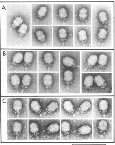
FIG. 4. Electron micrographs of virions produced in infections with
029+
(A), mutant 900(B), or mutant 914(C) and purified by cesium chloride density gradient centrifugation. Particles werefixedwithglutaralde-hydeand negativelystained for electron microscopy as described in the text. Fibers attached to the 4)29+
virionsshown in (A) are clearly absent on the virions shown in panels (B) and (C). The magnification bar represents100nm.
susl0(302) gave
comparable
average (n = 5)recombinationfrequencies of 1.8 + 0.21and 1.6
+ 0.26%, respectively (data not shown). The
results
shown
inTable
5areconsistent
with the
order
900-cistron9-cistron
10.Because
the
head protein
of
mutant 914has
a369
VOL. 24, 1977
on November 10, 2019 by guest
http://jvi.asm.org/
370 REILLY, NELSON, AND ANDERSON
__mm
4-Ap
p9
Hd
----m
host
p11
F
FIG. 5. Autoradiograph of "4C-labeled
029
pro-teins comprising mature virions produced by susl4(1241) and900 series mutant infections ofB. subtilis SpoA12 and isolated by sucrose gradient
centrifugation as shown in Fig. 3. Samples ofthe
peakfractions7(Aand BofFig. 3)and 3(Cand Dof Fig. 3)wereanalyzed bySDS-gel electrophoresisona
lineargradient of 16 to 20%polyacryalamide. (a) susl4(1241), (b) 903, (c) 900, (d) 914, and (e) susl4(1241)virionspurified bycesiumchloride den-sity gradientcentrifugation. The fiber (F)protein is present on virions from the susl4(1241) and 903 mutantinfections, but not on virions from the 900
and914mutantinfections.
distinctive electrophoretic
mobility,
afour-fac-tor cross was
possible.
The recombinant
sus9(756)-914
wasconstructed,
andthe
pheno-typewas
confirmed
by electrophoresis.
Aseries
of
sus8(769)-912 recombinants
wasconstructed.
One recombinant
gave an average (n =5)
re-combination
frequency
of 4.1 0.69%when
crossed with
sus9(756)
ascompared
withacon-trolseriesaverage(n=4) of 4.2 0.14%.
These
recombinants
wereused
toperform the
four-factor cross
depicted
inTable
6.Forty-eight
recombinant
clones chosen
at random wereprocessed
asdescribed
above, and the results
areconsistent withthe order
sus8(769)-914-912-sus9(756).A
series
of sus8(769)-900 recombinants
wasconstructed. One recombinantgaveanaverage
(n =4)
recombination
frequency of
5.2+ 0.76%when crossed with sus9(756)
ascompared with
a
control series average (n
=4)
of
4.6+ 0.57%.
This recombinant was used to perform the
four-factor
crossdepicted in Table
7.Forty-eight
recombinant
clones chosen
atrandom
wereprocessed as
described above, and the results of
the
electrophoresis are consistent with the
or-der sus8(769)-914-900-sus9(756).
DISCUSSION
Our
analysis of the role of the fiber
protein in
k29
replication
has been
complicated
by
the
fact that apparent mutation in the gene
that
codes
for the head fiber protein does not seem
tobe conditionally lethal
(Table 1) and because
anomalous
behavior in
genetic
crossesis
char-acteristic of mutants of the 900 series (Tables 3
and 4). We
have focused on the
mutant3us8.5(900) because this
mutantis
suppressible
by both the su+3 and su+44 hosts
(Fig. 2e and
Table 1), and an apparent
fragment of the fiber
is
produced.
After backcrossing, sus8.5(900)
could be used in three-
and four-factor
crosses(Tables
5
and 7). Our
evidence
suggests that
the properties of
the
900
particle are
character-istic of particles lacking the fiber protein.
We were
particularly interested in the
prop-erties of the
914particles that form in the
pres-ence
of
anapparently
normal fiber
protein (Fig.
ic, 5d, and 8c and Table 1). In these protein
profiles the protein plO has an altered
electro-phoretic mobility.
Backcrossing has restored
the
wild-type p10
inboth virion and
prohead
(data not
shown). Both
900and
914virions were
fast
sedimenting in
sucrosegradients relative
to
429+
(Fig. 3C and D). In contrast to
b29+,
900and 914 virions lacked fibers when
examined
by
electron
microscopy (Fig.
4Band C) or
electro-phoresis (Fig. 5c and d). All of these
particles
have approximately the same
absolute
effi-ciency of plating (Table 2). When b29+, 900, and
914
particles
were
heated
at
70'C in
TMS,
the
slopes
of inactivation were
indistinguishable
TABLE 2. Relationshipbetween
4b29
particlesand PFUPhage
PhagealMl
10-12 XPFUb/ml X
10-12 PFU/phage029+
3.9 ± 0.8 2.9 ± 0.3 0.73900 4.5 ± 1.3 4.8 ± 0.4 1.07
914 6.3 + 1.1 5.3 ± 0.8 0.84
a Based on five electron microscopic
determina-tions of the ratio ofphagetolatexparticles.Average countsof 179, 197, and58phage and 1,133, 909, and 397latexparticlesweremadefor
029+
900, and 914, respectively.bAveragePFUdeterminedonsixplatesofa sin-gle dilutionseries.
J.
on November 10, 2019 by guest
http://jvi.asm.org/
[image:8.504.94.235.89.336.2]MORPHOGENESIS OF BACTERIOPHAGE
029
371 IsI I' II II I I II II II I' II I I * 1 I I I 05
10
15
20
Fraction
Number
LV
_c
1.5E
E
1.0
''
c
C x0.5
u c. c 0Density
(g/cm3)
-153 ) -1.51 1.49 1.4 l145 ) 1.432.0
E
0-0 0 al C-,1.0
_0.5
vQ
5
10
15
20
[image:9.504.53.445.59.306.2]Fraction Number
FIG. 6. Fiberless virions have a buoyant densitygreater than that of 429'. Phage wereproduced in susl4(1241)- andmutant900-infectedB.subtilisSpoA12inthepresenceof14C-and3H-labeledaminoacids, respectively,andwerepurified by density gradient centrifugationincesium chlorideasdescribedpreviously (2).Some of the "IC-labeled14-virions weretreated withdimethylsulfoxidetoremovetheheadfibers(27).
"4C-labeled 14-particles with and withoutfiberswere mixedseparately with 3H-labeled900particles,and cesiumchloridewasaddedtoeach mixturetogiveanaveragedensity ofapproximately1.46glml.The result-ing mixtureswerecentrifugedinanSW50.1rotorat25,000rpm(60,000 xg) for38 h at250C.Fourteen-drop fractions were collected from each centrifuge tube, and the 14C and 3Hradioactivity in each fraction was
determinedby combustinga20-pI sampleinaPackard Tri-Carb oxidizer(model 306).Toobtain thedensity ofcertainfractions, therefractive indexwasdetermined byuseofanAbberefractometer. Therefractometer prism wasmaintained at25 + 0.10CbyuseofaLaudatypeK2-Rrecirculatingwater bath.Symbols: 0, 3H
radioactivity in mutant900virions lackingfibers (A)and(B); *, 14Cradioactivity in(A) 14-phagewith fibersand(B) dimethyl sulfoxide-treated14-phage lacking fibers; A,densityat250C(A)and(B).
(datanotshown). Survival of PFUin 1and2 M
sodium perchlorate for30minat370Coraftera
rapid 200-fold dilution from 2 M NaCl or 20% sucrose intoTMS gave comparable results for all threeparticles (data notshown). These
re-sults suggestthatifthe head of the 900or 914
particle is less stable than k29+, it is masked by the relativeinstability of the neck-tail complex
orthehead-neckjunction.
Apparently
T4phagecanalso beconstructedwithoutproteins normally found in the head of the virion. Infection with the T4 mutant IPO, defective for three internal head proteins,
re-sults in the production of active phage (7). Moreover, the hoc andsocproteins, which
to-gether with p23* form the lattice of theT4head shell and are distributed overthe entire head
surface,arenonessentialforphagegrowth (13).
The hoc andsoc proteinsform the six bridges
and centerunit, respectively, in the basic hex-americ repeating unit. In addition, p24*,
cleaved fromp24 during normal head assembly andthoughttoplayasignificant structural role
inthe T4head, has recently been shownto be dispensable withasingle compensating
muta-tioningene23 known asbypass 24 (N.
Stern-berg, D.Tiemeier, and L. Enquist, Abstr. Fifth Biennial Conf. Bacteriophage Assembly, Snow-bird, Utah, 1976; L. Black, personal communi-cation). As another example, a coliphage X
vectorconstructed bySternberg, Tiemeier, and EnquisttocloneEcoRI fragments contains the Dam15 mutation, yet plates on ansupO host
(Snowbird, Abstr. Fifth Biennial Conf. Bac-teriophage Assembly). The Xgene D protein is
amajorexternal capsid protein.
Someof the T4 particles assembled without the above proteins differ in certain respects
fromthe normalparticles. Forexample, phage lackingp24 (am24byp24) have apparently
nor-malmorphology and heat and pH stability, but
are osmotic shock resistant. Also, the am24
(A)
4.0E
Q.~0
10 0 w >u 3.0-2.0 [
1.0
p
(B)
S~~~
I' U'~~~~~~~~~~~~~~~~~~~~~~~~~~~~~~~~~~~~ I2.0
00E
1.0
E> 4-u 0 00.5
to v0
MVOL. 24, 1977
n)
e
19
r
A
on November 10, 2019 by guest
http://jvi.asm.org/
372
REILLY, NELSON, AND ANDERSON 1614
_(A)
12
10 5
8-8
o2
3 16
14
(B
0
-a
12-10
8
6 4-2 0
0 5 10 15 20 25 30
Fraction Number
FIG. 7. Sedimentation properties ofcontrol 14-andmutant900proheads. UV-irradiated B. subtilis
SpoA12 (12) was infectedwith susl4(1241) or mu-tant900phage (MOIof 20), andafter a5-min ad-sorptionperiodat23°Cthecultureswereincubatedat
37°C with shaking. The infectedcultures were la-beledcontinuously with 14C-amino acids (5
,uCiml)
from6to 100 min after infection.Labelingwas ter-minated andlysates werepreparedasdescribed pre-viously (29). Proheadswere isolated by
centrifuga-tionof thelysatesin5 to20%sucrosegradients,and representativegradient profilesareshownin(A) and (B).CentrifugationwasinanSW50.1rotor at35,000
rpm(120,000 xg)for 30 minat23°C. Sedimentation isfrom righttoleft, and the radioactivities in
frac-tions1to30of42fractions, determinedasdescribed previously (29), areplotted. Proheads arefound in the majorpeak of each gradient profile (20). (A)
susl4(1241)-infected cells; (B) mutant 900-infected
cells.
byp24 phage
areformed more slowly than
wild-type
phage, and protein cleavage occurs at a
greatly reduced rate. hoc- phage are more
re-sistant
toosmoticshock than
wild-type T4, but
soc-
hoc-
phage
aremuch
more sensitive toosmotic
shock
than is T4+. We have not de-tected differences between029+
and
thefiber-less
particles
insensitivity
toheat
or osmoticshock.
The prohead of
029
contains the head, fiber,
plO, and p7 proteins.
Assuming that the
num-ber of copies of head
proteinis
comparablein
the
prohead and virion, at least 50 copies of the
fiber protein are present in the virion (11, 30)
and
atleast
30copies
are present in the prohead(20).
The proheads
of 900 and 914 lack the fiberprotein
(Fig. 8). With other mutants, if anal-tered fiber protein can be detected on the
vir-ion, it is present in the prohead. In contrast, if
the altered fiber is absent from the virion, it
cannot
be
detected in the prohead. This
indi-cates a very
specific interaction
in aprimitive
organizing structure, the prohead.
We
have
observed
bromodeoxyuridine-generated
mutantsthat contained
more thanone point mutation (25; this study, data not
shown). Nevertheless,
the anomalous behaviorof
the recombinants containing mutations of the
900
series
(Tables 3 and 4) was unexpected. In
addition,
oneof four reciprocal three-factor
A
P
p9
Hd
-plO
J1i
F
-
I., [image:10.504.96.233.72.381.2]p7-a
b
c
d
e
FIG. 8. Autoradiograph of '4C-labeled proteins comprising proheads isolated by sucrose gradient centrifugation. Samples from the peak fractions23 (Fig. 7A) and21 (Fig. 7B) containing susl4(1241) andmutant900proheads, respectively, were exam-inedfor viral protein composition along with pro-headsfrom mutant 904- and 914-infected cells ob-tainedsimilarly(gradient profilesnotshown). SDS-gel electrophoresis was in 12%polyacrylamide. (a) Purified429+virions;(b)900proheads;(c)914 pro-heads; (d) 904proheads; (e) susl4(1241) proheads.
J. VIROL.
on November 10, 2019 by guest
http://jvi.asm.org/
[image:10.504.291.438.304.558.2]MORPHOGENESIS OF BACTERIOPHAGE
429
373crosses
that
gaveanomalous recombination
fre-quencies
(Table
3) gaveinconsistent results
when the recombinants
wereexamined
by
elec-trophoresis
(data
notshown).
For this reasonwe
focused
onrecombinants that
were"typical"
by the
criterionof recombination
frequency.
The
four
multiple-factor
crossesemploying
elec-trophoretic
analysis
(Tables
5,6, and 7)
arecon-sistent
with
the
order
sus8(769)-914-912-sus9(756)
orsus8(769)-914-900-sus9(756).
Mel-lado
etal.
(15a)have
isolated and
mapped
the
mutation sus8(22) (19).
Wehave
confirmed the
position
of sus8(22)
by four-factor
crosses(data
not
shown). During restrictive infection with
sus8(22),
neither
head protein
norfiber
ispro-Ap
P9
eM -b.m
Hd-
_m
-
--P11
'x
p1O7~
F
p15-duced; however,
anapparent
fragment
with amolecular
weight
of25,000
can be detected.Both
head protein and
fiber areproduced
by
revertants.
The tryptic
peptides of the
fragment
are
consistent
with a mutation in the cistroncoding for the head protein, and
the absence offiber
may
be the result ofpolarity
of transla-tion.Taken
together,
these results arecon-sistent
with ascheme
inwhich
cistron 8 codesfor the
major
headprotein
andcistron 8.5 codes for thefiber.
Therefore, the mutation
900 could be namedsus8.5(900).
There are two views as to the composition
of
the
429
head andhence
thestructure
of
429.
Mendez
etal.
(18)
report sevenpolypeptides
in429,
withthree being
present inthe
head.
The
results of
Pene
et al.(22) are
inagreement.
The
structure
proposed by Vifiuela
etal.
(30)con-tains five
subunits of the minor
head
protein
HP2
per phage.
Carrascosa et al. (9) now
detect
a
fourth
headprotein,
H00,
in thevirion.
Our view is that the virus
head
consistsof
the
major
head protein
(HP1), the fiber (HP3), and
the
upper
collarprotein p10 (NP2) and that
the
fiber is
nonessential.
Wehave
notobserved
HPO or HP2 in any
lysate,defective particle,
intermediate particle, or virus,
employingslab
discontinuous SDS-gel
systems with singlecon-centrations of
polyacrylamide (e.g.,
10and
12%)
or
with
linear
gradients
ofpolyacrylamide
ranging from
16to 20% to
12 to 19% (5, 6).HP2
has
never been detected
when
cistron 8mu-tants
have been
grownin the su- host
(5,9,15).
IfHP2
exists, then its absence must reflect
the
polarity
of the cistron
8mutation
[e.g.,
sus8(769)]
asdoes the
absence of fiber protein.
LM3-p7LM_
L
M6
[image:11.504.49.236.244.650.2]abcd
e
f g
FIG. 9. Autoradiograph of "4C-labeled proteins presentin lysates and proheads from susl4(1241)-,
901-, 908-, and 909-infected B. subtilis SpoA12. Phage infection, radioactive labeling, and prohead isolation were as described in the Fig. 7 legend. Preparation of cell lysates, SDS-gel electrophoresis (12 to 19%lineargradient ofpolyacrylamide), and autoradiography were as described in the text. (a) susl4(1241)lysate; (b)901 lysate; (c) 908lysate; (d) 909 lysate; (e) 909 proheads; (/) 908proheads; (g) 901 proheads; (h) susl4(1241) proheads. (i) repre-sentsanuninfected control culture, and (j) is a pro-file of purified 429+ virions. Symbols, names, and molecularweights of the429 structural proteins are asdescribedinthelegend to Fig.1.Otherviral pro-teins indicated here are the low-molecular-weight proteinsLM3andLM6having molecular weights of 16,700 and 13,000, respectively, and the prohead
"scaffolding"
protein p7 having a molecular weight of 14,200 (5).VOL. 24, 1977
'"Mmob . 4.
on November 10, 2019 by guest
http://jvi.asm.org/
374
REILLY, NELSON, AND ANDERSONTABLE 3. Anomalousbehavior of mutant 900 in three-factor crosses
Parental genotypes No. of tests
Recombination
cies frquen- Ratioa7(614) + + 23 0.90 ± 0.24 1.00
+ 8(769) +
7(614) + + 5 0.68 ± 0.05 0.76
+ 8(769) 900
7(614) + 900 6 0.32 ±0.08 0.36
+ 8(769) +
8(769) + + 33 5.3 ± 0.95 1.00
+ + 9(756)
8(769) + + 6 3.8 ± 0.34 0.72
+ 900 9(756)
8(769) 900 + 10 1.3 ± 0.16 0.25
+ + 9(756)
7(614) + + 24 4.5 ±0.95 1.00
+ + 9(756)
7(614) + + 8 3.3 ±0.88 0.73
+ 900 9(756)
7(614) 900 + 8 0.85 ± 0.20 0.19
+ + 9(756)
aThe ratioreflects thecomparisonof therecombinationfrequencyobtainedinathree-factorcrossbythat
obtained with thecorrespondingcontrol two-factorcross.
J. VIROL.
on November 10, 2019 by guest
http://jvi.asm.org/
MORPHOGENESIS OF BACTERIOPHAGE
029
375TABLE 4. Anomalous behavior of mutants of the 900 series in three-factor crosses Parentalgenotype
7(614) + +
+ + 9(756)
Recombination frequency of fibermutantX Rat
900 907 908 909 911 912
4.2 5.0 4.6 5.4 4.9 3.8 1.00
7(614) + +
+ (X) 9(756)
7(614) (X) +
+ + 9(756)
3.8 5.0 3.5 6.6 8.3 3.7 1.07 + 0.27
0.85 1.1 0.81 1.0 1.2 1.0 0.22 + 0.09
8(769) + +
+ + 9(756)
8(769) + +
+ (X) 9(756)
8(769) (X) +
+ + 9(756)
5.0 6.4 5.3
3.8 7.1 3.9
1.3 0.90 1.2
5.7 5.5 4.2 1.00
7.7 6.2 4.7 1.04 + 0.14
1.9 2.4 1.0 0.27 + 0.06
aEachcross wasmadeinquadruplicate, and theaveragerecombination frequencyis listed.
bThis ratio reflects the recombination frequency obtained byagiventhree-factorcrosscomparedwiththe control two-factorcross.The ratiolisted reflects theaverageof the six values obtained.
TABLE 5. Resultsof three-factorcrossestoposition the 900fibermutation Progeny phage
Parentalgenotypes Recombination Fiber in recombinant'
frequency Parental
types
8(769) + + 6.7 49 11 9
+ 900 9(756) 51
900 9(756) + 2.0 50 18 2
+ + 10(302) 50
a Twenty recombinantswerestudiedby SDS-polyacrylamide gel electrophoresis. Recombinantsareable
(like
029+)
or unable (like 900) to produce normal fibers in su- infections. The gels contained 12% polyacrylamide.tiob
VOL. 24, 1977
on November 10, 2019 by guest
http://jvi.asm.org/
[image:13.504.50.447.436.540.2]376
REILLY, NELSON, AND ANDERSONTABLE 6. Results of a four-factor cross to orient the cistrons coding for the major head protein and the fiber
Parentalgenotype Progeny
genotypea
combinants8(769) + 912 +b + + 912 + 17
+ 914 + 9(756) + 914 + + 27
+ 914 912 + 4
+ + + + 0
a The sus8(769)-912 parent constituted
53%
of the progeny phage. Therecombination frequency was 6.7%. The phenotype of 48 recombinants was studied by SDS-gel electrophoresis in 12% polyacrylamide.bThe 912 mutant is unable to synthesize the fiber protein during restrictive infection.The 914mutantis
[image:14.504.61.463.236.310.2]characterized by a major head protein of altered electrophoretic mobility, and the phenotype isassumed to reflect mutation in the cistron coding for the head protein.
TABLE 7. Results of a four-factor cross to orient the cistrons coding for the major head protein and the fiber
Parentalgenotype Progenygenotype0
No.
ofre-8(769) + 900 +b + + 900 + 16
+ 914 + 9(756) + 914 + + 18
+ 914 900 + 13
+ + + + 1
aThe sus8(769)-sus8.5(900) parent constituted 50% of the progeny phage. Therecombination frequency
was 4.2%. The phenotype of 48 recombinants wasstudied by SDS-gel electrophoresisin 12%polyacrylamide.
bThe 900 mutant is unable to synthesize the fiber proteinalthough an apparentfragment of the fiber is produced during restrictive infection. The 914 mutant ischaracterized by a major head protein of altered electrophoretic mobility, and the phenotype is assumed to reflect mutationinthe cistroncodingfor thehead protein.
The position of the major head protein after 914
infection would be expected to permit the
visu-alization
of HP2. It does
not.The putative
pro-tein
HPO bands at the approximate position of
the
head protein of 914. We suspect that both
HPO and HP2 could be generated by a low level
of
misreading and/or proteolytic cleavage and
for this
reasoncould appear at random in any
particular lysate.
We
believe that
the
genes
coding
for
struc-tural
proteins
of
429
have
been identified.
The
results of Yanofsky et al. (31) suggest that the
DNA
"linker" protein(s) described by Ortin et
al. (21) is
the product
of cistron
3and that
the
remaining
structural proteins are coded for by
cistrons
8, 8.5, 9, 10, 11, and
12(5, 9, 15, 16). We
have found no evidence for any function of the
fiber
protein except adornment under the
arti-ficial
laboratory
conditions
conventionally
used.
ACKNOWLEDGMENTS
We gratefully acknowledge the technical assistance of Richard Rau, Charlene Peterson, and Viola M. Zeece.
This work was aided by grant GB-29393 fromthe Na-tional Science Foundation andbyPublic Health Service grantsDE-3606 and GM-19743 from theNational Institute of Dental Research and the National Instituteof General MedicalSciences, respectively.
LITERATURE CITED
1. Adams, M. 1969. Bacteriophages. John Wiley & Sons, Inc., New York.
2. Anderson, D. L., D. D. Hickman, and B. E. Reilly. 1966.Structure ofBacillus subtilisbacteriophage 429 and thelengthof429deoxyribonucleicacid. J. Bacte-riol. 91:2081-2089.
3. Anderson, D.L., and E. T. Mosharrafa. 1968.Physical andbiological properties of phage 429 deoxyribonu-cleic acid. J. Virol. 2:1185-1190.
4. Anderson, D. L., M. E. Pollock, and L. F. Brower. 1965.Morphology ofMycoplasmalaidlawii type A. I. Comparison of electron microscopiccountswith col-ony-forming units. J.Bacteriol.90:1764-1767.
5. Anderson, D. L., and B. E. Reilly. 1974. Analysisof bacteriophage429genefunction:proteinsynthesisin
suppressor-sensitive mutant infection ofBacillus sub-tilis.J.Virol.13:211-221.
6. Anderson, D. L., andB. E. Reilly. 1976. Analysis of gene functioninBacillussubtilisbacteriophage429, p. 254-274. In D. Schlessinger (ed.), Microbiology -1976. AmericanSociety forMicrobiology, Washing-ton, D.C.
7. Black, L. W. 1974. Bacteriophage T4 internal protein mutants: isolation and properties. Virology
60:166-179.
8. Camacho, A., E. Moreno, J. L.Carrascosa, E.Vifnuela, and M.Salas. 1974.Asuppressor ofnonsense
muta-tions inBacillus subtilis. Eur. J. Biochem. 47:199-205.
9. Carrascosa,J.L.,A.Camacho,F.Moreno,F.Jimenez, R. P.Mellado, E.Vifnuela,andM.Salas.1976. Bacil-lus subtilisphage4)29-characterization ofgene prod-uctsand functions.Eur. J. Biochem. 66:229-241.
10. Carrascosa, J. L., E. Viftuela, and M. Salas. 1973.
J. VIROL.
on November 10, 2019 by guest
http://jvi.asm.org/
MORPHOGENESIS OF BACTERIOPHAGE 429 377 Proteins induced inBacillus subtilis infected with
bacteriophage429. Virology56:291-299.
11. Hagen, E. W., B. E. Reilly, M. E. Tosi, and D. L. Anderson. 1976.Analysisof gene function of bacterio-phage 429ofBacillus subtilis: identification of cis-tronsessential for viralassembly. J. Virol. 19:501-517.
12. Hawley, L.A., B. E. Reilly, E. W. Hagen, and D. L. Anderson. 1973. Viral proteinsynthesisin bacterio-phage +29-infected Bacillus subtilis. J. Virol. 12: 1149-1159.
13. Ishi, T., and M. Yanagida. 1975. Molecular organiza-tionof theshell of the Teven bacteriophagehead. J. Mol. Biol. 97:655-660.
14. Jimenez,F., A. Camacho, J. De La Torre, E. Vinuela, and M. Salas. 1977. Assembly of Bacillus subtilis phage4)29. 2.Mutantsinthe cistronscodingforthe non-structural proteins. Eur. J. Biochem. 73:57-72. 15. McGuire, J. C., J. J.Pene, and J. Barrow-Carraway.
1974.Gene expressionduring thedevelopmentof bac-teriophage4)29.III.Analysis ofviral-specificprotein synthesis with suppressible mutants. J. Virol. 13:690-698.
15a.Mellado, R. P., E.Mindez, E.Vifuela, and M. Salas. 1977. Order of the two major head protein genes of bacteriophage 429ofBacillus subtilLs. J. Virol. 24: 378-382.
16. Mellado, R.P., F. Moreno, E.Vinuela,M. Salas, B. E. Reilly, and D. L. Anderson. 1976. Genetic analysis of bacteriophage 4)29 of Bacillus subtilis: integration andmappingof reference mutants of two collections. J.Virol. 19:495-500.
17. Mellado, R. P., E.Viftuela, and M. Salas. 1976. Isola-tionof a strong suppressorof nonsense mutationsin
Bacillussubtilis. Eur. J. Biochem. 65:213-223. 18. Mendez, E., G. Ramirez, M. Salas, and E. Vinuela.
1971.Structuralproteins ofbacteriophage4)29. Virol-ogy 45:567-576.
19. Moreno, R., A. Camacho, E. Vinuela, and M. Salas. 1974.Suppressor-sensitive mutants and genetic map of Bacillussubtilis bacteriophage4)29.Virology 62:1-16.
20. Nelson, R. A., B. E. Reilly, and D. L.Anderson. 1976. Morphogenesis of bacteriophage4)29ofBacillus sub-tilis: preliminary isolation and characterization of intermediate particles of theassemblypathway. J. Virol. 19:518-532.
21. Ortin, J., C.Vasquez, E.Vinuela,and M. Salas. 1971. DNA-proteincomplex in circular DNA from phage 429.Nature(London) New Biol. 234:275-277. 22. Pene, J. J., P. C. Murr, and J. Barrow-Carraway. 1973.
Synthesis of bacteriophage 4)29proteins inBacillus subtilis. J. Virol. 12:61-67.
23. Polsinelli, M.,and M. Beretta. 1966. Genetic recombi-nation in crosses betweenStreptomyces aurofaciens andStreptomyces rimosus. J.Bacteriol.91:63-68. 24. Reilly, B. E., and J. Spizizen. 1965. Bacteriophage
deoxyribonucleate infection of competent Bacillus subtilis. J. Bacteriol. 89:782-790.
25. Reilly, B. E., M. E. Tosi, and D. L.Anderson. 1975. Genetic analysis of bacteriophage4)29ofBacillus sub-tilis: mapping of the cistrons coding for structural proteins. J. Virol. 16:1010-1016.
26. Reilly, B. E.,V. M. Zeece, and D. L. Anderson. 1973. A geneticstudy ofsuppressor-sensitive mutantsof the Bacillus subtilis bacteriophage4)29.J. Virol.
11:756-760.
27. Salas, M., C. Vasquez, E. Mendez, and E. Vinuela. 1972. Head fibers of bacteriophage 429. Virology 50:180-188.
28. Tosi, M., and D. L. Anderson. 1973. Antigenic proper-tiesofbacteriophage 429 structuralproteins. J. Vi-rol.12:1548-1559.
29. Tosi, M.E., B.E.Reilly, and D. L. Anderson. 1975. Morphogenesis of bacteriophage429of Bacillus sub-tilis: cleavage and assemblyof the neckappendage protein. J. Virol. 16:1282-1295.
30. Vinuela, E., A. Camacho, F. Jimenez, J. L. Carras-cosa,G. Ramirez, and M. Salas.1976.Structureand assembly of phage429.Philos. Trans. R.Soc. London Ser. B 276:29-35.
31. Yanofsky, S., F. Kawamura, and J. Ito. 1976. Thermo-labile transfecting DNA from temperature-sensitive mutantsof phage429.Nature (London) 259:60-63. VOL. 24, 1977