0022-538X/83/020634-14$02.00/0
Copyright © 1983, AmericanSocietyforMicrobiology
Detailed
Analysis
of the
Portion
of the
Herpes
Simplex
Virus
Type 1
Genome Encoding
Glycoprotein C
R.J. FRINK,1 R. EISENBERG,2G. COHEN,3ANDE. K.WAGNERI*
Department ofMolecularBiologyandBiochemistry, University ofCalifornia, Irvine, California 927171 and DepartmentofPathobiology, SchoolofVeterinaryMedicine,2andDepartmentofMicrobiologyand Center forOral HealthResearch, DentalMedicine,3 UniversityofPennsylvania, Philadelphia, Pennsylvania19104
Received 30 August1982/Accepted5November1982
Wepreviouslyshowedthat therightthird ofHindIIIfragmentL(0.59to0.65)of
herpessimplexvirustype 1(HSV-1)encodes afamilyof mRNAssomemembers of which appear to be related by splicing. In the experiments described in this
communication, we determined the nucleotide sequence of the DNA encoding
thismRNAfamily andprecisely located the mRNAs associated with this DNA
sequence. The major mRNA species is unspliced and encoded by a
2,520-nucleotideregion.Just upstream of the 5' endareTATA and CAT boxsequences
characteristic of HSV-1 promoters. The 3' end maps near a region containing a
nominalpolyadenylation signal.Three minorspecies (2,400, 2,200,and1,900bases,
respectively) appearto shareavery short leader sequence with the 5' end of the
majormRNA andarethen encoded byuninterruptedDNAsequences beginning about 100, 400,and 625 bases downstream of the 5' end of the majorunspliced mRNA. These positions map at or very near positions which agreereasonably wellwithconsensusspliceacceptor sequences. The fourth mRNA is encodedbya
contiguous730-nucleotide sequence atthe 3' end of the major unspliced mRNA
and has its 5' end just downstream of recognizable TATA and CAT box sequences. We suggest that this mRNA is controlled byits own promoter. The
nucleotide sequence data, in combination with the mRNA localization, demon-strate four potential polypeptides encoded by the region. The largest is 1,569
baseslongand defines a 523-amino acidproteinwithsequence features
character-istic of a glycoprotein. This was confirmed to be HSV-1 glycoprotein C by immune precipitation of the in vitro translation product of the major unspliced
mRNA,performedwithapolyspecificantibodytoHSV-1envelopeglycoproteins (anti-env-1 serum), and by comparison of tryptic peptides of this translation
product with thoseof authentic HSV-1 glycoproteinC. Polypeptides encodedby
someof the minor speciesalso weretentativelyidentified.
In anumber ofpreviouspapers, we reported
detailed mapping and characterization of the
major mRNAs encoded by about 20% of the herpes simplex virus type 1 (HSV-1) genome
(reviewed by E. Wagner, in G. Klein, ed., Ad-vances in Viral Oncology, vol. 3, in press). Although many HSV-1 mRNAs overlap others and thus form clusters or families, we find
detectable splicing to be rare, and the great
majority of abundant HSV-1 mRNAs so far
characterized appear to becontrolledby a pro-moterregionintheDNA, closely juxtaposed to thelocation of5' endsofthemRNAs. The sites
ofpolyadenylation for HSV-1 mRNAs appear
similartothose characterized for other
eucary-otic mRNAs; these HSV polyadenylation sites
often define the 3' termini of several partially colinear HSVmRNAs.
Despite the relative rarityof detectable
splic-ing during HSV-1 mRNA biogenesis, spliced
HSV-1 mRNAs do exist. The spliced family
investigated in this report is a prime example. Also, the a(immediate-early) mRNA encoding ICP-25 has a splice in its 5' region which appears tofunctionin theremovalof thetranscriptof the
reiterated terminal portion of the
IRS
region where it joins the UL region (41, 42). We also notedseveralHSV-1 mRNAs with 5' endsmap-ping differentlywhenS1 nuclease and
exonucle-ase VII digestion of DNA-RNA hybrids are
compared (15, 18). Splicing also appears to occurin the mRNA expression of other
herpes-viruses since potential splices have been
tenta-tively identified in certain Epstein-Barr virus mRNAs (22).
To date, the spliced HSV-1 mRNA family
mapping in HindlIl fragment L is the only
instance where differential splicing can be
in-634
on November 10, 2019 by guest
http://jvi.asm.org/
HSV gC mRNA 635
ferred toplayarole in HSV-1 gene expression. It is, of course, a notable curiosity that this mechanism soprevalent inthe gene expression of other nuclear replicating DNA viruses ap-pears toberarewith HSV-1. Forthisreason, we
carried out a detailed analysis of these HSV-1 mRNAs related by splicing. By correlating in vitro translation productswith thecoding
capac-ity ofvarious mRNAs and the predicted amino acid sequences ofproteins potentially encoded by these mRNAs, some statement could be made regarding the actual use of splicing in expression ofspecific HSV-1 gene products.
In addition tothis rather general question of the role of splicing in HSV-1 gene expression,
the detailed analysis described here was de-signed to answer specific questions regarding the expression ofa known HSV-1 polypeptide, glycoprotein C (gC). Thus, ouranalysiscan be related to theexpression ofaproteinof known biochemical properties. This polypeptide has been located in thegeneral regionof interestby
intertypic recombinant studies (19, 38). The ap-parent size of the major translation product
(69,000 daltons; 15) of the mRNA encoded by
this DNA correlates reasonably well with the size of the unglycosylated precursor for this glycoprotein (9, 34). Further, Lee et al. (24a) have shown that DNAfrom this precise region
canhybrid-select gC mRNA. We used the
anti-env-1 serumdescribedearlier (9)to immunopre-cipitate the in vitro translation product of the unspliced and most abundant member of the mRNA family, confirming its identification as
the mRNAfor HSV-1 gC. Thisantibodywasof value inpartiallydefiningthetranslation reading frames of other viral polypeptides which are
apparently encodedby thisregion of the HSV-1
genome.
MATERIALSANDMETHODS
Cellsand virus. Monolayer cultures of HeLacells were grownat37°C in Eagle minimal essential medium containing 10ocalf serum,penicillin,and streptomy-cin. Rabbit skin cells (agiftof J. Stevens ofUCLA)
weregrownin thesamemediumcontaining 5%serum.
Plaque-purifiedvirus of the KOS strain ofHSV-1was
used for all infections.
Enzymes. All restriction enzymes were obtained
from Bethesda ResearchLaboratories, Inc.;digestion was carriedout in buffers recommended by that sup-plier. PhageT4polynucleotide kinasewasusedfor5'
phosphate exchangeasdescribed byMaxamand Gil-bert(26).
Isolation, labeling,and sizefractionation of
polyribo-somal RNA.Monolayer cultures ofHeLacells(2x 10⁷ cellsperflask)wereinfected for30minat a multiplic-ity of10 PFUof virus per cell inphosphate-buffered saline containing 0.1% glucose and 1.09o fetal calf
serum. Polyribosomes were isolated from the cyto-plasm of HSV-1-infected cells by the magnesium
pre-cipitation method of Palmiter (32). mRNA containing polyadenylic acid [poly(A)] was isolated from total ribosomal RNA by oligodeoxythymidylic acid-cellu-lose (CollaborativeResearch, Inc.) chromatography. Details of this procedure have been presented else-where (1-4, 11, 15, 18, 20, 21). RNA wassize fraction-atedbyelectrophoresison1.4% agarosegels contain-ing 10 mM methyl mercuryhydroxide (5)aspreviously described.
Recombinant DNA. All recombinant DNA clones described in this paper were derived from either HindIIIfragment L (0.592to0.647)orBamHIfragment
1(0.602 to 0.643)cloned inplasmid pBR322 (2, 15). We usedtwo subclones: SaIl-EcoRIfragment T-A (0.621
to 0.633) and EcoRI-BamHI fragment I-I (0.633 to
0.643). ProceduresforcloningHSV-1 DNAfragments in thepBR322vector weredescribedpreviously (2, 6, 11). DNAfragments cloned were namedasdescribed previously and located by their map coordinates on the P arrangementof the HSV-1 genome (15).
In situ RNA blots. As described previously, 7-Rg samples of infected-cell polyadenylated mRNA were
fractionated on methyl mercury gels and dried onto Whatman 3MM paper withvacuum (18). The agarose film was floated off the paper in water and was
hybridized with appropriate32P-labeledDNA probes in 50% formamide containing 0.4 M Na+, 0.1 M
HEPES (N-2-hydroxyethylpiperazine-N'-2-ethanesul-fonic acid, pH 8.0), 0.005 M EDTA, and Denhardt solution (12)at50°Cfor 36 h. Blots were rinsedat50°C twice in 509o formamide-2x SSC (1x SSC is 0.15 M NaClplus 0.015 M sodiumcitrate)-0.1%sodium dode-cyl sulfate(SDS), thenoncein0.1x SSC-0.1% SDS. They were thenautoradiographed.
Invitro32P-labeledDNAwasmade by nick translat-ing appropriate DNA clones with DNA polymerase I (Boehringer Mannheim Corp.) and 50 XCi of
[ca-32P]dCTP(3,000Ci/mmol; Amersham Corp.).
Isolationofrestrictionfragment-specificmRNA. Re-striction fragment-specific mRNA was isolated from poly(A)polyribosomalRNAby preparative hybridiza-tion totheappropriate DNA, which had been
cova-lentlycoupledtocellulose.Details of theseprocedures were asdescribedpreviously (1-4,11,20, 21, 31).
Nuclease mapping of HSV-1 RNA. S1 nuclease and exonuclease VII analysis of RNA was carried out essentiallyasdescribedby Berk and Sharp (8) and by others(2, 11, 15). Appropriate HSV-1 DNA clones (2 p.g)werecleavedatthe desiredsites with the
appropri-aterestriction enzymes. The DNA was then 5' labeled with 32P by using polynucleotide kinase (26) to a specificactivity of 4,000 to 10,000 cpm/,Lg of DNA. The DNA fragment then was denatured and strand separated on 5% acrylamide gels as described by
Maxam and Gilbert (26). In one set ofexperiments,
DNA was uniformly labeled with 32p,(30 ,uCi/ml) in low-phosphate MOPS (morpholinepropanesulfonic acid) medium (30, 40).
The separated DNA strand (from 2 ,ug of cloned DNA) was hybridized with 10 ,ug of infected-cell
mRNAin0.1 MNa+-0.1 MHEPES(pH8.0)-0.01M
EDTA at65°C for6to 16 h in avolume of 30j±land
then wasdigested withS1nuclease orexonuclease VII
asdescribed. Materialwasfractionated on a denatur-ing 5% acrylamide gel with 5' end-labeled Hinfl-digested plasmid pBR322 DNA fragments or with nucleotide sequencegelsassize standards.All proce-VOL.45,1983
on November 10, 2019 by guest
http://jvi.asm.org/
dures were based on those of Maxam and Gilbert (26) Nucleotidesequencing. Asdescribed previously (13, 16), nucleotide sequenceanalysis was carried out by theprocedures of Maxam andGilbert (26).
Invitro translation. Size-fractionated viral mRNA wastranslated in vitro by using a micrococcal nucle-ase-treated rabbitreticulocyte system (New England NuclearCorp.) with[35S]methionine (675 Ci/mmol) as theradioactive amino acid. Details of the procedure and of fractionation of polypeptides in SDS-acryla-mide gels by the method of Laemmli (24) have been describedpreviously(10,13, 15). Gels were dried with vacuum, and radioactive bands were localized by autoradiography with Kodak NS-2T film.
Immune precipitation of in vitro translation prod-uctswasperformed asdescribed by J. T. Matthews, R. J. Eisenberg, and G. H. Cohen (submitted for publication). One-half of the RNAse-treated transla-tionproduct (14,ul)wasdiluted withanequal volume of 2xlysis buffer and incubated with4
RI
ofanti-env-l serum, a polyvalent antibody to HSV-1 envelope protein (9, 10).Lysis buffer is 20mM Tris-hydrochlo-ride(pH 7.4), 50mMNaCl, 10mMmethionine,0.5% Nonidet P-40, 0.5% sodium desoxycholate, and 0.1% SDS. After1 honice, 75,u1ofa10%suspensionof pro-ASepharosebeads(Pharmacia FineChemicals)in 50 mM Tris-hydrochloride (pH7.5)-150mM NaCl-5 mM disodiumEDTA-0.2% sodium azide wasadded, and thesuspensionwasincubatedafurther 30 minonice with frequentmixing.Thepro-ASepharosebeads with theimmune complex adsorbedwerethen deposit-edby1 minofcentrifugationinanEppendorf micro-centrifuge, and thepellet wassuspendedin 200 p.1of lysis buffer andcentrifugedfor 5 minthrougha1-ml pad oflysis buffer containing 1 M sucrose. The Se-pharosewasthenwashedby suspensionin 0.5 ml of 10 mM Tris-hydrochloride (pH 7.4)-S150 mM NaCl-10 mM methionine-0.2% Nonidet P-40-0.1% SDS fol-lowed by recentrifugation. After five washes, the Sepharose pelletwasincubated with 20 p.1 ofabuffer containing 0.1 M Tris-hydrochloride (pH 7.0), 3% SDS,10% 3-mercaptoethanol,and20%(vol/vol) glyc-erol. Thesuspensionwasheatedto95°Cfor 2min, and the Sepharose was pelleted by centrifugation. The supernatant containing releasedimmunoglobulin and anytranslationproductthatit had adsorbedwasthen loaded onto SDS-acrylamide gels for size fraction-ation.
Preparation ofsamples fortryptic peptide analysis. Forisolation oftheglycoprotein gC,KB cells
infect-ed with HSV-1 (strain HF) were pulse-labeled with
[methyl-3H]methionine (specific activity, 100 Ci/ mmol) as previously described (9). A cytoplasmic
extract was prepared and immunoprecipitated with anti-env-1 serum(9). Theglycoproteinswere separat-edbySDS-polyacrylamidegelelectrophoresisin slabs of10%acrylamidecross-linked with 0.4%
N,N'-dial-lyltartardiamide (9, 39). The gel was stained with Coomassie brilliant blue, dried on filter paper, and exposed to LKB Ultrafilm to locate tritium-labeled bands. The band corresponding to gC (110,000 dal-tons)wasexcised,dissolved in 2%periodicacidbythe methodof Gibson(17),oxidized, trypsinized (9), and chromatographedon acolumnof ChromabeadsPresin (Technicon Corp., Inc.; 9,14).
For tryptic peptide analysis of the 69,000-dalton protein,hybrid-selectedmRNAwastranslatedinvitro
in the presence of[355]methionine. The translation mix was immunoprecipitated with anti-env-1 serum, and thecomplex was collected with pro-A Sepharose as described above. The antigen-antibody complex was dissociated in SDS disrupting buffer and was centrifuged (13,000 xg, 3min) to remove the Sephar-ose. Bovine serum albumin was added as a protein carrier (1 mg/0.5 ml), and theprotein was precipitated with 25% trichloroacetic acid, oxidized, trypsinized, andchromatographed on Chromabeads P as described previously (14).
RESULTS
Thenucleotidesequence ofHSV-1DNA encod-ing the spliced mRNA family. We previously showed thattheDNA betweenca. 0.63and 0.65
in Hindlll fragment L (0.59to 0.65) encodes a
family of mRNAs related by splicing (15). A
high-resolution restriction endonuclease cleav-agemapof this region isshowninFig. 1A. Sites areshown for theenzymesEcoRI,SalI, BamHI,
PvuII,XbaI, SmaI, TaqI,SstII, andHinfl. The
HindIII site at0.647 isnotshown, but isca. 60 bases to the right of therightmost Sall site. It canbe seen thatthese sites are arranged so as to provide convenient sites for 5', andoccasionally 3', end labeling of DNA fragments for Maxam
and Gilbert chemical sequencing. Our general method wasto use strand-separatedDNA frag-ments, and our DNA sequencing overlaps are
shown in Fig. 1A. We numbered the sequence
bylocatingthe 5' endoftheunspliced andmost abundant mRNA at position +1. Our
experi-ments fixing the location of this mRNA with
respect tospecific restriction sitesaredescribed below.
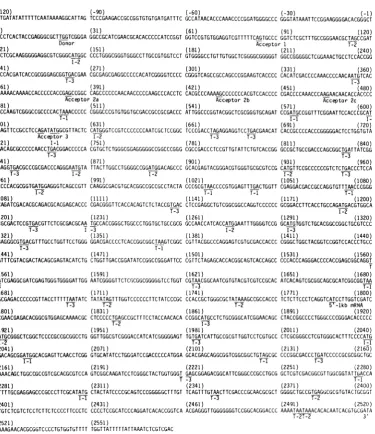
The nucleotide sequence of the noncoding
strandof the DNA in theregion studiedisshown
in Fig. 2.This strand has thesame sense asthe
mRNAs encoded by the region. Note the
se-quence TATAAATT between nucleotides -27
and -19 and thesequence CATTA from -96to
-92. Such putative TATA and CAT boxes are
generally found at these positions upstream of
the 5' end ofunspliced HSV-1 mRNAs (7, 16,
28)and cantentatively define HSV-1promoters. The location of polypeptide chain initiation (ATG) andchain termination (TAG, TGA, and
TAA) codons are indicated, as arethe
transla-tion reading frames they define. These data,
shown schematically in Fig. IC, are discussed below in relation to the in vitro translation
productsoffractionatedmRNAspeciesencoded
by thisDNA.
The coterminal 3' end of the mRNAs was
located very near nucleotide 2520, which is in the sequence ATAAAAA. A similar sequence
ATAATAAA was found to be very near the
coterminal 3' ends of a series of colinear mRNAs mappingbetween 0.554 and 0.600 (13)
on November 10, 2019 by guest
http://jvi.asm.org/
A. 0.633 0.639 0.643 0.646
Sm E Hf Sm Hf Sm X Hf B P S Tq S
SmTq St
I
Tq SmSmj
Sm TqFq
StI
St
Hfl Hf Tq SmStISnl
S-100 1 250 500 750 1000 1250 1500 1750 2000 2250 2500
S o F_ 1 _,,b, ei-)
B.
V
C. Translation Frame
2
11
"ezzz1II
3 " 1Z -l 11 IIiH 11 I
4 1
] I 11
[image:4.496.51.445.58.267.2].
FIG. 1. High-resolution restriction endonuclease cleavage map and sequence strategy for the HSV-1 DNA region encoding the major late 2.7-kilobase (kb) mRNAfamily.(A) Individual sequenceruns(waveylineswith arrowheads showing direction and extent) andrestriction sites utilized and listed in thetext.Numberingsystem takes the 5' terminal base of the mRNAs as +1. (B) Overlapping mRNA species described in this report, including major unspliced2,520-basespecies (dark bar) andintrons for the 2,400-,2,200-,and1,900-base spliced species(gaps).The 3' coterminal730-basemRNAalsois shown.(C)Available open translationreading frames,
including positions of translation initiation(0-.)andtermination(I)codons.
and is similar to the putative eucaryotic poly-adenylation signalAATAAAA (35). This region between nucleotide -2,500 and the Sanl site at
position 2575 (not shown) contains the3' end of
mRNAs from both DNA strands (15), and its
stretchesof AT-richsequencesisreminiscent of
asimilar structure around position 0.600 noted
byuspreviously (13).
The relative abundance of members of the mRNAfamily.Weused in situNorthernblots to measurethe relativeabundance of various
mem-bers of the mRNAfamily. Polyribosomal
poly-adenylated mRNA (5 ,ug) from cells 6 or 11 h postinfection(p.i.)wassizefractionated by
elec-trophoresis in 1.4% agarose gels containing 10 mMmethylmercuryhydroxide. These gelswere
dried, and theagarosefilmwas hybridized with
[32P]dCTP-labeled
DNA from EcoRI-BamHIfragment I-I (0.633 to 0.643), which had been
cloned inplasmidpBR322 (Fig. 3A). The major
mRNA species is indicated as having a size of
2,520 bases,based on ourSi data(see below). It migrates with a rate corresponding to a size of
2,700 bases. This size isexpectedforan mRNA
with a DNA-encoded size of 2,500 bases and a
200-nucleotidepoly(A) tail (Wagner, in press). It
is clear that this mRNA species is the most
abundantatboth 6 and 11 h p.i.
Two smaller mRNA species migrate with a rate corresponding to a size of about 2,500
bases,and another with a rateofca. 2,000 bases isalsoseen.Averyminorband migrating with a ratecorrespondingto a size of ca. 900 to 1,000
bases can be detected with some difficulty. These species are indicated by the lengths of their encoded sequences (see below) as 2,200,
1,900, and730bases, respectively. The relative
intensity ofthe smallest mRNA species is
proba-blyanunderrepresentation of the abundance of this mRNA because the probe used is
homolo-gous to only about 40% of the length of this mRNA.
Inafurtherexperiment (datanotshown), we
found anidenticalpattern by using polyadenyl-ated mRNAfrom rabbit skincells infected with HSV-1. This indicated thatthe mRNA
distribu-tion is generally not dependent on the type of cellinfected.
We used a
[32P]dCTP-labeled
probe covering theregion between nucleotides-30and+256 todemonstrate that the smaller mRNAs of2,200
and1,900 basescanbedetected withDNA from a region of the genome nominally upstream of
theirpredicted 5' ends (Fig. 3B). Although the
2,200-base species is notclearlyresolved in the
photo, it can be seen in the original
autoradio-graph, and the 1,900-base species is readily apparent. The 730-base species could not be
detected, evenafterverylongexposure times. Two faint bands of mRNA migrating with rates correspondingto sizes between 4,000 and
5,000 bases were consistently seen with both
probes. The small amounts of these RNA
spe-ciesprecluded their furthercharacterization. Preciselocation of the mRNAs.Wepreviously reportedthe grosslocalization ofthemRNAsof
I
I
HSV gC mRNA 637
VOL.45, 1983
on November 10, 2019 by guest
http://jvi.asm.org/
VIROL.
(-120) (-90) (-60) (-30) (-1)
ATTGATATATTTTTCAATAAAAGGCATTAG TCCCGAAGACCGCCGGTGTGTGATGATTTC GCCATAACACCCAAACCCCGGATGGGGCCCGGGTATAAATTCCGGAAGGGGACACGGGCT
(1) (31) (61) (91) (120)
ACCCTCACTACCGAGGGCGCTTGGTCGGGA GGCCGCATCGAACGCACACCCCCATCCGGT GGTCCGTGTGGAGGTCGTTTTTCAGTGCCCGGTCGrbGTbTGCCGGGAACGCTAGCCGAT
Donor Acceptor 1 T-2
(121) (151) (181) (211) (240)
CCCTCGCAAGGGGGAGGCGTCGGGCATGGC CCCTGGGCGGGTGGGCCTTGCCGTGGTCCT GTGGGGCCTGTTGTGGCTCGGGGCGGGGGT GGCCGGGGGCTCGGAAACTGCCTCCACCGG
1-2
(241) (271) (301) (331) (360)
GCCCACGATCACCGCGGGAGCGGTGACGAA CGCGAGCGAGGCCCCCACATCGGGGTCCCC CGGGTCAGCCGCCAGCCCGGAAGTCACCCC CACATCGACCCCAAACCCCAACAATGTCAC
T-3 1-3
(361) (391) (421) (451) (480)
ACAAAACAAAACCACCCCCACCGAGCCGGC CAGCCCCCCAACAACCCCCAAGCCCACCTC CACGCCCAAAAGCCCCCCCACGTCCACCCC CGACCCCAAACCCAAGAACAACACCACCCC
Acceptor2a Acceptor2b Acceptor 2c
(481) (511) (541) (571) (600)
CGCCAAGTCGGGCCGCCCCACTAAACCCCC CGGGCCCGTGTGGTGCGACCGCCGCGACCC ATTGGCCCGGTACGGCTCGCGGGTGCAGAT CCGATGCCGGTTTCGGAATTCCACCCGCAT
T-1 1-1
I-(601) (631) (661) (691) (720)
GGAGTTCCGCCTCCAGATATGGCGTTACTC CATGGGTCCGTCCCCCCCAATCGC TCCGGC TCCCGACCTAGAGGAGGTCCTGACGAACAT CACCGCCCCACCCGGGGGACTCCTGGTGTA
2 Acceptor3 I-2 1-3 T-3
(721) I-1 (751) (781) (811) (840)
CGACAGCGCCCCCAACCTGACGGACCCCCA CGTGCTCTGGGCGGAGGGGGCCGGCCCGGG CGCCGACCCLrcCGbTGTATTCTGTCACCGG GCCGCTGCCGACCCAGCGGCTGATTATCGG
T-3 T-3
(841) (871) (901) (931) (960)
CGAGGTGACGCCCGCGACCCAGGGAATGTA TTACTTGGCCTGGGGCCGGATGGACAGCCC GCACGAGTACGGGACGTGGGTGCGCGTCCG CATGTTCCGCCCCCCGTCTCTGACCCTCCA
T-3 1-2 1-2 1-2 T-3
(961) (991) (1021) (1051) (1080)
GCCCCACGCGGTGATGGAGGGTCAGCCGTTCAAGGCGACGTGCACGGCCGCCGCCTACTA CCCGCGTAACCCCGTGGAGTTTGACTGGTT CGAGGACGACCGCCAGGTGTTTMCCCGGG
1-2 T-1 T-1 T-1
(1081) (1111) (1141) (1171) (1200)
CCAGATCGACACGCAGACGCACGAGCACCC CGACGGGTTCACCACAGTCTCTACCGTGAC CTCCGAGGCTGTCGGCGGCCAGGTCCCCCC GCGGACCTTCACCTGCCAGATGACGTGGCA
T-3 1-2
(1201) (1231) (1261) (1291) (1320)
TCGCGACTCCGTGACGTTCTCGCGACGCAA TGCCACCGGGCbTGGCCTGGTGCTGCCGCG GCCAACCATCACCATGGAATTTGGGGTCCG GCATGTGGTCTGCACGGCCGGCTGCGTCCC
T-3 -3 I-2 1-3
(1321) (1351) (1381) (1411) (1440)
CGAGGGCGTGACGTTTGCCTGGTTCCTGGG GGACGACCCCTCACCGGCGGCTAAGTCGGCCGTTACGGCCCAGGAGTCGTGCGACCACCC CGGGCTGGCTACGGTCCGGTCCACCCTGCC
T-3 f--1
(1441) (1471) (1501) (1531) (1560)
CATTTCGTACGACTACAGCGAGTACATCTG CTGGTTGACCGGATATCCGGCCGGGATTCC CGTTCTAGAGCACCACGGCAGTCACCAGCC CCCACCCAGGGACCCCACCGAGCGGCAGGT
f
(1561) (1591) (1621) (1651) (1680)
GATCGAGGCGATCGAGTGGGTGGGGATTGGAATCGGGGTTC1CGCGGCGGGGGTCCTGGT CGTAACGGCAATCGTGTACGTCGTCCGCAC ATCACAGTCGCGGCAGCGCATCGGCGGTM
-3 T-3 T-1
(1681) (1711) (1741) (1771) (1800)
CGCGAGACCCCCCGTTACCTTTTTAATATC TATATAGTTTGGTCCCCCCTTCTATCCCGC CCACCGCTGGGCGCTATAAAGCCGCCACCC TCTCTTCCCTCAGGTCATCCTTGGTCGATC
T-3 T-2 T-2 5-lkbmRNA
(1801) (1831) (1861) (1891) (1920)
CCGAACGAGACACGGCGTGGAGCAAAACGC CTCCCCCTGAGCCGCTrTCCTACCAACACA CCGGCATGCCTCTGCGGGCATCGGAACAGC CTACCGGCCCCTGGGCCCCGGGACACCCCC
T-2 i-3
(1921) (1951) (1981) (2011) (2040)
CATGCGGGCTCGGCTCCCCGCCGCGGCCTG GGTTGGCG1CGGGACCATCATCGGGGGAGT TGTGATCATTGCCGCGTTGGTCCTCGTGCC CTCGCGGGCCTCGTGGGCACTTTCCCCATG
I-2 T I-1
(2041) (2071) (2101) (2131) (2160)
CGACAGCGGATGGCACGAGTTCAACCTCGG GTGCATATCCTGGGATCCGACCCCCATGGA GCACGAGCAGGCGGTCGGCGGCTGTAGCGC CCCGGCGACCCTGATCCCCCGCGCGGCTGC
1-1 T-1 T-3
(2161) (2191) (2221) (2251) (2280)
CAAACAGCTGGCCGCCGTCGCACGCGTCCAGTCGGCAAGATCCTCGGGCTACTGGTGGGTGAGCGGAGACGGCATTCGGGCCCGCCTGCG GCTCGTCGACGGCGTTGGCGGTATTGACCA
T1-3 T-1
(2281) (2311) (2341) (2371) (2400)
GTTTTGCGAGGAGCCCGCCCTTCGCATATG CTACTATCCCCGCAGTCCCGGGGGCTTTGT TCAGTTTGTMCTTCGACCCGCAACGCGCT GGGGCTGCCGTGAGGCGCGTGTAC TGCGGT
I-1 1T-2
(2401) (2431) (2461) (2491) (2520)
CTGTCTCGTCTCCTCTTCTCCCCTTCCCTC CCCCTCUCGATCCCAGGATCACACCGGTCA ACGAGGGTTGGGGGGGTCCGGCACGGACCC AAAATAATAAACACACAATCACGTGCGATA
T-2T-2 3'
(2521) (2551)
[image:5.496.71.447.72.507.2]AAAAGAACACGCGGTCCCCTGTGGTGITTT TGGTTATTTTTATTAAATCTCGTCGAC
FIG. 2. DNA sequence of thenoncoding(message sense)strandof the HSV-1region encodingthe colinear
mRNAs understudy.The 5'terminalbaseof the2,520-basemRNAis takenas +1.Characterizedsplicedonor
andacceptor sitesare indicated. Initiation (I)and termination (T) codons aremarked for the three potential
translationreading frames (seeFig.1C). Additionally,the 5'endforthe 3'coterminal 730-basemRNAand the3'
coterminal endregionfor all mRNAspeciesareshown.
interest (15). The unspliced mRNAspecieswas bases to the left of the Sall site at nucleotide
found to haveits 5' end about 600 basesto the number 2255.
leftof the EcoRI siteat0.633(Fig. 2,nucleotide In anotherexperiment (data not shown), we
number 588). The members 2,200 and 1,900 found that DNA 5' end labeledatthe Hinfl site nucleotides longwerefound tohave the 5' ends at nucleotide 709 and extending about 1,400 of their contiguously encoded regions about bases to the left of this site (corresponding to 1,400to1,500 basestotheleftofthe BamHI site approximatenucleotide number-700) protected at 0.643 (nucleotide number2084). Finally, the a fragment of DNA about 700 bases longafter
730-nucleotide mRNA was located with the 5' either Si nucleaseorexonucleaseVIIdigestion. endofitscontiguouslyencodedregionabout 500 Less abundant DNAfragmentsofca. 600 bases
on November 10, 2019 by guest
http://jvi.asm.org/
VOL. 45,1983
2~~~~~~~~~'
730
FIG. 3. Insitu RNA(Northern)blots.(A)Samples
(5 ~.g)ofpolyadenylatedRNAisolatedat6hp.i.(track
i) and 11 hp.i. (track ii) werefractionatedonmethyl
mercury agarosegels byelectrophoresisandprepared
forhybridization(seetext).The blotswerehybridized
with cloned EcoRI-BamHl fragment I-I (0.633 to
0.643) which had been made radioactive by nick translation with [ax-32P]dCTP. (B) RNA (7 ~Lg) from cells 6 h p.i. was hybridized with the Smal-Sstll fragment indicated, encompassing onlythe 5' terminal
portion of the 2,520-base unspliced species, and
la-beled as above. The positions of these hybridized RNAspeciesare asshowninFig. B.Sizes of mRNAs
werebasedonthemigrationof 28S and 18S rRNArun
in paralleltracks (5.2and 2.0kb).
(very minor), ca. 300 bases, and ca. 90 bases
were seen with Si nuclease digestion, but not
withexonuclease VIIdigestion.These data
con-firmedacommon5'end for all these members of the mRNA family and also located the splice
acceptor sites ofthe contiguously encoded
re-gionsofthe2,200- and1,900-base-longmRNAs
as well as a more minor species about 2,400
bases long.
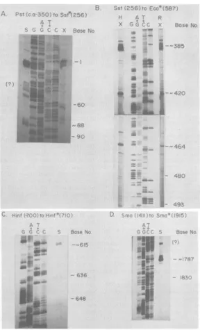
We precisely located the 5' end of the 2,520-base-long unspliced and most abundant mRNA
by the Si nuclease and exonuclease VII
diges-tion of hybrids formed by infected-cell poly-adenylated mRNA and the DNA fragment
ex-tendingbetweenthePstlsiteatnucleotide-350 and the Sstll siteatnucleotide +256, which was
5' end labeledatthis latter site. The major
nude-ase-resistant DNA fragment migrated
corre-sponding to a size ofca. 260 bases.
Fraction-ationof suchaDNAfragmenton asequencegel
localized the 5' end within the sequence TACC
(Fig. 4A). We arbitrarily set the A residue as
nucleotide number 1. Such stuttering of
nudle-ase-treatedhybridsonsequencing gels hasbeen reportedbyusandotherspreviously(15, 18, 27)
andcanreflect both someambiguityintheexact
start of an mRNA and some variability in the extentofdigestion. Smallamounts ofother
51-and exonuclease VII-resistant DNA fragments
HSV gC mRNA 639
are also seen; however, for most of them the amounts variedfrom experiment to experiment, and some could be seen when DNA was incubat-ed without infectincubat-ed-cell RNA. We concludincubat-ed that such DNAfragments were not due to
spe-cific RNA species hybridizing to the DNA. An
example is indicated by the question mark (?) in Fig. 4A. One faintSi-resistantband was consis-tently seen around base numbers 85 to 90 and was only seen in the presence of infected-cell polyadenylated mRNA. This would locate the splice acceptor of the least abundant spliced mRNAspecies in the sequence TCAGTG. The AG dinucleotide at bases 94 and 95 is indicated asacceptor 1 in Fig. 2. We positioned all splice acceptorsites at AGdinucleotides because this isthe canonical splice acceptor site (25, 29).
We carried out a similar experiment with strand-separated DNA 5' end labeled at the
EcoRIsite at base 587 andextending leftward to the SstII site at base 256. In addition to full-length DNA, a relatively major band about 200 bases long and two less abundant bands about 150 and 120 bases long were seen with Si nuclease and exonuclease VII digestion. The location of such bands on a sequence ladder extending to the left of the EcoRI site at base 587 is shown in Fig. 4B (track HX). In this experi-ment, exonuclease VII digestion is shown be-causeittended to produce less stutter. Howev-er,Si nuclease digestion (data not shown) gave
fragments about fivebases shorter than did the exonuclease VII digestion. In this experiment,
we included a track of DNA-protected HSV-infected rabbit skin cell polyadenylated mRNA
(Fig. 4B, track RX) to demonstrate that the
mRNA species are processed the same in these
cellsasin cellsofhumanorigin.The dataofthis experiment located three splice acceptor sites
forthe 2,200-base mRNA. One was within the
sequence CCGAGC (acceptor 2A, 381 to 386),
one in the sequence AAAAGCC (acceptor 2B, 428 to 434),and one in the sequenceCAAGAAC (acceptor 2C, 463 to 469). These sites are indi-cated in Fig. 2 as the AGdinucleotides at base numbers 384 to 385, 431 to 432, and 465 to466,
respectively.
We used strand-separated DNA 5' end labeled
attheHinflsite at base 710 andextending to the
Hinflsite at about base -700 to locate the splice acceptor site of the 1,900-base mRNA. This mRNA protected a DNA fragment about 90 baseslong from
Si
nucleasedigestion and locat-ed theacceptor within the sequence CCAGAT(613 to 618) (Fig. 4C). We indicated this as acceptor 3 on the AG dinucleotide atpositions
615and 616 inFig. 2.
Welocated the 5' endofthe730-base mRNA
about 120bases to the left of the SmaI site at
position1915. Wepreciselylocated it within the
on November 10, 2019 by guest
http://jvi.asm.org/
[image:6.496.40.235.64.220.2]FRINK ET AL.
C.
4,
9~~~~~~~r
'8~ ~ ~ n eKN.-,f> 53q
Q~~~~
*W
4pa ~ ~ 64
FIG. 4. Preciselocalizationof the 5' termini of thecontiguousregionsof theoverlappingmRNAspecies. (A)
HSV-1 DNAextendingfromtheSallsiteat0.621(about1,250basesupstreamof the mRNA 5'end)tothe EcoRI
site atbase 587wasdigestedwith bothSstIIandPstIIandwas sizefractionated. Thefragment encompassing bases -350to +256(Fig.tA)waseluted,5'endlabeled,strandseparated,andsequenced. Samplesofthe DNA
strandencodingthe mRNAwerealsousedtohybridizelatepolysomal polyadenylatedmRNA, andthehybridswere
digestedwithSinuclease(5)orexonuclease VII(X).Thepositionofthe 5'endof the2,520-baseunsplicedand
mostabundant mRNA and thespliceacceptor of the2,400-basesplicedspecieswerefixedbyfractionation of Si-and exonuclease VII-resistantfragmentsinparallelwith theDNA sequence determinedfrom the labeled SstII site. Thepositionof the 5' endwasfixedasposition+1(Fig. 2),and theacceptor site for the2,400-basemRNAis
nearposition 88. Other bands ofradioactivity were not reproducibly seen (seetext); oneis indicatedby (?). Thelocations ofsomeotherspecificbases in thissequenceladderareindicated andarecomplementarytothose
showninFig.2.(B)HSV-1DNAasdescribed in thelegendto(A)wasdigestedwith bothSstIIand EcoRI. The fragment extendingfrom +256 to +587(Fig. lA)wasusedasin(A)forsequenceandhybridization analysis.The
three splice acceptor sites for the2,200-basemRNA(positions,ca. 385,420, and464)werelocated with both
on November 10, 2019 by guest
http://jvi.asm.org/
[image:7.496.116.405.68.544.2]HSV gC mRNA 641
sequence CATCCT (1788 to 1791) by S1 nucle-ase digestion of hybrids of infected-cell poly-adenylated mRNA and strand-separated DNA 5' end labeled at theSmaI site atposition 1915 and extending leftward to the SmaI site at position 1411 (Fig. 4D). This positions the 5' end about 30bases below the sequence TATAAA (1755 to 1760).The similarity of these sequences to iden-tified HSV-1 promoters suggests that this mRNA is under its own promoter control. The fact that this mRNA cannot be detected in Northernblots hybridized with probesfromthe 5' region of the major mRNA supports this conclusion. We cannot completely exclude the
possibility that this mRNA is generated by a
splice since there is an AG dinucleotide at
nucleotides 1780 and 1781 which could be a splice acceptor site. Again, we cannot exclude the possibility that both mechanisms operate.
We located the 3' end of the total mRNA family about 260 bases to the right of the Sall site at base number 2255. This was done by S1 nuclease digestion ofhybrids between
infected-cell polyadenylated mRNA and
strand-separat-ed DNA 3' end labelstrand-separat-ed atthe SalI site at base 2255 andextendingrightward to theSal siteat
base 2574 (data not shown). Only one Si nucle-ase-resistant band was seen, and its size was
determined by denaturing acrylamide gel elec-trophoresis with plasmid pBR322 DNA frag-ments produced by Hinfl digestion as a size standard. As notedabove, this locates the 3' end of the mRNA family at or near the sequence ATAAAAA (2518 to2524).
Partial characterization of a splice donor se-quence. The lowabundance ofsplicedforms has precluded our full characterization of the splice
donor. The Northernblot data (Fig. 3) locate a noncontiguous (leader) portionof the2,200-base
and the 1,900-basemRNAs within 250 bases of
the 5' endof the 2,520-base mRNA. Any leader for the 2,400-base mRNA must, of course, be withinapproximately 100 bases of the 5' end of the majorunspliced mRNA. Our previous data
comparing the size of
Si
nuclease-protectedDNAfragmentson neutralandalkaline gels also indicated that the total length of the leader sequences for all spliced mRNA species could benolongerthan 100bases(15). The size of the spliced mRNAs on Northern blots (Fig. 3A),
including poly(A) tails, suggests that the leader isevenshorter than this.
Weuseduniformly32P-labeled DNAspanning
the region from -30 to +296 by using SmaI to
digest 32P-labeled SalI-EcoRI fragment T-A (0.621 to 0.633) and isolating the fragment in question by preparative acrylamide gel electro-phoresis. The strand complementary to the mRNA was isolated on a strand separation gel and hybridized with infected-cell
polyadenylat-ed mRNA. Size fractionation of Si nuclease-digested hybridized material yielded a major band 310 bases long corresponding tofull-length
DNA, a minor band due to protection by the 2,400-base mRNA, and a veryfaint band about 30 bases long. We suggest that this corresponds to DNAprotected by the leader sequence of the spliced mRNAs, although the band was not
intenseenough to photograph.
Weperformedaprimerextension experiment with avian myeloblastosis virus reverse
tran-scriptase to determine that the 1,900-base mRNA contains a noncontiguous fragment ca. 25baseslong at its 5' end (data not shown). We used the strand-separated DNA fragment con-tainedbetweentheHinfl site at position 710 and the SmaI site at 777. This 67-nucleotide DNA
fragmentwith the 3' end at aposition 96 bases below the nominal splice acceptor sitewas hy-bridized toinfected-cellpolyadenylated mRNA. The cDNA produced by avian myeloblastosis virus reverse transcriptase was fractionated ona
denaturing acrylamide gel. We consistently
foundaDNA bandmigratingwitha rate expect-ed for afragment 120 to 130 bases long. The very small amount of this cDNA precluded further characterization.
Wehaveidentified two potential splice donor sites in theregion betweenbases1and 100. One has the sequence TTGGT (21 to 25), and the otheris GAGGT (71 to 75). We tested the latter siteforbeingapotential splice donorby
isolat-ingstrand-separated DNA 3' end labeled at the TaqIsite at nucleotide 40 and at the SstII site at base 256. In several experiments, the only size ofDNAprotectedby thehybridizationwas200 bases long, corresponding to the full length of thefragment (data not shown). Since we would expectafragment35baseslong to be protected ifthesequence GAGGT were serving as a major
HeLa cell(H) and rabbit skin cell (R) late polysomal polyadenylated mRNA. In the experiments shown, hybrids
weredigested with exonuclease VII. (C) HSV-1 DNA extending from theSalI site at 0.621 to theBamHIsite at 0.643(Fig. 1A) was digested withHinfl.Thefragment between base -700 and base +710 was selected and used
asin(A)for sequence and hybridization analysis. The portion of the original autoradiograph shown indicates a bandatbase number 615, corresponding to the splice acceptor site for the 1,900-base mRNA species. Digestion ofhybridswithS1 nuclease is shown. (D) The HSV-1 DNA fragment between nucleotides 587 and 2085 (Fig. 1A)
was digested with SmaI, and the fragment between bases 1411 and 1915 was selected and used for DNA sequence andhybridization analysis. This fragment located the 5' end of the 730-base mRNA very near base
1787. The band marked with (?) may be analternatelength form or an artifactual band (see text).
VOL. 45,1983
on November 10, 2019 by guest
http://jvi.asm.org/
642
A. PREDICTEDAMINO ACID SEQUENCE OFHSV-1 (KOS)gC
1 5 10 15 20 25 30 35
M:A:P:G:R: L:A:V: :: :L:W:L:G:A:G.:UA:G:G: :E:T:A:S:T:G:P:T:1:
36 40 45 50 55 60 65 70
T:A:G:A:V:T: N:A:SE:A:P:T:S:G:S:P:G:S:A:A:S:P:E:V:T:P:T:S:T:P:N:P:NNg
71 75 80 85 90 95 100 105 : Q:N:K: T:P:T:E:P:A:S:P:P:T:T:P:K:P:T:S:T:P:K:S:P:P:T:S:T:P:D:P:K:
106 110 115 120 125 130 135 140 P:KN:N:T: P:A:K:S:G:R:P:T:K:P:P:G:P:V:W:C:D:R:R:D:P:L:A:R:Y:G:S:R:V:
141 145 150 155 160 165 170 175
Q:I:R:C:R:F:R.S:R:M:E:F:R:L:Q:I:W:R:Y:S:M:G:P:S:P:P: I:A:P:A:P:D:L: 176 180 185 190 195 200 205 210 E:E:V:L:T: 1: A:P:P:G:G:L:L:V:Y:D:S:A:P:F:L:T D:P:H:V:L:W:A:E:G:A:G: 211 215 220 225 230 235 240 245
P:G:A:D:P:P:L:Y:S:V:T:G:P:L:P:T:Q:R:L:I:I:G:E:V:T:P:A:T:Q:G:M:Y:Y:L:A: 246 250 255 260 265 270 275 280
W:G:R:M:D:S:P:H:E:Y:G:T:W:V:R:V:R:M:F:R:P:P:S:L:T:L:Q:P:H:A:V:M:E:G:Q:
281 285 290 295 300 305 310 315
P:F:K:A:T:C:T:A:A:A:Y:Y:P:R:N:P:V:E:F:D:W:F:E:D:D:R:Q:V:F:N:P:G:Q:1:0: 316 320 325 330 335 340 345 350 T:Q:T:H:E:H:P:D:G:F:T:T:V:S:T:V:T:S:E:A:V:G:G:Q:V:P:P:R:T:F:T:C:Q:M:T: 351 355 360 ....365 370 375 380 385
W:H:R:D:S:V:T:F:S:R:R iN:A:TG:L:A:L:V:L:P:R:P:T:I:T:M:E:F:G:V:R:H:V:V:
386 390 395 400 405 410 415 420
C:T:A:G:C:V:P:E:G:V:T:F:A:W:F:L:G:D:D:P:S:P:A:A:K:S:A:V:T:A:Q:E:S:C:D:
421 425 430 435 440 445 450 455
H:P:G:L:A:T:V:R:S:T:L:P:I:S:Y:D:Y:S:E:Y: I:C:W:L:T:G:Y:P:A:G: I:P:V:L:E: 456 460 465 470 475 480 485 490
H:H:G:S:H:Q:P:P:P:R:D:P:T:E:R:Q:V:I:E:A:I:E:U::G:I:G:I:G:V:L:A:A:G:V:
491 495 500 505 510 515 520 523
L:V:V:T:A:I:V:Y:V:VRT:S:Q:S:R:Q:R:I:G:G:N:A:R:P:P:V:T:F:L:I:S:I:TERM
B. PREDICTED AMINO ACIDSEQUENCE OF PROTEIN ENCODED BY 730 basenRNA
1 5 10 15 20 25 30 35
M:R:A:R:L:P:A:A:A:W:V:G:V:G:T: I:I:G:G:V:V: I: I:A:A:L:V:L:V:P:S:R:A:S:W: 36 40 45 50 55 60 65 70
A:L:S:P:C:D:S:G:W:H:E:F:N:L:G:C: I:S:W:D:P:T:P:M:E:H:E:Q:A:V:G:G:C:S:A:
71 75 80 85 90 95 100 105
P:A:T:L:I:P:R:A:A:A:K:Q:L:A:A:V:A:R:V:Q:S:A:R:S:S:G:Y:W:W:V:S:G:D:G:I:
106 110 115 120 125 130 135 140
R:A:R:L:R:L:V:D:G:V:G:G:I:D:Q:F:C:E:E:P:A:L:R:I:C:Y:Y:P:R:S:P:G:G:F:V: 141 145 150 153
Q:F:V:T:S:T:R:N:A:L:G:L:P:TERM
C. AMINO ACIDSEQUENCE PREDICTED FOR FRAME 1 POLYPEPTIDE
1 5 10 15 20 25 30 35
M:P:V:S:E:F:H:P:H:G:V:P:P:P:D:M:A:L:L:H:G:S:V:P:P:N:R:S:G:S:R:P:R:G:G:
36 40 45 50 55 60 65 70
P:D:E:H:H:R:P:T:R:G:T:P:G:V:R:Q:R:P:Q:P:D:G:P:P:R:A:L:G:G:G:G:R:P:G:R:
71 75 80 85 90 95 100 105
R:P:S:V:V:F:C:H:R:A:A:A:D:P:A:A:D:Y:R:R:G:D:A:R:D:P:G:N:V:L:L:G:L:G:P: 106 110 115 120 125 130 135 140
D:G:Q:P:A:R:V:R:D:V:G:A:R:P:H:V:P:P:P:V:S:D:P:P:A:P:R:G:D:G:G:S:A:V:Q:
141 145 150 G:D:V:H:G:R:R:L:L:P:A:TERM
D. AMINOACIDSEQUENCE PREDICTED FORFRAME3 POLYPEPTIDE
1 5 10 15 20 25 30 35
M:S:H:K:T:K:P:P:P:P:S:R:P:A:P:Q:Q:P:P:S:P:P:P:R:P:K:A:P:P:R:P:P:P:T:P:
36 40 45 50 55 60 65 70
N:P:R:T:T:P:P:P:P:S:R:A:A:P:L:N:P:P:G:P:C:G:A:T:A:A:T:H:W:P:G:T:A:R:G:
71 75 80 85 90 95 100 105
C:R:S:D:A:G:F:G:I:P:P:A:W:S:S:A:S:R:Y:G:V:T:P:W:V:R:P:P:Q:S:L:R:L:P:T:TERM
FIG. 5. Predicted amino acid sequences for
puta-tive polypeptides encoded by the overlappingmRNA sequences. (A) Predicted amino acid sequence of HSV-1 gC. The 523-amino acid polypeptide containsa
membrane insertion sequence (boxed) occurring
be-tweenamino acids6and 25. Themembrane-anchoring
sequence occurs between amino acids 478 and 501. Potential glycosylation sites of the form N:amino
acid:SorT(36) are alsoindicated.These are features
of a glycosylated membrane-associated protein. (B) Amino acid sequencepredicted forthe 3' coterminal
730-basemRNA.ThismRNAencodesa17,800-dalton polypeptide byin vitro translation(Fig. 6A, track V). (C) Amino acid sequence predicted for translation
readingframe 1. (D) Amino acid sequence predicted for translationreading frame3.
splice donor, we concluded that this sequence does not, infact, servewith any frequency.
Taken together, all of these data do not rule out otherpotentialleader sequences or
noncontigu-ous onesfunctioning in the region upstream of the splice acceptor sites of the spliced mRNA
species, but they do suggest that the DNA
between nucleotides 1 and 25 can serve to
generate asplice donor.
Correlation oftranslation reading frames with the in vitro translation products of size-fractionat-edmRNAs. The locationof translation initiation
and termination signals in the nucleotide se-quenceof Fig. 2defines fourtranslation reading
frames of appreciable size(Fig. 1C).The longest
frame (frame2) extendsfromthe ATG at bases 146 to 148 to the terminator TAG at bases 1715 to 1717. This 1,569-nucleotide reading frame
predictsapolypeptide of 523 amino acids whose sequence is shown in Fig. 5A. The anhydrous
molecular size of this polypeptide predicted
from its amino acid composition is 60,000 dal-tons, but its high proline content(12.5%)would cause it to migrate at a rate corresponding to a
somewhat larger protein in SDS-acrylamide gels. This polypeptide contains a hydrophobic membrane insertion or signal sequence (23, 33,
34) between amino acids 6(valine) and 25 (gly-cine) and a hydrophobic membrane anchoring
sequence (37) between amino acid 478
(trypto-phan) and 500 (valine) followed at position501
by anarginine residue. Itthus has theform ofa
membrane-associated protein. Further, there are eight potential glycosylation sites
(aspara-gine-amino acid-serine or threonine; 36), seven of which lie in the N-terminal half ofthe poly-peptide.
These properties suggest that this reading frame encodes HSV-1 gC, and the position of the reading frame predicted that only the
un-spliced 2,520-base mRNA orthe minor spliced 2,400-base mRNA could encodeit. We isolated
a mixture of the unspliced 2,520-base mRNA and the unresolvable2,400-baseand 2,200-base minor species by size fractionation of mRNA
selected by the hybridization of total infected-cell polyadenylated mRNA to EcoRI-BamHI
fragment I-I (0.633 to 0.643) DNA (see above). ThismRNA wastranslatedinvitroandyieldeda
major product migrating witha rate
correspond-ing to an apparent size of 69,000 daltons on
SDS-acrylamide gels (Fig.6A, track I). A portionof this material was incubated with a polyclonal antibodyto HSV-1 envelopeglycoproteins pre-viously shown to precipitate HSV-1 gA/gB,
gC, gD, and gE (anti-env-1 serum; 9) and the
unglycosylated precursors to these
glycopro-teins isolated from tunicamycin-treated cells. This antiserum specifically reacted with the
69,000-dalton polypeptide (Fig. 6A, track II). A smaller amountof apolypeptidemigratingwitha nominal size of 50,000 daltons also could be seen, and a very small amount of apolypeptide just smaller than 69,000 daltons could be seen
withdifficulty afterlong exposures.
Weconfirmed that themajortranslation
on November 10, 2019 by guest
http://jvi.asm.org/
HSV gC mRNA 643
2520 900 73C
base base base
RFNA mRNA mRNA
T*C.;+ PP Totat PrT?3Toroj __P
W ..:..
551D'0C)Od _>_40
4...
43*.
mm~~~~~~~~~~~Am "E
A .~
! ' ..V. . / .
FIG. 6. In vitro translation products of isolated mRNA species identified by immunoprecipitation. (A) Polyribosomal polyadenylated 32P-labeled RNAwashybridizedwith the HSV-1 DNAfragment extendingfrom
base 587tobase 2085 (Fig. 1A) coupledtocellulose.The mRNA selectedby hybridizationwasfractionatedby electrophoresis on methyl mercury agarose gels. The gel was sliced, slices were counted, and peaks of
radioactivity correspondingtothe 2,500-base species plus theunresolvable 2,400-and2,200-base species, the
1,900-base species, and the 730-base specieswereeluted and translated. The translationproductswereequally
divided; onehalfelectrophoresed withoutfurthertreatment, the other halfprecipitatedwithanti-env-1 serum,
andboth portions electrophoresed on aSDS-polyacrylamide gel in parallel. Track Ishows the major 69,000-dalton productaswellas anendogenous bandof50,000 daltonsfound after translation of thelargestmRNAs.
Track II shows that the69,000-dalton polypeptidereactswith anti-env-1 serum. Smalleramountsofa
50,000-daltonproductarealsoseen.Track IIIshows only theendogenous 50,000-daltonband foundupontranslation of the 1,900-base mRNA. Averyfaintpolypeptide band of 50,000 daltons precipitatedwithanti-env-1 serumis
detectable in trackIV.TrackVdemonstratesthepresenceofa17,800-dalton polypeptideupontranslation ofthe
730-base mRNA. Track VI shows that this polypeptide does not react efficiently with anti-env-1 serum.
Exposure wasfor 30 days on Kodak XARP film. (B) Comigration ofin vitro translated HSV-1 gC andthe
unglycosylated form ofgCisolated fromtunicamycin-treated cells is shown. Polyribosomal polyadenylated
32P-labeled RNAwasselectedby hybridizationasin(A)butwasnotsize fractionated. The mRNAthenwastranslated,
left untreated, or immunoprecipitated as in (A), and then the translation products were size fractionated by electrophoresis. Track VII shows the translation productsof total lateHSV-1poly(A)+RNA.Track VIII shows
theendogenous productspresentinacontrolwithout RNA. Track IX contains the products of hybrid-selected
RNA. The two endogenous translation products are seen along with the 69,000-dalton and 17,800-dalton polypeptides expected. Track X shows thetranslation products ofhybrid-selected RNAimmunoprecipitated withanti-env-1serum. Weobserved the69,000-daltonpolypeptide andafaintbandataround 50,000daltons, whichwould normally be hidden bytheendogenous band.TrackXIshows the products of total HSV-1 poly(A)+
RNAtranslationimmunoprecipitated by anti-env-1serum. Besides the bands for glycoproteins gA/gB, gC, and
gD,anumber of other bandsarepresentwhicharecharacteristicallyseenwithimmunoprecipitationoftotal in
vitrotranslation products. For comparison, track XII shows envelope proteins isolated fromtunicamycin-treated
HSV-1infected cellsandimmunoprecipitated withanti-env-1serum(9). Thedoublingof thegC bandis duetoan
artifact from drying the gel.
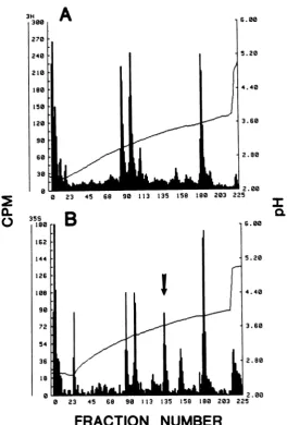
uct was, indeed, HSV-1 gC by comparing the
tryptic peptides of the immunoprecipitated in vitrotranslation product with trypticpeptides of authentic HSV-1 gC. We used the column chro-matography procedures describedearlier (9 and above) to do these experiments. Authentic gC
was eluted from a polyacrylamide gel before
trypsinization, and therecoveryofeach peptide
varied somewhatamongexperiments. However,
therelative elution position of peptides is
invari-ant.Atypical profile is shown in Fig. 7A. We isolated about 0.1 ,ug of mRNA hybrid selected asdescribed above and
immunoprecip-itated the in vitro translation product with anti-env-1 serum. When a portion was fractionated
on an SDS-acrylamide gel, only the
69,000-dalton polypeptidewasefficiently recovered by
suchtreatment (Fig. 6B, track X). We took the
A B
VOL. 45,1983
on November 10, 2019 by guest
http://jvi.asm.org/
[image:10.496.102.386.77.300.2]644 FRINK ET AL.J.Vo.
3H 300 270 240 210 180 150 120
s0
60 30 0
Ca.
A
23 45 60
B
6. 00
I
0.6. 00
5.20
4.40
3.60
2.800
LU - a -* E H 2.00
0 23 45 68 90 113 135 158 100 203 225
[image:11.496.124.389.75.466.2]FRACTION NUMBER
FIG. 7. Tryptic peptide analysis ofgCand the69,000-dalton polypeptide. (A)gCwas isolatedfrom a10%y
SDS-polyacrylamide gel, trypsinized, andchromatographedon aChromabeadsPcation-exchangecolumn. (B)
The69,000-daltonpolypeptidelabeled with [35S]methioninewasimmunoprecipitatedwith anti-env-1 serum(9) from an in vitro translation mix by using hybrid-selected mRNA. The precipitate was trypsinized and
chromatographedonChromabeads P. Thearrowindicatesapeptidewhich ispresentin theprofilefor the 69,000-dalton polypeptideand absentfrom theprofileforgC.
remainder of the
immunoprecipitated
translation product anddigested
and fractionated it; the profileis verysimilartothatseenwithauthentic gC exceptforone major additional methionine-containing peptide,whichprobably corresponds tothepost-translationallycleavedportionofgC containing the signalsequence (Fig. 7B).These data confirm the identification of the
majortranslationproductof mRNA encodedby
thisregionasHSV-1gC. Wecaninfer thatgCis
encoded by the unspliced 2,520-base mRNA
because of the relative abundance of this
mRNA. We cannot determine whether the
mi-nor2,400-base spliced species also encodes gC
or oneof the other smallerpolypeptides onthe basis of its position relative to the translation reading frames. However, the translation read-ingframe for theglycoproteinsshown inFig.5A
is not completely contained by the 2,200-base
splicedmRNA (Fig. 1C), sothis mRNAcannot
serve asmRNAforgC, althoughitcouldencode
a truncated immunologically reactive polypep-tide.
Although the immunoprecipitation of the
69,000-daltonpolypeptidewithanti-env-1 serum
and the tryptic peptide data demonstrate thatit was,infact,HSV-1gC, theapparentsize of this
polypeptide is somewhat at variance with the
J. VIROL.
on November 10, 2019 by guest
http://jvi.asm.org/
HSV gC mRNA 645
size of 74,000 to 85,000 daltons reported for the unglycosylated precursors to gC (33, 34), al-though the apparent size of 69,000 daltons is consistent with the amino acid composition shown in Fig. 5A. We demonstrated that the invitro translation product is about the same size as the unglycosylated precursor of gC found in tunicamycin-treated cells. As described, we translated total mRNA isolated by hybrid selec-tion and immunoprecipitated the gC polypeptide with anti-env-1 serum. The immunoprecipitated translation product was subjected to SDS-acryl-amide gel electrophoresis along with immuno-precipitated unglycosylated envelope proteins isolated from tunicamycin-treated cells as de-scribed previously (9; Fig. 6B, tracks X and XII). Although the envelope protein bands are distorted because of an artifact from drying the gel, the comigration of the in vitro translation product of gC mRNA and the unglycosylated gC is readily apparent.
As an added control, we used anti-env-1
se-rum to immunoprecipitate the in vitro transla-tion products of total polyadenylated mRNA from HSV-1-infected cells. Translationproducts
comigrating with the unglycosylated forms of
gA/gB,gC, and gD were readily apparent(Fig. 6B,track XI). Other faint bands alsowere seen.
Some, such as those marked agg, are probably
aggregations of one or more of the nonglycosy-lated polypeptides also seen in the envelope protein immunoprecipitate. Other bands could result from one or more of the following: (i) internaltranslation initiation, (ii) premature
ter-mination, (iii) translation of mRNAs encoding
minor membrane components, and(iv) proteins
immunologically cross-reacting with HSV-1
membraneproteins.
There are two otherpolypeptides potentially
encodedby one or anotherofthespliced mRNA
speciesthatare2,400 and 2,200 baseslong.One is defined by a 453-base openreading frame(Fig. 1C, frame 1) between nucleotides 574 and 1027. The sequence predicted for the 151-amino acid
polypeptide encoded is shown in Fig. 5C. We
would expect such a polypeptide tomigratewith
anapparentsizeof 18,000to22,000daltons, but
noproductthis size isobviouslyapparent in the in vitro translation of the mix of the unspliced 2,520-base and spliced 2,400- and 2,200-base mRNAs shown in Fig. 6A (track I) or in the
translation of totally hybrid selected mRNA
(Fig. 6B, track IX). A small amount ofa
poly-peptideabout this size can be seen invery long exposures of total translation products of hy-brid-selectedmRNA, but we cannot rule out its being due to the very low level of endogenous translationalactivity of the commercial reticulo-cyte lysate.
A third polypeptide of 105 amino acids,
en-coded by translation reading frame 3 between nucleotides 354 and 669, is also a potential
translation product of the mixture of the2,520-, 2,400-, and 2,200-base mRNAs. Its predicted amino acid sequence is shownin Fig. SD. Again, however, no obvious polypeptide migrating with asize of 12,000 to 16,000 daltons was apparent in the in vitro translation products of this mixture (Fig. 6A, track II) or in longer exposures.Here, however, the very intense band of radioactivity migrating at about 12,000 daltons could obscure such a product.
From these considerations, it is clear that we cannot determine whether any of the spliced mRNAs of 2,400 to 2,200 bases encodes these alternate reading frame polypeptides. We can statethat the 1,900-base spliced mRNA encodes atruncated form of HSV-1 gC. This comes from the fact that a 50,000-dalton polypeptide immunoprecipitable with the anti-env-1 serum was translated by the size-fractionated 1,900-base mRNA. This band isfaintlyvisible in Fig. 6A, track IV. The position of the 1,900-base mRNA in relation to the potential polypeptide initiation signals suggests that this polypeptide is
initiatedat one of theframe 2 initiators between nucleotides 630 and 980. Its precipitability by the anti-env-1 serum is due to its sharing a sizable C-terminal portion (60 to 70%) of the amino acid sequence ofgC and does notimply
thatthis polypeptide is itself a membrane-associ-atedprotein. It already has been noted that this 50,000-dalton polypeptide was detectable in the
immunoprecipitatedin vitro translationproducts of the mixture of the mRNAs of 2,520, 2,400, and 2,200 bases (Fig. 6A, track II), so one or more of the longer spliced mRNAs could also serve to encode it; however, this suggestion is notdirectly testable since the polypeptide could be translated by an in vitro interior initiation of the2,520-base mRNA.
The730-base mRNA located at the 3' end of
the longer mRNAs encompasses a 459-base
reading frame (frame 2) between the ATG
se-quence atnucleotides1922 to 1924 and the TGA
terminator at nucleotides 2381 to 2383. The
predicted amino acid sequence for this
153-aminoacid polypeptide is shown in Fig.SB. The calculated molecular size of this polypeptide is 16,204 daltons, and a polypeptide migratingwith an apparent size of 17,800 daltons is readily apparent in the in vitro translation products of thepurified730-basemRNA (Fig. 6A, track V). This polypeptide does not react significantly with the antibody to HSV-1 membrane glyco-proteins (Fig. 6A, track VI). This is not surpris-ing since it shares no amino acid sequence homology with HSV-1 gC and has no features of
a membrane-associated protein. It is readily detectable whenunfractionated mRNA
homolo-VOL.45,1983
on November 10, 2019 by guest
http://jvi.asm.org/
646 FRINK ET AL.
gousto thisregion of HSV-1 DNA is translated (Fig. 6B, track IX), but its function is unknown.
DISCUSSION
Thedatapresentedin thisreportsuggestthat splicing has a role in late HSV-1 gene
expres-sion. They also define rigorously the DNA se-quenceencodingaknown HSV-1 protein, gC.
The fact that the 1,900-base spliced mRNA encodesatruncatedform of gC
immunoprecipi-table withanti-env-1 serum does notrigorously
require that this polypeptide be expressed in infected cells, but it is strongly suggestive evi-dence. This is especially true since thistype of differential splicing ingeneratingmRNAs encod-ingproteins that share amino acid sequences is wellproveninother DNA viruses. We havenot
yet identified the translation products of the 2,200-base spliced mRNA. Its multiple splice acceptor sites could lead to variable mRNA function. At present, however, we cannot defi-nitelyclaim that either this mRNA speciesorthe
rarer2,400-base species hasadefined biological
function.
The730-base mRNA underlying the 3' end of thelonger species appears to be under its own
promotercontrol-a situation analogous to
sev-eral other regions of the genome (13, 15, 18).
Although this suggests that long intervening
sequences between the leader and the mRNA body are even more rare than HSV-1 mRNA
splicing itself, it should not be taken to imply thatHSV-1 musthave short introns. Costa and Draper, working in this laboratory, recently characterizedanmRNAspecies encoded bytwo exonsofnearlyequal length separated bya4-kb intron (unpublished results). Further, the fact that the spliced mRNA family members are of
lowabundance shouldnotbe takentoimplythat
a splice suppression mechanism is operating during HSV-1 infection. In otherwork (unpub-lished), CostacharacterizedanmRNAfamilyof which the most abundant member is spliced. From these considerations, it should be clear that there are no obvious mechanistic reasons
for the paucity of detectable splicing in HSV-1 mRNAbiogenesis.
The full characterization of an HSV-1 gene
whose biochemical properties, ifnotbiological function, areknownisclearlyof value. Like all well-characterized glycoproteins, HSV-1gChas
hydrophobicmembrane insertionandanchoring
sequencesaswellasreadilyapparent
glycosyla-tion sites. The fact that the HSV-1 gCgene is
expressed with no detectable splicing makes it
potentially convenientfor in vitro modification forstructure-function studies. Further, the fact that this gene has the capacity for alternate expression via splicing means that it will be
useful for manipulating DNA sequences to
de-terminetheeffectsofsequenceonmRNA proc-essing.
Although thebiochemical properties of
HSV-1 gC are known in that it is a
membrane-associated glycoprotein, the biological function of this protein isnot atall clear. Certainly, in its
mature form ofabout523 amino acids, it is found
only on the membranes of HSV-1 (10). It has
beenreportedthat the HSV-1 gC and the immu-nologically non-cross-reactive HSV-2 protein identified as HSV-2 gC map in different
loca-tions on the viral genome (19). Thus, these
membrane glycoproteins differ from most HSV
proteins, which seem to map in homologous locations on the HSV-1 and HSV-2 genomes. This makes HSV-1 gC aconvenient and poten-tially exploitable type-specific marker. Since it
isnotfoundexpressed withallstrains ofHSV-1, itis clearlynotrequiredper se for thereplication of HSV-1 in cell culture. Certainly ourfinding that HSV-1 gCcomaps with other polypeptides relatedby mRNAprocessingwillsuggest further investigations of the molecular reasons why
HSV-1 gC isnotexpressedin allHSV-1 strains
and why it lacks an obviously immunologically cross-reactive homolog expressed during HSV-2 infection.
ACKNOWLEDGMENTS
This work wassupported by Public Health Service grant CA-11861 from the National Cancer Institute and by grant MV-159fromthe American Cancer Society to E.K.W. and Public Health Service grant DE-02623 from the National Institute of Dental Researchto R.E. andG.C.
WethankP.G. Spearand L.Pereira forcommunicating the manuscript by Leeet al.(24a)to usbeforeitspublication.
LITERATURE CITED
1. Anderson, K. P., R. H. Costa,L.E.Holland, and E. K. Wagner. 1980. Characterization of herpes simplex virus type 1 RNA present in theabsence of de novoprotein synthesis. J. Virol. 34:9-27.
2. Anderson, K. P., R. J. Frink, G. B.Devi,B. H.Gaylord, R. H.Costa,and E. K.Wagner.1981.Detailed character-izationof the mRNAmapping in theHindlIl fragment K region of the herpes simplex virus type1genome. J. Virol. 37:1011-1027.
3. Anderson,K.P., L. E.Holland,B.H.Gaylord,and E. K. Wagner. 1980. Isolation and translation ofmRNA en-codedbyaspecific region oftheherpessimplexvirustype 1genome.J.Virol.33:749-759.
4.Anderson,K.P., J. R.Stringer,L.E.Holland,and E. K. Wagner.1979.Isolationandlocalization of herpessimplex virustype1 mRNA.J.Virol.30:805-820.
5. Bailey, J. M., and N.Davidson.1976.Methylmercuryasa reversibledenaturingagent foragarosegel electrophore-sis. Anal. Biochem. 70:75-85.
6. Bedbrook, J. R., R. Kolodner, and L.Bogorad. 1977. Zea mays chloroplast ribosomal RNA genes are part ofa
22,000basepairinvertedrepeat.Cell 11:739-749. 7. Benoist,C., K. O'Hare, R.Breathnach,and P.Chambon.
1980. Theovalbumingene-sequenceofputative control regions. Nucleic AcidsRes. 8:127-142.
8. Berk,A.J., and P. A. Sharp. 1977.Sizing andmapping of early adenovirus mRNAs by gel electrophoresis of S-I
on November 10, 2019 by guest
http://jvi.asm.org/
HSV gC mRNA 647
endonuclease-digestedhybrids. Cell 12:721-732. 9. Cohen, G. H., D. Long, and R. J. Eisenberg. 1980.
Synthesis andprocessing of glycoproteins gD andgC of herpes simplex virus type 1. J. Virol. 36:429-439. 10. Cohen, G. H., M. Katze, C.Hydrean-Stern, and R. J.
Eisenberg. 1978. Type-common CP-1 antigen ofherpes simplex virus is associated with a 59,000-molecular-weight envelopeglycoprotein. J. Virol. 27:172-181. 11. Costa, R. H., B. G. Devi, K. P. Anderson, B. H.Gaylord,
and E. K. Wagner.1981.Characterization ofamajorlate herpessimplex virus type 1 mRNA. J. Virol. 38:483-4%. 12. Denhardt, D. T. 1966. Amembrane-filter techniquefor the detection ofcomplementary DNA. Biochem. Biophys. Res.Commun.23:641-646.
13. Draper, K. G., R. J. Frink, and E. K. Wagner. 1982. Detailed characterization of an apparently unspliced , herpessimplexvirustype 1 genemappingin the interior of another.J.Virol.43:1123-1128.
14.Eisenberg, R. J., D. Long, L. Pereira, B. Hampar, M. Zweig, and G. H. Cohen. 1982. Effect of monoclonal antibodies on limited proteolysis of nativeglycoprotein gD of herpessimplex virustype 1.J. Virol. 41:479-488. 15. Frink, R.J.,K. P. Anderson, and E. K.Wagner. 1981.
Herpessimplexvirus type 1HindIIl fragmentLencodes spliced and complementary mRNA species. J. Virol. 39:559-572.
16. Frink, R., K. Draper, and E. Wagner. 1981. Uninfected cellpolymerase efficientlytranscribes earlybutnot late HSV-1 mRNA. Proc. Natl. Acad. Sci.U.S.A. 78:6139-6143.
17.Gibson, W. 1974.Polyoma virusproteins.Adescriptionof the structural proteinsof thevirionbasedon polyacryl-amide gelelectrophoresisandpeptideanalysis.Virology 62:319-336.
18. Hall, L. M., K. G.Draper,R.J. Frink,R.H.Costa,and E. K.Wagner. 1982.Herpessimplex virus mRNAspecies mappingin EcoRIfragmentI. J. Virol. 43:594-607. 19. Halliburton, I.1980. Review article:intertypic
recombi-nantsofherpessimplexvirus. J. Gen. Virol. 48:1-23. 20. Holland,L., K. Anderson,C. Shipman, Jr., andE.
Wag-ner.1980.ViralDNAsynthesisisrequired fortheefficient expressionofspecific herpessimplexvirus type1mRNA species. Virology101:10-24.
21. HoUand, L. E., K.P.Anderson,J.R.Stringer,and E.K. Wagner. 1979.Isolation andlocalizationofherpessimplex virus type 1 mRNA abundant before viral DNAsynthesis. J. Virol. 31:447-462.
22. Hummel, M., and E. Kieff.1982.Epstein-Barrvirus RNA. VIII. ViralRNAinpermissivelyinfected B95-8 cells. J. Virol. 43:262-272.
23. Kreil,G. 1981. Transfer ofproteins acrossmembranes. Annu. Rev.Biochem. 50:317-348.
24. Laemmli, U. K. 1970. Cleavage of structural proteins during the assembly ofthe head ofbacteriophage T4. Nature(London) 227:680-685.
24a.Lee, G.T.-Y.,K. L.Pogue-Geile, L.Pereira,and P. G. Spear. 1982.Expressionofherpessimplexvirus glycopro-tein Cfrom a DNAfragment insertedinto thethymidine kinase gene of this virus. Proc. Natl. Acad. Sci. U.S.A. 79:6612-6616.
25. Lewin, B. 1980.Alternativesfor splicing: recognizing the
ends of introns. Cell 22:324-326.
26. Maxam, A., and W.Gilbert. 1980.Sequencingend-labeled DNA with base-specific chemical cleavages. Methods Enzymol.65:499-559.
27. McKnight, S. L. 1980. The nucleotide sequence and transcript map of the herpes simplex virus thymidine kinase gene. Nucleic. Acids Res. 8:5949-5964. 28. McKnight, S. L., and R. Kingsbury. 1982. Transcriptional
control signals of a eukaryotic protein-codinggene. Sci-ence217:316-324.
29. Mount, S.1982. A catalogueof splice junction sequences. Nucleic Acids Res. 10:459-472.
30. Neidhardt, F. C., P. L. Bloch, and D. F. Smith. 1974. Culture media forenterobacteria. J. Bacteriol. 119:736-747.
31. Noyes, B. E., and G. R. Stark. 1975.Nucleic acid hybrid-ization usingDNAcovalentlycoupled tocellulose. Cell 5:301-310.
32. Palmiter, R. D. 1974.Mg+2precipitationof ribonucleotide protein complexes. Expedient techniques for theisolation ofundegraded polysomes and messengerribonucleic acid. Biochemistry13:3606-3614.
33. Peake, M. L., P.Nystrom, and L. I.Plzer.1982. Herpesvi-rusglycoprotein synthesis and insertion into plasma mem-branes.J.Virol.42:678-690.
34. Pizer, L. I., G. H.Cohen, and R. J. Eisenberg. 1980. Effect oftunicamycinonherpessimplex virus glycoproteinsand infectious virusproductioh. J. Virol. 34:142-153. 35. Proudfoot,N., and G.Brownlee. 1976. 3' noncoding region
sequences in eucaryotic mRNA. Nature (London) 263:211-214.
36. Rose,J.K., R. F.Doolittle,A.Anillonis,P.J.Curtis, and W. H. Wunner.1982. Homology between the glycopro-teins of vesicular stomatitis virus and rabies virus. J. Virol. 43:361-364.
37. Rose,J.K., W.J.Welch, B. M. Safton, F. S.Esch,and N. C. Line. 1980.Vesicular stomatitis virusglycoprotein is anchored in the viral membrane by a hydrophobic domain neartheCOOH terminus. Proc. Natl. Acad. Sci. U.S.A. 77:3884-3888.
38. Ruyechan, W. T., L. S. Morse, D. M. Knipe, and B. Roizman. 1979. Molecular genetics of herpes simplex virus. II. Mapping of the major viral glycoproteins and of thegeneticlocispecifyingthe social behavior of infected cells.J. Virol. 29:677-697.
39. Spear,P.G. 1976.Membraneproteinsspecifiedby herpes simplex viruses. I. Identification of four glycoprotein precursors andtheir products in type-1 infected cells. J. Virol. 17:991-1008.
40. Summers, W. C., I. Brunovkis, and R. Hyman. 1973. The process ofinfection withcoliphage T7.VII. Characteriza-tion and mapping of the major in vivo transcripCharacteriza-tion products of the early region. J. Mol. Biol. 74:291-300. 41. Watson, R.J.,C. M. Preston, andJ.B. Clements.1979.
Separationandcharacterization of herpes simplex virus type 1immediate-earlymRNA's. J.Virol. 31:42-52. 42. Watson, R.J.,M.Sullivan,and G. F. Vande Woude.1981.
Structures of two spliced herpes simplex virus type 1 immediate-earlymRNA'swhich map at the junctions of the uniqueand reiterated regions of the virus DNA S component. J. Virol. 37:431-444.
VOL.45,1983
on November 10, 2019 by guest
http://jvi.asm.org/