RESEARCH NOTE
Ability of real‑time PCR for differential
diagnosis of various forms of cutaneous
leishmaniasis: a comparative study
with histopathology
Maryam Fekri‑SoofiAbadi
1, Meisam Fekri
3, Alireza moradabadi
1, Reza Vahidi
1, Simin Shamsi‑Meymandi
2,
Donya Dabiri
1and Shahriar Dabiri
1*Abstract
Objective: Histopathological studies suggest that parasite load is different between acute and chronic forms of cutaneous leishmaniasis (CL). However, highly sensitive detection methods are still needed to distinguish different forms of leishmaniasis. In the present study, we developed a quantitative real‑time polymerase chain reaction (PCR) to detect and quantify Leishmania tropica parasites in paraffin‑embedded tissue samples.
Results: The ability of real‑time PCR for leishmania detection was higher than histopathological evaluation. The quantitative real‑time PCR (qPCR) quantified parasite loads were highly correlated with microscopic results (r = 0.598; P < 0.001). Among patients, the parasite load was inversely correlated with disease duration (acute CL lesions had very higher parasite load than chronic CL lesions), but there was no difference in the parasite load according to the patients’ age and sex as well as location of the lesions. In contrast to Ridley scoring system (P < 0.001), there were no statistically significant differences in the relative number of parasites among the lupoid and non‑lupoid forms of chronic lesions in real‑time PCR (P = 0.549), which indicates the superiority of histopathological evaluation for chronic forms differentiation.
Keywords: Leishmaniasis, Real‑time PCR, Ridley scoring system
© The Author(s) 2019. This article is distributed under the terms of the Creative Commons Attribution 4.0 International License (http://creat iveco mmons .org/licen ses/by/4.0/), which permits unrestricted use, distribution, and reproduction in any medium, provided you give appropriate credit to the original author(s) and the source, provide a link to the Creative Commons license, and indicate if changes were made. The Creative Commons Public Domain Dedication waiver (http://creativecommons.org/ publicdomain/zero/1.0/) applies to the data made available in this article, unless otherwise stated.
Introduction
Dry cutaneous leishmaniasis (CL) caused by Leishma-nia tropica is a significant parasitic disease in Iran [1]. The clinical phenotype, histopathology, and the number of organisms are diverse among acute, chronic lupoid, and chronic non-lupoid forms of this infectious disease [2]. In histopathology of acute CL, plasma cells, histio-cytes, epithelioid cells, and occasionally eosinophils and giant cells, and dense dermal infiltrate of lymphocytes are seen. Also, numerous intracytoplasmic Leishman bodies parasitized macrophages and sometimes neutrophils are
seen throughout the reticular dermis. A small number of infected macrophages and multifocal small tuberculoid granulomas composed of epithelioid cells, histiocytes, and occasional giant cells are seen more in chronic form. In addition, mild to moderate mononuclear infiltrates (lymphocytes and plasma cells) adjacent to the granu-loma along with fibrosis and telangiectasia are present. Low numbers of organisms, erythematous papules at the periphery of a scar of a healed acute lesion, and granulo-mas consisting of tubercles surrounded by lymphocytes, histiocytes, and giant cells are the most pathological find-ings in the lupoid forms of the disease; although, because of scant organisms in cutaneous lesions specifically in chronic leishmaniasis, microscopic studies has less sen-sitivity [2–9].
Open Access
*Correspondence: dabiri12@yahoo.com
1 Pathology and Stem Cell Research Center, Department of Pathology, Afzalipour Medical School, Kerman University of Medical Sciences, 22 Bahman Blvd, 7616913555 Kerman, Iran
Laboratory diagnosis of CL relies on either the micro-scopic detection of Leishman bodies in cutaneous tis-sue or the culture and isolation of parasites from lesions biopsy samples [10, 11]. Apart from high specificity, inad-equate sensitivity, difficulty, and time consuming nature are among disadvantages of these methods [12]. Nowa-days, PCR-based testing of skin lesion biopsies is known as a sensitive and specific test for diagnosis and quantifi-cation of leishmaniasis [13–16]. The analysis of the load of leishmania parasites within the skin lesions would be important not only for diagnostic purposes, but also for an eventual follow-up of a patient’s response to treat-ment [17]. Accordingly, in the present study we applied a standardized qPCR assay to detect Leishmania tropica load in paraffin blocks of various CL forms. The differen-tiation ability of this quantitative method was compared with semi-quantitative pathological scoring system.
Main text
Materials and methods Patients and sampling
Forty patients presenting with acute (n = 10), chronic
lupoid (n = 16), and chronic non-lupoid (n = 14) forms of
CL who were referred to the Dermatopathology Depart-ment of Afzalipour Hospital (2010–2013) were selected to participate in our study. Patient selection was per-formed after evaluation of inclusion/exclusion criteria. The patients were considered to be included in the study if they have confirmed and long-term CL (≥ 3 years), had
received at least 3 times glucantime treatment, and were able to give contact information for the follow-up. We excluded patients with other skin diseases or with small biopsy samples.
Histopathology
Skin biopsies were fixed in formalin, routinely processed, and after embedding in paraffin, sections were stained with hematoxylin and eosin, based on general approach. After grouping of cutaneous lesions according to Aza-deh [18] classification (Anergic macrophage reaction, Focalized histiocytic reaction, Diffuse necrotizing reac-tion, Diffuse lympho-histiocytic reacreac-tion, and Lupoid granulomatous reaction), the Ridley scoring system [19] was applied for determination of parasite load, as follows from 0 to + 4:
0: None amastigote
+ 1: One or more amastigotes
+ 2: 10 or more amastigotes
+ 3: 100 or more amastigotes
+ 4: 1000 or more amastigotes.
In should be noted that for uniform inflammatory cell-counting in all samples, it was decided to count the cells in inflammatory centers near the parasite and around granulomas. In addition, histopathologic alterations including necrosis, unorganized or organized granuloma, cellular (polymorphonuclears, eosinophils, giant cells, lymphocytes, plasma cells, and macrophages) infiltration and parasite index were estimated through an arbitrary semiquantitative procedure.
DNA extraction
For DNA extraction, 5 μm sections from paraffin-embedded blocks were cut using disposable blades and deparaffinized by hot xylene and then, were hydrated (descending grades of alcohol) and incubated in pro-teinase K (20 μg/μL, at 60 °C). After digestion com-pleted (3 days), the DNA was isolated using a QIAamp® DNA Mini Kit (QIAGEN, 51304), according to the manufacturer′s protocol.
Real‑time PCR assay
We applied a probe-based assay targeting rRNAITS region to detect and quantify parasites in the samples (Table 1). PCR amplification reaction was fulfilled using ABI StepOne system (Applied Biosystems, USA) and in a 25 μL of reaction mixture, containing 12.5 μL of master mix, 2 μL of forward and reverse primers for beta-actin and rRNAITS regions, 1.5 μL probe, 2 μL of H2O, and 5
μL of extracted DNA. Thermal cycling conditions started at 95 °C for 2 min followed by 95 °C for 20 s
(denatura-tion), and 60 °C for 30 s (annealing and extension), which
were programmed for 45 cycles. A cycle threshold (Ct) for each sample was determined based on the required cycles for the fluorescent signal to cross the background level.
Quantification of parasite DNA load
For absolute quantification, the standard strain (MHOM/ Sudan/58/OD) of L. tropica was cultured in RPMI1640 medium and serial dilutions (10 to 107) were prepared.
Table 1 Primers used in our study
F forward, R reverse, P probe, L.ITS leishmania ITS (internal transcribed spacer) gene
Primers Sequences (5′–3′)
L.ITS.F 5′‑CAA ATA CAC GCA TGC ACT CTC‑3′ L.ITS.R 5′‑TTT AAT AAT CCT GGT CAC AGCC‑3′
L.ITS.P FAM‑5′AGC GTC GAA ACT CCT CTC TGG TGC 3′‑TAMRA Actin.F 5′‑ACC ACC TTC AAC TCC ATC ATG‑3′
Actin.R 5′‑CTC CTT CTG CAT CCT GTC G‑3′
[image:2.595.305.540.610.706.2]Subsequently, a standard curve was set by plotting the Ct values against different standards with known concentra-tion of the parasite’s DNA.
Statistical analysis
The differences between experimental groups were ana-lyzed using the ANOVA (Tukey test). The Spearman’s rank correlation coefficient was used for evaluation of the relationship between real-time PCR and histopathologi-cal results. The SPSS software (version 22) was applied in this study.
Results
Histopathology and real-time PCR results in studied patients with different forms of CL are summarized in Table 2 and 3. Forty patients with confirmed CL were enrolled: 25 (62.5%) men and 15 (37.5%) women, with mean age of 32 years (range 6–73 years). To evaluate the correlation between the qPCR assay and histopathologi-cal evaluation, collected samples were analyzed in par-allel by both methods. The linearity of qPCR results was approved (diagram slope of − 3.23 and correlation coef-ficient (r2) ≥ 0.997) [20]. This assay allowed the
quan-tification of the parasite load in all samples, while the microscopic evaluation allowed this in 32 samples (80%, 8 negative samples corresponded to lupoid patients), which is indicating that the former method is more sensi-tive than the latter.
As presented in Tables 2 and 3, acute form has higher parasite load than chronic ones (P < 0.001) by real-time PCR. The mean parasite load in chronic lesions (n = 30) was 0.08 × 10³ parasites, compared with 13.064 × 10³ in
acute lesions (n = 10, P < 0.001). Interestingly, there was no significant difference in parasite load among lupoid and non-lupoid lesions by real-time PCR (P = 0.549). According to histopathological analysis, there were sta-tistically significant differences in the relative number of parasites among the acute and chronic (P< 0.01) and chronic-lupoid and non-lupoid forms (P < 0.001). These results indicate the superiority of histopathological
evaluation (Ridley scoring system) for differentiation of various forms of CL.
Discussion
In order to accurately and confidently quantify parasites in paraffin-embedded biopsy samples, we evaluated the parasitic load in acute and chronic forms using real-time PCR and histopathological scoring system. The focus of the present study was to compare the diagnostic ability of two common methods in a relatively large number of patients with CL. The power of the used qPCR assay [21] has allowed the quantification of a broad range of parasite load levels in tissue lesions. In terms of diagnostic sensi-tivity, our results confirmed that the sensitivity of real-time PCR is indeed higher than histopathological scoring system. Our findings are also consistent with the findings of previous studies that focused on different abundance of parasite in various forms of CL, pointing to inversely correlation of parasite load with the disease duration. Namely, in both methods of this study, acute form has higher parasite load than chronic ones. Interestingly, in contrast to Ridley scoring system (P < 0.001), there were no statistically significant differences in the relative num-ber of parasites among the lupoid and non-lupoid forms of chronic lesions in real-time PCR (P = 0.549), which indicates the superiority of histopathological evaluation in differentiation of chronic forms. It should be noted that the analysis performed here revealed no significant differences in parasite load with regard to the age, sex, and location of skin lesions. These findings were consist-ent with other studies [22–25]. For example, Mashayekhi et al. in a study on 11 male and 9 female patients with a mean age of 17.5 years showed that PCR was positive in 60% of the samples and no correlation was found between the results of PCR and age, sex, duration, and location of the lesions [26]. Venkataram et al. indicated that 65% of acute, subacute, and chronic lesions manifested leishma-nia parasites in tissues. But they could not find the rela-tionship between the duration of lesions and PCR results [25]. Weigle and others showed that PCR sensitivity was higher than the conventional assays for the diagnosis of
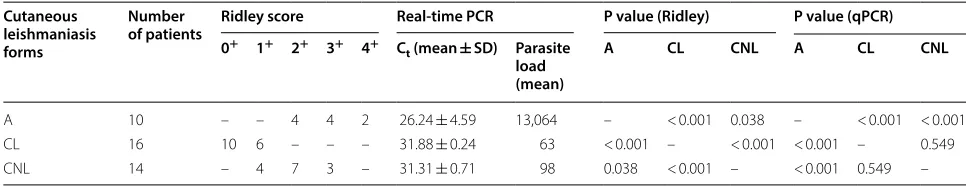
Table 2 Summary of patients informations
A acute form, CL chronic lupoid form, CNL chronic non‑lupoid form
Cutaneous leishmaniasis forms
Number
of patients Ridley score Real-time PCR P value (Ridley) P value (qPCR) 0+ 1+ 2+ 3+ 4+ C
t (mean ± SD) Parasite load (mean)
A CL CNL A CL CNL
A 10 – – 4 4 2 26.24 ± 4.59 13,064 – < 0.001 0.038 – < 0.001 < 0.001
CL 16 10 6 – – – 31.88 ± 0.24 63 < 0.001 – < 0.001 < 0.001 – 0.549
[image:3.595.55.538.623.716.2]acute lesions while for chronic samples, the sensitivity of PCR was much higher than the conventional assays [27]. In this regard, Verma et al. conducted real-time assay to estimate parasite burden in clinical samples of visceral leishmaniasis and patients with post kala-azar
dermal leishmaniasis. Concurrent diagnostic and prog-nostic ability of this assay, provide a simple molecular instrument to detect parasite and show the efficacy of anti-leishmanial drugs or vaccines [28]. In line with this, Dabiri et al. compared the effect of different treatments on DNA load of leishmania using real-time PCR method [29]. Jara et al. improved a quantitative real-time PCR (qPCR) method targeting mini-circle kinetoplast DNA (kDNA) to find and quantify Leishmania (Viannia) para-sites. According to the parasite species, the patients’ age, and number or area of lesions, there was no difference in parasite load [17]. Sirian et al. conducted a compari-son between conventional, molecular, and immunohis-tochemical methods for CL detection and reported that immunohistochemical and molecular techniques were more sensitive [30–32].
Our observations support the validity of using real-time PCR to simultaneously detect and quantify the leishmania load in human lesions, particularly in chronic lesions. This highly sensitive quantitative technique [10,
20, 21] can be employed also for monitoring the parasite load during treatment and follow-up as a way to assess the outcome of treatment.
Limitations
Accurately and confidently quantify parasites in biopsy samples help to evaluated the parasitic load in acute and chronic forms using real-time PCR in scoring CL. Using the paraffin-embedded biopsy samples make our samples collection time short. But it’s make the DNA extraction laboratories and effect on the quality of extracted DNA.
Abbreviations
CL: cutaneous leishmaniasis; PCR: polymerase chain reaction; Ct: cycle thresh‑ old; CNL: chronic non‑lupoid form; qPCR: quantitative real‑time PCR.
Acknowledgements
The authors would like to thank all friends and colleagues of Pathology and Stem Cell Research Center of Kerman University of Medical Sciences for their relentless efforts.
Authors’ contributions
SD and SM proposed the original concept and designed the experiment and supervised all aspects of the work. MAF, MEF, AM, RV, and DD equally partici‑ pated in the data acquisition and analysis. All authors contributed to writing the manuscript. SD and SM provided critical reviews in order to promote the manuscript. All authors read and approved the final manuscript.
Funding
No funding sources used in this study.
Availability of data and materials
Please contact corresponding author (S.D) for data requests.
Ethics approval and consent to participate
The study approved in Ethical Committee of Kerman University of Medical Sci‑ ences and the ethic approval code is IR.KMU.REC.1397.813. Informed consent was obtained from all the participants prior to enrolment.
Table 3 Detailed analysis of each patient
A acute form, CL chronic lupoid form, CNL chronic non‑lupoid form, M male, F
female
Patient Form Lesion site Ridley score Parasite load with qPCR
1 A Hand 2+ 63,096
2 A Hand 3+ 16,982
3 A Lower lip 3+ 4163
4 A Hand 4+ 5495
5 A Hand 3+ 3708
6 A Face 2+ 1942
7 A Face 2+ 6598
8 A Hand 2+ 2459
9 A Lower lip 3+ 1485
10 A Face 4+ 24,712
11 CNL Forearm 3+ 186
12 CNL Hand 1+ 89
13 CNL Ankle 1+ 161
14 CNL Nose 2+ 78
15 CNL Hand 1+ 72
16 CNL Hand 2+ 56
17 CNL Face 3+ 115
18 CNL Leg 1+ 76
19 CNL Ankle 2+ 85
20 CNL Forearm 2+ 74
21 CNL Hand 2+ 90
22 CNL Face 2+ 96
23 CNL Hand 2+ 84
24 CNL Forearm 3+ 110
25 CL Hand 1+ 48
26 CL Hand 0 59
27 CL Forearm 1+ 39
28 CL Face 0 95
29 CL Face 0 68
30 CL Hand 0 91
31 CL Hand 0 64
32 CL Hand 1+ 59
33 CL Face 0 72
34 CL Face 1+ 87
35 CL Hand 1+ 99
36 CL Forearm 1+ 68
37 CL Face 0 47
38 CL Face 0 40
39 CL Face 0 37
[image:4.595.57.287.106.616.2]Consent for publication Not applicable.
Competing interests
The authors declare that they have no competing interests.
Author details
1 Pathology and Stem Cell Research Center, Department of Pathology, Afzalipour Medical School, Kerman University of Medical Sciences, 22 Bah‑ man Blvd, 7616913555 Kerman, Iran. 2 Dermatology Department, Afzalipour Hospital, Kerman University of Medical Sciences, Kerman, Iran. 3 Department of Medicine, Montefiore New Rochelle Hospital, Albert Einstein College of Medicine, New York, USA.
Received: 8 August 2019 Accepted: 18 September 2019
References
1. Murray HW, Berman JD, Davies CR, Saravia NG. Advances in leishmaniasis. Lancet. 2005;366(9496):1561–77.
2. Meymandi S, Dabiri S, Dabiri D, Crawford RI, Kharazmi A. A quantitative study of epidermal Langerhans cells in cutaneous leishmaniasis caused by Leishmania tropica. Int J Dermatol. 2004;43(11):819–23.
3. Choi CM, Lerner EA. Leishmaniasis as an emerging infection. Journal of investigative dermatology symposium proceedings. New York: Elsevier; 2001.
4. Lallas A, Apalla Z, Argenziano G, Longo C, Moscarella E, Specchio F, et al. The dermatoscopic universe of basal cell carcinoma. Dermatol Pract Conceptual. 2014;4(3):11.
5. Salman SM, Rubeiz NG, Kibbi A‑G. Cutaneous leishmaniasis: clinical features and diagnosis. Clin Dermatol. 1999;17(3):291–6.
6. Zvulunov A, Cagnano E, Frankenburg S, Barenholz Y, Vardy D. Topical treatment of persistent cutaneous leishmaniasis with ethanolic lipid amphotericin B. Pediatr Infect Dis J. 2003;22(6):567–9.
7. Oliveira‑Neto MP, Mattos M, da Silva C, de Souza F, Fernandes O, Pirmez C. Leishmaniasis recidiva cutis in New World cutaneous leishmaniasis. Int J Dermatol. 1998;37(11):846–9.
8. Gurel MS, Ulukanligil M, Ozbilge H. Cutaneous leishmaniasis in Sanliurfa: epidemiologic and clinical features of the last four years (1997–2000). Int J Dermatol. 2002;41(1):32–7.
9. Ardehali S, Sodeiphy M, Haghighi P, Rezai H, Vollum D. Studies on chronic (lupoid) leishmaniasis. Ann Trop Med Parasitol. 1980;74(4):439–45. 10. Sundar S, Rai M. Laboratory diagnosis of visceral leishmaniasis. Clin Diagn
Lab Immunol. 2002;9(5):951–8.
11. Ramírez JR, Agudelo S, Muskus C, Alzate JF, Berberich C, Barker D, et al. Diagnosis of cutaneous leishmaniasis in Colombia: the sampling site within lesions influences the sensitivity of parasitologic diagnosis. J Clin Microbiol. 2000;38(10):3768–73.
12. Beheshti N, Ghafarifar F, Dalimiasl A, Eslamirad Z, Sharifi Z, Farivar SM. Detection of cutaneous leishmanioasis isolated from Iranian patients by using ITS1 gene and apol enzyme via PCR‑RFLP molecular method. Sci J Ilam Univ Med Sci. 2013;20(4):71–8.
13. Moradabadi AL, Farsinejad A, Fekri SM. Fast method for diagnosis of leish‑ mania by PCR and FLASH PCR. J Arak Univ Med Sci. 2017;19(11):79–86. 14. Al‑Jawabreh A, Schnur L, Nasereddin A, Schwenkenbecher J, Abdeen Z,
Barghuthy F, et al. The recent emergence of Leishmania tropica in Jericho (A’riha) and its environs, a classical focus of L. major. Trop Med Int Health. 2004;9(7):812–6.
15. Khosravi S, Hejazi H, Hashemzadeh‑Chaleshtori M, Eslami G, Yousofi Darani H. Molecular diagnosis of Old World leishmaniasis: real‑time PCR based on tryparedoxin peroxidase gene for the detection and identifica‑ tion of Leishmania spp. J Vector Borne Dis. 2012;49(1):15–8.
16. Nasreen SA, Hossain MA, Paul SK, Mahmud MC, Ahmed S, Ghosh S, et al. PCR‑based detection of Leishmania DNA in skin samples of post kala‑azar
dermal leishmaniasis patients from an endemic area of Bangladesh. Jap J Infect Dis. 2012;65(4):315–7.
17. Suárez M, Valencia BM, Jara M, Alba M, Boggild AK, Dujardin J‑C, et al. Quantification of Leishmania (Viannia) kinetoplast DNA in ulcers of cutaneous leishmaniasis reveals inter‑site and inter‑sampling variability in parasite load. PLoS Negl Trop Dis. 2015;9(7):e0003936.
18. Azadeh B, Samad A, Ardehali S. Histological spectrum of cutaneous leishmaniasis due to Leishmania tropica. Trans R Soc Trop Med Hyg. 1985;79(5):631–6.
19. Hassan AE, Kadaru A, Khalil E, Fadl A, Hassan ME. The pathology of cutaneous leishmaniasis in the Sudan: a comparison with that in other geographical areas. Ann Trop Med Parasitol. 1996;90(5):485–90. 20. Fekri SM, Dabiri S, Fotouhi AR, Fani ML, Amirpoor RS, Ziasistani M, et al.
Design and validation of real‑time PCR: quantitative diagnosis of com‑ mon leishmania species in Iran. J Clin Microbiol. 2016;19:496–501. 21. Jara M, Adaui V, Valencia BM, Martinez D, Alba M, Castrillon C, et al. A real‑
time PCR assay for detection and quantification of Leishmania (Viannia) in skin and mucosal lesions: an exploratory study of parasite load and clinical parameters. J Clin Microbiol. 2013;51:1826–33.
22. Noazin S, Khamesipour A, Moulton LH, Tanner M, Nasseri K, Modabber F, et al. Efficacy of killed whole‑parasite vaccines in the prevention of leishmaniasis—a meta‑analysis. Vaccine. 2009;27(35):4747–53. 23. Momeni AZ, Yotsumoto S, Mehregan DR, Mehregan AH, Mehregan DA,
Aminjavaheri M, et al. Chronic lupoid leishmaniasis: evaluation by poly‑ merase chain reaction. Arch Dermatol. 1996;132(2):198–202.
24. El‑On J, Weinrauch L, Livshin R, Even‑Paz Z, Jacobs G. Topical treatment of recurrent cutaneous leishmaniasis with ointment containing paromomy‑ cin and methylbenzethonium chloride. Br Med J (Clinical Research ed). 1985;291(6497):704.
25. Venkataram M, Moosa M, Devi L. Histopathological spectrum in cutane‑ ous leishmaniasis: a study in Oman. Indian J Dermatol Venereol Leprol. 2001;67(6):294.
26. Mashayekhi V, Mahmoudi M, Rastin M, Tayebi N, Taheri AR, Tavakoli M. Detection of Leishmania DNA in paraffin embedded specimens of chronic lupoid leishmaniasis using polymerase chain reaction. J Infect Public Health. 2012;9:557–63.
27. Weigle KA, Labrada LA, Lozano C, Santrich C, Barker DC. PCR‑based diagnosis of acute and chronic cutaneous leishmaniasis caused by Leish‑ mania (Viannia). J Clin Microbiol. 2002;40(2):601–6.
28. Verma S, Bhandari V, Avishek K, Ramesh V, Salotra P. Reliable diagno‑ sis of post‑kala‑azar dermal leishmaniasis (PKDL) using slit aspirate specimen to avoid invasive sampling procedures. Trop Med Int Health. 2013;18(3):268–75.
29. Dabiri S, Manafi Anari H, Shamsi Meymandi S, Fotouhi Ardakani R, Amirpour Rostami S, Meymandi MS, et al. DNA load analysis using real time PCR in comparison with immunohistochemical findings of dry type cutaneous leishmaniasis; before and after treatment by imiquimode, glu‑ cantime and combination of both drugs. Iran J Pathol. 2013;8(4):247–54. 30. Shirian S, Oryan A, Hatam G‑R, Panahi S, Daneshbod Y. Comparison of
conventional, molecular, and immunohistochemical methods in diagno‑ sis of typical and atypical cutaneous leishmaniasis. Arch Pathol Lab Med. 2014;138(2):235–40.
31. Shirian S, Oryan A, Hatam GR, Daneshbod Y. Three Leishmania/L. species– L. infantum, L. major, L. tropica—as causative agents of mucosal leishma‑ niasis in Iran. Pathog Glob Health. 2013;107(5):267–72.
32. Daneshbod Y, Oryan A, Davarmanesh M, Shirian S, Negahban S, Aledavood A, et al. Clinical, histopathologic, and cytologic diagnosis of mucosal leishmaniasis and literature review. Arch Pathol Lab Med. 2011;135(4):478–82.
Publisher’s Note