Dissertation
“
STUDY ON NORMAL AND INFECTION
RESPONSE AFTER SPLENECTOMY FOR
TRAUMA
”
M.S. BRANCH - I
GENERAL SURGERY
MADRAS MEDICAL COLLEGE
THE TAMILNADU
Dr. MGR MEDICAL UNIVERSITY
CHENNAI – TAMILNADU
CERTIFICATE
This is to certify that, the dissertation entitled “STUDY ON
NORMAL AND INFECTION RESPONSE AFTER
SPLENECTOMY FOR TRAUMA” is the bonafide work done by
Dr.KANNAH.E during his MS (General Surgery) course 2012-2015,
done under my supervision and is submitted in partial fulfillment for the
requirement of the M.S.(BRANCH-I)- General Surgery, April 2015
examination of The Tamilnadu Dr.MGR Medical University.
Prof. P. RAGUMANI, M.S., Prof. A. AFFEE ASMA, M.S.,
Professor & Director I/C Professor of Surgery
Institute of General Surgery Institute of General Surgery Madras Medical College Madras Medical College Chennai-3 Chennai-3
Dr. P. VIMALA, M.D., THE DEAN
DECLARATION
I, declare that this dissertation titled “STUDY ON NORMAL
AND INFECTION RESPONSE AFTER SPLENECTOMY FOR
TRAUMA” represents a genuine work of mine. The contributions of any
supervisors to the research are consistent with normal supervisory
practice, and are acknowledged.
I also affirm that this bonafide work or part of this work was not
submitted by me or any others for any award, degree or diploma to any
other University board, either in India or abroad. This is submitted to The
TamilNadu Dr. M.G.R Medical University, Chennai in partial fulfillment
of the rules and regulations for the award of Master of Surgery Degree
Branch I (General Surgery).
Date:
ACKNOWLEDGEMENT
As I walk down the memory lane, I realize with a deep sense of
humility that what I have done now would not have been possible, but for
certain luminaries, who have enlightened my path to wisdom.
“Surgery is learnt by apprenticeship and not from textbooks, not
even from one profusely illustrated” – Ian Aird.
While I put these words together it is my special privilege and great
pleasure to record my deep sense of gratitude to my revered Professor and
Guide Prof. T. Bavanisankar, M.S., and Prof. A. Affee Asma. M.S.,
but for whose constant guidance, help and encouragement this research
work would not have been made possible. The unflinching academic,
moral and psychological support will remain ever fresh in my memory
for years to come. Words cannot simply express my gratitude to them for
imparting to me the surgical skills that I have acquired.
It is my pleasure to record my profound gratitude and indebtness to
Prof.S.Deivanayagam M.S., and Prof. P. Ragumani M.S., for their
support, keen interest and the constant encouragement they have given
during the course of this thesis work.
I would like to express my sincere thanks to Dr. A. Anandi, M.S.,
Professors of Surgery, for all of them have given me invaluable advice,
guided me and have been most kind and patient to me.
My heartfelt thanks to the entire Institute of Biochemistry for
granting me permission and helping me to conduct this study.
All along the way, I have been supported and encouraged by all my
associate professors and assistant professors who helped me to reach
where I am.
I also thank my fellow postgraduates, friends and colleagues who
have extended their co-operation in my work.
I thank the Dean, MMC & RGGGH for permitting me to conduct
this study.
I would be failing in my duty if I do not show my deep sense of
gratitude to all the patients who had helped me to become a surgeon and
especially those who consented to be part of this study.
With deep reverence, I salute my parents and I thank the Almighty
for blessing me with a wonderful family to whom I have dedicated this
CONTENTS
S.No. TITLE PAGE No.
1 INTRODUCTION 1
2 AIMS AND OBJECTIVES 2
3 REVIEW OF LITERATURE 3
4 MATERIALS AND METHODS 75
5 DATA ANALYSIS AND RESULTS 76
6 DISCUSSION 99
7 CONCLUSION 100
8 BIBILIOGRAPHY 101
9 PROFORMA 112
MASTER CHART
KEY TO MASTER CHART
INTRODUCTION
Spleen is the most common organ involved in injuries to abdomen.
Though many patients with splenic trauma are managed
non-operatively, larger percentage are managed by splenectomy..
Early complications seen in the patients undergoing splenectomy
include hematoma, leak from pancreas, and post-operative infections such as
surgical site infection, lower respiratory tract infection and abdominal
abscesses.
It is very challenging for practitioners to identify infections in the
immediate post-operative period after splenectomy because there is an
unusual physiologic response to total count and platelet count.
The aim of this study is to assess the three risk factors i.e. total count,
platelet count / total count ratio and Injury Severity Score for post-operative
AIMS & OBJECTIVES
1. To study the total count and platelet count (PC) / total count ratio in
patients undergoing splenectomy for trauma and their response in the
infected individuals.
2. To study the association of the three risk factors i.e. total count, PC/ total
count ratio and Injury Severity Score in those individuals who have
underwent splenectomy for trauma and their role in post operative
REVIEW OF LITERATURE
HISTORICAL BACKGROUND
:The spleen in the ancient times was famously known to have had
different purposes by different people, for Gable it was an “organ of
mystery”; it was considered as an unwanted organ by Aristotle; Pliny
regarded it as an organ that produced laughter and mirth, which was again
reinforced by the Babylonian Talmud. Pliny also regarded it as an organ that
hindered with the speed of professional athletes.
The first known splenectomy was performed in 1549 on a 24 yrs old
female by Adrian Zacarelli for splenomegaly. The first successful partial
splenectomy was recorded in the year 1590 by Franciscus Rosetti for
trauma.
The total splenectomy for trauma was first done by Nicolaus Matthias
in a patient whose spleen protruded through the flank wound. It was
performed in Capetown, South Africa in 1678 and partial splenectomy was
replaced by total splenectomy in trauma cases.
The first splenectomy in the United States was done in the year 1816,
by a Royal Navy Surgeon, O’Brien. In England, Sir Thomas Spencer Wells
In an autopsy done in 1881, Billroth stated that a splenic injury might
heal spontaneously, but in 1927, Hamilton Bailey asserted the need for
surgical intervention.
During the first two decades of the 20th century, many theories began
to appear regarding the purpose and use of judicious tamponade of the organ
and its first successful suture repair was reported by Ziskoff in Russia in the
year 1895 for a case of lacerated spleen.
Campos Christo in 1962 reported the first successful partial
splenectomy for the modern times.
The role of spleen in fighting infections has always been under
consideration.
Experiments by Bullock and Morris with rat plague bacillus in the
year 1919 proved that by removing the spleen, we are robbing the body of its
resistance.
Most physicians and surgeons have considered that splenetomy
doesn’t hamper the host defense system, until recently when in 1965
Shumacker and Kling elicited that immunity is compromised in children
whose spleen has been removed in cases of hematological disorders.
Singer’s review of literature in 1973 further elicited the incidence of
ANATOMY:
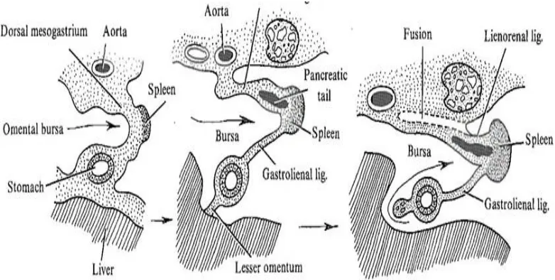
EMBRYOLOGY:
The spleen is formed by the mesenchymal differentiation along the
left side of the dorsal mesogastrium in juxtaposition to the anlage of the left
gonad in the 8-mm embryo as shown in Fig 1.
STRUCTURE AND POSITION:
In a healthy adult the spleen weighs from approximately 75 to 100
gms and is placed deep to the ninth, tenth and eleventh ribs in the posterior
portion of the left upper quadrant with its long axis corresponding to the
tenth rib.
[image:13.612.119.510.471.668.2]
Its superior, convex and the lateral surface lies adjacent to the
underside of the left leaf of the diaphragm. Its medial surface is concave due
to the impressions made on it by the stomach, pancreas, splenic flexure and
the kidneys as shown in Fig 2.
Several suspensory ligaments help in keeping the spleen in position.
These include gastrosplenic, splenophrenic, splenocolic, and splenorenal
ligaments (Fig 3). Usually these ligaments are avascular except the
gastrosplenic ligament which contains the short gastric vessels that course to
the splenic hilum from the greater curvature. But these ligaments may
become vascular in patients with portal hypertension and myeloproliferative
disorders.
BLOOD SUPPLY:
The splenic artery provides the main blood supply to the spleen. It is a
branch of the coeliac trunk. Further branching of the arteries occur proximal
to the hilus which includes a frequent branch to the inferior pole ehich
originates centrally.
VENOUS DRAINAGE:
The major venous drainage occurs through the splenic vein, which
usually receives the inferior mesenteric vein centrally, and then joins the
[image:15.612.92.564.205.601.2]
ACCESSORY SPLEEN
(Fig 4)
:
The classification includes two types of accessory spleens made of
blood, sinuses and Malphigian bodies:
(1)The most common is a separate distinct mass usually found in patients
with hematological disorders. It has been reported in about 14-30%
of the patients.
(2)The uncommon is the constricted part of the main organ, to which it is
attached by a fibrous tissue
The splenic artery also supplies the accessory spleen. They occur in
decreasing order of frequency in the hilus of the spleen, the gastrosplenic
ligament, splenorenal ligament, and the great omentum.
In females, accessory spleen may be seen in the pelvis, either in the
presacral region or adjacent to the left ovary, and in males, in the scrotum in
juxtaposition to the left testicle.
The spleen is surrounded by a capsule that is 1-2 mm thick and made
up of trabeculae that invaginate the pulp. The parenchyma of the spleen acts
as the immunological organ and is made of white pulp. The red pulp
phagocytizes the particulate matter from the marginal zone and the blood.
The white pulp is made of lymphatic nodules with germinal centers and
Fig 4- Usual location of accessory spleens: (1) gastrosplenic ligament, (2)
splenic hilum, (3) tail of the pancreas, (4) splenocolic ligament, (5) left
transverse mesocolon, (6) greater omentum along the greater curvature of
the stomach, (7) mesentery, (8) left mesocolon, (9) left ovary, (10) Douglas
lymphocytes and macrophages. It lies centrally and surrounds a central
artery (Fig 5).
The marginal zone contains lymphocytes and macrophages and red
blood cells (RBCs) that have exited from terminal arteries. The marginal
zone also contains the marginal sinus that filters material from the centrally
located white pulp.
It lies peripheral to the white pulp and contains end arteries arising
from the central artery and from peripheral penicilliary arteries. Locally
produced immunoglobulins enter the marginal zone, eventually coursing to
the blood stream. Peripheral to the marginal zone lies the red pulp. This pulp
consists of cords and sinuses that contain cellular elements of blood in
transit. Most of the blood flow passing through the spleen courses through
an "open" circulation in which the blood passes from arterioles to reticular
cell-lined networks of the splenic cords and to the sinuses.
PHYSIOLOGY
:
The spleen though is not a life saving organ, it performs some major
functions that can be grouped into two categories:
(1)Those that are immunological in nature
Fig 5 - Splenic compartments and potential vascular supply routes.
The cellular functions mainly concern hematopoiesis, storage,
"pitting" and "culling". Hematopoiesis, essentially ceases by the seventh
month in intrauterine life. It supplies erythroid, myeloid, lymphoid cells, and
important reservoir for blood cells, except platelets. At any given time, about
one third of the total platelet mass is in the spleen.
“Pitting” involves the removal of rigid structures from red cells such
as Heinz bodies, Howell-Jolly bodies, and hemosiderin granules. This
process concerns the elimination of nondeformable intracellular substances
from deformable cells. The rigid body is phagocytized while the deformable
cytoplasmic mass passes into the sinus and returns to the general circulation.
The postsplenectomy blood smear is usually characterized by circulating
erythrocytes with Howell-Jolly and Pappenheimer bodies (siderotic
granules). Nucleated cells also have their nuclei removed in the same
manner.
“Culling” refers to the spleen's ability to remove red cells that are
aged or abnormal. Normally, as the red cell ages after a life span of
approximately 120 days, it loses osmotic balance and membrane integrity,
and therefore deformability. When these cells lose their deformability they
are phagocytized by native macrophages. The spleen does not represent the
only site for red cell destruction, and there is no difference in red cell
survival following splenectomy. Naturally deformed cells and red cells that
are affected by disease states are also removed by phagocytosis. In those
cells are being remodelled in the spleen and exit as mature cells. In the
normal adult, the spleen is the most important site of selective erythrocyte
sequestration. During its 120-day life cycle, the red cell spends an estimated
minimum of 2 days within the spleen which, when normal, contains about
25 mL RBCs.
The neutrophil has a half-life of about 6 hours; thus 85% of
neutrophils either emigrate at random into tissues or are destroyed within 24
hours. The role of the spleen is amplified in some hypersplenic states, with
resulting neutropenia though the role of the spleen in the destruction of
neutrophils under normal conditions is not well quantified. This removal can
occur because of splenic enlargement and accelerated sequestration of
granulocytes or because of enhanced splenic removal of altered
granulocytes, as witnessed in immune neutropenias.
There is an accountable relation between the platelets and splenic
cells. Normally, about one third of the platelet mass is pooled in the spleen,
and this pool exchanges freely with the circulating platelets that have a life
span of about 10 days. With splenomegaly, a large proportion of platelets is
sequestered in the spleen (up to 80%) and this, coupled with accelerated
phagocytosis of platelets occurs in normal states, but in pathologic states,
such as immune thrombocytopenia, it is greatly increased.
Apart from the phagocytosis of antibody-coated cells, the
immunologic functions of the spleen also include generation of
lymphocytes, production of tuftsin, opsonins, properdin, and interferon and
antibody synthesis (especially immunoglobulin M [IgM]).
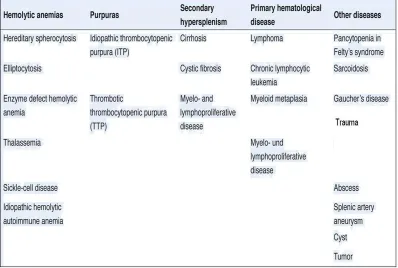
[image:23.612.126.524.303.571.2]The general indications for splenectomy are discussed in Table 1.
ABBREVIATED INJURY SCALE
[1]In 1969 an anatomical scoring system Abbreviated Injury Scale (AIS)
was introduced. The score has been revised and updated to provide an
accurate ranking of the severity of injury. The latest revision of the AIS score
was made in 1998. Association for the Advancement of Automotive
Medicine monitors the AIS.
Injuries are given a score from 1 to 6, with 1 being minor, 2 moderate,
3 serious, 4 severe, 5 critical and 6 un-survivable. The score indicates the
'threat to life' related to the injury and does not mean to represent a
comprehensive measure of severity. The AIS cannot be defined as an injury
scale, in that the difference between AIS1 and AIS2 is not the same as that
between AIS4 and AIS5. There are many identical features between the AIS
scale and the Organ Injury Scales of the AAST.
INJURY SEVERITY SCORE (ISS) & NEW INJURY
SEVERITY SCORE (NISS)
[2]The Injury Severity Score (ISS) is a versatile scoring system based on
anatomic classification that provides an overall score for those patients who
have sustained multiple injuries.
Each injury is allotted AIS and is assigned to any one of six body
External). Only those with the highest AIS score in each region of the body
is taken into account. Sum of the squares of the three most severely injured
organs will provide the injury severity score.
The ISS score ranges from 0 to 75. If an injury is associated with an
AIS of 6 (un-survivable injury), the ISS score is by default assigned to 75.
The ISS score is the only scoring system in use and correlates proportionally
with hospital stay, mortality, morbidity and other measures of severity.
Its disadvantage is that any fault in the AIS scoring increases the error
in ISS scores exponentially. Many different injury patterns can result in the
same ISS score and injuries to different body regions are not taken into
account. Since the full description of the patient injuries is incomplete prior
to complete investigation & operation, the ISS (along with other anatomical
scoring systems) is not very useful as a triage tool.
Since multiple injuries within the same body region are given a single
score, a modification of the ISS, the "New Injury Severity Score" (NISS),
has been given shape. Three most severely injures organs are assigned a
score and their sum of squares will yield the New Injury Severity Score.
SPLENIC INJURY
In the national trauma data bank, spleen is the most common organ
50.7% of blunt abdominal trauma patients. Similarly, 2.6% of injured
persons sustained splenic injuries in a multicentre series with data from 1993
to 1997 [3].Anyone who treats blunt abdominal injury should have the ability
to manage splenic injuries, since it has a mortality of 10.8%. This is mainly
caused by associated injuries and pre-hospital delay.
A deceleration mechanism that tears the splenic capsule or
parenchyma, at areas fixed to the retro-peritonium or direct compression of
spleen in the left upper quadrant is the patho-physiology of splenic injuries.
A ruptured spleen, at the time of presentation can have ongoing bleed
or the bleed would have stopped, which allows these injuries to be managed
non-operatively. The re-initiation of bleeding can be delayed in splenic
injuries, which is a concern among patients treated non-operatively. 10.6%
have late bleeding in a large series, though it varies with the grade of injury
[3]
.
Penetrating splenic trauma is less common. But still In national
trauma data bank, it represents 14.5% of all penetrating abdominal injuries.
Of the penetrating injuries involving spleen, this is higher compared to 9.2%
and 7.6% reported in a large series from Grady memorial and Ben Taub
Spleen is the most commonly bleeding intra-abdominal organ, as
noted in unstable patients with intra-abdominal fluid on focused assessment
with sonography in trauma (FAST). Splenic injuries are identified during
laparotomy in unstable patients taken to operating room emergently. In
stable patients, the mainstay of diagnosing splenic injuries is by abdominal
CT with IV contrast.
The splenic parenchyma is maximally enhanced with the contrast in
portal venous phase, while still the vasculature is visualised and the images
are taken. Disruptions in the splenic parenchyma represent splenic injuries,
with surrounding hematoma and free intra-abdominal fluid.
Active extravasation of contrast, seen as a high-density blush,
contained within a pseudoaneurysm or bleeding into the peritoneal space can
be identified occasionally. Injury to the hilar vessels causing complete
splenic devascularization or subcapsular hematoma can be other findings.
Splenic injuries are graded by the American Association for the
Surgery of Trauma organ injury scaling system, which relies on the
parenchymal or subcapsular characteristics and the vascular involvement.
Angiography has been the recent advance in splenic injury
management to evaluate further and also to treat at times. This is used in
bleeding in non-operative management, some centres use angiography in all
high grade injuries.
Specific sites of bleeding from the splenic parenchyma, segmental or
trabecular vessels can be identified by angiography. Though the splenic
parenchymal injury cannot be characterized by angiography, still it can be
complementary to CT.
The ability to obstruct sites of bleeding endovascularly using
angioembolization is the major benefit of angiography. Patients with active
extravasation by CT, but candidates for non-operative management are
benefited by angiography with embolization to eliminate splenic
pseudoaneurysm. With evidences, there is increase in the rate of safety of
managing splenic injuries non-operatively [5]. Angiographic evaluation and
angioembolic treatment can be considered only in patients who demonstrate
hemodynamic stability and not in shock.
Patients with blunt splenic trauma can be managed non-operatively
with approppriate patient selection and it should not be overraated.
Splenectomy does not have a risk profile when compared to the implications
of hemorrhage, and should not be overlooked as a definite treatment for
GRADING OF SPLENIC INJURIES
Grade Description
1 Hematoma Sub-capsular,<10% surface area
Laceration Capsular tear, <1 cm parenchymal depth
2 Hematoma
Sub-capsular, 10-50% surface area
Intra-parenchymal, <5 cm in diameter
Laceration Capsular tear, 1-3 cm parenchymal depth
which does not involve a trabecular vessel
3 Hematoma
Sub-capsular, >50% surface area or
expanding
Ruptured sub-capsular or parenchymal
hematoma
Intra-parenchymal hematoma >5 cm or
expanding
Laceration
>3 cm parenchymal depth or involving
trabecular vessels
4 Laceration
Laceration involving segemental or hilar
vessels producing major devascularization
(>25% of spleen)
5 Laceration Completely shattered spleen
Vascular
Hilar vascular injury which devascularizes
Fig 6- Grade I: sub-capsular fluid involving < 10% of the splenic surface.
Fig 7- Grade I: sub-capsular fluid involving < 10% of the splenic
[image:30.612.188.424.416.591.2]
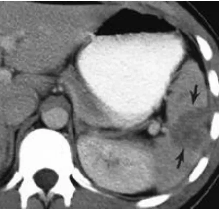
Fig 8- Grade II: sub-capsular hematoma, 10% to 50% surface area;
intra-parenchymal hematoma, < 5 cm in diameter
Fig 9- Grade II: capsular tear, 1 to 3 cm parenchymal depth that does
not involve a trabecular vessel
[image:31.612.217.417.359.592.2]
Fig 10- Grade III: capsular hematoma, laceration and
sub-capsular contrast extravasation.
Fig 11- Grade III: laceration of more than 3 cm in depth radiating
from the splenic hilum.
[image:32.612.230.434.386.581.2]Fig 12- Grade IV: laceration involving segmental or hilar vessels
producing major devascularization (>25% of spleen).
[image:33.612.211.465.489.660.2]
Fig 14- Splenic injury with sub-capsular hematoma. Despite only a 1-cm
In an attempt to push the non-operative envelope, no patients with
bleeding should be managed without splenectomy. It is possible to find
appropriate candidates for non-operative management of splenic injury
based on patients’ physiology. Non-operative management also requires
infrastructure to provide the ongoing surveillance.
To manage a patient without surgery, hemodynamic stability is a
prerequisite and is indicated by absence of tachycardia, normal blood
pressure, no findings suggestive of shock and absence of metabolic acidosis.
Until intravascular equilibrium occurs, the haemoglobin levels cannot reflect
the blood loss. Candidates with mild hemodynamic instability, but
responding to crystalloid infusion can be considered for non-operative
management.
There are other factors which have an impact while considering
non-operative management, though the patient's physiological status is the most
important. On comparing failure rates between groups older and younger
than 55 years, two retrospective studies have provided opposite conclusions
[6, 7]
.
A rate of 19% versus 10% failure of non-operative management in
patients older than 55 years was demonstrated by larger of these studies [7]
non-operatively. This proves age alone is not a contraindication for managing
[image:36.612.140.552.135.649.2]splenic injuries without surgery.
Another factor which has an impact on decision making is the grade
of splenic injury identified on imaging during admission, but there is no
prospective study to provide guidelines. A failure rate of 33.3% in grade IV
and 75% in grade V injuries, with 8% of failures happening more than 9
days after injury has been reported by one multi-institutional retrospective
study conducted by EAST. While all high grade injuries failed with
non-operative management in another multicenter study.
Some believe that splenectomy does not have a very high morbidity,
as there are unacceptably high failure rates with high grade splenic injuries,
and almost one in ten after hospital discharge. Others believe it to be
managed non-operatively, despite high failure rates. Non-operative
management is reserved for grade I, II injuries and isolated grade III injuries.
The decision is mainly a personal preference and is guided by surgical
intuition.
In case of instability at admission, unknown bleeding site, after failed
non-operative management, when spleen is suspected to be the cause
preoperatively, operative management is considered.
Splenic injury secondary to penetrating abdominal trauma is usually
or absence of ongoing bleeding. In damage control, splenic injury can be
packed but, more commonly splenectomy is performed.
OPEN SPLENECTOMY
A midline incision with packing of all four quadrants is the best
approach when instability is present. Left upper quadrant is exposed with a
fixed retractor.
The attachments are exposed by retracting the spleen
postero-medially, and dividing the peritoneum laterally marks the beginning of
splenectomy. The peritoneum is divided at the white line of Toldt and the
dissection begins at the splenocolic ligament extending superiorly, until
encountering the short gastric vessels.
After taking down the peritoneum, a blunt plane is created posterior to
the spleen in the medial direction that extends behind the tail of pancreas.
The spleen is delivered in to the visualized wound after mobilising the entire
spleen and distal pancreas.
The short gastric vessels are identified and ligated, avoiding injury to
greater curve of stomach. The tail of pancreas is protected while the hilar
vessels are clamped and ligated. Drains are placed only if the tail of the
To protect against infections by encapsulated bacteria including
Streptococcus pneumoniae, Neisseria meningitidis and Haemophilus
influenzae, postsplenectomy vaccines must be provided. Since patients who
usually benefit from splenic salvage techniques are managed
non-operatively, these techniques are less commonly used now.
[image:39.612.155.466.298.533.2]
Fig 16- Three-clamp method of Federoff for transecting the splenic hilum.
POST-SPLENECTOMY LYMPHOCYTOSIS
Pathophysiology
The kinetic aspects of lymphocyte trafficking is responsible for
post-splenectomy leukocytosis. The mounting of an efficient immune response
requires fast mobilization and distribution of lymphocytes. Lymphocytes
migrate from the blood cross a specialized endothelium of post-capillary
venules into lymph nodes [8, 9] and exit from lymphoid tissue via efferent
lymphatics and the thoracic duct. Most lymphocytes circulate through the
spleen, where direct access to the marginal zones does not require their
migration across a specialized endothelium.
The total lymphocyte pool in is estimated to be approximately 50*1010
cells out of which only 10-15*1010 lymphocytes circulate through lymphoid
tissues [41]. At a given time only a small proportion of 1*1010 lymphocytes
circulate in the blood of an average adult with a short transit time of 20-36
minutes for exchangeable cells as shown in fig 17 [10]. This results in a high
turnover of migratory blood lymphocytes of 50 times per day and a totally
daily exchange of approximately 50*1010 lymphoid cells between blood and
Fig 17- Lymphocyte traffic between blood and organs
As only 6% of the exchanging cell pool passes through the thoracic
duct, the majority of cells migrate to tissues other than lymph nodes after
leaving the bloodstream.
The spleen is the organ with the highest lymphocyte uptake during the
early stages of recirculation. Animal studies have demonstrated a splenic
uptake of approximately 40% of radiolabelled cells within the first 2 hours
of injection [11, 12]. Between 18 and 24 hours of injection labeled
through lymph nodes and a higher number of lymphocytes pass through the
spleen than through the thoracic duct.
Hence splenectomy results in a slower overall clearance of
lymphocytes from peripheral blood. Moreover neutrophils are destroyed in
spleen, though not quantified, splenectomy may result in neutrophilia. As a
result slower clearance of lymphocytes and decreased destruction of
neutrophils, there is physiological leukocytosis post-splenectomy.
Post-splenectomy thrombocytosis
Spleen plays major role in regulation of platelets. It is the site where
platelets are being destroyed. Hence there is physiologic thrombocytosis
after splenectomy predisposing to thrombocytosis in later stages.
COMPLICATIONS OF SPLENECTOMY
Intra-operative
Haemorrhage
Injury to tail of pancreas
Colon and gastric injuries
Early
Haematoma
Pancreatic leak
Pneumonia
Intra-abdominal abscess
Wound infections
Thrombotic complications
Late
Overwhelming post-splenectomy infection (OPSI)
Splenosis
POST_OPERATIVE INFECTIONS
Surgical infections were earlier considered as those that require
surgical therapy (e.g., complicated intraabdominal infections) and skin or
soft tissue infections. Since patients undergoing surgery suffer from
nosocomial infections post operatively, the definition now includes any
infection that affects post operative patients. Surgical site infections, central
line associated blood stream infections, urinary tract infections, hospital or
Inherent invasiveness of surgery is responsible for creating portals of
entry for the pathogens to break the natural epithelial barriers and invade the
host. Surgical illness is an immunosuppressive state similar to the
therapeutic suppression following organ transplantation.
Endotracheal intubation and mechanical ventilation following general
anaesthesia associated with a period of reduced consciousness poses a risk of
aspiration pneumonitis. Since the development of infections post surgery
impedes the recovery it is necessary to recognize and minimize the risks and
follows an aggressive approach to diagnose and treat post operative
infections.
Though infections are preventable it is prudent for every physician in
contact with his patients, do their utmost to prevent infections. Since no
method is universally effective an ensemble of prevention method is
required. It is necessary to take good and strict aseptic care of surgical
wounds regularly and dressing done as and when necessary under strict
aseptic precautions.
Moreover, one must avoid drains and catheters and if placed it must
be removed as early as possible. Role of antibiotics as therapeutic and
prophylactic modality must be well understood and used judiciously to
RISK FACTORS FOR INFECTION
Host Factors
Innate immunity is responsible for providing epithelial barrier
preventing the invasion of foreign antigens.
Potential pathogens are present universally and though colonization
may occur even in healthy host, it is necessary for a portal of entry to be
present for invasion of organisms to occur which may be seen in surgical
patients with surgical wound and intravenous catheters.
Inflammatory response stimulates autonomic nervous system which
promotes gluconeogenesis and glycogenolysis, promotes coagulation
pathway, and proinflammatory cytokines are stimulated to begin the tissue
repair process [13]. Innate and adaptive immunity are depressed by action of
cortisol [14].
Age > 64 years is the most important risk factor for adverse outcomes
post operatively especially post operative infections which are mainly due to
immune senescence [15]. Hyperglycemia induces immune cell dysfunction
Factors causing increased risk of post operative infections
[4]• Extremes of age (neonates, very old adults) • Malnutrition
• Obesity
• Diabetes mellitus • Prior site irradiation • Hypothermia • Hypoxemia
• Coexisting infection remote to surgical site • Corticosteroid therapy
• Recent operation, especially of chest or abdomen • Chronic inflammation
• Hypocholesterolemia
Genetics and Genomics of Trauma and Sepsis
Controversies exist whether there is any sex predilection for post
operative infections. Various studies that have been conducted and no study
could conclude about sex predilection for post operative infections [15, 16].
Genomic variability may be correlated with disease susceptibility in
infections. Nucleotide structures of genes containing single nucleotide
polymorphisms (SNPs) and single point mutations related to inflammation
(e.g., tumor necrosis factor-α [TNF-α], interleukin [IL]-1, IL-6, and IL-8),
innate immune response (e.g., Toll-like receptor 4), and the coagulation
system (e.g., factor V, plasminogen activator inhibitor-1) have been
associated with a predisposition to sepsis [17].
`Due to heterogeneity in the infection severity and resultant mortality,
as well as the immune response and infection predisposition, makes it
difficult to make conclusions that a single nucleotide polymorphism will
characterize the risk of infection in an individual.
INTERACTION BETWEEN THE THERAPY AND THE
HOST
Impairment of host defences, resuscitation and definitive care along
with the injury itself increases the risk of infection.
Hypothermia
Hypothermia occurs due to exposure, infusion of huge quantities of
unwarmed intravenous fluids and blood products or evaporative losses that
occur due to intra-cavitatory surgery, especially when abdomen and chest are
opened.
aggravated by hypovolemia, inflammatory response, coagulation pathway
activation and decreased transfused red blood cell deformability [18]. Thus
hypothermia is immunosuppressive, affects cardiovascular performance and
increases mortality after trauma and surgery [19, 20].
Tissue hypoxia
Injury to the face, airways, lungs, or chest wall, inability to secure the
airway, massive blood loss, cardiovascular instability, disruption of the
microcirculation and acute respiratory distress syndrome (ARDS) are few
factors that are responsible for tissue hypoxia after trauma. This hypoxia in
turn predispose to surgical site infections [21]. Oxygen administration post
surgery may reduce the risk of surgical site infection after elective surgery
[20]
.
Manner of Resuscitation
Intravenous fluid resuscitation is very important to restore
hemodynamics and microcirculatory perfusion, but the amount and nature of
fluid to be administered is still under discussion. Earlier crystalloids were
preferred over colloids due to cost effectiveness and almost equivalent
seventeen trials, eight trials were exclusively of crystalloid versus colloid for
the treatment of patients in sepsis. Results showed decreased mortality with
colloid resuscitation [23]. To sum up resuscitation of immune system is the
most important determinant, failure of which may lead to increased risk of
nosocomial infections and death [24].
Blood Transfusion
Though blood transfusion is life saving after trauma or hemorrhage,
there is increased risk of infection post transfusion. Altered leukocyte
antigen presentation and a shift to the T helper 2 cell phenotype causes
immuno-suppression following blood transfusion. An exponential
relationship between transfusion and infection risk among trauma patients,
detectable even after single unit transfusion, and becoming a near certainty
after more than 15 units of transfused blood has been suggested by Claridge
and his associates [25, 26].
Another study has suggested risk of infection is increased three times
in surgical patients following transfusion and five times increased risk in
trauma patients following transfusion. Ventilator associated pneumonia,
catheter line associated blood stream infections and critically ill patients are
Prolonged storage of banked blood results in the loss of high energy
membrane phosphates which in turn leads to impaired red blood cell
deformability, disruption of microcirculation, and impaired oxygen delivery
[28]
. As a result blood transfusion does not increase oxygen consumption [29]
but instead increase organ dysfunction [30]. So it is better to be conservative
while deciding on blood transfusion to stable patients in intensive care unit
[31]
.
Control of blood sugar
Hyperglycemia reflects the catabolic state and surgical stress
associated insulin resistance along with impairing the immune function of
the host. Inadequate glycemic control during the pre-operative and
post-operative period increases the risk of infection and worsens the outcome
from sepsis in both diabetic and non diabetics.
Hyperglycemia that is more than 200 mg/dl is associated with four
times increased risk of surgical site infection in both cardiac and non cardiac
surgery. Insulin infusion kept to maintain the blood glucose level less than
110 mg/dl was associated with 40% decrease in mortality among critically ill
post-operative patients and also lesser incidence of nosocomial infections
Effects of Hyperglycemia upon Immune Cell Function
• Decreased respiratory burst of alveolar macrophages
• Decreased insulin-stimulated chemokinesis
• Glucose-induced protein kinase C activation
• Increased adherence
• Increased adhesion molecule generation
• Spontaneous activation of neutrophils
Effects of Stress Response on Carbohydrate Metabolism
• Enhanced peripheral glucose uptake
• Hyperlactatemia
• Increased gluconeogenesis
• Depressed glycogenolysis
• Peripheral insulin resistance
Glycemic control in post-operative infection has become controversial
because of nonconfirmation of a salutary effect in intensive care patients.
Because of the risk of hypoglycemia following insulin therapy, the
maintenance blood glucose level has been increased from 110 mg/dl to
140-180 mg/dl [31]. Greisdale and associates conducted a meta-analysis and found
that intensive insulin therapy to maintain blood glucose level has been found
regardless of diabetes status. Certain contradicting opinions are still
prevailing [21].
Nutritional support in general is very crucial, keeping in mind that
restoration of anabolism requires calories and nitrogen in excess of basal
requirements of 25 to 30 kcal and 1 g nitrogen/kg/day. It is very difficult to
provide adequate calories and protein without producing hyperglycemia.
Parentral feeding has no advantage over other modes of feeding
because of inherent central line associated complications [34]. Enteral feeding
within 2 days, perhaps as quickly as possible if gut is functional, is
beneficial except in those patients where we suspect intestinal ischemia and
in those patients where we would like to prevent aspiration pneumonia. In a
meta-analysis of 15 randomized control trials it was found that there was
55% reduction in the risk of infection in those patients taking enteral feeding
following trauma, surgery or burns [35].
INFECTION CONTROL
It is very important to abide by the general principle of surgical care,
critical care and infection control at all times. One must rapidly resuscitate
the patients, either over resuscitation or under resuscitation increases the risk
pathology and treat it as soon as possible. Central venous catheters must be
removed and replaced if inserted under sub optimal conditions (e.g., lack of
cap, mask, sterile gown, and sterile gloves for the operator and a full bed
drape for the patient). This should be followed by re-insertion in a new site
as soon as the patient’s condition permits. Avoid placing drains and if placed
it must be removed as quickly as possible [36].
Control of infections is both individual and collective responsibility.
The most effective way of controlling infection is hand hygiene, compliance
of which is very challenging. Universal methods of precaution must be
carried out. Alcohol based hand cleansers are the most effective in clearing
all organisms except the clostridium spores for which washing with soap and
water is found to be very effective [37].
The source of most bacterial pathogens is found to be endogenous
flora. Skin surfaces, artificial airways, gut lumen, wounds, catheters, and
inanimate surfaces (e.g., bed rails, computer terminals) may become
colonized [38]. The most common mode of entry of pathogens is fecal-oral
route but health care workers transmit the organisms by their hands.
Isolation of contacts is the most important part of infection control
and must be used selectively to prevent pathogen spread such as Methicillin
multidrug resistant gram negative bacilli. There should be balance attained
because reduced manpower in ICU will result in increased incidence of
nosocomial infections [39].
Catheter care
Proper catheter care includes avoidance when not needed, proper skin
preparation, protecting the barriers during insertion, appropriate catheter
selection (antiseptic or antimicrobial coated), neat and sterile dressing of
indwelling catheters and removal of catheters as soon as when no longer
needed, or as is practicable, but no longer than 24 hours, after insertion of
catheters under less ideal circumstances.
Advantages and disadvantages of catheter insertion must be weighed
including the risk of infection. Though all the indwelling catheters carry the
risk of infection, non-tunneled central venous catheter and pulmonary artery
catheters pose the highest risk for local site infections and central line
associated blood stream infections. Other catheters associated with increased
risk of infection includes endotracheal tubes, intercostal thoracostomy
catheters, ventriculostomy catheters for intracranial pressure monitoring and
urinary bladder catheters. Risk of pneumonia increases by 1% to 3% for
Most common skin antiseptic that is used is chlorhexidine gluconate,
a phenolic biguanide derivative, in concentrations of 0.5% to 4% alone or in
lower concentrations in combination with an alcohol. This antiseptic has
cidal activity i.e., bactericidal, viricidal and fungicidal which is slow but
persistent. Chlorhexidine has been most commonly used for vascular
catheter insertion and it has been found to be superior to povidone iodine
solution. It is also being recommended for surgical site preparation, topical
bathing of critically ill patients and as an antiseptic coating for indwelling
catheters. For microbicidal effect of povidone iodine solution, one must
apply the solution and allow it to dry. Unless a mucous membrane has to be
prepared its use has been discouraged [41, 42, 43, 44, 45].
It is mandatory to have full barrier precautions during bedside
catheterization procedures except for arterial and urinary bladder
catheterization for which sterile gloves and field is more than enough if
maintained meticulously.
If a central venous catheter is inserted under suboptimal conditions,
then ensure that the catheter is changed to a different site as soon as patients’
hemodynamic condition improves, but not more than 24 hours of insertion.
following tube thoracostomy or ventriculostomy, but is not indicated for
vascular or bladder catheterization.
It is necessary to maintain dressings carefully which becomes
challenging in cases of agitated patients and irregular body surface.
Mentioning the date and time of dressing change over the dressing itself is
simple and effective.
One should not shift dressing cart from patient to patient, instead
sufficient dressing materials should be kept in the patient’s room. Inanimate
fomites such as scissors can transmit pathogens from one patient to another.
Hence it is prudent to implement care bundles and catheter care teams to
reduce the risk of catheter line associated bloodstream infections and urinary
tract infections [46, 47].
Catheter choice also plays an important role in reducing the risk of
infection related to endotracheal tubes, central venous catheters and urinary
catheters. Those areas that cannot be reached by routine endotracheal
suctioning such as the sub-glottic region may be cleared by continuous
aspiration of sub-glottic secretions through an endotracheal tube provided
with an extra lumen which opens to the airway just above the balloon.
Continuos aspiration of subglottic secretions decrease the incidence of
with silver are highly effective in reducing the risk of ventilator associated
pneumonia and mortality. Catheter related infections in high prevalence
units can be reduced by antibiotic coated or antiseptic coated tubes. Silver
coated urinary catheters are associated with decreased incidence of catheter
related bacterial cystitis [48, 49].
SPECIFIC INFECTIONS
Surgical site infections
Surgical procedures are classified into
Clean procedures
Clean contaminated procedures
Contaminated procedures
Dirty procedures
Clean surgical procedures affect only skin structures and soft tissue.
Under controlled setting when a hollow viscus is opened it is termed clean
contaminated procedure (e.g., elective aero-digestive or genitourinary tract
surgery). Contaminated procedures include those procedures where a large
inoculum of microorganism is introduced for a short duration into an
otherwise sterile body cavity for an infection to get established during
for mechanical bowel obstruction). In cases of established contamination
and infection if a surgery is performed it is termed dirty procedure (e.g.,
colon resection for perforated diverticulitis).
Factors determining microbiology of surgical site infections include
the nature of the procedure, whether a body cavity or a hollow viscus is
entered during surgery and location of the incision. Most surgical site
infections are the result of microorganisms that enter through the surgical
incision wound. Hence the most common organism responsible for surgical
site infection includes all the gram positive organisms - Staphylococcus
epidermidis, Staphylococcus aureus, and Enterococcus species.
For those surgeries that are done through infrainguinal incision and
intracavitatory surgery, gram negative organisms such as Escherichia coli
and Klebsiella spp are the most common pathogens. Anaerobic organisms
are the potential pathogens in pharynx, female genitourinary and lower
gastrointestinal surgeries. Hence antibiotic prophylaxis must be directed
appropriately against these antigens. Statistics indicate that the incidence of
surgical site infections vary from less than 5% for clean surgeries to about
20% for dirty procedures. Due to increase in number of ambulatory surgeries
the incidence of surgical site infections is under-estimated since the infection
Risk factors for development of surgical site infections includes
(1)Patient Factors
Ascites (for abdominal surgery)
Chronic inflammation
Corticosteroid therapy (controversial)
Obesity
Diabetes
Extremes of age
Hypocholesterolemia
Hypoxemia
Peripheral vascular disease (for lower extremity surgery)
Postoperative anemia
Prior site irradiation
Recent operations
Remote infection
Skin or nasal carriage of staphylococci
Skin disease in the area of infection (e.g., psoriasis)
Undernutrition
Inadequate disinfection/sterilization
Inadequate skin antisepsis
Inadequate ventilation
(3)Treatment Factors
Drains
Emergency procedures
Hypothermia
Inadequate antibiotic prophylaxis
Oxygenation (controversial)
Prolonged preoperative hospitalization
Prolonged operative time [50]
The factors included under National Nosocomial Infections
Surveillance System (NNIS) and its successor program, the National
Healthcare Safety Network (NHSN) is
1. Wound classification
2. ASA class 3 or higher
3. Prolonged operative time, where time is longer than the 75th percentile
According to NNIS-NHSN risk of surgical site infection increases
with increase in the number of risk factors irrespective of the type of surgery
performed. Laparoscopic surgeries are associated with decreased incidence
of surgical site infection. Factors responsible for decreased incidence of
surgical site infection includes decreased wound size, limited use of cautery
in the abdominal wall and a diminished stress response to tissue injury.
A study was conducted in 5031 patients who underwent non-cardiac
surgeries and incidence of surgical site infection was found to be 3.2%.
Ascitis, diabetes mellitus, post-operative anaemia and recent weight loss
were considered independent risk factors responsible for surgical site
infections. However chronic obstructive pulmonary disorders, tobacco use
and corticosteroid use did not contribute to the above incidence.
In another study conducted prospectively it was found that 12.5% of
the 9016 patients included in the study became infected within 28 days of
surgery. Analysis revealed that certain factors like low serum albumin,
tracheostomy, old age and amputation were responsible for early infections
whereas a shunt for dialysis, vessel repair and early infection were
responsible for hospital readmission. Hence risk factors for 28 day mortality
are increased age, hypoalbuminemia, increased serum creatinine levels and
Hypothermia is another important risk factor for surgical site infection
which occurs because of water loss due to evaporation, administration of
normothermic fluids and other factors [81]. It is said that there is increased
incidence of surgical site infections during elective colon surgery and
diverse operations due to mild intra-operative hypothermia [54].
Controversies exist whether peri-operative oxygen administration is a boon
for infection prevention [55]. The ischemic milieu of fresh surgical incision is
vulnerable to bacterial invasion. Moreover administration of oxygen is found
to have a beneficial antibacterial effect. Though there are no convincing
studies to suggest the usefulness of oxygen in preventing surgical site
infections, but there exists one meta-analysis suggesting the advantage of
oxygen in reducing the risk of infection.
Closure of a contaminated or a dirty wound is associated with very
high risk of surgical site infection. Now there has been a rapid increase in
open abdomen techniques of temporary abdominal closure for managing
trauma and severe peritonitis. From the results of a retrospective analysis it
has been found that antibiotic prophylaxis has no role in treating open
abdomen [56] but inability to close the abdomen is linked with increased
It has been found that drains instead of preventing infections, is seen
to increase the risk of infection. Drains prevent wound epithelialisation and
become a conduit, creating a portal of entry for the pathogens that has been
colonising the skin. Several studies conducted on placing the drains in clean
or clean contaminated procedures has shown that they increase the chances
of infection rather than decreasing the risk [58, 59]. As a result drains must be
avoided as much as possible and removed as early as possible. Moreover in
cases of retained drains prolonged antibiotic prophylaxis is not
recommended to cover the indwelling drains.
Regarding wound irrigation there are controversies regarding their
role in reducing the risk of infection. It has been found that compared to
routine low pressure saline irrigation high pressure irrigation may be
beneficial. Risk of surgical site infections can be reduced by application of
intra-operative topical antibiotics. To prevent the development antibiotic
resistance it is better to use an anti septic rather than an antibiotic [60, 61, 62].
Diagnosis of surgical site infections is established clinically. Clinical
signs and symptoms depend on the depth of infection which may occur as
early as post-operative day 4 or 5, though necrotising fascitis of surgical site
range from simple induration to pain, warmth, erythema, edema and
tenderness, finally ending in wound discharge.
In those patients with deep incisional surgical site infections,
tenderness may reach beyond the margins of erythema, and crepitus, vesicles
and bullae may develop in the skin. Systemic inflammatory response
syndrome which is defined by the presence of two or more of fever,
leukocytosis, tachycardia and tachypnea herald the development of sepsis in
patients with ongoing sepsis. Symptoms correspond to organ system
involved in intracavitary infections i.e. ileus, respiratory distress and failure,
altered sensorium.
For superficial surgical site infections it is not necessary to take
wound cultures especially in those cases where wound care and drainage are
adequate enough to treat the wound site infection. Even if superficial wound
swabs are collected, they may be easily contaminated with skin colonists. In
patients suffering from deeper infections or hospital acquired infections, it
becomes mandatory to send discharge samples for culture and sensitivity as
opposed to open wounds which are already colonised.
Immediate surgical attention is needed for severe forms of surgical
infection, especially necrotising soft tissue infections. Any delay in
Study conducted by Freischlag [63] and his co-workers has shown
increase in mortality from 32% to 70% when the treatment is delayed for
more than 24 hours. Patients with necrotising soft tissue infection must be
subjected for immediate surgical debridement irrespective of identification
of the causative pathogen or development of specific symptoms. Sequential
debridement might be needed to control the infection.
The initial step in the treatment of surgical site infection is to expose
the surgical wound and plan the further management [104]. In cases of
superficial infections, providing local wound care is more than enough to
treat the patient. Antibiotics are indicated only for those patients where
erythema has extended beyond wound margins or in those scenarios where
systemic signs of infection are present.
In cases of deeper infections it is necessary to do surgical exploration
and wound debridement to achieve local control of wound infection.
Surgical site infection must be considered as one of the factors responsible
for delayed wound healing.
Organ or space surgical site infections are directly associated with
surgical procedures and occur within the body cavity. These infections
infection and ending in an inadequate initial treatment. Such infections
become apparent only when complications develop.
Organ or space surgical site infections are diagnosed by some form of
imaging which confirms the site and extent of infection. Percutaneous or
open drainage is necessary to rapidly control and treat the infection.
Vacuum assisted wound closure is a newer technique developed first
by Morykwas and his colleagues. Vacuum assist control therapy helps in
optimizing the blood flow, reduces the edema and sucks out the accumulated
fluid providing clearance of microorganisms causing infection. Also it
promotes healing of wound by contraction and promotes cellular
proliferation. It is now being used to treat sternal infections following
cardiac surgery, laprostomy wounds, complex perineal wounds and
management of skin grafts [64, 65].
Various tactics for control of surgical site infections were brought
together in a single project known as Surgical Care Improvement Project,
the effectiveness of which at present is in doubt.
Previously another program named National Surgical Infection
Prevention Project focused mainly on the antibiotic prophylaxis including
An audit found that this antibiotic prophylaxis was inappropriate and
timing of administration was suboptimal and about 60% percent of the
patients had the antibiotics continued for more than 24 hours. Hence it is
advised to start antibiotic prophylaxis 60 minutes prior to incision and must
be continued for no longer than 24 hours.
Surgical Infection Prevention Project was incorporated into Surgical
Care Improvement Project with additional recommendations which includes
the following
1. Antibiotic Prophylaxis
Proportion of patients who have their antibiotic dose initiated within 1
hour before surgical incision (2 hours for vancomycin or a
fluoroquinolone)
Proportion of patients who receive an approved antibiotic agent for
prophylaxis consistent with current recommendations.
Proportion of patients whose prophylactic antibiotics were
discontinued within 24 hours of the surgery end time (48 hours for
cardiac surgery)
Clindamycin use is preferred for patients allergic to β-lactam
Vancomycin is allowed for prophylaxis of cardiac, vascular, and
orthopaedic surgery if there is a physician-documented reason in the
medical record or documented β-lactam allergy.
2. Glucose Control (Cardiac Surgery Patients)
Blood glucose concentration must be maintained <200 mg/dl for the
first 2 days after surgery.
Blood glucose determination closest to 6 am on postoperative days 1
and 2 (surgery end date is postoperative day 0) is monitored.
3. Hair Removal
No hair removal should be performed; if hair is removed, clippers or a
depilatory agent should be used immediately prior to surgery. Razors
are not to be used.
4. Normothermia (Colorectal Surgery Patients)
Core body temperature should be between 96.8° to 100.4°F within the
Though these factors have been enlisted, it does not cover all the factors
responsible for controlling infection. Hence to bring about effective surgical
site infection control it is necessary to execute an ensemble of prevention
tactics apart from those enlisted above.
Post-operative Pneumonia
Post-operative patients especially patients requiring ventilators are
susceptible to pneumonia. Ventilator associated pneumonia (VAP) is defined
as pneumonia presenting 48-72 hours after intubation. Being one of the most
common infections in ICU its incidence is steadily increasing due to
increasingly ill patients, incomplete diagnostic criteria, poor antibiotic
choice and failure to define therapeutic end point. Presence of multidrug
resistant pathogens has made it even more difficult to start empirical therapy
and thereby resulting in inadequate antibiotic therapy and thereby increased
mortality.
Early onset ventilator associated pneumonia is defined as that
occurring within 5 days of intubation. It is most commonly seen in trauma
patients mainly due to aspiration of gastric contents. Causative organisms
Late onset ventilator associated pneumonia is defined as that
occurring on or after 5 days after intubation. Most common organisms
involved in causing late onset pneumonia are the multidrug resistant
pathogens. For e.g. Acinetobacter, Pseudomonas aeruginosa, MRSA.
The risk of ventilator associated pneumonia is underestimated when
clinical criteria alone is taken into consideration. Hence it is necessary to
include even microbiological examination. Moreover the risk of infection
increases with increase in the number of days of intubation [66, 67]. Risk
factors associated with ventilator associated pneumonia are