Copyright © 2002, American Society for Microbiology. All Rights Reserved.
Alpha/Beta Interferon and Gamma Interferon Synergize To Inhibit the
Replication of Herpes Simplex Virus Type 1
Bruno Sainz, Jr., and William P. Halford*
Department of Microbiology and Immunology, Tulane University Health Sciences Center, Program in Molecular Pathogenesis and Immunity, New Orleans, Louisiana 70112
Received 23 May 2002/Accepted 14 August 2002
In vivo evidence suggests that T-cell-derived gamma interferon (IFN-␥) can directly inhibit the replication of herpes simplex virus type 1 (HSV-1). However, IFN-␥is a weak inhibitor of HSV-1 replication in vitro. We have found that IFN-␥synergizes with the innate IFNs (IFN-␣and -) to potently inhibit HSV-1 replication in vitro and in vivo. Treatment of Vero cells with either IFN-or IFN-␥inhibits HSV-1 replication by <20-fold, whereas treatment with both IFN- and IFN-␥ inhibits HSV-1 replication by⬃1,000-fold. Treatment with IFN-and IFN-␥does not prevent HSV-1 entry into Vero cells, and the inhibitory effect can be overcome by increasing the multiplicity of HSV-1 infection. The capacity of IFN- and IFN-␥ to synergistically inhibit HSV-1 replication is not virus strain specific and has been observed in three different cell types. For two of the three virus strains tested, IFN-and IFN-␥inhibit HSV-1 replication with a potency that approaches that achieved by a high dose of acyclovir. Pretreatment of mouse eyes with IFN- and IFN-␥ reduces HSV-1 replication to nearly undetectable levels, prevents the development of disease, and reduces the latent HSV-1 genome load per trigeminal ganglion by⬃200-fold. Thus, simultaneous activation of IFN-␣/receptors and IFN-␥receptors appears to render cells highly resistant to the replication of HSV-1. Because IFN-␣or IFN-
is produced by most cells as an innate response to virus infection, the results imply that IFN-␥secreted by T cells may provide a critical second signal that potently inhibits HSV-1 replication in vivo.
Herpes simplex virus types 1 and 2 (HSV-1 and HSV-2) establish lifelong, latent infections in the peripheral nervous system of their human hosts (3, 27, 50). Latent HSV genomes periodically reinitiate productive phase gene expression and the de novo synthesis of infectious virus; however, the majority of these reactivation events are intercepted by the host’s im-mune system before the secondary infection can cause disease (11, 29, 62, 63). In experimental animals, T cells are prominent in HSV-1 latently infected ganglia and appear to be critical in the rapid suppression of HSV-1 reactivation events (5, 7, 17, 35, 51, 52). In humans, the arrival of CD8⫹T cells in recurrent
genital lesions correlates with clearance of HSV-2 (28, 44). In contrast, suppression of T-cell function allows recurrent HSV infections to present as severe, disseminated lesions of the skin and mucosa (56, 57). Although T cells are essential, the mech-anism(s) by which T cells terminate HSV-1 and HSV-2 infec-tions in vivo are not well defined.
IFN-␣, IFN-, and IFN-␥are secreted proteins that play important roles in host resistance to viral infections. The 22 known isotypes of IFN-␣share 20 to 30% amino acid homol-ogy with IFN-. IFN-␣and/or IFN-(IFN-␣/) are secreted by most cells as an innate response to viral infection, and both bind to IFN-␣/receptors that are expressed on all cell types (47). Activation of IFN-␣/receptors modifies the transcrip-tional and translatranscrip-tional environment in cells inducing an “an-tiviral state” (61). In contrast, IFN-␥does not share amino acid homology with IFN-␣/. IFN-␥binds to a distinct receptor, and its production is restricted to activated T cells and natural
killer (NK) cells (10). While IFN-␥is a potent immunoregu-latory cytokine, IFN-␥also directly inhibits the replication of hepatitis B virus, measles virus, and murine cytomegalovirus (CMV) in vivo (14, 42, 43, 48).
IFN-␣/are not effective inhibitors of HSV-1 replication in vitro. Although pretreatment with IFN-␣or IFN-reduces the pathogenesis of HSV-1 infections in mice and humans (21, 41), IFN-␣and IFN-are not effective in treating established oc-ular herpes infections in humans (4, 26). It has become evident that the resistance of HSV-1 to IFN-␣/is an active process that is dependent on the expression of at least two viral pro-teins (22, 39). The immediate-early (IE) protein ICP0 is re-quired to prevent IFN-␣/from silencing HSV-1 IE gene ex-pression (22, 38), and ICP34.5 is necessary to prevent the cellular double-stranded RNA-dependent protein kinase (PKR) from inhibiting protein translation in virus-infected cells (8, 31).
IFN-␥is an important component in the adaptive immune response to HSV-1. Administration of neutralizing antibodies to IFN-␥increases the severity of HSV-1 infections in mice (55). Likewise, knockout mice that lack IFN-␥or its receptor are impaired in their capacity to control HSV-1 infection as measured by higher mortality, prolonged duration of acute HSV-1 infection, and higher frequencies of induced reactiva-tion (5, 6, 32). These phenotypes are consistent with a hypoth-esis that IFN-␥directly inhibits HSV-1 replication in vivo but may also be explained by a general impairment of T-cell-de-pendent responses in IFN-␥-knockout mice.
Recombinant IFN-␥is a weak inhibitor of HSV-1 replication in vitro (22). Although this fact appears to be incompatible with the hypothesis that IFN-␥inhibits HSV-1 replication in vivo, the in vitro test is not biologically relevant because T cells * Corresponding author. Mailing address: Department of
Microbi-ology and ImmunMicrobi-ology, Tulane University Medical School, 1430 Tu-lane Ave., SL-38, New Orleans, LA 70112. Phone: (504) 988-1376. Fax: (504) 588-5144. E-mail: [email protected].
11541
on November 8, 2019 by guest
http://jvi.asm.org/
do not secrete IFN-␥ into environments that are devoid of other cytokines. Secretion of IFN-␣/, proinflammatory cyto-kines, and chemokines probably occurs prior to, and during, the recruitment of T cells into sites of viral infection. There-fore, a more accurate test of the in vivo situation would be to treat cell cultures with IFN-␥and other cytokines of the innate immune response.
In testing this principle, we have found that IFN-␥acts in concert with the innate IFNs, IFN-␣ and IFN-, to potently inhibit HSV-1 replication in vitro and in vivo. Treatment of Vero cells with either IFN-or IFN-␥inhibits HSV-1 replica-tion by ⬍20-fold. However, treatment with both IFN- and IFN-␥inhibits HSV-1 replication by⬃1,000-fold. This inhibi-tion is neither cell type specific, nor is it virus strain specific. Moreover, pretreatment of mouse eyes with IFN-and IFN-␥ potently inhibits HSV-1 replication in vivo and prevents the development of viral pathogenesis. Because most cell types in the body secrete IFN-␣/ in response to viral infection (59), the results suggest that T-cell-secreted IFN-␥may provide a second signal that suppresses HSV-1 replication in animals and thus facilitates the establishment of viral latency.
(This work was presented in part at the 2002 Experimental Biology Meeting in New Orleans and at the 27th International Herpesvirus Workshop in Cairns, Australia.)
MATERIALS AND METHODS
Cells, viruses, and IFNs.Vero and SK-N-SH cells (American Type Culture Collection, Manassas, Va.) were propagated in Dulbecco modified Eagle me-dium (DMEM) containing 0.15% HCO3⫺supplemented with 10% fetal bovine
serum, penicillin G (100 U/ml), and streptomycin (100 mg/ml), hereafter referred to as complete DMEM. Cultures of primary mouse kidney (PMK) cells were established from female ICR mice by standard dissociation methods and were propagated in complete DMEM.
Wild-type HSV-1 strains KOS (54), McKrae (25), and TU1-1 were propagated in Vero cells. TU1-1 is a clinical isolate obtained at Tulane University that is two passages removed from an oral herpes lesion. TU1-1 was confirmed as HSV-1 by immunocytochemical staining of plaques with a monoclonal antibody that is specific for HSV-1 gG, whereas no staining was observed with an HSV-2 gG-specific monoclonal antibody (Rumbaugh-Goodwin Institute, Plantation, Fla.). A recombinant strain of KOS that expresses green fluorescent protein (GFP) was kindly donated by John Balliet and Priscilla Schaffer (Harvard University, Bos-ton). This virus, KOS-GFP, was constructed by the insertion of a CMV imme-diate-early promoter-GFP gene cassette into the intergenic region between the UL26 and UL27 genes of HSV-1 strain KOS (John Balliet, unpublished data). Recombinant human (hu) universal IFN-␣(A/D), hu IFN-, hu IFN-␥, mu-rine (mu) IFN-, and mu IFN-␥(PBL Biomedical Laboratories, New Brunswick, N.J.) were used at concentrations of 100 U/ml, unless indicated otherwise. In all in vitro experiments, IFNs were added to cultures 12 h prior to infection, and IFN treatment was maintained continuously.
Plaque reduction and virus replication assays.For plaque reduction assays Vero cells were seeded in 12-well plates at a density of 9⫻104cells per well, and
6 h later 100 U/ml of IFN-␣, IFN-, or IFN-␥or both IFN-and IFN-␥(100 U/ml of each) were added to the culture medium. Vero cells were inoculated with HSV-1 12 h later, and 1 h later the medium was replaced with complete DMEM containing 0.5% methylcellulose and the same IFNs used in the pre-treatment. Plaques were counted 2 to 3 days later.
For viral replication assays, Vero, PMK, or SK-N-SH cells were seeded in 24-well plates at a density of 5⫻104cells per well, and 6 h later cultures were
treated with vehicle, 100 U/ml of IFN-, 100 U/ml of IFN-␥, or both IFN-and IFN-␥(100 U/ml of each). Cells were inoculated 12 h later with HSV-1 at the indicated multiplicity of infection (MOI). One hour later cells were rinsed twice with 0.5 ml of complete DMEM, and the well was treated with complete DMEM containing the same IFN(s) present during the pretreatment. The cultures were freeze-thawed 24 h after infection, and the viral titer was determined on Vero cells by a 96-well microtiter plaque assay.
PCR analysis of HSV-1-infected Vero cells.Vero cells were plated in 24-well plates, and 6 h later the cells were treated with vehicle or both IFN-and IFN-␥
(100 U/ml of each). Cells were inoculated 12 h later with HSV-1 strain KOS at an MOI of 0.1 to 20 PFU per cell. One hour later, cells were washed twice with 0.5 ml of complete DMEM, and DNA was isolated from each culture by a phenol-chloroform extraction procedure (60). PCR was performed, as described below, on DNA samples to amplify a 243-bp fragment of the HSV-1 ribonucle-otide reductase (RR) gene by using the oligonucleribonucle-otide primers RR-a (5⬘-ATG CCAGACCTGTTTTTCAA) and RR-b (5⬘-GTCTTTGAACATGACGAAGG). The yield of 243-bp PCR product amplified from the DNA samples was quan-tified by densitometric analysis of ethidium bromide-stained agarose gels (Alpha Innotech Corp., San Leandro, Calif.).
Flow cytometry of KOS-GFP-infected Vero cells.Vero cells were seeded in 12-well plates at a density of 105cells per well, and 6 h later cultures were treated
with vehicle, 100 U/ml of IFN-, 100 U/ml of IFN-␥, or both IFN-and IFN-␥
(100 U/ml of each). Cells were inoculated 12 h later with KOS-GFP at an MOI of 0.03. One hour later the cells were rinsed twice with 0.5 ml of complete DMEM, and the medium was replaced with complete DMEM containing the same IFN(s) present during the pretreatment. Half of the cultures were second-arily treated with 300M acyclovir. At 1, 12, 24, and 36 h postinfection, Vero cells were treated with trypsin, resuspended in phosphate-buffered saline con-taining 10% fetal bovine serum, and then analyzed by using a FACSCalibur and CellQuest Pro software (Becton Dickinson Biosciences, San Jose, Calif.). At each time point,n⫽3 samples were analyzed per treatment group, as well asn ⫽3 negative controls (uninfected Vero cells) andn⫽3 positive controls (Vero cells infected with 30 PFU/cell of a GFP-expressing adenovirus, Ad.CMV-GFP [19]). Of the 25,000 events evaluated per sample, 24,000⫾550 (mean⫾the standard deviation) had forward-scatter and side-scatter properties indicating that they were single, viable cells. The results are presented as the net percentage of GFP-positive cells above background fluorescence, where this background is defined as the average number of uninfected Vero cells whose fluorescence exceeded the threshold necessary to be considered GFP positive (i.e., 48⫾5 per 24,000 cells).
Analysis of HSV-1 replication in IFN-treated mice.Female ICR mice (3 to 5 weeks, 22⫾2 g) were obtained from Harlan-Sprague Dawley (Indianapolis, Ind.) and handled in accordance with theNIH Guide for the Care and Use of Laboratory Animals. At 12 h before infection, mice were anesthetized by intra-peritoneal administration of xylazine (6.6 mg/kg) and ketamine (100 mg/kg), their corneas were scarified with a 26-gauge needle, tear film was blotted from the eyes, and the eyes were treated with 4l of DMEM alone or 4l of DMEM containing IFN-(200 U), IFN-␥(200 U), or IFN-and IFN-␥(200 U of each). These IFN treatments were repeated 8, 4, and 0 h before infection. At these subsequent time points, mice were anesthetized with 6.6 mg of xylazine and 50 mg of ketamine/kg (i.e., a half-dose). Mouse eyes were inoculated with 2⫻105
PFU of HSV-1 strain KOS in a volume of 4l. The ocular surface of both eyes was swabbed for seven consecutive days after infection with cotton-tipped ap-plicators. The tips were placed in 0.4 ml of complete DMEM, and viral titers were determined by a microtiter plaque assay.
Measurement of HSV-1 DNA load in latently infected mouse trigeminal gan-glia.DNA was isolated from the combined left and right trigeminal ganglia (TG) of mice by a phenol-chloroform extraction procedure (60), and the number of HSV-1 genomes per TG was determined by competitive PCR as previously described (20). A solution containing 1⫻Taqbuffer, 50M concentrations of each deoxynucleoside triphosphates, 0.25M concentrations of each RR primer, and 160 fg of an RR competitor template per ml (⬃1,400 competitors per 50-l reaction) was prepared. Then, 42l of this master mix was placed in 0.65-ml tubes and overlaid with mineral oil, and 100 ng of TG DNA (3l) was added to each tube. The tubes were heated to 90°C in a thermal cycler, and 2.5 U ofTaq
polymerase diluted in 5l ofTaqbuffer was added to each sample. PCR samples were incubated for 35 cycles of 94°C for 1 min 15 s, 57.7°C for 1 min 30 s, and 72°C for 40 s. Measurement of RR gene and competitor PCR product yields was performed as previously described (20, 23).
Statistics.Analysis of numerical data and statistical analyses were performed with the software packages Microsoft Excel and Modstat (Modern Microcom-puters, Mechanicsville, Va.). Data are presented as the mean⫾the standard error of the mean (SEM). All viral titers were transformed by adding a value of 1 such that all data could be analyzed on a logarithmic scale. Statistical compar-ison of viral titers (or HSV-1 genome loads) in multiple treatments was per-formed by one-way analysis of variance (ANOVA), followed by comparison of individual treatments to vehicle-treated controls by using Duncan’s multiple range test or Tukey’s post hocttest. Regression analysis was performed by the least-squares method. Two-way ANOVA was used to compare the course of viral shedding over multiple days in mice treated with vehicle or IFNs. The polynomial trendline feature of Microsoft Excel was used to describe the sigmoidal relation-ship between logarithm of viral genomes per TG (x, input) and the ratio of RR
on November 8, 2019 by guest
http://jvi.asm.org/
to competitor PCR products (y, output) amplified from viral DNA standards (e.g.,x⫽0.328y3⫹0.551y2⫹1.4851y⫹3.4423).
RESULTS
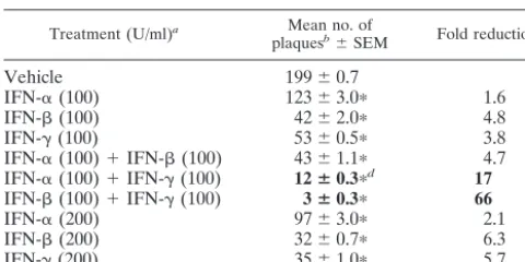
IFN-␣/and IFN-␥synergize to inhibit HSV-1 plaque for-mation on Vero cell monolayers.The efficiency of HSV-1 strain KOS plaque formation was compared in Vero cell monolayers treated with IFN-␣, IFN-, IFN-␥, or combinations of these IFNs. Plaque formation was reduced from an average of 199 plaques on vehicle-treated monolayers to 123, 42, and 53 plaques on Vero cell monolayers treated with IFN-␣, IFN-, or IFN-␥, respectively (Table 1). In contrast, HSV-1 plaque formation was reduced by ⬎90% in Vero cells treated with both IFN-␣/and IFN-␥. An inoculum of 200 PFU formed an average of 12 plaques on cell monolayers treated with both IFN-␣and IFN-␥. Likewise, only 3 of 200 PFU formed plaques on Vero cells treated with both IFN-and IFN-␥(Table 1). Comparable levels of inhibition could not be obtained by dou-bling the concentration of any one IFN from 100 to 200 U/ml (Table 1). Likewise, simultaneous treatment of Vero cells with both IFN-␣and IFN-did not decrease HSV-1 plaque forma-tion any more than IFN-alone (Table 1).
If this phenomenon was receptor dependent, then one would predict that synergy would first be observed at the min-imum biologically active dose of each IFN (1 U/ml, as defined by the manufacturer). Vero cells were treated with a constant, high dose of IFN-and 0.5-log dilutions of IFN-␥(Fig. 1A). An average of 32 plaques (out of 200 PFU) formed on Vero cells treated with IFN- alone. When IFN- was combined with ⬍0.3 U/ml of IFN-␥, the number of HSV-1 plaques formed on Vero cells did not change (Fig. 1A). However, as the concentration of IFN-␥was increased from 1 to 100 U/ml, the number of plaques that formed decreased linearly from 32 to 3 plaques per culture (Fig. 1A). In a second experiment, Vero cells were treated with 0.5-log dilutions of IFN-and a constant, high dose of IFN-␥ (Fig. 1B). An average of 55 plaques (out of 200 PFU) formed on Vero cells treated with
IFN-␥alone. When IFN-␥was combined with less than 0.3 U/ml of IFN-, the number of HSV-1 plaques formed on Vero cells did not change (Fig. 1B). However, as the concentration of IFN- was increased from 1 to 100 U/ml, the number of plaques that formed decreased linearly from 55 to 3 plaques per culture (Fig. 1B). The results suggest that simultaneous activation of IFN-␣/ receptors and IFN-␥ receptors is re-quired to synergistically inhibit HSV-1 plaque formation.
[image:3.603.328.506.70.450.2]IFN-and IFN-␥synergize to inhibit HSV-1 replication in Vero cells. HSV-1 replication was compared in Vero cells treated with vehicle, acyclovir (ACV), IFN-, IFN-␥, or both FIG. 1. IFN-and IFN-␥synergize to inhibit HSV-1 plaque for-mation on Vero cells. (A) Efficiency of HSV-1 plaque forfor-mation on Vero cells treated with 100 U/ml of IFN-and variable doses of IFN-␥ (n⫽3 per group). The dashed line indicates the number of plaques that formed on vehicle-treated cells. Significant reductions in plaque counts relative to cells treated with 100 U/ml of IFN- alone are denoted by a single asterisk (P⬍0.05, one-way ANOVA and Duncan’s multiple-range test). (B) Efficiency of HSV-1 plaque formation on Vero cells treated with variable doses of IFN-(n⫽3 per group) and 100 U/ml of IFN-␥. The dashed line indicates the number of plaques that formed on vehicle-treated cells. Significant reductions in plaque counts relative to cells treated with 100 U/ml of IFN-␥ alone are denoted by a single asterisk (P⬍0.05, one-way ANOVA and Duncan’s multiple-range test).
TABLE 1. IFN-␣/and IFN-␥synergize to inhibit HSV-1 plaque formation on Vero cells
Treatment (U/ml)a Mean no. of
plaquesb⫾SEM Fold reductionc
Vehicle 199⫾0.7
IFN-␣(100) 123⫾3.0ⴱ 1.6
IFN-(100) 42⫾2.0ⴱ 4.8
IFN-␥(100) 53⫾0.5ⴱ 3.8
IFN-␣(100)⫹IFN-(100) 43⫾1.1ⴱ 4.7 IFN-␣(100)⫹IFN-␥(100) 12ⴞ0.3ⴱd 17
IFN-(100)⫹IFN-␥(100) 3ⴞ0.3ⴱ 66
IFN-␣(200) 97⫾3.0ⴱ 2.1
IFN-(200) 32⫾0.7ⴱ 6.3
IFN-␥(200) 35⫾1.0ⴱ 5.7
aVero cells were treated continuously with IFN-␣, IFN-, IFN-␥, or
combi-nations of these cytokines from 12 h before infection until the end of the experiment.
bThat is, the number of plaques formed in Vero cells inoculated with 200 PFU
of HSV-1 strain KOS (n⫽4 per group).ⴱ,P⬍0.05, as determined by one-way ANOVA and Tukey’s post hocttest comparison of this treatment to vehicle.
cThe fold reduction in each group was calculated as follows: number of
plaques in vehicle/number of plaques in treatment.
dBoldface type indicates a⬎10-fold reduction in HSV-1 plaque formation.
on November 8, 2019 by guest
http://jvi.asm.org/
[image:3.603.43.283.89.209.2]IFN- and IFN-␥. In vehicle-treated cultures, HSV-1 strain KOS replicated to a titer of 3.5 ⫻ 106 PFU/ml over a 24-h period of incubation (Fig. 2). HSV-1 replicated to titers of 3.1 ⫻105and 2.3⫻105PFU/ml in cultures treated with 100 U of IFN-or IFN-␥/ml, respectively (Fig. 2). Thus, treatment with IFN-or IFN-␥inhibited HSV-1 replication by⬍20-fold. In cultures treated with both IFN-and IFN-␥, HSV-1 replicated to titers of 4.8 ⫻103 PFU/ml and was inhibited by 800-fold relative to vehicle-treated cultures (Fig. 2). Comparable levels of inhibition could not be obtained by increasing the concen-tration of either IFN-or IFN-␥alone to 316 or 1,000 U/ml (Fig. 2). Whereas the level of inhibition achieved by both IFN-and IFN-␥was far greater than additive, it was consid-erably less than the ⬃20,000-fold inhibition achieved by the HSV DNA synthesis inhibitor, ACV (Fig. 2).
Treatment with IFN-and IFN-␥ does not inhibit HSV-1 adsorption to Vero cells.PCR was used to compare the relative efficiency with which HSV-1 adsorbed to Vero cells treated with (i) vehicle or (ii) IFN-and IFN-␥. One hour after inoc-ulations with MOIs of 0.1 to 20 PFU/cell, DNA was isolated from HSV-1-infected Vero cells, and PCR was used to amplify a 243-bp fragment of the HSV-1 genome (Fig. 3A). The HSV-1 RR PCR product yield increased as a function of viral MOI in Vero cells treated with vehicle (Fig. 3B; r2 ⫽ 0.89) or both IFN-and IFN-␥(Fig. 3B;r2⫽0.98). Thus, PCR provided a valid basis for comparing the relative amount of HSV-1 DNA that entered Vero cells. The considerations that account for the inherent quantitative capacity of such noncompetitive PCR assays are addressed elsewhere (15, 16).
At all MOIs tested, the amount of RR PCR product
ampli-fied from Vero cells treated with IFN-and IFN-␥was com-parable to or greater than the amount of RR PCR product amplified from vehicle-treated Vero cells (Fig. 3B). Likewise, the correlation coefficient between MOI and PCR product yield did not differ between the treatments (P⫽0.87, Fisher two-sidedztest). Therefore, treatment with IFN-and IFN-␥ does not prevent the adsorption of HSV-1 to Vero cells.
Treatment with IFN-and IFN-␥does not prevent HSV-1 entry into Vero cells. Fluorescence microscopy was used to compare the efficiency with which a recombinant virus entered Vero cells and expressed a GFP reporter in the presence of vehicle, IFN-, IFN-␥, or both IFN- and IFN-␥. In cells infected with 0.03 PFU/cell of KOS-GFP, treatment with ei-ther IFN-or IFN-␥did not prevent virus from spreading to the majority of Vero cells by 36 h after infection (Fig. 4A). In contrast, treatment with both IFN-and IFN-␥resulted in a gross decrease in the extent of GFP expression observed 36 h after infection (Fig. 4A). The presence of many single GFP⫹
cells and several small foci of GFP⫹cells suggested that IFN-
[image:4.603.68.258.68.276.2]and IFN-␥did not prevent KOS-GFP from entering Vero cells but rather inhibited a subsequent step in viral replication (Fig. 4A).
FIG. 2. IFN-and IFN-␥synergize to inhibit HSV-1 replication in Vero cells. Virus yield in Vero cells treated with vehicle, IFN-(100, 316, or 1,000 U/ml), IFN-␥(100, 316, or 1,000 U/ml), IFN-and IFN-␥ (100 U/ml of each), or ACV (300M). Virus titers were determined 24 h after infection with 0.1 PFU of HSV-1 strain KOS/cell (n⫽4 per group). Treatments that significantly reduced virus titer relative to vehicle-treated cultures are indicated with an asterisk (P⬍0.05, one-way ANOVA and Tukey’s post hocttest). The dashed line represents the lower limit of detection of the plaque assay used to measure viral
titers. cells. (A) Ethidium bromide-stained RR PCR products amplified fromFIG. 3. IFN-and IFN-␥do not inhibit HSV-1 adsorption to Vero HSV-1-infected Vero cells treated with either vehicle (upper panel) or IFN-and IFN-␥(lower panel, 100 U/ml of each). From left to right, PCR tubes received no template (H2O), 100 ng of uninfected (UI)
Vero cell DNA, or 100 ng of HSV-1-infected Vero cell DNA harvested from cells inoculated with 0.1 to 20 PFU/cell. (B) RR PCR product yield plotted as a function of viral MOI in Vero cells treated with vehicle or both IFN-and IFN-␥. The correlation coefficient between PCR product yield and viral MOI wasr⫽0.81 in vehicle-treated cells and wasr⫽0.84 in cells treated with IFN-and IFN-␥.
on November 8, 2019 by guest
http://jvi.asm.org/
[image:4.603.317.523.69.354.2]The kinetics of KOS-GFP spread in Vero cells treated with vehicle, IFN-, IFN-␥, or IFN-and IFN-␥were compared by flow cytometry. One hour after infection with KOS-GFP (MOI ⫽0.03), the frequency of GFP-positive cells in virus-infected cultures was indistinguishable from uninfected cultures of Vero cells. At 12, 24, and 36 h after infection, GFP expression was detectable in an average of 2.6, 46, and 98% of vehicle-treated cells, respectively (Fig. 4B). Although treatment with IFN-or IFN-␥slowed the spread of KOS-GFP, 64%⫾3% of IFN--treated cells and 77%⫾3% of IFN-␥-treated cells were found to be GFP positive by 36 h after infection (Fig. 4B). In contrast, only 4.0%⫾0.3% of cells treated with both IFN- and IFN-␥were GFP positive 36 h after infection (Fig. 4B).
In the presence of ACV, KOS-GFP was unable to replicate, and thus GFP expression was restricted to the⬃3% of cells that were initially infected (Fig. 4A). In cells treated with ACV only, flow cytometry demonstrated that GFP expression was detectable in an average of 3.1, 3.2, and 3.6% of cells by 12, 24,
and 36 h after infection, respectively (Fig. 4C). When com-bined with ACV, treatment with IFN- slightly delayed the expression of GFP in KOS-GFP-infected cultures (Fig. 4A and C). When combined with ACV, treatment with IFN-␥had no apparent effect on GFP expression. When combined with ACV, treatment with IFN-and IFN-␥delayed GFP expres-sion, and an average of 0.9, 1.0, and 2.2% of cells were found to be GFP positive by 12, 24, and 36 h after infection, respec-tively (Fig. 4A and C). Based on the frequency of GFP expres-sion observed 36 h after infection, KOS-GFP entered Vero cells treated with IFN-and IFN-␥with at least 60% of the efficiency observed in vehicle-treated cells (i.e., 2.2%⫼3.6%). Therefore, treatment with IFN-and IFN-␥does not prevent HSV-1 entry into Vero cells.
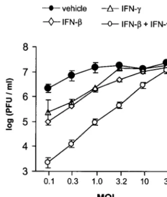
Increased multiplicity of infection overcomes the inhibitory effect of IFN-and IFN-␥.The effect of viral MOI on inhibi-tion by IFN-, IFN-␥, or both IFN-and IFN-␥was compared in Vero cells inoculated with 0.1 to 32 PFU/cell. In vehicle-FIG. 4. Effect of IFNs and/or ACV on the entry and spread of KOS-GFP in Vero cells. (A) Representative photomicrographs of Vero cells taken 36 h after infection with KOS-GFP (MOI⫽0.03), as seen when illuminated with the 360- to 400-nm spectrum of light that excites GFP fluorescence. Magnification,⫻10. Vero cells were treated with vehicle (VEH), IFN-(100 U/ml), IFN-␥(100 U/ml), or IFN-and IFN-␥(100 U/ml of each) and were treated with either no ACV (⫺ACV) or 300M ACV (⫹ACV) after infection with KOS-GFP. (B and C) Flow cytometric analysis of GFP fluorescence in Vero cells 1, 12, 24, and 36 h after infection with KOS-GFP (MOI⫽0.03). IFN treatments were the same as described above, and cells were secondarily treated with either (B) no ACV (⫺ACV) or (C) 300M ACV (⫹ACV). The mean percentage⫾the standard error (SE) of GFP-positive cells in each treatment was determined in three replicate cultures per time point. The results are presented as the mean percentage⫾the SE of GFP-positive cells after subtracting out the background frequency of fluorescence observed in uninfected cultures of Vero cells (i.e., 48⫾5 per 24,000 cells evaluated).
on November 8, 2019 by guest
http://jvi.asm.org/
treated cultures inoculated with 0.1 PFU/cell, HSV-1 strain KOS replicated to titers of 2.2⫻106PFU/ml (Fig. 5). At an MOI of 0.1, treatment with IFN-or IFN-␥reduced HSV-1 titers by 10- to 20-fold, and treatment with IFN-and IFN-␥ reduced HSV-1 titers by 980-fold (Fig. 5). Inhibition of HSV-1 replication by IFN-or IFN-␥was overcome by increasing the MOI to 3.2 PFU/cell. Although inhibition by both IFN-and IFN-␥was only completely reversed when the MOI was in-creased to 32 PFU/cell, virus yield inin-creased in direct propor-tion to MOI over the entire range tested (Fig. 5;r2⫽0.99).
Synergistic inhibition of HSV-1 replication is not cell type or virus strain specific.The effect of IFN-and IFN-␥on HSV-1 replication was compared in Vero cells, SK-N-SH cells, and PMK cells inoculated with 0.1 PFU/cell. In each cell type, HSV-1 strain KOS replicated to titers of greater than 5.6⫻105 PFU/ml in vehicle-treated cells (Table 2.) Treatment with IFN- or IFN-␥ reduced HSV-1 titers in each cell type by ⱕ20-fold (Table 2). Treatment with both IFN- and IFN-␥
reduced HSV-1 titers in Vero, SK-N-SH, and PMK cells by 930-, 620-, and 2,600-fold, respectively (Table 2). In PMK cells, the efficiency with which IFN- and IFN-␥inhibited HSV-1 replication was 24% of that achieved by ACV (Table 2).
The effect of IFN-and IFN-␥on the replication of HSV-1 strain KOS in Vero cells was compared to two other HSV-1 strains, McKrae and TU1-1. All three HSV-1 strains replicated to titers of greater than 2.5⫻ 105PFU/ml in vehicle-treated cells inoculated with 0.1 PFU/cell (Table 3). Treatment with IFN-or IFN-␥reduced titers of KOS, McKrae, and TU1-1 by ⱕ20-fold. In contrast, treatment with both IFN-and IFN-␥ reduced the titers of KOS, McKrae, and TU1-1 by 1,000-, 1,400-, and 1,800-fold, respectively (Table 3). The efficiency with which IFN- and IFN-␥ inhibited the replication of McKrae and TU1-1 was 25% of that achieved by ACV (Table 3).
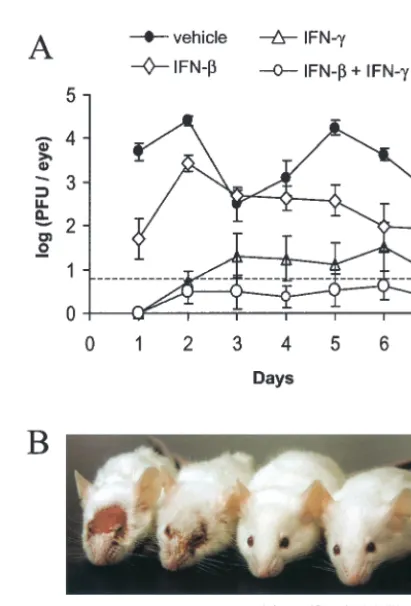
Pretreatment with IFN-and IFN-␥inhibits HSV-1 repli-cation in mouse eyes.Viral replication was compared in mouse eyes that were pretreated with vehicle, IFN-, IFN-␥, or both IFN-and IFN-␥at 12, 8, 4, and 0 h prior to HSV-1 infection. In vehicle-treated mice, an average of 5,000 PFU/eye was de-tected 24 h after infection, and average titers ranged from 300 to 25,000 PFU/eye over the next 6 days (Fig. 6A). Treatment with IFN-caused an ⬃100-fold reduction in ocular HSV-1 shedding 24 h after infection, but HSV-1 titers increased to 2,700 PFU/eye by 48 h after infection (Fig. 6A). Treatment with IFN-␥reduced ocular HSV-1 shedding to undetectable levels 24 h after infection, and five of eight mice shed 10 to 40 PFU/eye between 2 and 7 days after infection. (Fig. 6A). Treat-ment with both IFN-and IFN-␥reduced ocular HSV-1 shed-ding to undetectable levels 24 h after infection, and only two of eight mice shed detectable levels of virus between 2 and 7 days after infection. (Fig. 6A). HSV-1 infection killed one of eight vehicle-treated mice and none of the IFN-treated mice. Vehi-cle-treated mice developed extensive periocular skin lesions by 8 days after infection (Fig. 6B). IFN--treated mice developed periocular skin lesions, but the severity of the lesions was reduced (not shown). Only two of eight IFN-␥treated mice developed visible skin lesions. None of the eight mice treated with IFN-and IFN-␥developed periocular skin lesions by 8 days after infection, or at any other subsequent time during the 40-day course of the experiment (Fig. 6B). These results are representative, as similar results were obtained in two inde-pendent experiments.
Pretreatment with IFN-and IFN-␥inhibits the establish-FIG. 5. Inhibition by IFN-and IFN-␥is overcome by increasing
[image:6.603.76.243.71.267.2]the MOI. Virus yield was plotted as a function of MOI. Vero cells were treated with vehicle, IFN-(100 U/ml), IFN-␥(100 U/ml), or IFN- plus IFN-␥(100 U/ml of each). Virus titers were determined 24 h after infection with 0.1 to 32 PFU/cell of HSV-1 strain KOS (n⫽4 per group). Regression analysis demonstrated that the titer of virus recov-ered from cells treated with IFN-and IFN-␥was linearly dependent on MOI (r2⫽0.99,P⫽10⫺6).
TABLE 2. Effect of IFN-and IFN-␥on HSV-1 replication in three different cell types
Treatmenta Mean HSV-1 log titer⫾SE (fold inhibition) b
Vero cells SK-N-SH cells PMK cells
Vehicle 6.31⫾0.9 6.19⫾0.3 5.75⫾0.1
IFN- 5.01⫾0.3ⴱc(20) 5.47⫾0.1 (5) 5.16⫾0.2ⴱ(4)
IFN-␥ 5.37⫾0.5ⴱ(14) 5.99⫾0.1 (2) 5.19⫾0.1ⴱ(4)
IFN- ⫹IFN-␥ 3.34⫾0.2ⴱ(930) 3.36⫾0.2ⴱ(620) 2.33⫾0.1ⴱ(2,600)
ACV 1.96⫾0.2ⴱ(22,000) 2.65⫾0.2ⴱ(5,400) 1.69⫾0.2ⴱ(11,000)
aVero and SK-N-SH cells were treated with vehicle, 100 U/ml of hu IFN-, 100 U/ml of hu IFN-␥, hu IFN-and hu IFN-␥(100 U/ml of each), or with ACV (300
M). PMK cells received identical treatments with murine IFNs. Viral titers were determined 24 h after infection with 0.1 PFU/cell of HSV-1 strain KOS.
bHSV-1 titers (log [PFU/ml]) recovered from Vero, SK-N-SH, or PMK cells (n⫽4 per group). The fold inhibition was calculated within each cell type as the titer
in vehicle/titer in treatment.
cⴱ,P⬍0.05, as determined by one-way ANOVA and Tukey’s post hocttest comparison of this treatment to vehicle-treated cultures of the same cell type.
on November 8, 2019 by guest
http://jvi.asm.org/
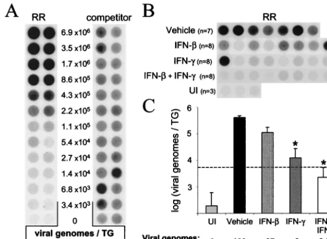
[image:6.603.47.544.602.682.2]ment of latent HSV-1 infection. The efficiency with which HSV-1 established latent infections in the TG of mice treated with vehicle, IFN-, IFN-␥, or both IFN- and IFN-␥ was compared 40 days after infection. HSV-1 RR and competitor PCR products were coamplified from viral DNA standards in order to define the relationship between RR PCR product yield (normalized to the competitor) and the number of HSV-1 genomes per TG (Fig. 7A). In parallel reactions, RR and competitor PCR products were coamplified from TG DNA samples isolated from HSV-1-infected mice and uninfected controls (Fig. 7B; competitor product yields not shown). Based on the yield of RR PCR products (normalized to the compet-itor), the average number of latent HSV-1 genomes in vehicle-treated mice was 4.3⫻105viral genomes per TG (Fig. 7C).
Pretreatment of mouse eyes with IFN-or IFN-␥reduced the number of latent HSV-1 genomes per TG by 4- and 33-fold, respectively (Fig. 7C). An average of 2.2 ⫻ 103 viral genomes per TG were detected in mice treated with both IFN-and IFN-␥(Fig. 7C). This latter value may be somewhat of an overestimate because the amount of amplified RR PCR product was slightly below the linear range of the competitive PCR assay (dashed line in Fig. 7C). Therefore, pretreatment with IFN- and IFN-␥reduced the number of latent HSV-1 genomes per TG by ⬃200-fold relative to the number of HSV-1 genomes detected in TG of vehicle-treated mice.
DISCUSSION
Although HSV-1 is highly resistant to the antiviral activities of IFN-␣/or IFN-␥, it is possible that the combined activities of IFN-␣/and IFN-␥could synergize to inhibit HSV-1 repli-cation. Several reports from the 1980s contain limited evidence that is consistent with this hypothesis (9, 40, 64). Balish et al. (2) provide definitive evidence that IFN-␣and IFN-␥synergize to inhibit HSV-1 strain KOS replication in human corneal fibroblast cells, but the generality of the effect was not further investigated (2). Therefore, the combined effect of IFN-␣/ and IFN-␥ on HSV-1 replication has never been rigorously evaluated.
The results of the present study establish that the combined activities of IFN-␣/ and IFN-␥inhibit HSV-1 replication in vitro with a potency that approaches that of ACV. The results of dose-response analyses with recombinant IFN proteins in-dicate that (i) the effect is far greater than additive and (ii) the synergy appears to be dependent on the simultaneous
[image:7.603.42.542.82.161.2]activa-tion of IFN-␣/receptors and IFN-␥receptors. Therefore, we propose that coactivation of IFN-␣/ and IFN-␥ receptors renders cells highly resistant to HSV-1 replication. The fact that this synergy is observed in multiple cell types and against
FIG. 6. Pretreatment with IFN-and IFN-␥inhibits HSV-1 repli-cation in vivo. (A) Virus titers recovered from mouse eyes treated with vehicle, IFN-, IFN-␥, or both IFN- and IFN-␥(n⫽8 mice per group). Mouse eyes were scarified and treated 0, 4, 8, and 12 h later with vehicle, 200 U of IFN-, 200 U of IFN-␥, or IFN-and IFN-␥ (200 U of each). After the final treatment, mouse eyes were inoculated with 2⫻105PFU of HSV-1 strain KOS. The titer of virus recovered
[image:7.603.317.523.303.606.2]from mouse eyes was measured by plaque assay, and the dashed line indicates the lower limit of detection of this assay. Two-way ANOVA demonstrated that each IFN treatment significantly reduced the course of ocular HSV-1 shedding in mice relative to vehicle-treated mice (P⬍ 0.001). (B) Photograph taken 8 days after ocular HSV-1 infection of mice treated with vehicle or both IFN-and IFN-␥.
TABLE 3. Effect of IFN-and IFN-␥on the replication of three different HSV-1 strains
Treatmenta Mean HSV-1 log titer⫾SE (fold inhibition) b
KOS McKrae TU1-1
Vehicle 6.66⫾0.1 5.81⫾0.1 5.40⫾0.1
IFN- 5.38⫾0.2ⴱc(19) 4.52⫾0.1ⴱ(20) 4.30⫾0.2ⴱ(14)
IFN-␥ 5.94⫾0.2ⴱ(7) 4.87⫾0.1ⴱ(9) 4.32⫾0.1ⴱ(12)
IFN- ⫹IFN-␥ 3.70⫾0.1ⴱ(1,000) 2.78⫾0.3ⴱ(1,400) 2.15⫾0.1ⴱ(1,800)
ACV 2.64⫾0.1ⴱ(11,000) 2.07⫾0.1ⴱ(5,500) 1.58⫾0.1ⴱ(7,000)
aVero cells were treated with vehicle, 100 U/ml of hu IFN-, 100 U/ml of hu IFN-␥, hu IFN-and hu IFN-␥(100 U/ml of each), or ACV (300M). Viral titers
were determined 24 h after infection with 0.1 PFU/ml of HSV-1 strains KOS, McKrae, or TU1-1.
bHSV-1 titers (log [PFU/ml]) recovered from Vero, SK-N-SH, or PMK cells (n⫽4 per group). The fold inhibition was calculated within each cell type as the titer
in vehicle/titer in treatment.
cⴱ,P⬍0.05, as determined by one-way ANOVA and Tukey’s post hocttest comparison of this treatment to vehicle-treated cultures infected with the same HSV-1
strain.
on November 8, 2019 by guest
http://jvi.asm.org/
three different HSV-1 strains suggests that this is a generally applicable principal.
The results of the present study establish for the first time that the combined activities of IFN-␣/ and IFN-␥potently inhibit HSV-1 replication in vivo. The potency with which IFN-and IFN-␥inhibited HSV-1 replication in vivo appeared to be considerably greater than is typically achieved when an-imals are treated with ACV or other antiviral agents in vivo (12, 18, 30, 36, 58). In mice pretreated with IFN-and IFN-␥, HSV-1 replication was not detected in the eyes of six of the eight mice, and the average level of virus replication in the remaining two mice was⬃500-fold less than observed in vehi-cle-treated controls. Consequently, treatment with IFN-and IFN-␥ prevented HSV-1-associated disease in mice and re-duced the number of latent HSV-1 genomes per TG by⬃ 200-fold relative to vehicle-treated mice. While IFN-reduced the severity of disease, HSV-1 still replicated to high titers in mice treated with IFN-alone. Treatment with IFN-␥alone, how-ever, inhibited HSV-1 replication with nearly the same potency as that of IFN-and IFN-␥together. We hypothesize that the high level of inhibition achieved by exogenous IFN-␥alone is the result of synergy with endogenous IFN-␣/that is locally produced in response to HSV-1 infection. The validity of this hypothesis remains to be tested.
Mechanism(s) by which IFN-and IFN-␥may synergize to inhibit HSV-1 replication.The results of the present study do not establish the specific mechanism(s) by which IFN- and IFN-␥synergize to inhibit HSV-1 replication. In the absence of such knowledge, it is theoretically possible that IFN- and IFN-␥interfere with HSV-1 replication at one, or several, of the following steps: (i) adsorption, (ii) entry, (iii) viral gene expression, (iv) viral DNA replication, (v) virus assembly, or (vi) virus maturation and egress. The results of the present study argue strongly against the first two possibilities. PCR analysis of HSV-1 DNA levels associated with Vero cells 1 h after infection demonstrate that IFN-and IFN-␥do not pre-vent HSV-1 from binding to Vero cells. Likewise, fluorescence microscopy and flow cytometry indicate that IFN-and IFN-␥ have a⬍2-fold effect on the efficiency with which KOS-GFP enters Vero cells. Thus, it is highly unlikely that inhibition of viral adsorption and/or entry account for the mechanism(s) by which IFN-and IFN-␥achieve an⬃1,000-fold inhibition of HSV-1 replication in Vero cells. It remains to be formally proven that treatment with IFN-and IFN-␥does not inhibit HSV-1 entry into PMK, SK-N-SH, or mouse eye cells.
[image:8.603.124.458.72.316.2]If IFN-and IFN-␥inhibited an essential cellular function, viral replication would have been efficiently inhibited at all MOIs. However, the degree of inhibition achieved by IFN- FIG. 7. Pretreatment with IFN-and IFN-␥reduces the establishment of latent HSV-1 in the TG. (A) RR and competitor PCR products amplified from viral DNA standards. These DNA standards consisted of (i) twofold dilutions of HSV-1 genomes, (ii) a constant amount of competitor (⬃1,400 copies), and (iii) 100 ng of uninfected TG DNA. Duplicate blots of PCR products were hybridized to RR-specific or competitor-specific probes, and the ratio of RR to competitor PCR product was used to define the “normalized RR PCR product yield.” The relationship between the number of viral genomes per TG (x, input) and the normalized RR PCR product yield (y, output) was sigmoidal and was described by the polynomial equation:x⫽0.328y3⫹0.551y2⫹1.4851y⫹3.4423 (r2⫽0.99). The linear range of the standard curve was between
6.8⫻103and 1.7⫻106viral genomes/TG. Only⬃1/230th of the DNA from each TG was included in the PCR (i.e., 100 ng); thus, the lower limit
of accurate quantitation was 30 HSV-1 genomes per PCR. (B) Dot blot of HSV-1 RR PCR products amplified from TG of 3 uninfected (UI) mice and 31 HSV-1 latently infected mice (sacrificed 40 days after inoculation) that were treated prior to HSV-1 infection with vehicle (n⫽7), IFN- (n⫽8), IFN-␥(n⫽8), or both IFN-and IFN-␥(n⫽8). (C) Effect of IFN-, IFN-␥, or IFN-plus IFN-␥on HSV-1 genome load in latently infected TG. The dashed line indicates the lower limit of the linear range of the assay. Significant differences in viral genome load relative to vehicle-treated mice are indicated by an asterisk (P⬍0.05, one-way ANOVA and Tukey’s post hocttest).
on November 8, 2019 by guest
http://jvi.asm.org/
and IFN-␥was highly dependent on the multiplicity of HSV-1 infection (r2 ⫽ 0.99). Thus, we hypothesize that IFN- and IFN-␥synergize to inhibit the expression of at least one virus-encoded function that is necessary for HSV-1 replication. For example, if IFN-␣/ and IFN-␥ synergistically block the ex-pression of ICP0 or ICP34.5, HSV-1 would be highly sensitive to inhibition by IFN-␣/(22, 39). If this hypothesis is correct, then increasing the copy number of either ICP0 and/or the ICP34.5 genes should be sufficient to overcome inhibition by IFN-␣/and IFN-␥. Investigations are currently in progress to test these and other hypotheses regarding the mechanism(s) by which IFN-␣/and IFN-␥synergistically block HSV-1 replica-tion.
Implications. (i) Host immunity to HSV-1.Three pieces of evidence are consistent with a hypothesis that T cells inhibit HSV-1 replication in the peripheral nervous system through noncytolytic mechanisms. First, despite their best known role as cytotoxic T lymphocytes, CD8⫹T cells actually contribute to
the survival of HSV-1 infected neurons in mice (53). Second, during acute HSV-1 infection, ganglionic neurons surrounded by CD8⫹T cells remain healthy in appearance (35). Finally,
CD8⫹T cells are sufficient to block HSV-1 reactivation from
TG neurons ex vivo, and this activity can be blocked with neutralizing antibodies to IFN-␥(33, 34).
The results of the present study are consistent with a hy-pothesis that secretion of IFN-␥suppresses HSV-1 replication in vivo. If IFN-␣ and/or IFN- are produced as an innate response to viral infection (61), then it follows that IFN-␣/ are ubiquitous in virus-infected tissues. Extrapolating from the in vitro results, one can imagine that the⬃10-fold inhibition achieved by IFN-␣/would reduce the rate of HSV-1 replica-tion but would not stop the spread of virus in vivo. Secrereplica-tion of IFN-␥from T cells and/or NK cells, however, may provide a second signal that synergizes with IFN-␣/ and renders cells far less permissive to HSV-1 replication. Thus, the⬃1,000-fold inhibition achieved by the combined activities of IFN-␣/and IFN-␥may effectively stop HSV-1 infection from spreading in vivo.
Under this hypothesis, cells that are capable of secreting IFN-␥should be essential components of the host immune response to HSV-1. Depletion of either CD4⫹, CD8⫹, or␥␦⫹
T-cell subsets grossly impairs the host immune response to HSV-1 (37, 49, 53). Likewise, depletion of NK cells signifi-cantly increases the pathogenesis of HSV-1 infections (1, 45). Therefore, the primary cell types that are capable of secreting IFN-␥in vivo are all important for host immunity to HSV-1.
(ii) Establishment of latent infections.The fact that IFN- and IFN-␥synergize to inhibit the replication of HSV-1 has important implications for the coevolution of herpesviruses and their vertebrate hosts. Most viral infections have two pos-sible outcomes: (i) the virus kills the host or (ii) the host clears the virus. In vertebrate animals, it is the clonal proliferation and dissemination of virus-specific T cells that allows the host to clear the virus. The results of the present study raise the intriguing possibility that HSV, and perhaps other herpesvi-ruses, have evolved to respond to T-cell-secreted cytokines as cues that trigger the efficient suppression of viral replication at appropriate times. By coupling repression of virus replication to the infiltration of T cells into infected tissues, a third pos-sible outcome of viral infection may be created: the
establish-ment of latent infections that persist in an equilibrium with the host’s immune system. In light of this hypothesis, it is intriguing that secretion of IFN-␥from T cells also inhibits the replica-tion of hepatitis B virus, an unrelated persistent virus (13, 14, 46).
(iii) Clinical relevance.Although IFNs are not considered clinically effective in treating HSV-1 infections, only IFN-␣and IFN-have been tested in humans (24). In the present study, IFN-␥was a far more potent inhibitor of HSV-1 replication in vivo than IFN-. Recombinant IFN-␥has never been clinically evaluated for its potential in treating human HSV-1 infections, nor has recombinant IFN-␥been rigorously evaluated in ani-mal models of HSV-1 infection.
If IFN-␣/ are ubiquitous in HSV-infected tissues, then treatment with IFN-␣or IFN-can only increase the levels of a preexisting cytokine. However, if secretion of IFN-␥from T cells is truly rate limiting (particularly in the immunodeficient), then there is a clear theoretical rationale for determining if exogenous IFN-␥can be used to limit the spread of HSV-1 infection in vivo. Further investigation will be required to de-termine whether IFN-␥has any potential application in limit-ing the replication and pathogenesis of HSV-1 in humans.
ACKNOWLEDGMENTS
This work was supported by a grant from the W. M. Keck Founda-tion of Los Angeles and the Louisiana Board of Regents Support Foundation (LEQSF-2001-2004-RD-A-33).
We are indebted to John Balliet and Priscilla Schaffer (Harvard University, Boston, Mass.) for kindly donating the recombinant virus KOS-GFP. We also thank Bryan Gebhardt, Daniel Carr, and Robert Hendricks for critical review of the manuscript.
REFERENCES
1. Adler, H., J. L. Beland, N. C. Del-Pan, L. Kobzik, R. A. Sobel, and I. J. Rimm.1999. In the absence of T cells, natural killer cells protect from mortality due to HSV-1 encephalitis. J. Neuroimmunol.93:208–213. 2. Balish, M. J., M. E. Abrams, A. M. Pumfery, and C. R. Brandt.1992.
Enhanced inhibition of herpes simplex virus type 1 growth in human corneal fibroblasts by combinations of interferon-alpha and -gamma. J. Infect. Dis.
166:1401–1403.
3. Buddingh, G. J., D. I. Schrum, J. C. Lanier, and D. J. Guidry.1953. Studies of the natural history of herpes simplex virus infections. Pediatrics11:593– 605.
4. Cantell, K.1995. Development of antiviral therapy with alpha interferons: promises, false hopes, and accomplishments. Ann. Med.27:23–28. 5. Cantin, E., B. Tanamachi, and H. Openshaw.1999. Role for gamma
inter-feron in control of herpes simplex virus type 1 reactivation. J. Virol.73:3418– 3423.
6. Cantin, E., B. Tanamachi, H. Openshaw, J. Mann, and K. Clarke.1999. Gamma interferon (IFN-␥) receptor null-mutant mice are more susceptible to herpes simplex virus type 1 infection than IFN-␥ligand null-mutant mice. J. Virol.73:5196–5200.
7. Cantin, E. M., D. R. Hinton, J. Chen, and H. Openshaw.1995. Gamma interferon expression during acute and latent nervous system infection by herpes simplex virus type 1. J. Virol.69:4898–4905.
8. Cassady, K. A., M. Gross, and B. Roizman.1998. The second-site mutation in the herpes simplex virus recombinants lacking the␥134.5 genes precludes shutoff of protein synthesis by blocking the phosphorylation of eIF-2␣. J. Vi-rol.72:7005–7011.
9. Czarniecki, C. W., C. W. Fennie, D. B. Powers, and D. A. Estell.1984. Synergistic antiviral and antiproliferative activities ofEscherichia coli -de-rived human alpha, beta, and gamma interferons. J. Virol.49:490–496. 10. Farrar, M. A., and R. D. Schreiber.1993. The molecular cell biology of
interferon-gamma and its receptor. Annu. Rev. Immunol.11:571–611. 11. Feldman, L. T., A. R. Ellison, C. C. Voytek, L. Yang, P. Krause, and T. P.
Margolis.2002. Spontaneous molecular reactivation of herpes simplex virus type 1 latency in mice. Proc. Natl. Acad. Sci. USA99:978–983.
12. Field, H. J., S. E. Bell, G. B. Elion, A. A. Nash, and P. Wildy.1979. Effect of acycloguanosine treatment of acute and latent herpes simplex infections in mice. Antimicrob. Agents Chemother.15:554–561.
13. Guidotti, L. G., K. Ando, M. V. Hobbs, T. Ishikawa, L. Runkel, R. D.
on November 8, 2019 by guest
http://jvi.asm.org/
Schreiber, and F. V. Chisari.1994. Cytotoxic T lymphocytes inhibit hepatitis B virus gene expression by a noncytolytic mechanism in transgenic mice. Proc. Natl. Acad. Sci. USA91:3764–3768.
14. Guidotti, L. G., T. Ishikawa, M. V. Hobbs, B. Matzke, R. Schreiber, and F. V. Chisari.1996. Intracellular inactivation of the hepatitis B virus by cytotoxic T lymphocytes. Immunity4:25–36.
15. Halford, W. P.1999. The essential prerequisites for quantitative RT-PCR. Nat. Biotechnol.17:835.
16. Halford, W. P., V. C. Falco, B. M. Gebhardt, and D. J. J. Carr.1999. The inherent quantitative capacity of the reverse transcription-polymerase chain reaction. Anal. Biochem.266:181–191.
17. Halford, W. P., B. M. Gebhardt, and D. J. Carr.1996. Persistent cytokine expression in trigeminal ganglion latently infected with herpes simplex virus type 1. J. Immunol.157:3542–3549.
18. Halford, W. P., B. M. Gebhardt, and D. J. Carr.1997. Acyclovir blocks cytokine gene expression in trigeminal ganglia latently infected with herpes simplex virus type 1. Virology238:53–63.
19. Halford, W. P., C. D. Kemp, J. A. Isler, D. J. Davido, and P. A. Schaffer.2001. ICP0, ICP4, or VP16 expressed from adenovirus vectors induces reactivation of latent herpes simplex virus type 1 in primary cultures of latently infected trigeminal ganglion cells. J. Virol.75:6143–6153.
20. Halford, W. P., and P. A. Schaffer.2000. Optimized viral dose and transient immunosuppression enable herpes simplex virus ICP0-null mutants to es-tablish wild-type levels of latency in vivo. J. Virol.74:5957–5967. 21. Harle, P., E. Lauret, P. M. Pitha, E. De Maeyer, and D. J. Carr.2001.
Expression of human and macaque type I IFN transgenes interferes with HSV-1 replication at the transcriptional and translational levels: IFN-is more potent than IFN-␣2. Virology290:237–248.
22. Harle, P., B. Sainz, Jr., D. J. Carr, and W. P. Halford.2002. The immediate-early protein, ICP0, is essential for the resistance of herpes simplex virus to interferon-alpha/beta. Virology293:295–304.
23. Hill, J. M., W. P. Halford, R. Wen, L. S. Engel, L. C. Green, and B. M. Gebhardt.1996. Quantitative analysis of polymerase chain reaction products by dot blot. Anal. Biochem.235:44–48.
24. Ho, M.1990. Interferon as an agent against herpes simplex virus. J. Investig. Dermatol.95(Suppl. 6):158S–160S.
25. Kaufman, H. E., A. B. Nesburn, and E. D. Maloney.1962. IDU therapy of herpes simplex. Arch. Ophthalmol.67:583–591.
26. Kaufman, H. E., J. Sugar, and E. D. Varnell.1973. Effect of exogenous interferon on herpetic keratitis in rabbits and monkeys. Investig. Ophthal-mol.12:378–380.
27. Kinghorn, G. R.1994. Epidemiology of genital herpes. J. Int. Med. Res.
22(Suppl. 1):14A–23A.
28. Koelle, D. M., C. M. Posavad, G. R. Barnum, M. L. Johnson, J. M. Frank, and L. Corey.1998. Clearance of HSV-2 from recurrent genital lesions correlates with infiltration of HSV-specific cytotoxic T lymphocytes. J. Clin. Investig.101:1500–1508.
29. Koelle, D. M., and A. Wald.2000. Herpes simplex virus: the importance of asymptomatic shedding. J. Antimicrob. Chemother.45(Suppl. T3):1–8. 30. LeBlanc, R. A., L. Pesnicak, M. Godleski, and S. E. Straus.1999. The
comparative effects of famciclovir and valacyclovir on herpes simplex virus type 1 infection, latency, and reactivation in mice. J. Infect. Dis.180:594–599. 31. Leib, D. A., M. A. Machalek, B. R. Williams, R. H. Silverman, and H. W. Virgin.2000. Specific phenotypic restoration of an attenuated virus by knock-out of a host resistance gene. Proc. Natl. Acad. Sci. USA97:6097–6101.
32. Lekstrom-Himes, J. A., R. A. LeBlanc, L. Pesnicak, M. Godleski, and S. E. Straus.2000. Gamma interferon impedes the establishment of herpes sim-plex virus type 1 latent infection but has no impact on its maintenance or reactivation in mice. J. Virol.74:6680–6683.
33. Liu, T., K. M. Khanna, B. N. Carriere, and R. L. Hendricks.2001. Gamma interferon can prevent herpes simplex virus type 1 reactivation from latency in sensory neurons. J. Virol.75:11178–11184.
34. Liu, T., K. M. Khanna, X. Chen, D. J. Fink, and R. L. Hendricks.2000. CD8⫹T cells can block herpes simplex virus type 1 (HSV-1) reactivation
from latency in sensory neurons. J. Exp. Med.191:1459–1466.
35. Liu, T., Q. Tang, and R. L. Hendricks.1996. Inflammatory infiltration of the trigeminal ganglion after herpes simplex virus type 1 corneal infection. J. Vi-rol.70:264–271.
36. Loutsch, J. M., B. Sainz, Jr., M. E. Marquart, X. Zheng, P. Kesavan, S. Higaki, J. M. Hill, and R. Tal-Singer.2001. Effect of famciclovir on herpes simplex virus type 1 corneal disease and establishment of latency in rabbits. Antimicrob. Agents Chemother.45:2044–2053.
37. Mercadal, C. M., D. M. Bouley, D. DeStephano, and B. T. Rouse.1993. Herpetic stromal keratitis in the reconstituted scid mouse model. J. Virol.
67:3404–3408.
38. Mossman, K. L., H. A. Saffran, and J. R. Smiley.2000. Herpes simplex virus ICP0 mutants are hypersensitive to interferon. J. Virol.74:2052–2056. 39. Mossman, K. L., and J. R. Smiley.2002. Herpes simplex virus ICP0 and
ICP34.5 counteract distinct interferon-induced barriers to virus replication. J. Virol.76:1995–1998.
40. Neumann-Haefelin, D., R. Sundmacher, H. Frey, and W. Merk.1985. Re-combinant HuIFN-␥prevents herpes simplex keratitis in African green mon-keys: demonstration of synergism with recombinant HuIFN-␣2. Med. Mi-crobiol. Immunol.174:81–86.
41. Noisakran, S., I. L. Campbell, and D. J. Carr.1999. Ectopic expression of DNA encoding IFN-␣1 in the cornea protects mice from herpes simplex virus type 1-induced encephalitis. J. Immunol.162:4184–4190.
42. Orange, J. S., B. Wang, C. Terhorst, and C. A. Biron.1995. Requirement for natural killer cell-produced interferon gamma in defense against murine cytomegalovirus infection and enhancement of this defense pathway by in-terleukin 12 administration. J. Exp. Med.182:1045–1056.
43. Patterson, C. E., D. M. Lawrence, L. A. Echols, and G. F. Rall.2002. Immune-mediated protection from measles virus-induced central nervous system disease is noncytolytic and gamma interferon dependent. J. Virol.
76:4497–4506.
44. Posavad, C. M., D. M. Koelle, and L. Corey.1998. Tipping the scales of herpes simplex virus reactivation: the important responses are local. Nat. Med.4:381–382.
45. Rager-Zisman, B., P. C. Quan, M. Rosner, J. R. Moller, and B. R. Bloom.
1987. Role of NK cells in protection of mice against herpes simplex virus-1 infection. J. Immunol.138:884–888.
46. Robek, M. D., S. F. Wieland, and F. V. Chisari.2002. Inhibition of hepatitis B virus replication by interferon requires proteasome activity. J. Virol.76:
3570–3574.
47. Samuel, C. E.1998. Reoviruses and the interferon system. Curr. Top. Mi-crobiol. Immunol.233:125–145.
48. Schultz, U., and F. V. Chisari.1999. Recombinant duck interferon gamma inhibits duck hepatitis B virus replication in primary hepatocytes. J. Virol.
73:3162–3168.
49. Sciammas, R., P. Kodukula, Q. Tang, R. L. Hendricks, and J. A. Bluestone.
1997. T-cell receptor-gamma/delta cells protect mice from herpes simplex virus type 1-induced lethal encephalitis. J. Exp. Med.185:1969–1975. 50. Scott, D. A., W. A. Coulter, and P. J. Lamey.1997. Oral shedding of herpes
simplex virus type 1: a review. J. Oral Pathol. Med.26:441–447.
51. Shimeld, C., J. L. Whiteland, S. M. Nicholls, D. L. Easty, and T. J. Hill.1996. Immune cell infiltration in corneas of mice with recurrent herpes simplex virus disease. J. Gen. Virol.77(Pt. 5):977–985.
52. Shimeld, C., J. L. Whiteland, S. M. Nicholls, E. Grinfeld, D. L. Easty, H. Gao, and T. J. Hill.1995. Immune cell infiltration and persistence in the mouse trigeminal ganglion after infection of the cornea with herpes simplex virus type 1. J. Neuroimmunol.61:7–16.
53. Simmons, A., and D. C. Tscharke.1992. Anti-CD8 impairs clearance of herpes simplex virus from the nervous system: implications for the fate of virally infected neurons. J. Exp. Med.175:1337–1344.
54. Smith, K. O.1964. Relationship between the envelope and the infectivity of herpes simplex virus. Proc. Soc. Exp. Biol. Med.115:814–816.
55. Smith, P. M., R. M. Wolcott, R. Chervenak, and S. R. Jennings.1994. Control of acute cutaneous herpes simplex virus infection: T cell-mediated viral clearance is dependent upon interferon-gamma (IFN-␥). Virology202:
76–88.
56. Stanberry, L. R., S. A. Floyd-Reising, B. L. Connelly, S. J. Alter, M. J. Gilchrist, C. Rubio, and M. G. Myers.1994. Herpes simplex viremia: report of eight pediatric cases and review of the literature. Clin. Infect. Dis.18:
401–407.
57. Stewart, J. A., S. E. Reef, P. E. Pellett, L. Corey, and R. J. Whitley.1995. Herpesvirus infections in persons infected with human immunodeficiency virus. Clin. Infect. Dis.21(Suppl. 1):S114–S120.
58. Thackray, A. M., and H. J. Field.1998. Famciclovir and valaciclovir differ in the prevention of herpes simplex virus type 1 latency in mice: a quantitative study. Antimicrob. Agents Chemother.42:1555–1562.
59. Thomson, A.1998. The cytokine handbook, 3rd ed. Academic Press, San Diego, Calif.
60. Treco, D. A.1990. Preparation and analysis of DNA, p.2.0.3–2.2.3.InF. M. Ausubel (ed.), Current protocols in molecular biology. John Wiley & Sons, Inc., New York, N.Y.
61. Vilcek, J., and J. Sen.1996. Interferons and other cytokines, p. 375–400.In
B. N. Fields, D. M. Knipe, and P. M. Howley (ed.), Virology. Raven Pub-lishers, Philadelphia, Pa.
62. Wald, A., L. Corey, R. Cone, A. Hobson, G. Davis, and J. Zeh.1997. Frequent genital herpes simplex virus 2 shedding in immunocompetent women: effect of acyclovir treatment. J. Clin. Investig.99:1092–1097.
63. Wald, A., J. Zeh, S. Selke, T. Warren, A. J. Ryncarz, R. Ashley, J. N. Krieger, and L. Corey.2000. Reactivation of genital herpes simplex virus type 2 infection in asymptomatic seropositive persons. N. Engl. J. Med.342:844– 850.
64. Zerial, A., A. G. Hovanessian, S. Stefanos, K. Huygen, G. H. Werner, and E. Falcoff.1982. Synergistic activities of type I (alpha, beta) and type II (gam-ma) murine interferons. Antiviral Res.2:227–239.