A Study on Urinary Neutrophil Gelatinase Associated Lipocalin
(NGAL) and Clinical
Profile of Patients with
Acute Kidney Injury
(AKI) in Medical ICU
A dissertation submitted to the Tamilnadu
Dr. M.G.R. Medical University in partial fulfillment of
the University regulations for the award of D . M .
( B r a n c h –
I I I
) ( N e p h r o l o g y ) .
B
B
O
O
N
N
A
A
F
F
I
I
D
D
E
E
C
C
E
E
R
R
T
T
I
I
F
F
I
I
C
C
A
A
T
T
E
E
This is to certify that the work presented in this dissertation titled
“A Study on
Urinary Neutrophil Gelatinase Associated Lipocalin (NGAL) and Clinical Profile
of Patients with Acute Kidney Injury (AKI) in a medical ICU
”done towards
fulfillment of the requirements of the Tamilnadu Dr. M.G.R. Medical University,
Chennai for the D.M. (Branch–III) (Nephrology) exams to be conducted in August
2015, is a bonafide work of the candidate
Dr. Vibhanshu
Gupta
, Senior Post-graduate
student in the Department of Nephrology, Christian Medical College, Vellore under
my guidance and supervision. This dissertation has not been submitted, fully or in part
to any other board or University.
Guide & Head of Department
Dr. V Tamilarasi
B.A ,M.D., D.C.H, D.M.,
Professor and Head,
Department of Nephrology,
Christian Medical College,
DECLARATION
I ,
Dr. Vibhanshu Gupta
, Senior PG Registrar in the Department of Nephrology ,
Christian Medical College, Vellore under guidance of Professor
V.Tamilarasi
(Guide)
and
Dr. Shibu Jacob
(Co-guide) declare that the work presented in this Dissertation
titled
“A Study on Urinary Neutrophil Gelatinase Associated Lipocalin (NGAL) and
Clinical Profile of Patients with Acute Kidney Injury (AKI) in medical ICU
”done
towards fulfillment of the requirements of the Tamilnadu Dr. M.G.R. Medical
University, Chennai for the D.M. (Branch–III) (Nephrology) exams to be conducted in
August 2015, has not been submitted, fully or in part to any other board or University.
Candidate
Dr. Vibhanshu Gupta
Senior PG registrar,
Department of Nephrology,
Christian Medical College,
Turnitin Originality Report
A study of urinary NGAL and Clinical profile of patients with acute kidney
injury in medical ICU by 161213064. Dm-nephrology Dr Vibhanshu Gupta
From TNMGRMU EXAMINATIONS (The Tamil Nadu Dr.M.G.R.Medical Uty 2014-15 Examinations)
Processed on 26-Mar-2015 17:58 IST
ID: 515918479
Word Count: 15657
sources: 1
4% match (Internet from 24-Apr-2014)
http://www.ncbi.nlm.nih.gov/pmc/articles/PMC3919808/?report=reader 2
4% match (publications)
Charles L. Edelstein. "Biomarkers in Acute Kidney Injury", Biomarkers in Kidney Disease, 2011
3
1% match (Internet from 28-Oct-2014)
http://en.wikipedia.org/wiki/SOFA_score 4
1% match (publications)
&NA;. "THE TREATMENT OF MÉNIÈREʼS SYNDROME", The American Journal of the Medical Sciences, 10/1942
5
1% match (publications)
Acknowledgement
First and foremost I thank the Lord Almighty for providing me the opportunity and grace to conduct this dissertation.
This dissertation would not have been possible without the support, encouragement, timely help and advice from many people.
I am greatly indebted to Prof. Dr. V. Tamilarasi, Professor and Head, Department of Nephrology, Christian Medical College, Vellore for being instrumental in initiating this research venture, for her valuable inputs into the study, and guidance throughout the study. I owe a deep sense of gratitude to Prof. Dr. Santosh Varghese for his valuable inputs and encouragement in fulfilling the dissertation.
I extend my gratitude to my mentor Dr. Shibu Jacob for his valuable suggestions and guidance throughout the study and immense help in data analysis, write-up of the dissertation and moral support.
I extend my sincere thanks to Dr. Kishore Pichamithu, Dr.Nathaniel Samson, Dr.Atul More Critical Care Department for their valuable inputs and guidance and help.
I extend my sincere thanks to Dr. T S Vijaykumar for his valuable inputs and guidance regarding study .
I am very much thankful to my wife, Dr.Ankita and my parents for their inspiration and support towards the smooth completion of the dissertation.
CONTENTS
FRONT MATTER PAGE NO
Abbreviations
List of Tables
List of Figures
PART I
Introduction 1
Review of Literature 3
PART II
Aim and Objective 41
Materials and Methods 42
Observation and results 50
Discussion 77
Conclusions 82
ANNEXURES
Bibliography 83
Pro forma
List of patient’s
ABBREVIATIONS
AKI - Acute Kidney Disease
ICU – Intensive Care Unit
CKD – Chronic Kidney Disease
ARDS – Acute Respiratory Distress Syndrome
ADQI – Acute Dialysis Quality Initiative
RIFLE – Risk Injury Failure Loss ESRD
ESRD – End Stage Renal Disease
GFR – Glomerular Filtration Rate
RRT – Renal Replacement Therapy
AKIN – Acute Kidney Injury Network
CS-ICU – Cardiac Surgery ICU
BMT – Bone Marrow Transplant
LTx – Liver Transplant
ICU ECMO – ICU patients treated with Extracorporeal membrane oxygenation for
acute renal failure
TGFβ – Transforming Growth Factor β
ATN – Acute Tubular Necrosis
BrdU – Bromodeoxy Uridine
NKT – Natural Killer T cells
TCR – T Cell Receptor
IFNγ – Interferon γ
TNF – Tumour Necrosis Factor
ROS – Reactive O2 Species
TLR – Toll Like Receptors
HSP – Heat Shock Protein
MAPK – Mitogen Activated Protein Kinase
ERK – Extracellular Regulated Protein Kinase
SAPK – Stress Activated Protein Kinase
JNK – Jun N Terminal Kinase
IGF-1- Insulin Like Growth Factor 1
EGF – Epidermal Growth Factor
HGF – Hepatocyte Growth Factor
FGF – Fibroblast Growth Factor
BMP 7 – Bone Morphogenic Protein 7
Rv – Resolvin
PD – Protectins
I/R – Ischemic Reperfusion
NIH – National Institute Of Health
ROC – Receiver Operating Characterstic
AUC – Area Under Curve
IL 18 – Interleukin 18
CIN – Contrast Induced Nephropathy
KIM1 – Kidney Injury Molecule 1
NAG – N – acetyl β ( D) Glucosaminadase
NGAL – Neutrophil Gelatinase Associated Lipocalin
L – FABP – Liver Fatty Acid Binding Protein
GM CSF – Granulocyte / Monocyte Colony Stimulating Factor
APACHE-Acute Physiology and Chronic health evaluation
MMP 9 – Matrix Metalloproteinase 9
ATP – Adenosine Triphosphate
FENa – Fractional Excretional of Sodium
SIRS – Systemic Inflammatory Response Syndrome
CPB – Cardio Pulmonary Bypass
HGF – Hepatocyte Growth Factor
VEGF – Vascular Endothelial Growth Factor
BMI – Body Mass Index
SOFA – Sequential Organ Failure Assessment
ELISA – Enzyme Linked Immunosorbent Assay
CLD – Chronic Liver Disease
CAD - Coronary Artery Disease
CVA – Cerebro Vascular Accident
COPD – Chronic Obstructive Pulmonary Disease
GCS – Glassgow Coma Scale
HRP – Horse Raddish Peroxidase
TMB – Tetra Methyl Benizidine
ANOVA – Analysis Of Variance
ALT – Alanine Amino Transferase
AST – Aspartate Amino Transferase
NIH- National Institute of Health
MOF- Multiple Organ Failure
LIST OF TABLES
Table No. Title Page No Table 1. AKIN vs. RIFLE Classification 5 Table 2 Different Studies in Acute Kidney Injury 6 Table 3a SOFA Score -Respiratory System 45 Table3b SOFA Score - Nervous System 45 Table 3c SOFA Score - Cardio Vascular System 46 Table 3d SOFA Score - Liver 46 Table3e SOFA Score - Coagulation 46 Table 3f SOFA Score - Renal System 46 Table 4 Study population comorbidities 52 Table 5 Clinical Profile at admission 53 Table 6 Baseline comparison between AKIN 1,2 and 3 55 Table 7 Urinary NGAL between AKIN stages 56 Table 8 Baseline Characteristics of Transient and Persistent AKI 60 Table 9 Baseline Clinico-laboratory characteristics in Transient
and Persistent AKI
62
Table 10 Characteristic of patients with renal recovery vs non recovery at day 7
64
Table 11 Clinical and Lab parameters of patients with renal recovery vs non recovery at day 7
66
Table 14 Regression model predicting Renal outcome at day 7 69 Table 15 Clinico-biochemical parameters associated with RRT
requirement
70
LIST OF FIGURES
Figure Number
Title Page
no
Figure 1 RIFLE Criteria for Acute Kidney Injury 4
Figure 2 Renaloutcomes of AKI 12
Figure 3 Extension Phase of AKI 18
Figure 4 Timing Diagram of Acute Kidney injury 29
Figure 5 Urinary NGAL Vs. Plasma NGAL 36
Figure 6 STUDY ALGORITHM 44
Figure 7 Schematic overview of the NGAL Assay 48
Figure 8 Study Protocol 50
Figure 9 Age group vs. Sex distribution of study population 51
Figure 10 AKIN Stage at inclusion 54
Figure 11 Causes of Acute Kidney Injury 57
Figure 12 Transient & Persistent AKI Population distribution 59
Figure 13 Overall outcome at day 7 63
Figure 14 ROC curve for urinary NGAL for prediction of renal outcome at day 7
67
Figure 15 ROC curve for NGAL predicting RRT requirement 71
Figure 16 Mortality on days 1-7 72
1
Introduction
Acute Kidney injury (AKI) in the ICU set up has been a rising problem with the past few
decades showing a change in the profile of patients getting admitted to the ICU‘s. They
are more severely ill as compared to 10-15 years back and often have multiple organ
involvement and associated sepsis and other comorbidities.[1] AKI in ICU has a
significant effect on patient outcome and recently has been equated to other two
syndromes of Severe sepsis and Acute lung injury that determine patient prognosis in
ICU.
An understanding of factors affecting renal recovery might improve overall outcome.
The current study is undertaken to study the profile of AKI and factors which predict its
outcome in the ICU set up. Recently a number of novel biomarkers have not only been
shown to predict AKI but also its outcome. Neutrophil gelatinase associated Lipocalin is
one such biomarker that has also been called as the Renal Troponin due to its excellent
performance in predicting AKI prior to rise of serum creatinine but also in predicting
outcome. Few studies from South India have validated the utility of NGAL in AKI
.Present study also aims to look into the utility of urinary NGAL in predicting AKI
outcomes as a Pilot project in view of the costs incurred.
The Medical Intensive Care unit in CMC, Vellore has a monthly average admission rate
of 80-90 patients from all medical specialties. Hence the study is undertaken with an
2
outcome in the ICU of this tertiary care hospital of South India. Simultaneously the study
3
REVIEW OF LITERATURE
ACUTE KIDNEY INJURY: Acute kidney injury (AKI) is one of the common clinical syndromes in Nephrology with a host of underlying etiologic factors. It commonly occurs
in the in-hospital setting with increasing frequency as the admission place changes to the
Intensive Care Unit (ICU) [1]. It causes significant morbidity including increased length
of hospital stay, progression to Chronic kidney disease (CKD) as well as being
associated with in hospital mortality.[1,2] In fact with the requirement of Renal
replacement therapy mortality rates can go as high as 60%. In the ICU setting AKI has
emerged as a powerful clinical syndrome besides Sepsis and Acute Respiratory Distress
Syndrome (ARDS) to predict patient outcome [3].
DEFINITIONS OF ACUTE KIDNEY INJURY:
Until 2004 there was no standard definition of Acute Kidney Injury .In fact when applied
to the same cohort, different definitions resulted in different incidence and prevalence
rates for AKI thus adding to confusion. It was in 2004 when Acute Dialysis Quality
Initiative (ADQI) group came up with the RIFLE classification of AKI with an aim to
standardize its definition for clinical and research purposes [4]. As depicted in Fig.1
RIFLE includes three AKI severity classes i.e. ‗Risk‘, ‗Injury‘ and ‗Failure‘ in increasing
order of severity and two outcome classes : ‗Loss‘ and ‗End stage kidney disease‘. The
severity classes are based on changes in urine output, serum creatinine and Glomerular
4
and End stage kidney disease depend on the duration of loss of renal function viz. 4
weeks and 3 months respectively.
Fig.1 RIFLE Criteria for Acute Kidney Injury (from Reference 4)
GFR: Glomerular Filtration RATE; ESRD: End Stage Renal Disease
AKIN STAGING : While the RIFLE criteria was a significant advancement towards standardization of the definition of Acute Kidney Injury ,it had certain drawbacks .First,
the Risk class definition of increase in serum creatinine to 1.5 times of baseline was
considered as too conservative as even smaller increases in creatinine were shown to
5
creatinine was associated with increased morbidity and mortality. Second the RIFLE
classification does not consider the requirement of Renal Replacement Therapy (RRT) in
defining the severity of AKI. Considering these drawbacks, in 2007 the AKIN group
suggested modifications in the RIFLE classification. These included broadening the
‗Risk‘ class to include an increase in serum creatinine of 0.3 mg% occurring within 48
hours and adding the need of RRT irrespective of serum creatinine to the ‗Failure‘ class.
Finally the RIFLE classes Risk‘, ‗Injury‘ and ‗Failure‘ have been replaced by AKIN
stage 1, 2 and 3 respectively 6 [Table 1], In fact modification of ‗Risk‘ class has increased
[image:24.612.68.546.370.529.2]the sensitivity for diagnosis of AKI further.
Table 1: AKIN vs. RIFLE Classification
KDIGO STAGING: In 2012 Kidney Disease: Improving Global Outcomes introduced the composite AKI staging which is almost the same as the AKIN staging except for in
patients younger than 18 years a decrease in GFR to less than 35ml/min/1.73m2 is also
6
EPIDEMIOLOGY OF ACUTE KIDNEY INJURY: Since the first publication of RIFLE in 2004 numerous studies have been done in different settings to validate it.
Accordingly the incidence of AKI varies depending on the cohort studied. Table 2
illustrates the incidence of AKI in different patient population along with the maximum
RIFLE class attained in each.
First Author (Reference No.)
Cohort Patients n
AKI% Risk% Injury% Failure, %
Cruz (20) ICU 2,164 10.8 2.1 3.8 4.9
Heringlakeb (29) CS-ICU 29,623 16 9 5 2
Uchinob (10) Hospital 20,126 18 9.1 5.2 3.7
Kuitunen (30) CS-ICU 813 19.1 10.8 3.4 4.9
Lopes (16) BMT 140 33.5 13.5 10 14.3
Lopesb (15) Burn 126 35.7 14.3 8.7 12.7
Ostermann(22) ICU 41,972 35.8 17.2 11 7.6
O’Riordan(13) Liver Tx 359 35.9 NA 10.9 25.1
Lopes(17) Sepsis 182 37.4 6.0 11.5 19.8
Lopes(28) HIV-ICU 97 47.4 12.4 9.6 25.8
Ahlstrom (31) ICU 685 52.0 25.5 15.2 11.2
Guitard(14) Liver Tx 94 63.8 NA 41.5 22.3
Hoste (27) ICU 5,383 67 12.4 26.5 28.1
Lin (32) ICU-ECMO 46 78.2 15.2 39.1 23.9
Abosaif (21) ICU-AKI 183 86.9 32.8 30.6 23.5
Bell(33) ICU-RRT 207 90.8 8.2 24.2 58.5
[image:25.612.68.527.231.651.2]Maccariello (34) ICU-RRT 214 100 25.0 27.0 48.0
Table 2: Different Studies in Acute Kidney Injury
7
treated with extracorporeal membrane oxygenation for acute renal failure; ICU-AKI,ICU patients with AKI;ICU-RRT,ICU patients requiring renal replacement therapy
Population based studies : Few studies have looked at the incidence of AKI in general population .One of such studies was done in Northern Scotland by Ali et al [8] using the
RIFLE criteria and included a population base of 523,390.The annual incidence of AKI
was 2147 per million population with sepsis being the commonest etiology of renal
dysfunction. Another study done by Hsu et al [9] in Northern California from 1996-2003,
reported the incidence of AKI to be 4085 per million population (pmp) although it used
somewhat different criteria than RIFLE for diagnosis of AKI. They also reported an
increase in the use of acute RRT from 195 pmp/yr in the period 1996–1997 to 295
pmp/yr in the period 2002–2003.
Hospital Studies: Uchino et al. [10] studied AKI using the RIFLE classification in a cohort of 20,126 patients admitted to a teaching hospital for more than 24 hour over a
period of 3 years. Overall incidence of AKI was 18%. The maximum RIFLE class
achieved was Risk in 9.1%, Injury in 5.2 % and Failure in 3.7% of patients. In
multivariate logistic regression analysis ‗R‘ carried an odds ratio of in- hospital mortality
of 2.5, class ‗I‘ of 5.4, and class ‗F‘ of 10.1. Similarly Fang et al. [11] studied 176,155
patients from a tertiary hospital in Eastern China over a 4 year period. Using the AKIN
staging the incidence of AKI was 3.19% and higher AKIN stages had greater association
with in –hospital mortality. Recently Murugan et al [12] in a prospective multicenter trial
8
different hospitals in USA for Community Acquired Pneumonia .Overall incidence of
AKI was 34% , with 58% occurring in patients with severe sepsis and 24% in patients
without severe sepsis.
O‘Riordan et al [13]
and Guitard at al [14] studied the incidence of AKI in Liver transplant
in 359 and 94 patients and found the incidence to be 35.9% and 63.8% respectively .Of
note all the patients were in RIFLE class I or F.
Lopes et al has studied AKI in specific population groups including Burns [15], Bone
marrow Transplant [16] and Sepsis [17] and has found the incidence to be 33.5%, 35.7%,
37.4% respectively.
Acute Kidney Injury In ICU Set Up:
In different ICU studies AKI occurs in up to two thirds of patients. Approximately, 5% of
ICU patients develop AKI severe enough to require RRT [18, 19]. Also mortality in these
ICU patients requiring RRT is heavily dependent on other organ dysfunction and
comorbidities amounting to 50-60% [18,19].
NEiPHROS-AKI was a prospective multicenter AKI study, carried out in 19 ICUs in
northeastern Italy over a 3-month period [20]. Using the RIFLE criteria, 234 (10.8%) out of
2164 patients developed AKI of which 19% were in R class, 35% in I, and 46% were in F
class. Overall mortality was 20%, 29.3% and 49.5% in R, I and F class respectively
.Similarly in 2005, Abosaif et al [21] found that RIFLE class predicted mortality: 74.5% in
9
Ostermann and Chang [22] in 2007 retrospectively analyzed the incidence of AKI in
41,972 patients admitted between 1989 and 1999 to 22 ICUs in UK and Germany. AKI
as defined by the creatinine criteria of RIFLE, occurred in 15, 019 (35.8%) patients. Of
these AKI patients 7207 (48 %) were in R, 4613 (30.7%) had I, and 3199 (21.3%) were
in F class. In - hospital mortality rates were highest in F (56.8%), followed by I (45.6%)
and 20.9% in R class as compared to 8.4% in non AKI patients .Similarly, from January
2000 to December 2005, Bagshaw et al. [23], studied AKI by RIFLE using only creatinine
in 120, 123 patients admitted in 57 intensive care units across Australia for at least 24 h
period. Category wise AKI class was R in 16.3%, I in 13.6%, and F in 6.3% patients,
cumulating to 36.1% overall. In multivariate analysis, each RIFLE category had
independent association with hospital mortality with Odds Ratio of 1.58, 2.54, and
3.22 for R,I,F classes respectively.
In one of the largest ICU studies Thakar et al. [24] retrospectively analysed the data of 191
ICU‘S comprising 323, 395 patients across USA and used the AKIN staging (Modified
RIFLE) for the diagnosis of AKI. He found the overall incidence of AKI to be 22%
(71,486 patients) after excluding many cases of community acquired AKI. Of these
17.5% had stage 1, 2.4% had Stage 2 and 2% had stage 3 AKI. The odds ratio of
mortality after adjusting for severity of illness was 2.2 for stage 1, 6.1 for stage 2 and 8.6
for stage 3. In 2009 Joannidis et al. [25] compared the RIFLE criteria with and without the
modification of 0.3 mg/dl increase in serum creatinine within 48 h as proposed by AKIN
in 16,784 patients admitted across 330 ICUs in Austria. They found that using the AKIN
10
showed that by excluding the patients who met the RIFLE criteria of 1.5 times increase in
serum creatinine compared to baseline but did not exhibit an increase of serum creatinine
of at least 0.3 mg/dl over 48 h or less, they would not be able to diagnose AKI in 10% of
patients. Thus a combination of both change from baseline creatinine and an absolute rise
of 0.3 mg% in less than 48 hours is the best combination for the diagnosis of AKI.
NEFROINT [26] was a prospective observational multicenter study to evaluate the profile
of AKI in incident patients admitted to 10 ICU‘s in Italy. It studied 576 patients (25
patients with End stage renal disease were excluded) of which 42.7% (246) had AKI at
admission while 133 developed new AKI during stay. RIFLE-initial class was Risk in
54.1% (205) patients, Injury in 26.1% (99) and Failure in 19.8% (75) patients.AKI further
progressed to higher class in 30.8% (114) patients. Forty eight patients (8.3%) required
renal replacement therapy of which significant patients had sepsis. AKI patients had
significantly higher mortality (28.8%) vs. non AKI patients (8.1%) with
P<0.001.Complete renal recovery occurred in 205(59.7%) of AKI, partial recovery in 51
(13.5%) while 103 (27.2%) did not recover renal function at ICU discharge/ death. In
another study Hoste et al [27] studied 5383 critically ill patients in 7 ICU‘s with AKI
occurring in 67% of them. Of these 12% achieved a maximum of R class, 27% reached I
class and 28% attained F class. Patients with no AKI had 5.5% mortality while those with
R had 8.8%, I had 11.4% and those in F class had 26.3% mortality rate.
Lopes et al studied 97 HIV patients admitted to ICU and found the incidence of AKI to
be 47.4%, not much different from general population [28]. Heringlake et al [29] and
11
incidence of 16% and 19.1% respectively .In both studies more than 50% of patients were
in Risk class (Table 2)
RECOVERY FROM ACUTE KIDNEY INJURY:
Besides the increased risk of in hospital mortality, AKI can cause persistent loss of renal
function including a dialysis dependant state. This includes both development of
progressive Chronic Kidney Disease (CKD) and increased rate of progression of
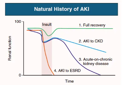
pre-existing CKD. (Fig.2)
Recent studies have associated AKI survival to subsequent CKD or end-stage renal
disease (ESRD) development. Chawla et al [35] assessed 11,589 patients for AKI and
evaluated the risk for CKD progression in AKI patients. A total of 5351 patients had AKI
of which14% (728) entered stage 4 CKD. Patients with more severe AKI especially those
requiring RRT and subsequently recovered were at higher risk for progression to CKD.
Acute Renal Failure Trials Network (ATN) study of intensity of dialytic support only
53% of surviving patients had complete or partial recovery of renal function by 28 days
while by day 60 only 16% could be discharged off dialysis [36]. It is possible that the renal
recovery in AKI survivors is dependent on a number of factors including the underlying
12
Fig .2: Renal outcomes of AKI (Amdur R etal KI 2009)
A large study by Ishani et al in 2009 in U.S. cohort of medical beneficiaries (age ≥ 67
years) without prior evidence of CKD found that an AKI episode resulted in an eightfold
increased risk of CKD G5.37 To estimate the risk of progressive CKD in known CKD
population Hsu et al studied patients with preadmission baseline eGFR < 45 ml/min/1.73
m2 and found that those who developed an episode of AKI requiring dialysis had very
high risk of developing CKD 5G[38].Another U.S. study evaluated the risk of progressive
CKD after AKI in patients with a baseline eGFR more than 45 ml/min/1.73 m2 [39].
Patients who required dialytic support for AKI but became dialysis independent within 30
days of discharge had a 28-fold increased risk of progressing to CKD G4 or worse.
Coca SG et al in 2012 did a meta-analysis of 13 cohort AKI studies and reported
13
and 8.6 per 100 person-years, respectively [40]. Pooled adjusted relative risks in these AKI
patients for subsequent CKD and CKD G5 compared to those without AKI were 8.8
(95% CI 3.1-25.5) and 3.1 (95% CI 1.9- 5.0) respectively. Depending on the severity of
AKI older adults have 2-13 times the risk for development of ESRD after a single
episode of AKI. Also after a single AKI episode the annual absolute risk of CKD G5
ranges from 0.6% to 1.2% in those who experience mild AKI and up to 9% in those with
preexisting CKD [41].
Age, unmeasured comorbidity and fraility might be confounding factors for the
association between AKI and subsequent CKD. However CKD development has been
linked to AKI in pediatric patients who are generally not affected by these confounding
factors. In this regard126 critically ill pediatric AKI survivors who had completely
recovered their kidney function were studied [42] .This study revealed that overall 10%
patients developed CKD over 3 years of follow up (albumin to creatinine ratio less than
or equal to 30mg/g or eGFR less than 60ml /min /m2) .Also it is noteworthy that 38% of
cohort had mildly decreased eGFR 60-90 ml/min and 3.2% had hypertension both being
risk factors for future CKD)
Link between AKI and CKD: Incomplete repair or pathological transition of the repair process can result in renal non recovery and progression to CKD. Nath et al [43] in his
work on rats showed that repeated cutaneous glycerol injection would result in recurrent
AKI with reversible decrease in creatinine clearance but subsequently it led to slow loss
14
interstitial fibrosis. In ischemia/ reperfusion and cisplatin induced AKI in rats chronic
reductions in concentrating ability have been observed even after single episode of insult
[44,45] .Following a single bout of ischemia reperfusion in rats the proximal tubule repair
usually is complete by 4-6 weeks while increased expression of TGF β also normalizes in
4 weeks .In case where the repair process is delayed to 16-40 weeks ,features of CKD
including proteinuria, interstitial fibrosis manifest along with secondary increase in TGF
β [45].
In case AKI occurs in the presence of reduced renal mass there are greater chances
of incomplete repair and progression to CKD [38] . Patgulanan et al. did morphometric
analysis of ultra thin serial sections in AKI patients to show that an AKI episode resulted
in nephron dropout which was presumed to be due to loss of contact between the tubule
and the parent glomerulus [46]. AKI in reduced renal mass models has been associated
with dilated tubules which contain differentiated cells expressing profibrotic factors like
Platelet Derived Growth Factor which may initially help in physiological repair, however
with sustained expression may cause fibrosis [47].Also renal vascular epithelium repair
seems to be reduced as compared to tubular epithelium as capillary microfill experiments
show a reduction in capillary density of 30-50% most pronounced in inner stripe of outer
medulla following recovery from AKI[45].The role of inflammatory cells and immune
system in progression to CKD has been demonstrated in several studies. Forbes et al,
showed that blockade of ED-1 positive macrophages by treating with an endothelin
antagonist early decreased the late complications of acute kidney injury [48]. The role for
T cells in CKD development was demonstrated by Chandrakar et al, who showed that
15
reduced renal mass, resulted in a substantial decrease in progressive proteinuria and
interstitial fibrosis with recovery occurring in up to 6 months [49]. Burne-Taney et al
demonstrated that splenocytes from injured mice transferred to non injured recipients
promoted proteinuria within 12 weeks of injury, suggesting the role of immune responses
in CKD progression [50]. Finally, in a study by Pechman et al, it was shown that blocking
the lymphocyte function with mycophenolate mofetil from 5 weeks after recovery from
I/R injury prevented the building up of hypertension and interstitial fibrosis associated
with elevated sodium intake [51]
PATHOPHYSIOLOGY OFACUTE KIDNEY INJURY:
Pathophysiologic classification of AKI broadly divides it into pre-renal, intrinsic renal
and post renal mechanisms. [52] While post renal AKI accounts for about 10% of cases
secondary to intrinsic or extrinsic obstruction the remainder is taken care of by Pre-renal
and intrinsic renal mechanisms. Post renal AKI is characterized by acute obstruction to
urinary flow and rise in intratubular pressures and decrease in glomerular filtration rate
(GFR). It can also cause reduction in renal blood flow as well as trigger inflammatory
processes resulting in further reduction in GFR [53, 54]. Pre-renal AKI occurs secondary to
a reduction in renal perfusion and a fall in Glomerular filtration rate thus resulting in a
‗functional‘ decline without any histopathological evidence of renal injury [55].
The
various causes of such decline include decreased cardiac output ,systemic vasodilation
16
Since the tubular function is maintained hence conservation of Sodium and water results
to maintain intravascular volume and renal perfusion in low urinary sodium and high
urine osmolarities [56].
As compared to the pre-renal AKI, intrinsic AKI may affect different compartments of
the kidney which include the glomeruli, tubules, interstitium and the vascular
compartment. Hence it may manifest in acute tubular necrosis or intratubular obstruction,
acute interstitial nephritis ,acute glomerulonephritis or may involve the renal
micro/macrovasculature causing thrombotic microangiopathies and vascular
thrombosis/embolism respectively.
Most cases of hospital acquired AKI are secondary to Acute tubular necrosis (ATN)
which occurs due to ischemic or nephrotoxic insult. The etiology of AKI in the ICU is
again a combination of decreased renal perfusion, sepsis and nephrotoxic agents resulting
in prerenal injury to frank ATN. The S3 segment of proximal tubule followed by the
medullary thick ascending limb of loop of Henle have been considered to be the most
important sites of injury in ATN. This is due to the chronic low Oxygen concentration in
the medullary region as well as high metabolic rates [57].
Clinically ATN can be divided into initiation, extension, maintenance, and recovery
phases. The initiation phase starts with reduction in renal blood flow severe enough to
cause ATP depletion and cellular injury. This sublethal injury of tubular cells results in
disruption of the filamentous actin architecture [58] and translocation of the
17
is decreased salt and water retention in the proximal tubule which activates the
tubuloglomerular feedback. Recent evidence shows that there may be activation of
epithelial and possibly endothelial cells resulting in upregulation of inflammatory
cytokines [59].
The extension phase is characterized by continued hypoxia and inflammatory response
both of which are predominant at the corticomedullary junction and outer medulla. The
cells in the outer medulla predominantly continue to undergo injury and death via
necrosis and apoptosis [60] .Cellular detachment leads to intraluminal cast formation and
obstruction resulting in further fall in GFR. There is further amplification of the
inflammatory cascade due to production of chemokines and cytokines [61].Fig .3 shows
the interplay between inflammation and tubular and vascular injury in the extension
phase of ATN.
During the maintenance phase to maintain tubular integrity the cells undergo
proliferation, migration, apoptosis and repair with the GFR remaining stable at a level
determined by the severity of initial insult. Lin et al. used a transgenic cre-lox approach
to label renal tubular epithelial cells and track their progeny following I/R injury. They
were able to demonstrate that Bromodeoxyuridine (BrdU) localized in Cre-induced
transgene expressing dedifferentiated cells signifying that the source of regenerating cells
could be resident, non lethally injured tubular epithelial cells [62].Other groups have also
confirmed that the primary source of regenerating new cells in repair is sublethally
18
phase characterized by increased cell proliferation , restoration of polarity and return of
organ function [64].
Fig.3: Extension Phase of AKI
The hemodynamic changes in ATN consist of sustained increase in renal vascular
resistance caused by number of mediators including increased sympathetic nervous
system activity [65], ,endothelin [66], Platelet activated factor[67], adenosine as well as direct
ISCHEMIA
TUBULAR CELL INJURY
LETHAL SUBLETHAL
1. Apoptosis Cytoskeleton
2. Necrosis disruption
Loss of Cell Polarity + Cell Shedding
Altered Vectorial transport Tubular Obstruction
Backleak
DECREASED GFR
INFLAMMATION
MICROVASCULAR
INJURY
IMPAIRED FLOW
↑
Leukocyte Adhesion
↑ Permeability
19
injury to vessel walls leading to decreased vascular responsiveness. In fact alteration in
vascular responsiveness has been shown to occur even one week after recovery from
Ischemia/Reperfusion injury when total renal blood flow has returned to normal[68].
Role of inflammatory cells and cytokines: A variety of cells including neutrophils , T cells, macrophages and dendritic cells have been implicated to play important role in the
inflammatory cascade in AKI. While the role of neutrophils is unclear as they are not
prominent in biopsies of patients with AKI, recent evidence suggests that T cells may
play important role in ATN. While T cells are found in the medullary vasa recta region in
animal models of AKI and also in biopsies of patients with AKI [69, 70] , there role in
pathogenesis has been supported by studies which have demonstrated that depleting the T
cells using the antibody neutralizing [71] or genetic approach [72] results in reducing the I/R
injury. However the fact that CD4+ T cells are cells of antigen specific adaptive
immunity and require 2-4 days of processing puts their role in ATN to question.
However, natural killer T (NKT) cells express both the TCR (T cell receptor) and the
marker NK1.1 thus representing a unique smaller subset of CD4+ cells. These cells are
capable of producing large amounts of cytokines both Th1 type such as Interferon γ
(IFNγ) and Tumor Necrosis Factor (TNF) or Th2 type like IL-4 and IL13. In fact , NKT
producing IFNγ are present within 3 hours in the post-I/R kidney injury [73]. Blockade of
activation of NKT cell with the anti-CD1d mAb or NKT cell depletion with anti-NK1.1
mAb in wild-type mice, or use of iNKT cell deficient mice (Jα18−/−) inhibits the
20
Macrophages are found to infiltrate the injured kidney within 1 hour of ischemia
reperfusion in response to expression of fractalkine (CX3CL1) by injured endothelial
cells which is a potent chemoattractant and adhesion molecule for them . Blockade of
CX3C receptor-1 on macrophages by specific antibody reduces the severity AKI in mice
[74]
. In the setting of Acute kidney injury, macrophages are most abundant during the
repair phase with a distinct population, referred to as M2 macrophages being active .
These M2 macrophages secrete anti inflammatory cytokines such as IL-10 and TGF-β
and may help in tissue repair by secreting potential trophic growth factors and
angiogenic factors [75,76]. Wang et al., demonstrated that ex vivo programming of
macrophages to an M2 phenotype would reduce chronic renal inflammation [77].Also
administration of macrophages during the repair phase of ATN hastens recovery
[78]
.Dendritic cell activation has been suggested to lead to TNF α production in ischemic
AKI [79], but there are few functional studies to support it. That dendritic cells may have a
protective role was supported by a study which showed that ablation of CD11c +
Dendritic cells increased the sensitivity to cisplatin induced injury [80].
Both tubular cells and leukocytes produce a number of inflammatory mediators to
promote postitive feedback loop of inflammation producing further kidney injury.
Tubular epithelial cells can produce IL-1, IL-6, IL-8, TNF-α, Monocyte Chemotactant
Protein 1 (MCP-1) ,Transforming Growth factor β (TGF-β), ENA78, RANTES and
fractalkines while leukocytes may produce IL-1, IL8, MCP-1, reactive oxygen species
21
IL1β is a chemotactic factor that recruits leukocytes to injured areas.The blockade
of IL-1β decreases the infiltration of neutrophils following ischemic injury, but does not
improve the consequent loss of renal function [82]. IL-18 is increased in AKI and is
considered an early biomarker of acute tubular injury [83].Injured proximal tubules
activate the expression of IL-6 on infiltrating macrophages and IL6 is elevated in mice
following AKI [84]. Other systems which have been implicated in AKI are the Toll like
receptors (TLR‘s) and complement system. In fact TLR 4 is expressed on capillaries of
vasa recta within 4 hours of ischemia reperfusion injury with secondary increases
occurring within 24 hours on the proximal tubular cells [85].There is evidence that
alternate pathway of complement may be active during ischemia reperfusion [86] with C3a
and C5a contributing to injury with activation of apoptosis and impairment of recovery
responses.
Repair and Regeneration:
Renal stress respone: Acute kidney injury activates cytoprotective pathways which can be broadly divided in three broad categories:
1.Heme Oxygenase (Inducible HO) and anti-oxidant genes: Heme Oxygenase is the rate limiting step in the heme catabolism producing biliverdin, iron, and carbon monoxide
(CO).The enzyme occurs in two isoforms : the inducible HO1 and the constitutive
HO2[87].In animal studies HO1 is rapidly induced following nephrotoxic [88],
glycerol-rhabdomyolysis [89], and ischemia/reperfusion [90] injury.HO-1 reduces oxidant stress by
22
peroxy radical scavenger that prevents lipid peroxidation. Besides CO can have potential
vasodilatory effect resulting in improvement of renal blood flow. Mice with a HO-1 null
mutation suffer from more severe AKI and have increased mortality in ischemia
reperfusion [91], glycerol [89], and LPS [92] AKI models while induction of HO-1
expression by viral delivery of HO-1 gene or by intravenous infusion of hemoglobin
prior to injury protects from AKI [93, 94].
2. Heat shock proteins (HSP): They belong to multigene protein family ranging in size from 10–150 kDa and are found in all major cellular compartments [95].The protective
properties of HSPs is widely ascribed to their activity as "molecular chaperones," which
are considered to aid in the assembly and repair of newly synthesized or degenerated
proteins [96]. An important role of HSPs is linked to the stabilization and revival of
cytoskeletal structure following AKI. During recovery after ischemia/reperfusion , the
Na, K, ATPase gets reincorporated in the cytoskeleton which is aided by HSP 70 [97].
Emami et al in one of the earliest reports suggested the role of heat shock proteins in
protection from acute kidney injury. Exposure to transient minimal ischemia of 15 min
resulted in upregulation of heat shock protein72 (HSP72), which resulted in protection
from later ischemia/reperfusion injury [98].Since then many more members HSP family
have been found. It is now clear that not only HSP 72, but also other members including
HSP10, HSP 25/27, HSP47, HSP70 , HSP90 as well as αβ-crystallin play important role
in diverse models of AKI including ischemia reperfusion, cisplatin toxicity and unilateral
23
HSP-chaperone activity may also aid in cytoprotection by manipulation of the
biochemical pathways leading toward cell death i.e. apoptosis or necrosis [102]. Heat
Shock Proteins may also be having role as cellular antioxidants. In MDCK cells exposed
to oxidant stress, HSP-70 controlled the superoxide production by increasing the activity
of enzymes glutathione reductase and glutathione peroxidase [103].
Recently, Kim et al., confirmed that increased expression of HSP-27 in proximal tubules
conferred protection from ischemic AKI [104]. Also higher basal levels of HSP-72 and
HSP-27 have been reported in Brown Norway rats which are more resistant to AKI than
the commonly utilized Sprague Dawley rats [105] .
3. Stress activated protein kinases : Cellular stress results in activation of two related parts of the mitogen-activated protein kinase (MAPKs) signaling pathway- the
extracellular-regulated protein kinases (ERKs), and the stress-activated protein kinase
(SAPK), also identified as Jun N-terminal kinase (JNK and p38).Both of these kinases
are activated in response to tubular stress /AKI [106] .Following ischemic injury JNK
activation occurs in both proximal and distal tubule [107]while its down regulation has
been shown to reduce the peroxide induced proximal tubular injury [108]. Inhibition of
ERK activity in proximal tubules of opossum has been shown to block apoptosis
associated with cisplatin nephrotoxicity [109].
Regeneration:
The restoration of structure and function of proximal tubule following renal injury
requires complex and coordinated cellular activity and not just proliferation [110] . The
24
markers and express vimentin [111]. They rapidly realign the denuded proximal tubule
basement membrane, undergo hyperplasia and differentiation with a decrease in cellular
number per cross sectional area towards normal. Concomitantly there is a wave of
apoptosis that restores tubular cell density to normal and may last from between one
week to several months of recovery [112].
Proximal tubule cells can proliferate on exposure to a variety of mitogens like
insulin-like growth factor-I (IGF-1) [113], epidermal growth factor (EGF) [114], hepatocyte
growth factor (HGF) [115] , and fibroblast growth factor (FGFs) [116].However not all of
them have been shown to play important roles in the regeneration process. While IGF 1
has been shown to be expressed in regenerating tubular cells [117] and HGF levels are
increased in urine of AKI patients [118], there is no definite evidence of their role in renal
repair. Recently Bone Morphogenic Protein 7 (BMP 7) has been found to have significant
potential of stimulating tubular cell proliferation and also acting as anti- fibrotic agent in
CKD. Exogenously administered BMP 7 helps in early recovery from AKI [119].
EGF is expression is decreased in multiple forms of AKI [120], still it is expressed and may
help in renal repair. Also different members of FGF family are increased after the
induction of ischemic and nephrotoxic injury.
Discovered by Serhan et al [121] Resolvins (Rv) and protectins (PD) are two novel families
of n–3 fatty acid docosahexaenoic acid metabolites shown to play important role in
resolution of AKI. Mouse kidneys have been shown to produce D series resolvins (RvDs)
25
to and after the injury has been shown to have protective effect on kidney by decreasing
inflammation as well as it reduces the resulting interstitial fibrosis.
BIO MARKERS OF ACUTE KIDNEY INJURY:
Biomarker-Definition: As per the NIH Biomarkers Definitions Working Group, a biological marker (biomarker) is defined as ―A characteristic that is measured objectively
and is evaluated as an indicator of normal biological processes, pathogenic processes or
pharmacologic responses to a therapeutic intervention.‖[123]
Characteristics of Ideal Biomarker: Althoughit is impossible to find a biomarker with all the characteristics as mentioned below still an approximation of them is desired
[123,124]
.
(1)It should be non invasive and easily measured, inexpensive and produce quick results;
(2) It should be tested from easily available sources, such as blood or urine;
(3) The biomarker should have a high sensitivity, allowing for early detection of disease,
without overlap between diseased patients and healthy controls;
(4) It should have a high specificity, being greatly affected in the diseased samples
specifically and not by comorbid conditions;
(5) The levels of biomarker should change quickly in response to treatment;
(6) Biomarker levels should help in risk stratification and provide prognostic information
regarding outcomes;
(7) It should be biologically plausible and should help to understand the disease
26
The receiver operating characteristic (ROC) curve depends on both sensitivity and
specificity and the area under the ROC curve (AUC) for a biomarker at specific cut off is
a good measure of its validity. While an AUC of 1 means a perfect biomarker but it is not
practically possible.An AUC of 0.75 represents a good biomarker while that of 0.9 is
considered excellent for all practical purposes [124].
Need for Biomarker in AKI: While serum creatinine and urine output have been established markers of renal damage since long time, serum creatinine cannot be
considered as a sensitive/specific marker for AKI. This is because of the presence of a
‗Window period‘ of 8- 48 hours when AKI continues without manifesting as increase in
serum creatinine which occurs later only[125] .In fact serum creatinine may show upward
trend only once about 50% of renal function has been lost .Also the production of
creatinine from muscle is decreased in AKI due to sepsis, thus rise in creatinine may not
be according to decline in GFR [126].Besides several non-renal factors can affect
creatinine levels irrespective of renal function - age, gender, race, nutritional status,
muscle mass, infection, total parenteral nutrition and certain drugs. In fact trimethoprim,
cimetidine and salicylates are known to alter the tubular secretion of creatinine causing
changes in its level independent of GFR [127].Fig.4 shows the window period where novel
biomarkers may have a potential role in early diagnosis and intervention.
Novel Biomarkers of AKI
27
has been implicated in the pathogenesis of a number of conditions including arthritis,
ischemic AKI, Acute coronary syndrome and others. Caspase 1 is the enzyme which
activates both IL-18 and IL-1 β [128]. In freshly isolated mice proximal tubules it was
shown that hypoxic tubules expressed high levels of IL-18 [129].Thereafter another
study demonstrated increased urine IL 18 in mice with ischemic AKI than sham operated
controls [128].Subsequently urine IL 18 was found to be raised in humans with ATN than
normal controls, patients with prerenal azotemia, urinary tract infection nephrotic
syndrome ,and chronic kidney disease.[130]
A nested case control study was performed within the ARDS network trial to determine
the utility of IL-18 in predicting AKI in ICU patients. Urine IL- 18 had an area under the
receiver operated characteristic (ROC)curve of 0.73 as a predictor of AKI in the next 24
hours thus establishing as a good biomarker [131]. In a pediatric cohort of 137 children
with an average age of 6.5 years (47% female) the peak IL-18 levels significantly
correlated with the severity of AKI as defined by the pediatric RIFLE (pRIFLE)
classification. Urinary IL-18 rose 2 days prior to significant increase in serum creatinine
in critically ill non septic AKI patients and were independent predictor of mortality.
Besides they were also increased in sepsis [132].In 55 children who underwent
cardiopulmonary bypass urinary IL-18 started increasing at 4-6 hours of procedure in
those who developed AKI. It peaked to almost 25 times at 12 hours and consistently
remained elevated upto 48 hours post procedure. In comparison serum creatinine
28
Haiyan he et al studied IL-18 in 180 patients who underwent coronary intervention and
received low osmolal contrast. Contrast induced nephropathy (CIN) as defined by
increase in serum creatinine by 0.5 mg% or ≥25% increase over baseline within 24-48
hours of procedure occurred in 16 (8.9%) patients.IL-18 levels increased in these patients
even after 2 hours of procedure though not significantly , but the rise became significant
at 6,12,24 and 48 hours as compared to pre procedural levels. Serum creatinine did not
show significant difference between pre procedure values and 24 hours after procedure.
The area under the ROC curve for IL-18 for predicting CIN was 0.811. [134]
Recently in a meta-analysis of 18 studies, IL-18 level predicted AKI with a sensitivity
and specificity of 0.58 and 0.75, respectively. Also the area under the ROC curve for
29
Fig. 4: Timing Diagram of Acute Kidney injury (From Ref. 125)
2. Kidney Injury Molecule 1:
Kidney injury molecule 1 (KIM 1) is a recognized epithelial cell adhesion molecule
which contains an immunoglobulin domain. In fact normal kidney expresses KIM 1
mRNA and protein at a low level in but its levels increase rapidly in ischemic AKI
[136]
Urinary KIM 1 have been showed to be elevated significantly in both cisplatin
30
and chronic kidney diseases, and eight normal controls urinary KIM 1 levels were
significantly higher in patients with ischemic ATN as compared to other
patients/controls.In all six patients with renal biopsy proven ATN there was widespread
expression of KIM 1 in proximal tubule cells [138] . In a study involving 90 patients
undergoing cardiac surgery ,urinary KIM 1, N acetyl beta (D) glucosaminidase (NAG)
and Neutrophil gelatinase associated lipocalin (NGAL) were measured immediately and
3 hours post surgery. Thirty six patients (40%) had AKI within 72 h of surgery. The area
under ROC curve for KIM 1 to predict AKI immediately and 3 h after surgery was 0.68
and 0.65 respectively. A combination of the three biomarkers, KIM 1, NAG and NGAL,
improved the sensitivity for early detection of AKI at 0 and 3 hours of procedure to 0.75
and 0.78 respectively[139].
In a meta-analysis of 11 studies involving a total of 2979 patients, urinary KIM-1
had a sensitivity of 74.0% and a specificity of 86.0% for the diagnosis of AKI. The area
under the ROC curve for KIM 1 to predict AKI was 0.86 (95% CI, 0.83–0.89). [140]
3. Cystatin C:Cystatin C is produced by all nucleated cells of the body .It is a polypeptide containing 120 amino acid residues .Its completely reabsorbed in the
proximal tubules and is not secreted anywhere in the nephron [141].
In 127 patients undergoing cardiac catheterization, Artunc et al studied serum creatinine,
serum cystatin C and the clearance of the iodinated contrast iopromide (reference
standard for GFR ). Serum cystatin C had a higher correlation (0.805) to the iopromide
31
than 1.3 mg/L confirmed renal failure with a sensitivity of 88%and 96% specificity as
defined by an iopromide clearance of less than 80 mL/min/m2.[142]
Rosenthal studied cystatin C and creatinine in live renal donors before and after
uninephrectomy. Serum cystatin C rose 1 day after uninephrectomy as compared to
serum creatinine that increased after 2 days of procedure[143] . Orlando et al studied
Inulin, serum cystatin C and creatinine clearances in 36 patients with decompensated
cirrhosis and 56 non cirrhotic controls .Plasma cystatin C and not serum creatinine or
creatinine clearance was an accurate marker of GFR in the cirrhotic patients. In fact
plasma creatinine and calculated creatinine clearance, varied with the severity of the
liver disease and did not correlate with GFR[144].
Cystatin C was compared to creatinine in detecting AKI as defined by the the RIFLE
classification in 85 high risk patients .Serum cystatin C rose by more than 50% at an
average of 0.6 days earlier than the increase in serum creatinine thus concluding that
serum cystatin C is a better marker for the detection of AKI than creatinine [145].To study
the use of urinary cystatin C for the early identification of AKI, plasma and urine samples
were collected from 72 adults undergoing elective cardiac surgery [146]. While plasma
NGAL and cystatin C levels did not predict AKI within the first 6 h of procedure but
urinary cystatin C and NGAL were elevated in 34 patients who later developed AKI,
compared to patients with no AKI. With urinary Cystatin C being the most useful at 6
32
Thus it is possible that Cystatin C may be an ideal endogenous marker as all nucleated
cells produce it at a constant rate, its levels are unaffected by changes in height, gender,
age and muscle mass, nutrition and is neither secreted nordegraded by renal tubules. In
fact in Germany in 2002 ,at a multinational meeting it was concluded that cystatin C was
equal if not superior marker of GFR than creatinine. It may actually be of benefit to use it
as a marker in children ,elderly people and reduced muscle mass patients [147].
4.Liver fatty acid binding protein (L-FABP): It acts as a carrier protein for fatty acids and other lipophilic substances like ecosanoids and retinoids .Some of the FABPs help in
transportation of lipophilic molecules from outer cell membrane to intracellular receptors
like PPAR and also in transportation of fatty acids between extra and intracellular
membrane.
In a model of Folic acid induced kidney injury in mice L FABP levels in both kidney and
urine was found to have a major correlation with the degree of tubulointerstitial damage
[148]
.In live related human renal transplants ,renal peritubular blood flow and ischemia
time had a significant correlation with the urinary L-FABP levels [149].
40 pediatric patients were assessed for urine L-FABP levels just before undergoing
cardio-pulmonary bypass surgery .Following surgery 21 patients developed AKI .
Enzyme linked immunosorbent assay analysis in these patients revealed increased
L-FABP levels of about 94 and 45 fold at 4 hrs and 12hrs, respectively which was
confirmed by western blot analysis. This study showed urine L-FABP to be a sensitive
33
Further in a study of 80 critically ill patients it was shown that urine L-FABP levels were
higher in patients with septic shock as compared to patients with severe sepsis alone [151].
To summarise , urine L-FABP is increased in patients with AKI. Also, it is the urinary
LFABP levels and not serum L-FABP levels that is increased in patients with septic
shock. L-FABP is also a biomarker for progression of chronic kidney disease.
5. IL-6 and IL-8:IL6 which is a proinflammatory cytokine plays an importrant role in immune response and haematopoesis .It is basically found in endothelium , macrophages
and fibroblast . IL8 is also a proinflammatory cytokine .It functions by activating and
recruiting the neutrophils .In a mouse model of AKI IL8, IL6 and IL12 levels were
significantly raised early after onset of ischaemia. Serum and urine levels of IL 8 were
highest after 3 hour of induction of ischemia and before occurrence of significant rise in
serum creatinine. There was a significant fall in the levels of IL1 , IL2,IL4 and IFN γ
after AKI while IL 1b, IL 3, IL 5, IL 10, IL 12 (p70), IL 17, GM CSF and RANTES did
not show any change as compared to no AKI group [152] .
In renal allograft recipients, urine IL 8 was found to be markedly elevated in patients who
had delayed graft function as compared to patients with immediate graft function [153].
In the PROWESS (Prospective Recombinant Human Activated Protein-C Worldwide
Evaluation in Severe Sepsis) trial, of the 547 patients from the placebo group 127
developed AKI . In a multivariable Cox regression model log IL 6 and APACHE II score
34
NEUTROPHIL GELATINASE ASSOCIATED LIPOCALIN (NGAL):
NGAL, also known as Lipocalin 2/Siderocalin is a 25 kDa glycosylated polypeptide
member of lipocalin superfamily of proteins. Human NGAL is made up of 178 amino
acid residues linked by a single disulfide bond and can exist as monomer with molecular
weight (MW) of 25kDa ,homodimer with MW of 35kDa or as heterodimer complexed
with Matrix Metalloproteinase 9 (MMP 9) [155]. In neutrophils it is present chiefly as
homodimer and small percent occurs as monomer or heterodimer while renal epithelium
secretes mainly monomers when under stress. [156,157].This is supported by the observation
that patients with urinary tract infection have raised homodimeric NGAL while those
with AKI have predominant monomeric molecule [157,158].
NGAL has capacity to bind siderophores and thus is involved in iron traffic to and
fro between cells. Because iron is important for bacterial proliferation and can also cause
tissue injury by free radical generation , hence by sequestering iron NGAL can act as
bacteriostatic agent as well as protect from inflammation associated injury [159,160].It has
also been shown to promote iron dependant differentiation of mesenchymal progenitors
into nephrons during renal development [161].
NGAL is expressed at very low concentrations in other human tissue like trachea ,colon,
lung, kidney etc. and its levels increase in stimulated epithelia due to inflammation [156].It
is also expressed in adenomas and inflamed bowel epithelium[162], adenocarcinoma
35
degradation NGAL is freely excreted in the urine both in free and complex form with
MMP 9.Thus urinary levels correlate with plasma levels, but specifically high NGAL
levels in urine can be found when released from ischemic tubules [165]. While both urinary
and plasma NGAL have been shown to be elevated in different forms of AKI including
post cardio-pulmonary bypass, Contrast induced Nephropathy, critically ill patients etc.
urinary NGAL may be better in characterizing AKI as has been suggested by study by
Mårtensson, Bell et al. They showed that mean plasma NGAL levels were more elevated
in patients with sepsis than without sepsis irrespective of GFR. They studied peak levels
of plasma NGAL (a) and urinary NGAL (b) in non-AKI patients (N=27) with SIRS, severe sepsis, and septic shock and in AKI patients with septic shock (N=18).Plasma
NGAL (pNGAL) were elevated in all four groups as shown in Fig.5 and was not
significantly different between Septic shock with and without AKI groups. Urinary
NGAL (uNGAL) was less confounded by sepsis being significantly elevated in AKI
patients with septic shock .It was good predictor of AKI in next 12 hours (AUC ROC of
36
Fig.5 Urinary NGAL Vs. Plasma NGAL (from Reference 166)
Study by Mishra et al showed that NGAL mRNA and protein was produced in cultured
human proximal tubule cells within 1 hour of ATP depletion due to Ischemia/reperfusion
injury. This confirmed that the source of urinary NGAL was kidney proximal tubule cells
and not merely activated neutrophils which are found to accumulate in ischemic AKI at
around 4 hours and peak at 12 hours of insult. Also these neutrophils have not been found
to express the NGAL mRNA in the renal tissue [167].
A large number of studies have analysed the utility of both plasma and urinary NGAL in
post Cardiopulmonary bypass AKI in both children and adults. In children urine and
plasma NGAL at 2 hours of surgery predicted AKI and plasma NGAL was also
associated with the duration of AKI and mortality [168, 169].Similarly in adults mean
urinary NGAL levels were significantly elevated at 3 and 18 hours after cardiac surgery
37
patients after cardiac surgery and found it to be independent predictor of AKI ,its
duration and severity as well as length of ICU stay [171,172].
In studies of Contrast induced nephropathy in both children and adults uNGAL and
pNGAL significantly rose much earlier i.e. 2-4 hours of cardiac catheterization than
serum creatinine which was found to increase at only 6-24 hours of procedure [173,174].
Urinary NGAL was studied in 140 critically ill children who were on mechanical
ventilation. The levels of uNGAL rose 48 hours before a rise in creatinine of 50% or
more occurred and they correlated well with worsening Pediatric RIFLE class. The area
under the ROC curve for development of AKI for uNGAL was 0.78 while its levels were
also predictors for AKI duration of more than 48 hours [175]. Hilde R et al studied 632
ICU patients of which 171 (27%) developed AKI. Plasma and urinary NGAL at
admission were significantly related to severity of Aki as determined by RIFLE scores.
Also NGAL was an independent predictor of RIFLE ‗F‘ class in multivariate logistic
regression model [176].
In a cohort of 635 patients admitted to the Emergency Department ,a single urinary
measurement of NGAL had high sensitivity and specificity in detecting AKI . In fact a
cutoff value of 130 μg/G creatinine had a sensitivity of 90% and specificity of 99.5% for
detecting AKI , which was much higher than N acetyl beta (D) glucosaminidase,
fractional excretion of sodium (FeNa) and serum creatinine. In regression analysis
uNGAL predicted requirement of dialysis and admission to ICU. Since only 6% of the
38
acting as a source of extrarenal NGAL and thus confounding measurements of uNGAL is
low [177].
Lately quite a few studies have investigated the role of NGAL in differentiating pre-renal
and intrinsic AKI and predicting its severity and outcome [178,179,180].Singer et al studied
147 hospitalized patients and found that a cutoff level of uNGAL of more than 104 ng/ml
predicted intrinsic AKI (Odds ratio 5.97) while a level of less than 47 ng/ml was in
favour of prerenal injury [1