“CYTOMORPHOL DIFFUSE THYROM
SYSTEM OF RE
D THE TAMIL
the
TIRUNEL
HOLOGICAL EVALUATION OF NODU YROMEGALY WITH EMPHASIS ON B OF REPORTING – A PROSPECTIVE S
DISSERTATION SUBMITTED TO
MILNADU DR.M.G.R. MEDICAL UNIVERS CHENNAI
in partial fulfilment of
the requirements for the degree of
M.D. (PATHOLOGY) BRANCH - III
UNELVELI MEDICAL COLLEGE HOSPITAL TIRUNELVELI
APRIL-2015
NODULAR AND ON BETHESDA
VE STUDY”
ERSITY
CERTIFICATE
This is to certify that this Dissertation entitled
“CYTOMORPHOLOGICAL EVALUATION OF NODULAR AND
DIFFUSE THYROMEGALY WITH EMPHASIS ON BETHESDA SYSTEM OF REPORTING – A PROSPECTIVE STUDY” is the bonafide original work of Dr.M.UMA DEVI, during the period of her Post graduate study from 2012 – 2015, under my guidance and supervision, in the Department
of Pathology Tirunelveli Medical College & Hospital, Tirunelveli, in partial
fulfillment of the requirement for M.D., (Branch III) in Pathology examination
of the Tamilnadu Dr.M.G.R Medical University will beheld in April 2015.
The DEAN
CERTIFICATE
I hereby certify that this dissertation entitled
“CYTOMORPHOLOGICAL EVALUATION OF NODULAR AND DIFFUSE THYROMEGALY WITH EMPHASIS ON BETHESDA SYSTEM OF REPORTING – A PROSPECTIVE STUDY ” is a record of work done by Dr. M.UMA DEVI, in the Department of Pathology, Tirunelveli Medical College, Tirunelveli, during her postgraduate degree course
period from 2012- 2015. This work has not formed the basis for previous award of any degree.
Dr.K.Swaminathan, M.D Dr.K.Shantaraman, M.D
Professor of Pathology, Professor and HOD of Pathology,
Department of Pathology, Department of Pathology, Tirunelveli Medical College, Tirunelveli Medical College,
DECLARATION
I solemnly declare that this dissertation titled
“CYTOMORPHOLOGICAL EVALUATION OF NODULAR AND DIFFUSE THYROMEGALY WITH EMPHASIS ON BETHESDA SYSTEM OF REPORTING – A PROSPECTIVE STUDY ” submitted by me for the degree of M.D, is the record work carried out by me during the
period of 2012-2015 under the guidance of Prof. Dr.K.SWAMINATHAN,
Professor of Pathology, Department of Pathology, Tirunelveli Medical College,
Tirunelveli. The dissertation is submitted to The Tamilnadu Dr. M.G.R.
Medical University, Chennai, towards the partial fulfilment of requirements for
the award of M.D. Degree (Branch III) Pathology examination to be held in
April 2015.
Place: Tirunelveli DR.M.UMA DEVI,
Date: Department of Pathology,
Tirunelveli MedicalCollege,
ACKNOWLEDGEMENT
I take immense pleasure at this opportunity to acknowledge all those who
have helped me to make this dissertation possible.
I express my heartfelt thanks to the Dean, Tirunelveli Medical College,
for permitting me to undertake this study. I express my profound sense of
gratitude to Dr.K.Shantaraman ,MD., my respected Professor and Head of Department of Pathology, Tirunelveli Medical College, Tirunelveli, for his
valuable advice, constant guidance and motivation in the preparation of this
work.
I consider it my privilege and honour to have worked under the unstinted
guidance, encouragement, and supervision of my respected guide
Dr.K.Swaminathan ,MD., Professor of Pathology.
I thank Dr.S.Vallimanalan MD, Dr.J.Suresh Durai, MD., Dr.Arasi Rajesh, MD., Professors of Pathology, for their constant support and inspiration. I also thank the Assistant Professors, for their encouragement and
support. I sincerely thank the Professors and faculties of the Department of
General Surgery for providing me the patients for my study. I take this
opportunity to thank all my postgraduate colleagues for their valuable support.
My thanks to all the technicians and other members of the Department of
Pathology for their kind help at different stages of this study.
LISTS OF ABBREVIATIONS USED
FNAC - Fine needle aspiration cytology
TBSRTC - The Bethesda system of reporting thyroid
cytopathology
ND /UNS - Non- diagnostic/Unsatisfactory
AUS/FLUS - Atypia of undetermined significance/ Follicular
lesion of undetermined significance
FN/SFN - Follicular neoplasm/ Suspicious for follicular
Neoplasm
SFM - Suspicious for malignancy
CAT –I - Category I
BFN - Benign follicular nodule
FNHCT - Follicular neoplasm of Hurthle cell type
SFNHCT - Suspicious for Follicular neoplasm of Hurthle cell type
HCN - Hurthle cell nodule
MEN - Multiple endocrine neoplasia
FMTC - Familial medullary thyroid carcinoma
MTC - Medullary thyroid carcinoma
CEA - Carcino embryonic antigen
TABLE OF CONTENTS
S.NO TITLES PAGE NO
1 INTRODUCTION 1
2 AIMS AND OBJECTIVES 3
3 REVIEW OF LITERATURE 4
4 MATERIALS AND METHODS 47
5 OBSERVATION AND RESULTS 49
6 DISCUSSION 87
7 SUMMARY 106
8 CONCLUSION 108
BIBLIOGRAPHY
APPENDIX
ABSTRACT
Swelling in thyroid are frequently encountered . Clinical evaluation helps
in diagnosis but is difficult to distinguish the early malignant lesions from the
more prevalent benign goiters. FNAC is a simple and safe procedure , carried
out in OPD with minimum equipment and has good patient compliance . As
FNAC is the primary investigation for the management of thyroid lesions , its
interpretation is very crucial . This study aims at classifying the
cytomorpholgical profile of nodular and diffuse thyromegaly cases with
emphasis on Bethesda system of reporting. The Bethesda System for Reporting
Thyroid Cytopathology ( TBSRTC) is used for clarity of communication and
recommends that each case should be reported in 1 of 6 diagnostic categories
which include Category I –Non –diagnostic , Category II- Benign, Category
III-Atypia of undetermined significance/ Follicular lesion of undetermined
significance (AUS/FLUS) ,Category IV- Follicular neoplasm / Suspicious for
follicular neoplasm (FN/SFN), Category V- Suspicious for malignancy (SFM),
CategoryVI – Malignant, thus facilitating a uniform communication among the
managing team of doctors and leaves no confusion regarding the management
of thyroid lesions.
A total of 300 cases with thyroid swellings were studied during
the period of January 2013 to June 2014 . FNAC of thyroid was done in all
and all cases were reported using TBSRTC. Of the total 300 cases , 3 cases(1%)
were reported in category I , 274 cases (91.3%) were reported in category II , 1
case(0.3%) in category III , 12 case ( 4%) in category IV , 2 cases (0.7%) in
category V, 8 cases (2.7%) in category VI. Major bulk of cases were seen in
category II.
Thus the application of TBSRTC bridges the gap in communication
between the clincians and helps in the proper patient management .
1
INTRODUCTION
Thyroid lesions are one of the common clinical conditions being encountered in clinical practice and are different from other diseases in terms of their ease of diagnosis, accessibility of medical treatment, and the relative visibility of the swelling that even a small swelling of the thyroid offers to the treating physician. Early diagnosis and treatment remains the cornerstone of management.
The annual incidence of thyroid malignancy ranges from 0.5 to 10 cases per 1,00,000 population.[1] With a wide spectrum of disorders and a considerable degree of variation in its presentation, it is often difficult to arrive at a correct diagnosis by clinical evaluation alone. So it is essential to have a battery of test done which include: thyroid hormonal profile, ultrasound imaging studies and Fine Needle Aspiration Cytology.
Fine Needle Aspiration Cytology is an outpatient procedure which is used as a primary investigation in the diagnosis of thyroid swellings. It is a simple, speedy, safe, cost effective and accurate technique being used worldwide for evaluation of thyroid swellings. The clinical value of thyroid FNAC is useful in the diagnosis of inflammatory, infectious and neoplastic conditions.[2]
2
The thyroid nodules have a reported prevalence of 4 to 7% in the general population.[3-6] Thyroid cancer is the most common type of endocrine malignancy. It constitutes 1.7% of all newly diagnosed cancer cases worldwide. The incidence of thyroid cancer is about three times higher among females than males.[7]
The incidence of thyroid cancer is rare in children, but its incidence begins to rise sharply in the second decade of life and peaks during the fifth and sixth decades of life. Papillary carcinoma of thyroid constitutes ~ 80% of all thyroid cancer cases, follicular carcinoma (15%), and medullary carcinoma (3%). Anaplastic carcinoma which accounts for < 2% of thyroid tumors, typically occurs in the older age group and its incidence continues to rise with age.[7]
3
AIMS AND OBJECTIVES
A prospective study is carried out on fine needle aspiration cytology of thyroid lesions with the following objectives.
1. To study the cytomorphological features of aspirated material from various thyroid lesions like nodular and diffuse thyromegaly.
2. To correlate the cytomorpholgical features and classify the thyroid lesions with emphasis on Bethesda system of reporting.
4
REVIEW OF LITERATURE
EMBRYOLOGY
The thyroid gland develops as an outpouching of the primitive foregut in early weeks of gestation at the site of foramen caecum in the base of the tongue. The endoderm cells which are seen lining the floor of pharyngeal anlage thicken and form the median thyroid anlage.The thyroid gland descends in the midline as a component of the thyroglossal duct to reach its final position in the mid neck. The second branchial arch forms the hyoid bone. The thyrogossal duct is situated anterior to the hyoid bone and is divided by the hyoid bone into a suprahyoid and an infrahyoid portion.In due course the thyroglossal duct is obliterated and leaves a vestige of pyramidal lobe in 40% of individuals. The paired lateral anlages originate from fourth branchial pouch and fuse with median anlage at fifth week of gestation.[8]
The lateral anlages are neuroectodermal in origin (ultimobranchial bodies) and contain the parafollicular or C cells which produce calcitonin [9] The cords and plates of thyroid follicular cells are formed by the end of the 9th week, the small follicular lumen is formed by 10th week, the colloid secretion occurs by 12th week and thyroglobulin positive colloid in lumen by 14th week.[10]
5
ANATOMY
Thyroid gland is placed in front of the neck, anterior to the trachea and composed of two lobes which are connected by isthmus. The two lobes of the thyroid are divided by fibrous septa into lobules, and each lobule contains about 30 to 40 follicles. The whole thyroid gland is enclosed in a true capsule. Mature thyroid gland measures about 6×3×2 cms approximately and weighs about 25-30 gms. Thyroid gland is a highly vascular structure and it is supplied by subclavian artey and carotid artery. There are four pea sized parathyroid glands embedded posteriorly in superior and inferior pole of thyroid gland.[8]
HISTOLOGY
6
PHYSIOLOGY
The main function of thyroid gland is secretion of thyroid hormones and calcitonin. Regulation of thyroid function is by the hormone TSH which is secreted from the anterior pituitary.The lesions in thyroid are largely divided into two major types: the lesions that show a diffuse pattern and the lesions that are associated with nodular pattern. The nodules in turn can be divided into solitary or multiple.
CLASSIFICATION OF THYROID SWELLING [12]
1. SIMPLE GOITRE(EUTHYROID)
Diffuse hyperplastic - (Physiological,Pubertal, Pregnancy) Multinodular goitre
2. TOXIC
Diffuse (Grave’s disease) Multinodular (Toxic adenoma) 3. INFLAMMATORY
Autoimmune - Hashimoto’s thyroiditis
- Classic lymphocytic thyroiditis Granulomatous - De Quervain’s thyroiditis Fibrosing - Riedel’s thyroiditis
Infective - Acute (bacterial thyroiditis, viral thyroiditis,) - Chronic (tuberculosis, syphilis)
7
NON –NEOPLASTIC LESIONS OF THYROID
DIFFUSE NON TOXIC (SIMPLE) GOITER OR COLLOID GOITER
The other names of this goitre include adenomatous goiter, diffuse or nodular goiter, endemic goiter, multinodular goitre. This type of goitre is usually caused by hyperplasia of the thyroid gland which is induced by iodine deficiency. In the early stages of this lesion there is a diffuse enlargement of the gland, which is made up of small follicles. In later stages, some of the follicles may become distended and may coalesce to form nodules ranging in various sizes. The degenerative changes, usually occur in the nodules such as haemorrhage, necrosis, cyst and scar formation. The colloid goitre is the most common lesion which is often referred for aspiration. The colloid appears as pink homogeneous material in microscopy and is usually surrounded by a few follicular cells.
GRAVE’S DISEASE [16]
8
INFLAMMATORY LESIONS
ACUTE THYROIDITIS
It can be suppurative or nonsuppurative.The etiology can be bacterial, fungal or it may be associated with radiation and is usually of infectious nature.[13]
HASHIMOTO’S THYROIDITIS
It is the most common cause of autoimmune thyroiditis reported by Hakaru Hashimoto in 1912. The other name of this thyroiditis is “ Struma lymphomatosa”. The etiology is mostly due to effect of autoantibodies that alter the thyroid function. The immune-mediated injury of the thyroid gland leads to diffuse or nodular enlargement of the gland and then leading to exhaustion atrophy which is then manifested by diffuse oxyphilia of the follicular epithelium. The etiological factors leading to autoimmune thyroiditis are of both humoral and cellular nature.There are circulating autoantibodies which are present against thyroglobulin and other follicular cell antigens, and other thyroid follicle receptors like thyrotropin (TSH) receptors. The main etiology of this type of thyroiditis is due to organ-specific defect in suppressor T lymphocytes. Therefore it is concluded that the etiology of autoimmune thyroiditis is multifactorial and is therefore of variable penetrance.[14]
9
thyroiditis may resemble an aspirate of a hyperplastic lymph node. There is striking, abundant, eosinophilic and granular cytoplasm and there is variability in nuclear sizes. The nuclei contains large nucleoli and contain large intranuclear cytoplasmic inclusions. Sometimes the follicular cells in clusters may mimic a papillary carcinoma.
The ‘lymphocytic’ type of HT is mostly seen in children and young adults. The smears show characteristic reactive lymphoid proliferation. The histiocytes have abundant amount of pale cytoplasm with granular chromatin. The florid type of HT occurs in older individuals who are hypothyroid.
The aspirate smear shows lymphocytes admixed with variable number of plasma cells. There is prominent oxyphilic change.
SUBACUTE GRANULOMATOUS THYROIDITIS (De QUERVAIN’S)
10
degenerating follicular cells, neutrophils, macrophages and lymphocytes in a dirty smear background that shows debris.
REIDEL’S THYROIDITIS
This type of thyroiditis presents with an extensive sclerosis of the thyroid gland. The sclerosis is sometimes also associated with a similar tissue reaction in the mediastinum, retroperitoneum, and orbit. The consistency of thyroid gland is rubbery or firm and the aspirations are very minimal due to extensive desmoplastic reaction in the thyroid stroma. Due to its firm nature this type of thyroiditis must be differentiated from infiltrating carcinoma. [2] [15]
TUMORS OF FOLLICULAR OR METAPLASTIC EPITHELIUM
FOLLICULAR ADENOMA
11
PAPILLARY CARCINOMA
They are the most common malignant tumors of the thyroid and usually presents as a palpable thyroid nodule. These malignancies are predominantly seen in females but sometimes may be seen in males and few cases also present in children.The smears are very cellular and the pattern of arrangement is predominantly in anastomosing papillary fragments but can be arranged as follicular structures, or in monolayered sheets generally free of colloid. The papillary pattern of arrangement is diagnostic of this tumor. It is common to find psammoma bodies in this cancer. The individual tumor cells, are usually larger. The cytoplasm is basophilic with sometimes discrete vacuoles can be seen. The nuclear features are very characteristics for this malignancy.The common nuclear features is the prescence of “ground-glass” nuclei and small central nucleoli. There is prescence of intranuclear cytoplasmic inclusions. Another characteristic nuclear feature is the prescence of nuclear grooves. The most important diagnostic feature is prescence of intracytoplasmic nuclear inclusions. The variants of papillary carcinoma include :
1. Cystic papillary carcinoma.
2. Follicular variant of papillary carcinoma. 3. Tall-cell variant of papillary carcinoma. 4. Warthin's-like variant of papillary carcinoma.
12
FOLLICULAR CARCINOMA
It is a rare neoplasm of thyroid showing invasion of capsule, blood vessels, or adjacent thyroid parenchyma. It also has predilection for women.The smears are cellular. Its microscopic appearance ranges from well formed follicles to solid growth pattern.[10] Thus, they may closely resemble the cytologic feature of follicular adenoma. In the well-differentiated form of follicular carcinoma, cellular atypia is minimal, and the smear may suggest a benign lesion. In such cases, the cytologic diagnosis should be “follicular neoplasm or tumor,” clearly indicating that surgical excision and histologic examination is mandatory for a confirmed diagnosis.
HURTHLE CELL (ONCOCYTIC) TUMORS
13
POORLY DIFFERENTIATED (INSULAR ) CARCINOMA
This tumor occurs in older age but can occur in adolescents also. This tumor shows a characteristic nesting (insular) pattern, small uniform tumor cells, variable mitotic activity, tumor necrosis resulting in a peritheliomatous pattern.[10]
UNDIFFERENTIATED CARCINOMA
This tumor most commonly presents in older individuals. Extrathyoidal extension is present in most cases. There are two forms of anaplastic carcinoma: a giant cell carcinoma and a small-cell-type carcinoma. [2] Smears of aspirates from the anaplastic giant cell carcinoma contain usually necrotic matter, cellular debris, inflammatory cells, mainly granulocytes, and large polymorphous, often multinucleated cells with large bizarre nuclei and very prominent nucleoli and in the small-cell variant of anaplastic carcinoma, the aspirate contains malignant cells with round or oval nuclei and scanty cytoplasm. [2] [10].
MEDULLARY CARCINOMA
14
LYMPHOMA
The primary lymphomas of thyroid,are of B-cell derivation, and is seen in patients with Hashimoto's thyroiditis.The smear contains lymphoma cells admixed with epithelial components.The majority of lymphomas arise primarily in the gland itself but may later involve lymph nodes and other organs.
METASTATIC TUMORS IN THYROID
The clinical presentation under this category can present as nodular or diffuse pattern. In nodular presentation the nodules can present as single or multiple or the metastasis can involve the whole gland and present in a diffuse pattern involving the entire gland. The metastases may sometimes mimic primary tumors of the thyroid. The distinction between a metastasis and primary thyroid tumor may sometimes be difficult. For example, in metastatic renal cell carcinoma cells may be elongated, or spindle in shape which mimics the cells seen in medullary carcinoma.
15
[TABLE 1]: Approximate percentage of various thyroid malignancies[1]
TYPE OF MALIGNANCY PERCENTAGE
Papillary carcinoma 70-80 % Follicular carcinoma 10-20% Medullary carcinoma 5-10% Anaplastic carcinoma 2-10% Poorly differentiated carcinoma 0.4-10%
Lymphoma 4-5 %
Expressed as the proportions of all thyroid malignancies
The above table shows the distribution of percentage of various thyroid malignancies encountered.
FINE NEEDLE ASPIRATION CYTOLOGY
It is one of the primary investigation used for the management of thyroid lesions, and its interpretation is very crucial for diagnosis[18].
HISTORICAL HIGHLIGHTS
16
lesions harboring malignancy.[22] Various modifications have been introduced to the Fine needle aspiration cytology technique. Santos JEC and Leiman G in 1988 pioneered a technique, non aspiration of fine needle cytology to study nodular lesions. This technique eliminates the active aspiration which is replaced by the principle of capillary suction of fluid or semisolid material into thin channel of the needle.[23]. Another development is the ultrasound guided FNAC which has proved to be beneficial in non - palpable nodules. Liquid based preparation is advocated as an adjunct to conventional smears. Its main advantages are reduction in number of slides to be screened, ability to perform immunocytochemical and other special stains and enhancement of nuclear details and irregularities of papillary carcinoma.
Orell has defined the main indications for thyroid FNAC, they are [24] 1. Diagnosis of diffuse non – toxic goitre.
2. Diagnosis of solitary thyroid nodule. 3. Confirmation of a malignancy.
4. To obtain material for defining prognostic parameters. ADVANTAGES OF FNAC
1. The technique is simple, safe, cost effective and can be done as an out patient procedure.
17
3. Representative sample is drawn, while maintaining steady suction in various areas of the nodule.
4. Cysts can be aspirated completely and thus it acts as a therapeutic procedure for the patient.
5. Its main purpose is to provide a rational approach to management. DISADVANTAGES OF FNAC
Minimal material available for examination. Architecture,cell relationship are absent.[25]
1. The needle may miss important areas of the nodule despite a good attempt for a representative smear by aspiration from different areas. 2. Distinction between a follicular adenoma and follicular carcinoma is
difficult.[26] COMPLICATIONS
FNAC is considered simple and safe procedure. It is rarely associated with complications as described below.[27]
1. Local discomfort. 2. Minor hematoma.
3. Puncturing carotid artery, internal jugular vein, which is very rare. 4. Seeding of tumour along the needle track, though theoretically
18
PITFALLS OF FNAC
1. Inadequate sampling : The aspirate may be cell poor as in Reidel’s thyroiditis and a second attempt becomes mandatory.
2. Geographical misses: In such cases prior ultrasound may delineate the area of interest.
The lack of a standardized reporting format has caused confusion and ambiguity in interpreting FNAC results. To address this need, the 2007 National Cancer Institute State of the Science Conference proposed a uniform classification scheme named Bethesda with 6 distinct diagnostic categories for classifying the various thyroid disorders.
THE BETHESDA SYSTEM OF THYROID CYTOPATHOLOGY
[TABLE-2]
The National cancer institute (NCI) hosted the State Of Science Conference on thyroid fine needle aspiration. The meeting took place on October 22 and 23, 2007, in Bethesda. The result of the meeting proposed a uniform classification system for identifying the thyroid lesions and named the classification as Bethesda system for reporting thyroid cytology which had six diagnostic categories as mentioned in [TABLE-2].[32]
19
[TABLE -2 ] : The Bethesda system of classification
CATEGORY I
Non -diagnostic/ Unsatisfactory
Cyst fluid only
Virtually Acellular smear
Others (Obscuring blood/Drying artefact)
CATEGORY II
Benign Consistent with benign follicular nodule
(Adenomatoid nodule, colloid nodule etc)
Consistent with lymphocytic (Hashimoto) thyroiditis in proper clinical context
Consistent with granulomatous (subacute ) thyroiditis
Other CATEGORY
III
Atypia of undetermined significance or follicular lesion of undetermined significance CATEGORY
IV
Follicular neoplasm/ suspiciousof
follicular neoplasm CATEGORY
V
Suspicious for
malignancy
Suspicious for papillary carcinoma Suspicious for medullary carcinoma
Suspicious for metastatic carcinoma
Suspicious for lymphoma
Other CATEGORY
VI
Malignant Papillary thyroid carcinoma
Poorly differentiated carcinoma Medullary thyroid carcinoma
Undifferentiated (anaplastic carcinoma)
Squamous cell carcinoma Carcinoma with mixed features Metastatic carcinoma
20
CRITERIA FOR ADEQUACY
A thyroid FNA sample is considered adequate for evaluation if it contains a minimum of six clusters of well visualized thyroid follicular cells, and there should be atleast ten cells per cluster.
NON DIAGNOSTIC- UNSATISFACTORY FOR EVALUATION
A specimen is considered Non – diagnostic [31] if it does not contain the adequate clusters recommended for a definite diagnosis. An adequate FNA smear contains a minimum of six clusters of well visualized thyroid follicular cells, with atleast ten cells per cluster, a smear is considered non-diagnostic when it fails to meet the adequacy criteria. Few cystic lesions are also considered non-diagnostic when it shows only cyst macrophages and very fewer benign thyroid follicular cells.
BENIGN FOLLICULAR NODULE [BFN]
21
The arrangement of thyroid follicular cells is predominantly in monolayered sheets sometimes the follicular cells may be seen arranged in clusters also. The individual follicular cells have moderate amounts of cytoplasm with dark, round to oval nuclei and a granular chromatin. Anisonucleosis if present is very mild. The abundance of colloid with honeycomb-like arrangement of follicular cells and occasionally admixed with few Hurthle cells if any is the hallmark of BFN.
Sometimes the smear in BFN contains thyroid follicular cells which have nuclear features suggestive of a papillary thyroid carcinoma. Such smears are better interpreted as “Suspicious for malignancy” or “Atypia of Undetermined Significance (AUS)”, depending on the extent of atypia.[
33,34,35]
GRAVE’S DISEASE
22
LYMPHOCYTIC (HASHIMOTO’S ) THYROIDITIS
This thyroiditis is mostly seen in middle-aged females. The clinical presentation is mostly diffuse enlargement of thyroid, but sometimes can present as nodular swelling which then warrants a FNAC to be done.
DEFINITION
The term “Consistent with lymphocytic (Hashimoto’s) thyroiditis” refers to a smear which is exclusively composed of many lymphocytes admixed with few Hurthle cells along with the thyroid follicular cells.[37] CRITERIA
The smears are hypercellular. In this diagnostic category a minimum number of follicular/Hürthle cells is not needed for adequacy to interpret.[38] There is polymorphic lymphoid population admixed with occasional plasma cells and germinal center like lymphoid follicles may be seen sometimes in the background smear. The oxyphilic cells if they are present, they are usually arranged in sheets or seen as isolated cells. The oncocytes have abundant granular cytoplasm, large nuclei, and prominent nucleoli sometimes mild anisonucleosis is seen.
GRANULOMATOUS (SUBACUTE/De QUERVAIN’S THYROIDITIS)
23
absence of granulomas. Due to inflammatory condition the biopsy procedure may be quite painful for the patient and thus leading to inadequate sampling. CRITERIA
There is variable cellularity and the cellularity depends on the stage of disease. There is prescence of clusters of epithelioid histiocytes. [39] Different stages of presentation depends on different features in smears. In early stage of this type of thyroiditis there is lot of neutrophils seen in the smear and it resembles as that of acute thyroiditis and in the later stages the samples are hypocellular and they show giant cells surrounding the colloid and sometimes seen engulfing it , epithelioid cells, lymphocytes, macrophages, and scant degenerated follicular cells are also seen.[39] In the involutional stage of this disease the giant cells and inflammatory cells are absent in the smear.
ACUTE THYROIDITIS
This type of thyroiditis is most commonly seen in immunocompromised individuals and is considered as a infectious condition of thyroid and with very rare presentation.
CRITERIA
24
RIEDEL’S THYROIDITIS
This type of thyroiditis is very rare in presentation and there is extensive fibrosis of the thyroid and also involving the adjoining area in the neck thus leading to a consistency which is firm to stony hard.
CRITERIA
Due to the fibrosis involving the gland the cellularity is very scant and almost acellular in many. The smear shows few spindle cells admixed with collagen. Sometimes rare inflammatory cells may be seen in the smear. The thyroid follicular cells are almost absent in the smear. Colloid is also not discernible in the smear.
ATYPIA OF UNDETERMINED SIGNIFICANCE / FOLLICULAR
LESION OF UNDETERMINED SIGNIFICANCE
DEFINITION
25
CRITERIA
An AUS interpretation is appropriate in the cytological conditions which are mentioned below:
1. When the smear shows a predominant clusters of microfollicles but the cellularity is very little so that it does not fulfill the criteria for “Follicular Neoplasm/Suspicious for Follicular neoplasm, or when the sample is markedly cellular but the proportion of microfollicles is not adequate to classify the lesion under the category of “Follicular Neoplasm/Suspicious for Follicular Neoplasm.”[41]
2. The aspirate is very sparse and contains scant colloid admixed with a predominant population of Hürthle cells.
3. Prescence of artefact interfering with follicular cell atypia like the air-drying artefact which causes a slight nuclear and cytoplasmic enlargement and the clotting artifact associated with cellular crowding.
4. When there is predominant population of Hürthle cells in a cellular smear.
5. The smear shows nuclear features which are suggestive of papillary carcinoma like nuclear grooving and ground glass nuclei.
6. The smear shows a small proportion of thyroid follicular cells with nuclear enlargement.
26
FOLLICULAR NEOPLASM / SUSPICIOUS FOR A FOLLICULAR
NEOPLASM
DEFINITION
This diagnostic lesion implies to an aspirate which is markedly cellular and composed of thyroid follicular cells mainly arranged in a predominant microfollicle pattern.[40,43]
CRITERIA
27
aspirated the smears show crowded and overlapping follicular cells which are uniform in size so they are often misinterpreted as FN/SFN.
FOLLICULAR NEOPLASM, HURTHLE CELL TYPE/ SUSPICIOUS
FOR A FOLLICULAR NEOPLASM,HUTHLE CELL TYPE
The Hürthle cell is also called Askanazy cell because its named after its discoverer and the other names of Hurthle cells include oxyphilic cell, and oncocyte, the Hurthle cell is defined as a thyroid follicular cell which is large in size with abundant granular cytoplasm and have enlarged round to oval nucleus with prominent nucleoli. These cells are commonly seen in some reactive or hyperplastic conditions like lymphocytic (Hashimoto’s) thyroiditis (LT) and multinodular goiter (MNG) these cells are considered as metaplastic, non-neoplastic follicular cells in these conditions but the Hurthle cells can be seen in neoplastic conditions like (Hürthle cell adenoma and Hürthle cell carcinoma). [40,45,46]
DEFINITION
This category comprises of a smear that is markedly cellular and consists predominantly of Hürthle cells.
CRITERIA
28
predominantly seen as singly scattered cells, but sometimes these cells can also be seen in crowded clusters or in syncytial pattern.
There are three problematic features in diagnosing a minimum criteria of FNHCT/SFNHCT, these include
1. when the smear is sparsely cellular and is predominantly composed of Hürthle cells;
2. when the smea is moderately-to-markedly cellular and exclusively composed of Hürthle cells without atypia;
3. An abnormal smear which shows partial or minimal Hürthle cell differention.
In a FNA procedure when the smear shows <75% of the large cells are Hürthle cells it should be classified as “Follicular Neoplasm/Suspicious for a Follicular Neoplasm” rather than the FNHCT/SFNHCT. [47,48]
The differential diagnosis of FNHCT/SFNHCT includes the neoplasms like Papillary carcinoma of thyroid , the HCNs may show papillary pattern of arrangement with characteristic nuclear features of papillary carcinoma and conversely the cells in classic papillary carcinomas of thyroid often shows focal oncocytic differentiation. This feature is particularly seen in the oncocytic variant of papillary carcinoma [40,49]
29
contains prominent nucleoli which is almost absent in most medullary carcinoma.On staining with Romanowsky stains, the cytoplasmic granules of Hürthle cells are stained blue in color whereas in medullary carcinoma the cytoplasmic granules are stained usually red.
In parathyroid adenomas the cells mimicks like the cells of HCNs but the arrangement of the cells in parathyroid adenomas in monomorphous pattern and the nuclei showing features of “salt and pepper” chromatin are not seen in HCN.[50]
SUSPICIOUS FOR MALIGNANCY
DEFINITION
This category of suspicious for malignancy (SFM) is reported when the smear shows strong features of malignancy (mainly papillary thyroid carcinoma ) but the cytological findings present in the smear are not definite to make a conclusive diagnosis of malignancy.[51] Such diagnostic category is created with the aim of acheiving a very high positive predictive value of the malignant lesions
CRITERIA
SUSPICIOUS FOR PAPILLARY CARCINOMA[52,53]
Pattern A (Patchy Nuclear Changes Pattern)
30
features of papillary carcinoma such as nuclear grooves,nuclear molding, and nuclear pseudoinclusion.
Pattern B (Incomplete Nuclear Changes Pattern)[54]
In this pattern the aspirate is of variable cellularity.The smear obtained can be sparsely cellular, moderately, or highly cellular. The nucleus shows features of papillary carcinoma but in an incomplete pattern, there is prescence of mild nuclear atypia and pallor the nuclear grooves are seen but the characteristic intranuclear pseudoinclusions are not present.
Pattern C (“Sparsely Cellular Specimen Pattern”)
In this category the smear although it is sparsely cellular but it contains many features of papillary thyroid carcinoma.
Pattern D (Cystic Degeneration Pattern)[59]
31
SUSPICIOUS FOR MEDULLARY CARCINOMA
The smear is sparsely cellular. The smear shows a uniform population of single dispersed small to medium sized cells. The nuclear cytoplasmic ratio is high and the nuclei is eccentrically located, and shows smudged chromatin and admixed in a background of amorphous material.
SUSPICIOUS FOR LYMPHOMA
The smear can be cellular or sparsely cellular. When the sample is cellular it is composed of numerous population of monomorphous population of lymphocytes. When the smear is sparse in cellularity the atypical lymphocytes may be present.
SUSPICIOUS FOR MALIGNANCY, NOT OTHERWISE SPECIFIED
The other thyroid malignancies which are encountered in thyroid if present can cause poor cellularity and thus a diagnosis of SFM is opted in such conditions.
PAPILLARY THYROID CARCINOMA AND ITS VARIANTS[60,61]
32
DEFINITION
Papillary thyroid carcinoma is a malignant epithelial tumor which shows peculiar nuclear features. The pattern is papillary.[53]
CRITERIA
The thyroid follicular cells are arranged in papillary pattern or as in diffuse sheet like pattern. The nuclear features of papillary carcinoma are very characteristic and mentioned below:
1. Nuclei are enlarged which can be oval or irregularly shaped with some nuclei may show molding.
2. Longitudinal nuclear grooves are present. 3. Intranuclear cytoplasmic pseudoinclusions. 4. The nuclei is pale (“Orphan Annie” nuclei).
5. The nucleoli is small and can be solitary or multiple. 6. Psammoma bodies are sometimes present.
7. Multinucleated giant cells are common.
8. The colloid is variable and may be stringy, ropy, or “bubble-gum”-like consistency.
VARIANTS OF PAPILLARY CARCINOMA[60,61]
DEFINITION
33
colloid and its consistency and there is a lymphoplasmacytic infiltrate in the background of the smear which can be prominent if present or it can be absent.
FOLLICULAR VARIANT OF PAPILLARY CARCINOMA
DEFINITION
This variant is the most common variant of papillary carcinoma and is composed of thyroid follicles which are small to medium-sized and have peculiar nuclear features of papillary carcinoma of thyroid.There is a predominant microfollicle type of arrangement in the smear and sometimes the smear may be composed of normal sized follicles. And if in this variant the smear is composed predominantly of macrofollicle clusters it is considered as a different entity. [63,64]
CRITERIA
Smears are hypercellular, with arrangement of thyroid follicular cells in syncytial sheets and in microfollicles.Some thick colloid may be present within the neoplastic follicle. The nuclear changes are subtle.
MACROFOLLICULAR VARIANT OF PAPILLARY
CARCINOMA[65,66]
DEFINITION
34
CRITERIA
The smear consists of diffuse sheets of variably sized follicles. The nuclear features are essential for a conclusive evidence of malignancy. The background of the smear shows lot of thin or thick colloid. The differential diagnosis of this variant includes the nodular goiter in a benign follicular nodule and the follicular adenoma of the macrofollicular type[.66,67]
CYSTIC VARIANT OF PAPILLARY CARCINOMA[68]
DEFINITION
This variant is predominantly cystic and composed of thin cystic fluid, with abundant macrophages.[57]
CRITERIA
The thyroid follicular cells are seen as sheets, papillae, or follicles. Tumor cells are “histiocytoid” in appearance with hemosiderin laden macrophages admixed with thin colloid. The nuclear characteristics of papillary carcinoma are essential for a conclusive diagnosis of this malignancy .
ONCOCYTIC VARIANT OF PAPILLARY CARCINOMA[70]
DEFINITION
35
CRITERIA
In this category the smear shows a predominant population of oncocytic cells which are commonly arranged in papillary pattern or in diffuse sheets or as single cells but the nuclear peculiarity of papillary thyroid carcinoma are mandatory for a conlusive evidence of this variant.
[70,71]
WARTHIN LIKE VARIANT OF PAPILLARY CARCINOMA[72]
DEFINITION
This category presents as a circumscribed thyroid neoplasm with a papillary pattern of arrangement and usually admixed with lymphoid follicles that mimics the Warthin tumor of parotid gland.
CRITERIA
The follicles are arranged in papillary pattern and composed predominantly of thyroid follicular cells which are predominantly oncocytic and also some of the cells are present as singly dispersed cells admixed in a lymphoplasmacytic background. The nuclear characteristics of papillary thyroid carcinoma are a must for a conclusive evidence.
TALL CELL VARIANT OF PAPILLARY CARCINOMA[73,74]
DEFINITION
36
which have abundant cytoplasm which is dense and granular and contains the characteristic nuclear features of papillary thyroid carcinoma.
CRITERIA
Tall cells with abundant cytoplasm should be present in more than 50% of the tumor mass to allow it to be classified as a tall cell variant of papillary carcinoma with characteristic nuclear features are present for a definite diagnosis of this variant. The chromatin is more granular with few psammoma bodies and intranuclear inclusions are more frequently present. COLUMNAR CELL VARIANT OF PAPILLARY CARCINOMA [75]
DEFINITION
This variant is aggressive and is composed predominantly of columnar cells which have cytoplasmic vacuoles which can be supranuclear or subnuclear. The neoplastic cells are commonly seen as papillary pattern, but trabecular and follicular arrangement can also be seen.
CRITERIA
37
HYALINIZING TRABECULAR TUMOR[76]. DEFINITION
This tumor shows the thyroid follicular cells arranged in trabeculae and there is hyalinization in between the trabeculae growth and most important feature is that it contains the nuclear peculiarities of papillary thyroid carcinoma.
CRITERIA
The tumor cells are arranged around a hyaline stromal material.The nuclear features like intranuclear inclusions and nuclear grooving are present.
There is a confusion regarding considering this tumor as a variant of papillary thyroid carcinoma or as a form of follicular adenoma. Because of its nuclear features, these tumors are mostly interpreted as papillary thyroid carcinoma or “suspicious for papillary thyroid carcinoma.[77]
MEDULLARY THYROID CARCINOMA
38
includes mucosal neuromas and with somatic marfanoid habitus; and admixed with familial medullary thyroid carcinoma (FMTC).[78]
DEFINITION
It is a malignant neoplasm and is derived from the parafollicular cells of the thyroid gland.
CRITERIA OF MTC
The aspirates are moderate to markedly cellular. The thyroid follicular cells are single cells alternating with cells in syncytial clusters. The individual tumor cells are plasmacytoid, polygonal, round, and/or spindle-shaped. And a mild nuclear pleomorphism is usually seen. The nuclei are often round and the chromatin can be fine or coarsely granular (“salt and pepper”) chromatin. Occasionally the pseudoinclusions in nucleus are present and often the nucleus can be binucleated and multinucleated. There is inconspicuous nucleoli but sometimes it can be prominent also. The cytoplasm is granular and variable The amyloid is often present in the smear and is seen as a dense, amorphous material. There is strongly immunoreactivity in tumor cells for calcitonin, CEA, chromogranin, synaptophysin, and TTF-1, and are negative for thyroglobulin.[78,79]
39
Immunohistochemistry markers help in differentiation between these tumors.[79]
POORLY DIFFERENTIATED CARCINOMA
DEFINITION
In this category the thyroid follicular cells are arranged in insular, solid, or trabecular growth pattern. The pure form of poorly differentiated carcinoma lacks the characteristic nuclear features of papillary thyroid carcinoma but has peculiar features like mitoses, necrosis, or small convoluted nuclei. The most classic pattern of poorly differentiated thyroid carcinoma is the insular type of arrangement which is shown by cells arranged in nests or groups and shows a outlining of thin fibrovascular core. In a few cases poorly differentiated thyroid carcinoma can be seen admixed with well differentiated thyroid carcinoma like follicular carcinoma, papillary carcinoma.[80]
CRITERIA
The smears are cellular and shows a insular, solid, or trabecular pattern. The thyroid follicular cells are uniform with scant cytoplasm with occasional plasmacytoid appearance. The individual tumor cells show nuclear atypia which is variable.[81]
UNDIFFERENTIATED (ANAPLASTIC) THYROID CARCINOMA
40
nature and accounts for less than 5% of thyroid malignancy. Prognosis is poor compared to other thyroid malignancy and there is a female predominance. There is a hard, nodular thyroid gland, and most of th e patients presents with a rapidly growing mass. There is marked tumor growth which leads to neck enlargement and may present with or without reactive fibrosis, and this may infiltrate into surrounding soft tissues of the neck.[82]
DEFINITION
Undifferentiated thyroid carcinoma is a high grade thyroid malignancy that is pleomorphic and composed of epitheloid and spindle cell features. CRITERIA
41
SQUAMOUS CELL CARCINOMA OF THYROID
This thyroid cancer accounts for less than 1% of all thyroid cancers. Like undifferentiated thyroid carcinoma, it usually occurs in elderly and has a poor prognosis.
DEFINITION
This diagnostic category includes a malignant tumor in thyroid that shows exclusively squamous differentiation.
CRITERIA
The smears in this category are almost exclusively composed of large, pleomorphic keratinized cells admixed with few areas showing necrosis. METASTATIC TUMORS IN THYROID
42
1. METASTATIC RENAL CELL CARCINOMA
A majority of these lesions present as solitary or multiple nodules and the lesions from metastatic renal cell carcinomas are of clear cell type and they can occur after a long gap of even 20 years following the removal of the primary cancer in the patient.[82]
DEFINITION
The metastatic renal cell carcinoma is a malignant tumor that is seen arising from any one of the kidneys and subsequently involves the thyroid gland.
CRITERIA
The aspirates are markedly cellular. The tumor cells are arranged in small clusters, fragmented papillae in sheets or can be seen as single dispersed cells. The individual tumor cells have abundant granular, clear or vacuolated cytoplasm. The nuclei are round to oval with prominent nucleoli. The aspirates are haemorrhagic.[82,83]
2. METASTATIC MALIGNANT MELANOMA[84
It is a malignancy that is derived from the cells which are differentiating towards melanocytes that arises from the skin or, can arise from the extra-cutaneous sites and thus involves the thyroid gland.
CRITERIA
43
as anaplastic forms. The nuclei are large and are often eccentrically placed. Sometimes intranuclear cytoplasmic pseudoinclusions can be seen. The cells are immunoreactive for S-100 protein, melanA, and HMB45.[84]
METASTATIC ADENOCARCINOMA FROM BREAST
DEFINITION
It is an epithelial type of malignancy arising from the cells differentiating towards the ductal epithelium and involves the thyroid gland. CRITERIA
The aspirates are moderately cellular and composed of monomorphous cells.The cells can be seen in single or in small clusters. METASTATIC PULMONARY CARCINOMA
The smears from the bronchogenic carcinomas depends on the nature of primary tumor. The metastatic small cell carcinoma resembles the insular pattern of the thyroid carcinoma. The individual tumor cells in tumors like adenocarcinomas of pulmonary origin are composed of medium-sized to large sized cells that are seen in diffuse form or can be seen in clusters. The cells are columnar in appearance with round to oval and eccentric nuclei and with prominent nucleoli.
MALIGNANT LYMPHOMA IN THYROID
44
constitutes about 5% of the thyroid neoplasms.Plasma cell malignancy in thyroid and Hodgkin lymphoma are rare malignancy in thyroid.[85,86]
EXTRANODAL MARZINAL ZONE B-CELL LYMPHOMA
Aspirates are markedly cellular and there is prescence of lymphoid cells in clusters or as singly scattered. The individual cells are small in size and are about twice as bigger as that of a small lymphocyte. The individual tumor cells have moderate amount of cytoplasm vesicular nuclei and have open chromatin and nucleoli are present which are small. Another population of large cells are seen which have eccentric nuclei, coarse chromatin, and prominent nucleoli. These cells are often seen along with centrocytes,the monocytoid B-cells, and plasma cells. Sometimes the follicular and oncocytic type of thyroid epithelial cells are seen along with lymphoid cells[85]
DIFFUSE LARGE CELL LYMPHOMA
45
THE BETHESDA SYSTEM FOR REPORTING CYTOPATHOLOGY:
IMPLIED RISK OF MALIGNANCY AND RECOMMENDED
CLINICAL MANAGEMENT
[TABLE -3] : Risk of malignancy in different categories in Bethesda and
proper management.
Diagnostic Category Risk of
malignancy(%)
Usual management
Non- diagnostic or unsatisfactory
- Repeat FNA with ultrasound guidance
Benign 0-3 Clinical follow up
Atypia of undetermined significance / Follicular lesion of undetermined significance
5-15 Repeat FNA
Follicular neoplasm / Suspicious for follicular neoplasm
15-30 Surgical lobectomy
Suspicious for malignancy 60—75 Near –total thyroidectomy
46
47
MATERIALS AND METHODS
ETHICAL CONSIDERATION
The study was conducted after obtaining approval from the Institutional Ethical Committee of Tirunelveli Medical College, Tirunelveli. This prospective study was carried out in the Cytopathology Laboratory of the Department of Pathology Tirunelveli Medical College, Tirunelveli from January 2013 to June 2014. All the paients who reported to the department of pathology with a requisition to perform FNAC with clinically detectable thyroid swelling were included in the study.
A total of 300 cases was included in the study. The patients referred from Surgical OPD, Medicine OPD, ENT and other OPDs was included in the study.
A detailed clinical history was taken and recorded on a proforma with special emphasis on thyroid function.
A detailed general examination was also carried out. Examination of the thyroid done as per the standard protocol and recorded in the proforma. Fine needle aspiration was carried out in the cytopathology laboratory. INCLUSION CRITERIA
All patients who presented with thyroid swelling irrespective of age and sex.
EXCLUSION CRITERIA
48
PROCEDURE
All patients were seen and informed written consent was obtained from each patient prior to FNAC.
After obtaining the consent and giving a brief explanation about the procedure to the patient, aspiration was done with the patient in supine or sitting position with hyperextended neck, so as to make the thyroid swelling prominent.
Under aseptic precautions a 23 gauge needle with syringe was inserted into the lesion. Three to four to and fro movements were performed quickly. Under negative pressure, material gets collected in the needle. After collection of material, negative pressure was released, needle with syringe holder was removed. The material was spread over clean labelled slides and smears were prepared and stained by Hematoxylin and Eosin method(H&E). Whenever fluid was aspirated the whole content was aspirated and centrifuged and smears then made with the sediment and stained with H&E. In such cases whenever a residual mass was observed, it was reaspirated. In case of multiple nodules, more than one aspirate was done from prominent nodules. No serious complication occurred in our study.
OBSER
STUDY DESIGN [CHART
FNAC was done on 300 patients with the history of thyroid swellings a period from January 2013 to June 2014. Of the 300 aspirations aspirates were satisfactory
unsatisfactory.[CHART
CHART- 1 : Percentage of satisfactory and unsatisfactory smears in
present study
49
OBSERVATION AND RESULTS
[CHART-1]
300 patients with the history of thyroid swellings a period from January 2013 to June 2014. Of the 300 aspirations
aspirates were satisfactory smears and 3 smears were [CHART-1]
1 : Percentage of satisfactory and unsatisfactory smears in
300 patients with the history of thyroid swellings during a period from January 2013 to June 2014. Of the 300 aspirations, 297 smears and 3 smears were
50
AGE OF DISTRIBUTION [TABLE-4], [CHART -2]
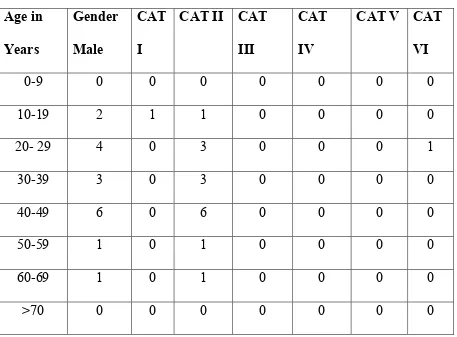
In this study it was noted that the maximum clustering of cases occurred in the age group from second to and fourth decade of life (68%) between the age group of 20- 49 years.
In this study the youngest patient with thyroid lesion was a 9 year old female and oldest one was a 85 years old female. The mean age is 38.3 years with a standard deviation of 14.917.
It was noted that 2 cases presented with thyroid lesions in the age group of 0-9 years and both were benign lesions.
The highest percentage of cases occurred in the age group of 30-39 years.
The age group of 20-29 yrs and 40-49 years had similar percentage of distribution of cases in this study. The remaining age groups had average number of cases. In the age group of > 70 years, 11 cases were present out of the total 300 cases in this study.
AGE WISE DISTRIBUTION
TABLE -4 : Total no of cases and percentage according to age group
0 10 20 30 40 50 60 70 80
0-9 10-19 2
27
0.7 9
C
a
se
s
&
P
e
rc
e
n
ta
g
e
CHART 2 : No of thyroid lesions in different
Age
(in years)
0-9
10-19
20-29
30-39
40-49
50-59
60-69
>70
TOTAL
51
DISTRIBUTION OF THYROID LESIONS
Total no of cases and percentage according to age group
20-29 30-39 40-49 50-59 60-69 >70 62
80
62
34
22
11 20.7 26.7 20.7
11.3 7.3
3.6
Age Group
CHART 2 : No of thyroid lesions in different
age group
in years) No : of cases Percentage
2 0.7
27 9
62 20.7
80 26.7
62 20.7
34 11.3
22 7.3
11 3.6
300 100
LESIONS
Total no of cases and percentage according to age group
CHART 2 : No of thyroid lesions in different
No : of cases
GENDER DISTRIBUTION
Among the 300 cases who presented wi study the majority of cases wer
of (5.7%) and the female : male ratio was 16.5: 1.
CLINICAL PRESENTATION
Out of the 300 cases who presented with thyroid swelling of the patients presented with lateral neck swelling (79%) rest of them presented as
depicted in CHART-4,
0 20 40 60 80 100
Male
P
e
rc
e
n
ta
g
e
o
f
th
y
ro
id
l
e
si
o
n
s
CHART
-according to gender distribution
52
DISTRIBUTION [ CHART- 3]
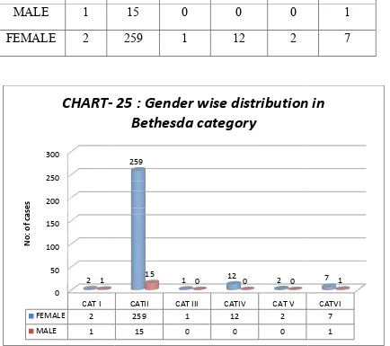
Among the 300 cases who presented with thyroid swelling the majority of cases were females (94.3%) and the males (5.7%) and the female : male ratio was 16.5: 1.
CLINICAL PRESENTATION [CHART -4]
who presented with thyroid swelling in this study presented with lateral neck swelling (79%) of thyroid
rest of them presented as a midline swelling (21%) in thyroid which is , mentioned below.
Male Female
5.7
94.3
Gender
-3 : Percentage of thyroid lesions
according to gender distribution
th thyroid swelling in this males comprised
in this study most of thyroid whereas in thyroid which is
3 : Percentage of thyroid lesions
according to gender distribution
Male
DURATION OF SWELLING
Out of 300 cases, who presented with thyroid swelling was noted it was
in 56.3% of cases, 1- 10 years in 13.3% Maximum number of
year which is depicted in the CHART
0 10 20 30 40 50 60 70 80
Lateral Neck
P
e
rc
e
n
ta
g
e
o
f
th
y
ro
id
s
w
e
ll
in
g
CHART 4 : Location of thyroid swelling in
53
DURATION OF SWELLING [CHART -5]
who presented with thyroid swelling,
it was < 6 months in 29.3% of cases, 6 months to 1 year 10 years in 13.3% cases and > 10 years in 1% of
Maximum number of cases presented with a duration of 6 months to one h is depicted in the CHART -5 mentioned below.
Lateral Neck Medial neck swelling 79
21
Axis Title
CHART 4 : Location of thyroid swelling in
present study
the duration of 6 months to 1 year > 10 years in 1% of cases. ation of 6 months to one
CHART 4 : Location of thyroid swelling in
RATE OF GROWTH
Out of 300 cases
it was noted that 95.3% of patients had gradual progression of disease 4.7% had a sudden progression of
study presented with a swelling.
0 10 20 30 40 50 60
Less than 6 Months
29.3
P
e
rc
e
n
ta
g
e
CHART -5 : Percentage of different thyroid
lesions with different duration
54
RATE OF GROWTH [CHART-6]
Out of 300 cases who presented with swelling of thyroid in this study 95.3% of patients had gradual progression of disease 4.7% had a sudden progression of disease. Most of the individuals in this
presented with a history of gradual onset of increase
6 Months to 1yr
1-10 Yr More than 10Yr 56.4
13.3
1
Duration of swelling
5 : Percentage of different thyroid
lesions with different duration
who presented with swelling of thyroid in this study 95.3% of patients had gradual progression of disease and . Most of the individuals in this increase in size of the
5 : Percentage of different thyroid
CLINICAL SYMPTOMS
Out of the total 300 cases who this study, it was noted
dysphagia in 26.39%
hoarseness of voice in 3% of patients rest of the patients presente
individuals who presented in this
CHART
55
CLINICAL SYMPTOMS [CHART -7]
Out of the total 300 cases who presented with the thyroid swellings it was noted that the most common clinical symptoms was
of patients followed by dyspnoea in 3% hoarseness of voice in 3% of patients and pain in 3.3% of patients
presented with no significant complaints.
individuals who presented in this study had no significant complaints.
95% 5%
CHART -6 : Percentage of thyroid lesions
showing rate of growth
thyroid swellings in l symptoms was yspnoea in 3% of cases, in 3.3% of patients and the no significant complaints. Most of the
complaints.
6 : Percentage of thyroid lesions
Gradual
FUNCTIONAL STATUS OF THE THYROID SWELLING
Out of the total euthyroid.
I.HYPOTHYROID FEATURES
Out of 300 cases hypothyroidism like bradycardia II.HYPERTHYROID FEATURES
Out of the total 300 cases hyperthyroidism which were tachycardia III. EUTHYROID
Out of the total 300 cases significant complaints.
0% 5% 10% 15% 20% 25% 30% 35% 40%
Dyspnoea 3%
CHART- 7 : Percentage of clinical features in
thyroid lesions in present study
56
FUNCTIONAL STATUS OF THE THYROID SWELLING
Out of the total 300 cases studied, majority of the patients
HYPOTHYROID FEATURES
300 cases, 12.3% cases presented with features of bradycardia and dry skin.
HYPERTHYROID FEATURES
Out of the total 300 cases, 1.7% cases presented with which were tachycardia and moist skin.
the total 300 cases, 86% cases were euthyroid and had no .
Dysphagia Hoarseness Pain
36.30%
3% 3.30%
7 : Percentage of clinical features in
thyroid lesions in present study
Series1
FUNCTIONAL STATUS OF THE THYROID SWELLING [CHART-8]
of the patients were
with features of
presented with features of
86% cases were euthyroid and had no
Pain 3.30%
HORMONAL LEVEL ESTIMATION
T3, T4 and TSH LEVELS
Among the 37 patients who presented with features of hypothyroidism clinically 35 cases (94.6%)
levels. These patients presented with like bradycardia and dry
Among the 5 patients clinically, 4 cases (80%) had decreased TSH levels
symptoms of hyperthyroidism
0 10 20 30 40 50 60 70 80 90
Hypothyroid 12.3
P
e
rc
e
n
ta
g
e
CHART 8: Percentage of functional status of
thyroid lesions in present study
57
HORMONAL LEVEL ESTIMATION
TSH LEVELS
Among the 37 patients who presented with features of hypothyroidism 94.6%) had decreased T3, T4 levels and
patients presented with clinical symptoms of hypothyroidism and dry skin.
Among the 5 patients who presented with features of hyperthyroidism 4 cases (80%) had increased T3 and T4 levels and a
decreased TSH levels and all of these patients presented with hyperthyroidism like tachycardia and moist skin
Hypothyroid Hyperthroid Euthyroid
12.3
1.7
86
Thyroid Status
CHART 8: Percentage of functional status of
thyroid lesions in present study
Among the 37 patients who presented with features of hypothyroidism increase in TSH clinical symptoms of hypothyroidism
features of hyperthyroidism and T4 levels and all of them had all of these patients presented with clinical
like tachycardia and moist skin.
Euthyroid
Majority of the individuals study were euthyroid
hormonal levels were in the normal range NATURE OF THYROID SWELLING
Of the total 300 study, 191 cases (63.7% 109 cases (36.3%) presented 191 cases of nodular swelling
cases (16.8%) presented with multiple nodules. presented with a nodular swellin
in nature.
0 10 20 30 40 50 60 70
Nodular Swelling
P
e
rc
e
n
ta
g
e
CHART
nodular and diffuse swelling
58
individuals who presented with thyroid swelling in our and as such had no significant complaints
were in the normal range.
NATURE OF THYROID SWELLING [CHART-9]
300 cases who presented with thyroid swelling 63.7%) presented with features of nodular swelling
presented with features of diffuse swelling
nodular swelling 159 cases (83.2%) had solitary nodule
presented with multiple nodules. Majority of the patients presented with a nodular swelling of thyroid and which was
Nodular Swelling Diffuse Swelling 63.7
36.3
Nature of swelling
CHART-9 : Percentage of patients with
nodular and diffuse swelling
who presented with thyroid swelling in our no significant complaints and their
d with thyroid swelling in our f nodular swelling, and th features of diffuse swelling and out of the had solitary nodule and 32 Majority of the patients and which was mostly solitary
9 : Percentage of patients with
59
CORRELATION OF MICROSCOPING FINDINGS WITH NATURE
OF THYROID SWELLING
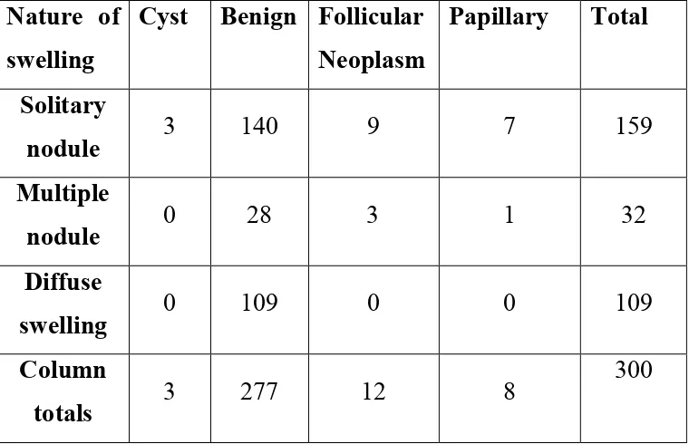
Of the total 300 cases who presented with thyroid swelling in this study , 159 cases (53%) presented with solitary nodule, 32 cases (10.7%) presented with multiple nodules, and the remaining 109 cases (36.3%) presented with diffuse swelling of thyroid.
SOLITARY NODULE – 159 (53%) [CHART-10]
Cystic lesion - 3 Benign - 140 Follicular neoplasm - 9 Papillary carcinoma - 7
MULTIPLE NODULE - 32 (10.6%) [CHART -11]
Benign - 28
Follicular