COMPARATIVE STUDY OF PRESERVATION VERSUS
ELECTIVE DIVISION OF ILIOINGUINAL NERVE IN OPEN
MESH REPAIR OF INGUINAL HERNIA
DISSERTATION SUBMITTED FOR
BRANCH-I M.S (GENERAL SURGERY)
APRIL 2015
THE TAMILNADU DR.M.G.R.MEDICALUNIVERSITY
Submission author: Assignment title: Submission title: File name: File size: Page count: Word count: Character count: Submission date: Submission ID:
Digital Receipt
This receipt acknowledges that Turnitin received your paper. Below you will find the receipt information regarding your submission.
The first page of your submissions is displayed below.
221211114.ms General Surgery SM… TNMGRMU EXAMINATIONS
COMPARATIVE STUDY OF PRESE… Copy_of_smitha.pdf
3.24M 133
19,646 99,936
DECLARATION
I, Dr.Smitha Nair, do hereby declare that I carried out this work on “comparative
study of preservation versus elective division of ilioinguinal nerve in open mesh
repair of inguinal hernia " at the Department of Surgery, Govt. Rajaji Hospital,
Madurai, under the guidance of Prof. Dr.A.M.Syed Ibrahim M.S., Professor of
Surgery, during the period of August 2013 to August 2014.
I also declare that this bonafide work has not been submitted in part or full by me
or any others for any award, degree or diploma to any other University or Board
either in India or abroad.
This is submitted to The Tamil Nadu Dr. M.G.R. Medical University, Chennai in
partial fulfilment of the rules and regulations for the M.S degree examination in
General Surgery (Branch I) to be held in April 2015.
Place :
ACKNOWLEDGEMENT
At the outset, I would like to thank our Dean for permitting me to use the facilities in Madurai Medical College and Government Rajaji Hospital for conducting my study.
I sincerely thank Prof. Dr.S.Sankaramahalingam M.S., Professor and Head of the Department, Department of General Surgery for his constant support, advice and co-operation in completing this study.
My Unit Chief Prof. Dr.A. M. Syed Ibrahim M.S. has always guided me, by example and valuable words of advice and has given me his moral support. I will be ever grateful to him.
I offer my heartfelt thanks to my unit Assistant Professors Dr.J.Amuthan M.S.,D.L.O., Dr.M. Lakshminarayanan M. S., and Dr.D. Geetha M.S.,D.G.O. for their constant encouragement, timely help and critical suggestions throughout the study and also for making my stay in the unit both informative and pleasurable.
Place: Madurai.
Date :
Dr.Smitha Nair
Post Graduate in General Surgery Department of General Surgery,
Madurai Medical College, Madurai
AD Anna domino :After Christ BC Before Christ
Cms Centimeter
ECG Electrocardiogram Gms Grams
H/O History of
Hb% Hemoglobin percentage HBsAg Hepatitis B surface antigen
HIV Human immuno deficiency virus Inj IM Injection , Intramuscular
ie, That is
IP No In patient number Lt Left
N Total number
P Probability (Significance of difference) RBS Random blood sugar
Rt Right
RCT Rondomized Controlled trial S/o Suggestive of
USG Ultrasonogram Vs Versus
Yrs Years
ABSTRACT
Background and Objectives: Chronic post herniorrhaphy groin pain is defined as
pain lasting for more than 3 months after surgery. It is one of the most important
complications occurring after inguinal hernia repair and it occurs with greater
frequency than previously thought .Majority of chronic pain has been attributed to
Ilioinguinal nerve entrapment.
Routine excision of the ilioinguinal nerve is an attempt to decrease the incidence of
chronic groin pain caused by nerve entrapment, inflammation and fibrotic reactions
around the nerve.
The purpose of the current study is to evaluate the effect of routine ilioinguinal nerve
excision compared to nerve preservation on chronic groin pain and other sensory
symptoms when performing Lichtenstein tension free inguinal he rnia repair.
Method: A total of 60 patients admitted for inguinal hernia at Government Rajaji
Hospital Madurai who met with the inclusion criteria underwent open mesh repair
of inguinal hernia over the study period from August 2013 to August 2014.The
ilioinguinal nerve was identified and preserved in 30 patients(group A) and
elective division of the ilioinguinal nerve was done in 30 patients(group-B).Patients
were evaluated for pain and other sensory symptoms at PoD-1, at one month, at
three months, and at six months after surgery by using 4 point verbal scale.
Results: 50 of 60 patients completed the study protocol fully, 26 patients ( 25 men
and 1 women ) with mean age 31± 20 belonged to group A, and 24 patients (all
The results of the follow up visits are 24 Vs 19 ( p>0.05 ) at POD-1 ; 1 3 V s 1 0
( p>0.05 ) at 1 month ; 1 0 V s 2 ( p>0.05 ) at 3 months ; and 8 V s 1 (
p<0.05 ) at 6 months in group A and group B respectively. The mean severity score
was 1.65±0.79 vs 1.37±0.92 at POD-1 ; 0.81±0.94 vs 0.42±050 at 1 month ;
0.58±0.81 vs 0.08±0.28 at 3 months ; and 0.39±0.09 vs 0.05±0.20 at 6 months
in group A and group B respectively. There was significant difference(p<0.05) at 3
and 6 months.
The results showed no significant difference in the neurosensory disturbances in
either Groups, that is, the incidence of hypoesthesia was 57.6% vs 62.% ; 26.9% vs
37.5% ; 19.2% vs 20.8% ; and 11.5% vs 16.6% at POD-1, 1, 3,and 6 months in
group A and B respectively. And the incidence of numbness was 19.2% vs 12.5% ;
23% vs 25% ; 15.3% vs 20.8% ; and 11.5% vs 12.5% at POD-1,1,3, and 6 months
in group A and b respectively.
Conclusion:The prophylactic excision of the ilioinguinal nerve during Lichtenstein
mesh hernia repair decreases the incidence of chronic groin pain after surgery.
Furthermore the procedure is not significantly associated with additional morbidities
in terms of local cutaneous neurosensory disturbances. So when performing
Lichtenstein inguinal hernia repair, routine ilioinguinal neurectomy is a reasonable
option.
Keywords: Inguinal hernia; Groin; Lichtenstein; Polypropylene mesh;
TABLE OF CONTENTS
1. INTRODUCTION 1
2. OBJECTIVES 3
3. REVIEWOF LITERATURE 4
4. METHODOLOGY 73
5. OBSERVATION ANDRESULTS 76
6. DISCUSSION 93
7. CONCLUSION 104
8. SUMMARY 105
9. BIBLIOGRAPHY 107
10. ANNEXURES
ANNEXURE-I:PROFORMA 116
ANNEXURE-II :KEYTO MASTERCHART 122
INTRODUCTION
Hernias may be generally defined as a “Protrusion of a viscus or part of a viscus
through an abnormal opening in the walls of its containing cavity’. 1
“A protrusion of any viscus from its proper cavity is denominated a hernia. The
protruded parts are generally contained in a bag by a membrane with which the
cavity is naturally invested”
– Sir Astley Cooper 1804.
Chronic post herniorrhaphy groin pain is defined as pain lasting for more than 3
months after surgery.
It is one of the most important complications occurring after inguinal hernia repair
a n d i t occurs with greater frequency than previously thought.
A review of studies published between 1987 to 2000 showed an overall incidence of
25% with 10% of patients having pain fitting a definition of moderate or severe. 2
Incidence of long term (≥1 year) post operative neuralgia reported for Lichtenstein
repair of inguinal hernia range from 6 – 29 %, 3
Inguinodynia is the recommended generic term for chronic groin pain after hernia
repair and should replace ‘neuralgia or mesh inguinodynia ‘ to promote uniformity
and avoid confusion in the literature.4
In cases that involves workman`s compensation issue, treating a post surgical
patient becomes complicated. Although most legal cases results in out of court
neuralgia will sue their surgeons. 5
The concept of routine neurectomy in surgery is not unique to inguinal hernia
repairs. Routine neurectomy is often performed during axillary and neck
dissections in which the Intercostobrachial and greater auricular nerves are
sacrificed. 6
Routine ilioinguinal nerve excision has been proposed as a means to avoid the
troubling complication of long term post herniorrhaphy neuralgia. 7 , 8
Theoretically excision of ilioinguinal nerve would eliminate the possibility of
inflammation neuralgia arising from entrapment, neuroma, fibrotic reactions yet
controversies persists and the procedure is not widely accepted. 9 ,10
The purpose of the current study was to evaluate the effect of routine ilioinguinal
nerve excision compared to nerve preservation on chronic groin pain and other
AIMSAND OBJECTIVES
1. To evaluate the effect of preservation versus elective division of the ilioinguinal
nerve on chronic groin pain a n d hypoesthesia after Lichtenstein tension free
REVIEW OFLITERATURE
HISTORICAL ASPECTS OF INGUINAL HERNIA
In the entire history of surgery, no subject has been as
Controversial as the repair of groin hernia.
C.B.McVay
The history of hernia, one of the most beautiful chapters in the triumphs of anatomy and
surgery, is replete with ideas and realities, myths and facts, transmutations and
Shadows. The history of hernia in toto is as old as human race. Inguinal hernias are
recorded since 1500 BC by the ancient Greeks. The term hernia is derived from
Greek meaning “offshoot, a budding or bulge.”11
Hippocrates has barely mentioned about hernia in his writings (500 BC).
Ancient Times 12, 13
The word hernia is derived from the Greek word "Hernios", meaning "Nad" or "Shoot".
Hernia is known to mankind since time immemorial. Shushruta in Vedic period
described hernia as "Antra-vriddi" and had thought it to be incurable. Hernia is barely
mentioned in the writings of Hippocrates (500 BC).The evolution of surgical
treatment of inguinal hernia encompasses the trials and errors of surgeons
practicing their art, for thousands of years. Most of the evidence obtained from
historical documents, suggest that throughout the ages, till the onset of 19th
century, the mainstay of treatment of hernia has been conservative and the evolution of
Egyptian physicians reported the management of hernia by conservative means that
included the snugly fitting bandage for reduction and support.
Figure-1. Hippocrates (460-377 B C) Figure-2. Aulus Celsus of Alexandria (50 A D)
Figure-3. Middle Dark Ages (476-15th Century) Figure-4. Ambrose Pare (1570-1590)
The treatment by taxis for irreducible hernia has been traced back twenty four
hundred years.The earliest mention of inguinal hernia is found in "Eber's papyrus"
(1500BC), in which it is apparent that the pre-Homeric physicians treated the inguinal
hernia by conservative methods.Aulus Celsus, the Greek encylopedist and
surgeon, documented (50AD) the use of transillumination to distinguish
hydrocoeles from hernias and described taxis for strangulation. Trusses and bandages
were used for reducible hernia. Incomplete hernias (Bubonocele) were distinguished
from Complete (Scrotal) inguinal hernias by Paul of Aegina, around 700AD.
MedievalPeriod 14
After the fall of Rome, religious prejudice against mutilation of the human body
caused regression of surgical technique. During the lengthy dark Middle Ages, two
important advances in herniology were made. Guy de Chauliac, from France (1363 AD),
in his text "Chirurgia Magna", distinguished femoral from inguinal hernias. He
developed taxis for incarcerated hernia, recommending the head down Trendelenberg
position. He considered operations doubtful and dangerous, preferred chemical
cauterization to burning skin, fascia and pubic bone. In 1556, Pierre Franco of
Switzerland was the first to work on strangulated hernia cases, as a routine procedure.
In the early stage of strangulation, cutting the constriction ring, using a grooved
director to protect the bowel, which was replaced in the abdomen using fine linen.
Ronald, of Parma (1383AD), recommended the use of Trendelenberg position in the
on strangulated hernia cases, as a routine procedure. In the early stage of
strangulation, cutting the constriction ring, using a grooved director to protect the bowel,
which was replaced in the abdomen using fine linen.
Post-Renaissance Period 15, 16
After the renaissance, autopsy and anatomic dissection spread throughout Europe.
Started in Bologna in 1200 AD, knowledge about herniation accumulated rapidly.
In 1700AD, Littre reported Meckel’s diverticulum in hernial sac. In 1721, William
Cheselden successfully operated on strangulated hernias. Heister (1724) in his
monograph distinguished indirect from direct inguinal hernias. Hunter and Percival
Pott of London pointed out the congenital natu re of some indirect inguinal hernia. In
1731, Gimbernat described the ligament that bears his name and advocated its
division in instances of strangulated femoral hernias, rather than an upward
incision of the Poupart ligament, which occasionally led to serious bleeding from an
aberrant inferior epigastric artery. 'Antonio Scarpa'1 (1752-1832), in his treatise on
hernia accurately described the sliding hernia, based on autopsy studies. Astley
Patson Cooper1 (1768-1841),described for the first time the superior pubic ligament,
which bears his name and transversalis fascia with full recognition of its role in the
pathogenesis of hernias. Frenz Casper Hesselbach(1759-1816), contributed anatomic
studies relative to groin hernia - Iliopubic tract and Hesselbach’s triangle. Jules
German Cloquet1 (1770-1883), dissected and sketched 345 cases of hernia.
In 1867, Joseph Lister, Professor of Orthopedic Surgery at Glasgow infirmary presented
his first paper on antiseptic surgery performed under carbolic acid spray. Prior to Lister,
all hernia repair were performed through the external ring incisions, fascial planes were
scrupulously avoided to prevent infection and its dire consequences. In 1871, Marcy,
published the first article in United States on Antiseptic herniorrhaphy, using
carbolised catgut ligature. In 1877, Czerny described pulling the sac down through the
external ring and excising it, allowing the ligated neck to retract and invert at the
internal ring. Lucas-championniere introduced antisepsis to France. In 1885, he incised
the external oblique aponeurosis, laid open the inguinal canal and imbricated the roof
in the closure. The period 1880-90, has rightly been termed as “The Decade of
Inguinal Hernia”,for the significant contributions made towards hernia surgery by
Lucas championniere, Marcy and Bassini. The credit for modern herniology should be
given to Marcy of United States. Marcy(1837-1924), was the first to indicate the
importance of the high ligation of the hernial sac and closure of dilated inguinal ring as
essential steps in the repair of inguinal hernia. He was also the first to describe, the
trans-abdominal approach.Edoardo Bassini 17(1844-1924) of Pavia, Italy
revolutionized the treatment of inguinal hernia by the introduction of a technique
designed to restore the conditions in the area of hernial orifice, which existed under
normal circumstances. He thought that recurrence was due to the inadequacy of mere
ligature of the sac without reconstructing the inguinal canal. In 1890, he published his
recurrence rates. He initiated the use of transversalis fascia, rectus sheath and
interrupted silk sutures. He used to do bilateral hernia repairs and surgery for
cryptorchidisim in the same sitting. Bassini advocated not to obliterate the inguinal
canal with deep suturing of the rings, but to reconstruct it physiologically,recreating the
internal and external openings with anterior and posterior walls. He sutured the
conjoined transversus abdominis and internal oblique to the inguinal ligament with
continuous silk sutures. His triple layer included transversalis fascia, which was
divided from pubis to an inch beyond the internal ring. He emphasized closing the floor
from below upwards to restore the valve-like mechanism.In 1940, McVay and Anson
pointed out that, the rectus fascia, a portion of the transversalis fascia that inserts
into the lateral border of rectus muscle, was strong enough to prevent incisional
herniation. Shouldice, Obney, Ryan 1950-1953 Performed multiple layer repair of
posterior inguinal wall under local anaesthesia (Shouldice technique).
Darn Repairs
McArthur in 1921 used the pedicled strips of external oblique aponeurosis woven
between the conjoint tendon and inguinal ligament. Gallie and Lemesurier in 1921,
published papers on using fascia lata strips as sutures woven into the muscles and the
inguinal ligament and the tissues of the posterior wall of the inguinal canal-- "Much
as one would darn a sock". Ogilvy in 1936,practiced floss silk lattice repairs with non
absorbable material which was followed by Maingot.Pratt, in 1948, used steel wire
the forerunner of the modern nylon darn technique.
PatchGraft Repairs
Whenever the local tissues were weak and attenuated, approximation of tissues
was under tension, the sutures did not hold and the hernia recurred. So the surgeons
thought of exogenous or endogenous prosthesis with good tensile strength. To start
with, patch in the form of sheets of natural tissues and biological materials or synthetic
sheets to fill the gap in the posterior wall of the canal were tried. Silver wire mesh used
in most of the cases, were corroded, fragmented and were rejected through chronic
sinuses and lead to recurrence. In 1940, Burke introduced tantalum metal sheets,
but due to fragmentation of metal, hernia recurred. Surgeons started getting sheets
of natural tissue flaps from fascia of thigh. Aponeurosis of external, internal
oblique or rectus sheath were turned downwards and sutured to inguinal ligament.
Mayr, in 1943, used skin graft. In 1958 Uscher introduced synthetic polyethylene mesh
prosthesis to buttress and reinforce previously sutured repair. A variety of mesh designs
and mesh placements have flourished since, Lichtenstein showed that mesh could be
used successfully Lichtenstein 1986 Introduced tension-free repair by reconstructing
floor of inguinal canal using prosthetic material, Concept of tension free repair
introduced by Lichenstein in 1989.In 1991, Gilbert used suture less repair of small to
moderate sized inguinal hernia by cones and swatch, i.e. a suture less patch was
placed over the whole of the posterior inguinal wall to reinforce this "Swatch".A
plug or suture less patch. In this, a roll of material is placed in the hernial orifice with
or without suture to obstruct the passage of hernia to the exterior, popularized by
Robbins and Rutkow in 1993.
Expended polytetrafluroethylene (ePTFE) has been adopted for both the external
and pre-
peritoneal approach, with good results. In recent years, sheaths of woven
monofilament polyamide or knitted monofilament polypropylene have been used
extensively. Recently, a bilayered patch device for inguinal hernia has been
introduced. The unique feature of this polypropylene mesh device is that it has three
components. Its underlay patch provides posterior mesh repair. Its connector has the
desirable attributes of the plug repair. Its Onlay patch covers the posterior wall up to
internal inguinal ring. 18
Pre-Peritoneal Repairs
Thomas Annandale of Edinburgh presented for the first time in 1876, the concept of
the pre-peritoneal approach. Lawson Tait of Birmingham, England, in 1883 reported
the advantages of treating hernias by a median abdominal section. Bates (1913),
repaired the defect from the posterior approach, using transversalis fascia. Cheatle
(1920) renewed the interest in the pre-peritoneal approach. Henry, in 1936, suggested
that this approach might facilitate the technical handling of inguinal and femoral
hernias. This approach was strongly recommended by 'Nyhus' in 1960. The foremost
for problematic cases in which repeated repairs of multiple recurrent hernias have been
carried out, and in which tissues have become scarred and weakened and the
normal anatomy is destroyed.Stoppa 1984 Devised procedure for reinforcing
peritoneum using large unslit prosthesis Rutkow 1993 Modified mesh-plug repair .
LaparoscopicInguinal HerniaRepair 20,21
y The wave of minimal access surgery has inevitably swept hernia repair along its
surge.Ger and his colleagues, in 1982, through laparoscope used Michel staple applied
with a Kocher clamp to close peritoneal opening of the hernia sac.
y Schultz & co workers (1990) described “plug & patch laparoscopic
inguinal herniorrhaphy.
y Spaw & co workers (1991) detailed anatomy with respect to laparoscopic approach.
Toy & Smoot (1991) described intra peritoneal on lay patch technique (IPOM).
y Dion & Morin (1992) described transabdominal preperitoneal patch (TAPP)
technique.
Figure-5. Antonio Scarpa(1752–1832)
Figure-7. August Richter (1742-1812)
Figure-6. Astley Cooper (1768-1841)
EMBRYOLOGY17 ,22
There is no doubt, that the first appearance of the mammal, with his unexplained need
to push his testicles out of their proper home into the air made a mess of the three
layered abdominal wall that had done the reptiles well, for millions of years. In a
highly synergistic way the skin, the parietal peritoneum and the embryologic and
anatomic entities between them, produce the future pathway for the testes. The skin
will form the scrotum in males and labia in females. The embryological entities
between the skin and the peritoneum, permit the processes vaginalis to penetrate
them and to form the inguinal canal. The downward journey of the testis to the
scrotum is thus allowed but, the descent of the ovary outside the peritoneal cavity is
forbidden.
InguinalRegion
The Testis originally lies on the posterior wall of the abdomen, at the level of the
upper lumbar vertebrae on the medial side of the mesonephros, near the lower pole
of the mesonephros by a peritoneal fold, called mesorchium. The descent or
migration of testis into its corresponding scrotal chamber is accomplished by
following the lead of the fibromuscular band – gubernaculums testis. It arises mainly
within a peritoneal fold called the plica inguinalis, which stretches from the inguinal
region to the lower end of the mesonephros. The gubernaculum attains greatest
attached, above to the lower end of the testis and below it pierces through the
abdominal wall, in its passage to the bottom of the scrotal pouch, thereby forming
the inguinal canal. Along with it, a process of peritoneum the processus vaginalis
descends into the scrotum, dragging with it thin fascial prolongations of the
layers of the abdominal wall. Thus, the processus vaginalis receives coverings
from the aponeurosis of the external oblique and internal oblique muscles and from
fascia transversalis. As the passage through the abdominal wall occurs, the testes
and cord structures are surrounded by vestiges of the external oblique (external
spermatic), internal oblique (cremasteric fascia and muscle) and transversalis fascia
(internal spermatic). The blind extremity of the processes vaginalis gets invaginated
in the form of a cup for the reception of the descending testis. As the migration of
the testis proceeds, the gubernaculum shortens and eventually atrophies, but some
trace of the gubernaculum persists at the bottom of the scrotum below tunica
vaginalis. The shortened remains of the gubernaculum form the scrotal
ligament,fixing the testis to the bottom of the scrotal pouch
ANATOMYOFGROIN ANDTHEINGUINALCANAL24 ,25,26, 27
No disease of the human body belonging to the province of
surgeon requires in its treatment, a better combination of accurate
anatomical knowledge with surgical skill than hernia, in all its
varieties.
The groin is that portion of the anterior abdominal wall below the level of anterior
superior iliac spines. For proper orientation, the groin is referred to as the surgeon
views the patient on the operating table. The pubis and superior pubic (Cooper's)
ligament are medial. The epigastric vessels and transversalis fascia condensation at
the internal ring are lateral. The anterior femoral sheath, iliopubic tract and
inguinal ligament are inferior and the transverses abdominis aponeurosis and its
arch are superior.
Skin
Langer's lines are transverse in the groin with convexity facing downwards. The
anterior superior spine of ileum is easily palpable in the lateral groin and pubic
tubercle on the lateral margin of the pubis. Spermatic cord is identified as it exits
from the external ring which overlies the lateral aspect of the pubic tubercle. The
deep ring is located approximately 2 cm above the skin crease between the thigh and
the abdomen and midway between anterior superior iliac spine and pubic
symphysis.The skin of groin is innervated by the ilioinguinal, iliohypogastric
and genital branch ofgenitofemoral nerves.
SubcutaneousTissuesof Groin
Divided into two strata - superficial fatty layer (Camper's fascia) and deeper
membranous layer (Scarpa's fascia), which continues into perineum as the Colles
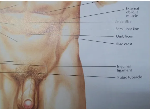
ExternalObliqueMuscleandAponeurosis
It arises from the lower 8 ribs (5 to 12) and its fibers are directed downwards,
forwards, and medially. From the anterior superior iliac spine to the pubic spine, the
aponeurosis forms a free border which is called inguinal ligament. The muscle
becomes totally aponeurotic in the groin with its fibers running obliquely
downwards. It becomes the external layer of the anterior rectus sheath and further
inserts on the pubis. The superficial inguinal ring is a triangular opening in the
external oblique aponeurosis, 1 to 1.5 cm lateral to the pubic tubercle. The opening is
formed by the splitting of external oblique.
InternalObliqueMuscle andAponeurosis
The internal abdominal oblique muscle lies between the external oblique and the
transverses abdominis muscles. It originates from the outer half of the inguinal
ligament, from the intermediate line on the iliac crests and from the posterior lamella
of the lumbodorsal fascia through which it gains attachment to the lumbar spines.
The anterior lamella accompanies the external oblique aponeurosis to form the
anterior rectus sheath and the posterior lamella accompanies the aponeurosis of the
transversus abdominis to form the posterior rectus sheath. They insert conjointly
with those of the transverses abdominis into the crest of the pubis. This fusion of
the tendinous portions of the internal oblique and transverses muscles that results in
TransversusAbdominis MuscleandAponeurosis
This is the most internal of the three flat muscles of the abdominal wall. It passes
medially in a transverse manner around the lateral aspect of the abdomen on to the
anterior abdominal wall.This is the key layer, because of its role in hernia repair. The
general layer of the muscle (lateral portion) and the aponeurosis (medial portion) is
towards the linea alba, where it forms the anterior rectus sheath below the
semicircular line of Douglas. In the groin it can be divided into continuous and
discontinuous portions.
A. The continuous portion is the extension of the main muscle and aponeurosis, the
lower border of which arches above and medial to cord structures and are called
Transverses abdominis arch,which in 10% of cases due to its dense nature and
insertion into the pubic tubercle and the crest is called falx inguinalis. In 3% of cases
the falx receives contribution from the internal oblique aponeurosis also thereby
forming the conjoined Tendon.
B. The discontinuous portion lies below the transverses arch, forms the posterior
wall of the inguinal canal, medial to the internal ring. One fourth of these fibers
show marked individual variations and most often is deficient, represented only
by the transversalis fascia, thereby forming a critical weak spot in the posterior wall
of the inguinal canal. The inferior most edge of this layer is formed by the "iliopubic
TransversalisFascia
This is a portion of the endo-abdominal fascia that encloses the abdominal
cavity and peritoneum. The portion which invests the transverses muscle and
aponeurosis is called Transversalis fascia. It is continuous with the lumbar, iliac,
psoas, obturator and rectus fascia. It is adherent to the transverses muscle -
aponeurosis due to the numerous slips of fibrous tissue that traverse the muscle and
attach to the deep interpareital fascia. Hence, practically it forms part of the
transverses muscle aponeurosis fascia complex. At the deep inguinal ring there is a
tubular projection of this fascia - internal spermatic fascia that extends outwards in a
blunt funnel like fashion to cover the ductus deferens and the spermatic vessels.
However, the blunt funnel is not perfectly conical, but is skewed and the axis of the
funnel is less oblique than the axis of the vessels through the deep inguinal ring. The
redundant transversalis fascia in the medial side of the deep ring is called
'Transversalis fascia sling'. The transversalis fascia is somewhat like the letter ‘V’
with the open end pointing superolaterally to the groin and the diverging ends are
called crurae. Most often, the posterior inguinal wall is represented only by this
fascia and leads to weak spot in the groin.
RectusSheath
In the groin aponeurosis of all the three flat muscles contribute to the anterior sheath.
In the groin as elsewhere, the peritoneum is a thin elastic membrane that serves only
to provide a lubricating surface for its contained viscera. Because of the elastic
character of the peritoneum it does not act in the prevention of hernia.
TheConjointTendon(FalxInguinalis)
The aponeurosis of the transverses abdominis and the internal oblique are fused
some distance lateral to the rectus sheath, the term conjoined tendon . This
anatomic configuration, The transversus muscle contributes 80% of the conjoint
tendon. The conjoint tendon lies lateral to the rectus muscle and immediately deep
to the superficial inguinal ring. It passes down to its insertion deep to the inguinal
and lacunar ligaments. The spermatic cord or round ligament of uterus lies anterior
to it while passing through the superficial inguinal ring. The conjoint tendon has a
very variable structure and in 20% of the subjects it does not exist as a discrete
anatomic structure - it may be absent or slightly developed or it may be replaced by
a lateral extension of the tendon or original ring, so that no interval is present
between the lower border of the transverses and the inguinal ligament.
Cooper’sLigament(Iliopectineal Ligament)
Cooper's ligament is remarkably constant in form and extent and represents the
strongly reinforced periosteum of the superior ramus of the pubis. On the superior
pectineal line, the periosteum is supplemented by a considerable quantity of dense
fibrous tissue so that it usually becomes 2 cm or even 3 cm thick. Laterally, it
continues posteriorly along the brim of the true pelvis, becoming progressively
thinner until it can no longer be distinguished from periosteum of ileum. Cooper's
ligament is particularly important in the surgical correction of femoral hernias and
large direct inguinal hernias, because it forms a solid anchor along the inferior or
posterior aspect of these hernial defects, through which sutures may be placed with
[image:30.612.126.410.409.615.2]confidence that they will hold.
Figure-10.SubcutaneoustissuesofGroin
[image:31.612.126.413.38.237.2] [image:31.612.141.427.331.538.2]Figure-12.Hasselbach`sTriangle
InguinalLigament (LigamentofPoupart)
It is the lower, thickened portion of external oblique aponeurosis extending from
the anterior superior iliac spine to the pubic tubercle. Its edge is rolled inwards to
form a gutter. The lower edge of the inguinal ligament is loosely bound to the fascia
lata by the Innominate fascia. This fascia also serves to bind together the
collagenous fibers of aponeurosis and inguinal ligament.Medially, the inguinal
ligament gets inserted on the pubic tubercle and fans downward to the superior
pubic ramus as the lacunar ligament.
LacunarLigamentofGimbernat
The ligament of Gimbernat is a triangular fascial extension of the inguinal ligament,
before its insertion to the pubic tubercle. It is inserted at the pecten pubis and its
lateral end meets the proximal end of the ligament of Cooper. It serves to broaden
[image:32.612.135.425.71.261.2]TheCremasterMuscle
The cremaster consists of a number of loosely arranged muscle fascicles lying
along the spermatic cord. They are united by areolar tissue to form the sac like
cremasteric fascia around the cord and testis, within the external spermatic fascia.
The lateral part of the muscle, arising from the inguinal ligament, has been variously
described as in continuity with the medial edge of the internal oblique, deep to the
internal oblique extending as far as the anterior superior iliac spine and in continuity
with either the internal oblique or transverses or as a pointed tendon from the middle
of the inguinal ligament piercing the internal oblique near its medial margin.The
fibres pass along the lateral aspect of the spermatic cord through the superficial
inguinal ring and then spread out into the fasciculi in loops, of increasing length along
its anterolateral aspect.
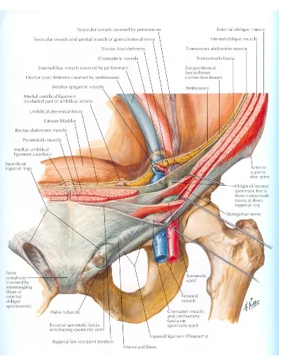
InguinalCanal
It begins at the site of emergence of the spermatic cord through the transversus
aponeurosis (internal ring) and ends at the pubic tubercle. It is oblique and
3.75 cm long, slanting downwards and medially, parallel with and a little above the
inguinal ligament. It extends from the deep to the superficial inguinal ring. The
boundaries are:
Anteriorly: Throughout by the skin, superficial fascia, external oblique aponeurosis,
in its lateral one third by the muscular fibres of the internal oblique.
present).
Superiorly: The arched fibers of internal oblique and transverses aponeurosis.
Inferiorly: The inguinal ligament and its continuation, lacunar ligament.
Hesselbach’sTriangle
It is bounded medially by the lateral border of the rectus sheath, laterally by
the inferior epigastric vessels and below by the inguinal ligament. Spermatic cord:
Originates at the deep ring and consists of
a. Arteries: Testicular, cremasteric and artery to vas.
b. Veins: Corresponding veins, mainly testicular (pampiniform plexus).
c. Nerves: Genital branch of genitofemoral nerve, cremasteric nerve, Sympathetic
plexus derived from Para aortic and pelvic plexus.
d. Lymphatics of the testes.
e. Vas deferens and areolar connective tissue.
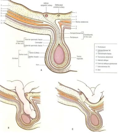
Coverings of the spermatic cord from within are: processus vaginalis internal
spermatic fascia
(Transversalis fascia), cremasteric fascia (Internal oblique muscle and fascia)
and external
spermatic fascia (External oblique muscle and fascia).
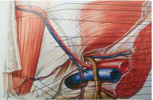
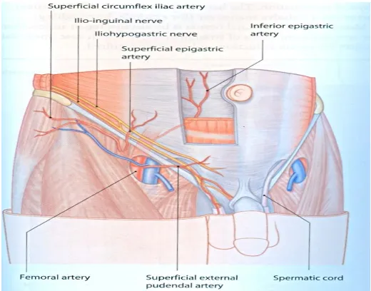
Blood Vessels and Nerves The external iliac artery gives off two major
branches, before
tributaries,the deep circumflex iliac and the inferior epigastric vessels, are not vital.
The latter, serves as the medial border of the deep ring, or the lateral border of the
direct triangle.There is, therefore an additional method of identifying the position of
the internal ring. The epigastric artery gives off two branches, the cremasteric and
the pubic. The testicular artery arises directly from the aorta to supply the testis. The
spermatic cord contains still one other small vessel, the umbilical artery, to supply the
ductus deferens.
Nerves28
The essential nerves of the groin are the ilioinguinal, the iliohypoga stric and the
genitofemoral.The last, arising form L1 and L2 supplies the cremaster muscle, the
skin of the scrotum and the medial aspect of the thigh ,in females to mons pubis and
labia majus. Its integrity is essential to the cremaster reflex.The ilioinguinal and
iliohypogastric nerves arise primarily from L1. Just medial to the anterior superior
spine these nerves traverse the internal oblique and come to lie beneath the
external oblique aponeurosis then Ilioinguinal nerve traverses the inguinal canal and
emerges through the superficial inguinal ring and supplies sensory innervation to
proximal and medial thigh,root of the penis and uppers scrotum.In females it
innervates mons pubis and labia majus. The hypogastric branch of the
iliohypogastric is mainly a motor nerve that supplies the abdominal muscles along
its course. It exits through the external oblique aponeurosis above the external ring.
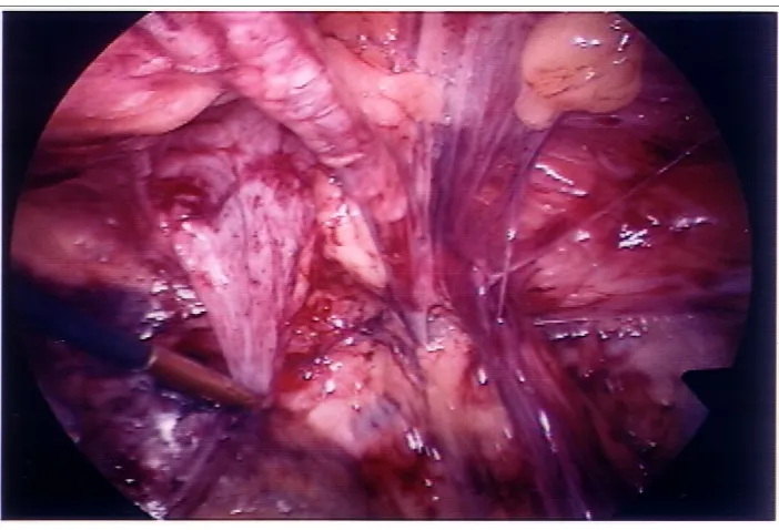
Figure-14.Inguinalcanalanditscontents
[image:37.612.153.479.65.291.2] [image:37.612.182.442.384.588.2]EPIDEMIOLOGY,ETIOLOGY ANDPATHOPHYSIOLOGY29 ,30 ,31 ,32
Seventy –five percent of all abdominal hernias are found in the groin; Of all groin
hernias,95% are hernias of the inguinal canal with the remainder being femoral hernia
defects.Inguinal hernias are 9 times more common in men than in women, the inguinal
hernia is still the most common hernia in women .The overall lifetime risk of
developing a groin hernia is approximately 15% in males and less than 5% in
females. There is clearly an association between age and hernia diagnosis. In the
same way the complications of hernias (incarceration , strangulation ,bowel
obstruction ) are found commonly at the exremes of age.Currently in this country
approximately 700,000 operations for inguinal hernia repair are performed
annually.The cause of hernia is multifactorial and it is assumed that the following factors
are involved.
1.Evolution
The absence of posterior rectus sheath below the arcuate line and only rather
substantial transversalis fascia unsupported by muscles or aponeurosis resisting the
intra-abdominal pressure and holding the breach between the abdomen and the thigh. It
is compounded by humans having adopted the upright posture and change from
quadrupedal to bipedal locomotion In man, the upright posture causes gravitational
stress to pass down to the lower abdominal wall, which is structurally not designed for
2.CongenitalandAnatomicalFactors
a. Patent Processes Vaginalis: Is the prime cause of indirect inguinal hernia in
infants and children. The development of processus vaginalis, its migration and its
final obliteration are intimately linked to the descent of the testis from the abdominal
cavity into the scrotum.The incidence of patent processus vaginalis in adults who do not
develop hernia during their life is up to 20%.
b.Subtlevarietiesin theattachmentandarrangementof abdominalmuscles.
c. Females are particularly free of direct inguinal hernia: The narrowness of the
interval between the transversus arch and the inguinal ligament and the hermetical
attachment of external oblique aponeurosis are the important factors in protecting
women against direct hernia. On the other hand, musculoaponeurotic attachments in
woman are such that they frequently develop femoral hernia. Other factors that are
significant in the etiology are the number of aponeurotic fibers in the transversus
aponeurosis which determines the intrinsic strength of the layer. The disposition of the
transversus arch in relation to the Iliopubic tract indirectly determines the size of the
inguinal gap or defect in the Hesselbach's triangle.
d. The obliquity of the inguinal canal: During sudden exertion increases the
intraperitoneal pressure, compresse in the anterior and posterior walls of the canal there
by occluding the canal.
3.ShutterMechanism
activated,when the abdominal muscles contract to raise the intra abdominal pressure
.As the internal oblique and transverse abdominis muscles contract, their
lower fibers forming the myoaponeurotic roof of the inguinal canal "the conjoined
tendon", that arches over the spermatic cord also sharply contracts and as the fibers
shorten, the arch straightens out and descends to come to lie close to or on the inguinal
ligament and so covers and protects the fascia transversalis. The shutter also passes
down in front of the internal ring and counteracts the pressure on the ring from inside
the abdomen. Contraction of the transverses abdominis muscle also pulls up and
tenses the crurae of the internal ring which make up the thickened bands of the
iliopubic tract and fascia transversalis causing the ring to close like a sphincter
snugly around the cord.
4.Integrityof theFasciaTransversalis31
The ability of the fascia transversalis to withstand physiologic and pathologic
elevations in the intra abdominal pressure is dependent on the state of the collagen
fibers that make up its tissues and give its strength. The factor which interferes with
normal production of collagen or causes its increased destruction or abnormal
production of collagen fibers decreases the strength of transversalis fascia. These
factors include congenital connective tissue disorders like Marfan's,Ehler-Danlos and
Hurler-Hunter syndromes and mesenchymal metabolic defects. It is found that
substances in cigarette smoke inactivate anti-proteases in lung tissues and so
and collagen of the rectus sheath and fascia transversalis and predispose to herniation in
smokers.
5.General ContributingFactors
Like weakening of muscle and fascia by advancing age, lack of physical exercise,
obesity and multiple pregnancies. Loss of weight and body fitness as may occur after
illness, operation or prolonged bed rest, very low and unduly long transverse
abdominal incisions for gynecological, urological and appendicectomy incision.
Pulmonary diseases like COPD and emphysema, prostatism, chronic constipation,
diverticular disease, genito-urinary causes like cystitis, cystocele and urethrocele
3.Coverings27
[image:42.612.106.489.165.601.2]Coverings in case of an indirect inguinal hernia are, from inside out, as follows:
Components ofInguinalHernia23
1.The sac consists of a diverticulum of peritoneum, which is divided into mouth, neck,
body and fundus.
Mouth : This is the part between the sac interior and the abdominal cavity.
Neck: This is the narrowest section between the mouth and the body of the sac.
Body: It lies between the neck and the fundus.
Fundus: This is the blind end or the distal most part of the sac.
2.Contentsof Hernia
These can be almost any abdominal viscera, except the liver. The commonest are;
a. Fluid - Derived from peritoneal exudates, usually in congenital hernias..
b. Omentum - Omentocele (Synonym -Epiplocele).
c. A loop of intestine - Enterocele (Usually small intestine, but in some instances large
intestine or vermiform appendix).
d. A portion of the circumference of the intestine - Richter's hernia.
e. A portion of urinary bladder wall or a diverticulum of the bladder.
f. Ovary with or without the corresponding Fallopian tube.
g. Meckel's diverticulum - Littre's Hernia.
h. Two loops of intestine in the manner of W - Maydl's hernia.
i. Rarely stomach, spleen or caecum may be found within the sac.
j. Sliding or Hernia-en-Glissade (Contents - Caecum, Urinary bladder).
l. Dual hernia (saddle or pantaloon). Hernia, on either side of the inferior epigastric
vessels.
CLASSIFICATION OFINGUINALHERNIAS 33 ,34
Clinical Classification
This is based on the clinical presentation of hernia:
Reducible hernia
Irreducible hernia
Obstructed hernia (Incarcerated hernia)
Strangulated hernia
Inflamed hernia
Gilbert’sClassification(Additionby RutkowandRobbins)
It is based on anatomical and functional defects established intra-operatively,
categorized groin hernias in to 5 types. Type1, 2 and 3 were indirect whereas type 4 and
5 were direct.
IndirectHernia
TypeI: Snug internal ring, intact canal floor.
TypeII: One finger breadth internal ring, intact canal floor. Not more than 4 cm.
Type III :Two-finger breadth internal ring. Canal floor is defective (Scrotal and sliding
hernias).
DirectHernia
internal ring.
TypeV: Diverticular defect, admitting no more than one finger, internal ring intact.
TypeVI: Consists of both direct and indirect components.
TypeVII: Covers all femoral hernias.
NyhusClassification ofGroin Hernias
Is based on strict anatomic criteria, focusing on functional state of the internal ring and
posterior wall of the inguinal canal.
TypeI: Indirect inguinal hernia -- internal inguinal ring normal (Congenital hernia).
Type II : Indirect inguinal hernia -- internal ring dilated but posterior inguinal wall
intact,inferior deep epigastric vessels not displaced.
TypeIII: Posterior wall defects
A. Direct inguinal hernia.
B. Indirect inguinal hernia - internal ring dilated, mediallyencroaching or destroying the
transversalis fascia of the Hesselbach’s triangle. (e.g. massive scrotal, Sliding or
Pantaloon hernias).
C. Femoral hernia.
TypeIV: Recurrent hernias
ClassificationasperthePatencyof ProcessesVaginalis
a.Vaginal hernia:
b.Infantile hernia:
BENDAVIDTSD CLASSIFICATION
Type Anterolateral (indirect)
Anteromedial (direct)
Posteromedial (femoral)
Posterolateral ( perivascular )
Stage I.Sac in canal
II.Sac outside external ring
III.Sac into scrotum
Anatomical Classification
a. Direct hernia
b. Indirect hernia
c. Femoral hernia
Classificationaccording to Descentof theSac
A. Bubonocele
B. Funicular
C. Complete
CLINICALFEATURES OFINGUINALHERNIA 35 ,36
History
Age: Inguinal hernias occur at all ages. They may be present at birth or appear suddenly
in an 80 year old. Peak times of presentation are in the first few months of life, in the
late teens and early 20's and between 10 and 60 years. Indirect hernias are seen in
Sex: Males are 20 times more commonly affected than females.
Occupation: Heavy work, especially lifting puts a great strain on the abdominal
muscles. If there is an underlying weakness, the appearance of a hernia may
coincide with strenuous physical effort. Hard labour workers, sportsmen and weight
lifters are more prone.
Associated diseases:
Many a times, hernia is due to diseases causing weakness of anterior abdominal
wall like obesity, previous lower abdominal operations, ascites and Malgaigne's
bulges. Certain diseases lead to increase in abdominal pressure such as prostatic
enlargement, stricture urethra, chronic cough and respiratory disorders and chronic
smoking.
Local symptoms:
Pain : The commonest symptoms are discomfort, heaviness and pain in the initial
stages. The patient complains of a dragging, aching sensation in the groin, which
gets worse as the day passes.
Lump : A lump in the groin is the second most common complaint. This may be a
small lump of 2-3 cm or a huge lump going down to the knee level. Patient feels that it
gets smaller when he lies down and bigger when he strains or stands.
Systemicsymptoms:
If the hernia is obstructing the lumen of a loop of bowel, the patient may complain
abdominal pain, vomiting,abdominal distension and absolute constipation. In late
cases of strangulation where gangrene has set in, patient can present with features of
peritonitis, more so if perforation of bowel has occurred.
Signs
Inspection: In standing position, a bulge or swelling will be seen in groin. This might
disappear on lying down, if the hernia is reducible spontaneously in direct hernia.
Impulse on coughing is present in reducible hernia. Loss of rugosities of scrotal skin in
large inguino-scrotal hernias is seen. Visible peristalsis is seen in enterocele.
Malgaigne's bulges are seen in patients with
lax
abdominal wall. An indirect herniais sausage or pear shaped and lies parallel to the inguinal ligament. After reduction it
reappears more laterally and runs down above the inguinal ligament towards the
scrotum. A direct hernia is more rounded, more medial, bulges forward and tends not to
go down to scrotum. After reduction it reappears in a forward direction.
Palpation: Reducing the hernia by manipulation is called taxis and it is performed in
lying down position of the patient. As the hernia is reduced following features are noted:
a. Gurgling sound is felt in enterocele.
b. In enterocele first part takes longer to reduce and in omentocele later part.
c. Impulse on coughing is felt.
Internal Ring Occlusion Test: Internal ring is occluded and patient is asked to cough.
If a bulge is seen medial to the occluding finger, then it is a direct hernia, if not an
External Ring Occlusion Test: After complete reduction, the external ring is
occluded with a finger and patient is asked to stand up gently. The reducible
inguinoscrotal swelling will not come down as its descent is prevented by occluding
finger, where as swelling fills gradually from below in case of varicocele and
lymphvarix.
Finger Invagination Test: After reduction of the hernia, this test may be performed to
palpate the hernial orifice. The skin is invaginated from the bottom of the scrotum by
little finger, which is pushed up to palpate the pubic tubercle. The finger is then rotated
and pushed further up into the superficial inguinal ring.Normal ring is a triangular slit
which admits only the tip of a finger. When the finger enters the ring, it goes directly
backwards in direct hernia and it goes upwards, backwards and outwards in indirect
hernia. The finger is again rotated so that the pulp of the finger faces backwards. The
patient is again asked to cough. If the impulse is felt on the pulp of the finger, the
hernia is a direct one, if it is felt on the tip, then it is an indirect hernia. “Sharma's
ring”, may be felt in the sac during finger invagination test.
Percussion: Over the swelling, tympanic, if it is an enterocele and impaired or dull in
Auscultation: Bowel sounds will be heard in enterocele.
Always examine
1.ExternalGenitalia
• Scrotum for thickened spermatic cord.
• Epididymis and Testes.
• Prepuce for phimosis and External urethral meatus for pinhole meatus. 2. PerRectalexamination
3. Per Abdomen Examination: To rule out any abdominal mass,ascites and
Divarification of recti.
4.RespiratorySystem: To rule out COPD and Koch's. Differential DiagnosisofInguinal Hernia
I. Whentheswellingis incompletei.e. an inguinal or a groin swelling:
a. Femoral hernia.
b. Enlarged Inguinal Lymph Nodes.
c. Saphena Varix:
d. Femoral Aneurysm.
e. Encysted Hydrocele of the Cord.
f. Lipoma of the Cord.
g. Undescended or Ectopic Testis.
i. Malgaigne Bulges.
j. Spermatocele.
k. Lymph Varix.
II. Whentheswelling iscomplete i.e., inguinoscrotal swelling.
a. Infantile Hydrocele.
b. Congenital Hydrocele.
c. Encysted Hydrocele of the Cord: Already discussed.
d. Varicocele.
COMPLICATIONSOF GROINHERNIA37
Certain complications are well recognized. Others are not. Irreducibility Incarceration Reduction-en-masse Strangulation Gangrene
Peritonitis due to perforation of the intestinal wall and
INVESTIGATIONS 38 ,39 ,40
Laboratory and radiological aids are of limited use in the diagnosis of inguinal
hernias.Routine laboratory investigations like Hb%, urine routine, blood urea, serum
Creatinine will aid in the search of normal parameters before taking the patient for
Surgery. Roentgenographic examination of the abdomen may reveal the patterns
characteristic of intestinal obstruction with air and fluid filled loops of intestine on
Plain x-ray erect abdomen as in complicated presentations of inguinal hernias.
Ultrasound of the abdomen to know the obstructive urinary outflow diseases and
Chest xray to find pulmonary pathology.
Herniography:
According to the literature, herniography is used primarily in patients with
unexplained groin pain, or to find nonpalpable, symptomatic cases of hernia
recurrence. The technique of examination is described by Gullmo. A 20 to 22
gauge Veress needle is used to puncture the midline below the umbilicus. The
catheter is guided into the lesser pelvis and 50 to 80 ml of contrast medium is
injected. As the patients turns form side to side in the prone position, the contrast
medium pools in the inguinal region. With the techniques now available, we believe that
TREATMENTOFINGUINAL HERNIAS41,42,43
Aim of treatment of inguinal hernia comprises of exposing the site of defect,
correcting the anatomical defect, strengthening or reinforcing the deficiency in the
posterior wall of the inguinal canal. Treatment of inguinal hernias is essentially
surgical, exceptionally temporarily conservative, when efforts are made to keep the
hernia in reduced state by clinical maneuvering,
till the time the patient becomes fit for surgery.
TYPESOFSURGICALTREATMENTFORINGUINALHERNIA
Herniotomy: This is the essential and basic operation and it entails dissecting out and
opening the hernial sac, reducing any contents and then transfixing the neck of the sac
and removing the remainder. It is employed either by itself or as the first step in
herniorrhaphy or hernioplasty. Herniotomy is sufficient for the treatment of hernia
in infants and adolescents. In High herniotomy, the sac is removed at the level of
deep inguinal ring.
Herniorrhaphy: refers to the strengthening or reconstruction of the posterior wall
of the inguinal canal.
Hernioplasty: is the addition of grafts or prosthetics to herniorrhaphy (Reinforcement).
Bassini'sRepair:
This classical operation was first described by Bassini in 1888.
Indications:
the deep ring is not stretched.
2. Adults in whom the internal ring is stretched.
3. Also suitable for large indirect inguinal hernia where the internal ring is stretched and
posterior inguinal wall is distorted.
Aim of the operation: To narrow the internal ring and to reinforce the posterior
wall of the inguinal canal with conjoint tendon.
Technique: Simple herniotomy is done. The lower part of the conjoint tendon and
upper surface of the inguinal ligament are carefully cleared off fat and areolar tissue.
The muscle and tendon are lifted forwards on finger and 4 to 5 stitches are inserted
at about one centimeter interval between conjoint tendon and the inguinal ligament at
medial end of the canal, since it is the site of maximum recurrence. To make sure of
closing the medial gap it is advised to take the first bite through the periosteum of the
pubic bone. The stitches should be introduced at different depths into the inguinal
ligament in order not to cause splitting of the inguinal ligament along the line of suture.
In placing sutures in the inguinal ligament, care should be taken not to injure the
external iliac vessels, which lie immediately deep to the inguinal ligament.
Non-absorbable monofilament suture (prolene) is usually used but any other suture material
of surgeon's choice can be used. It is particularly important that the stitches should not
be too tight. Care should be taken not to include the iliohypoga stric nerve. The
conjoint muscle should lie snugly around the internal ring. Care should be taken not
wall and external oblique aponeurosis, sutured with interrupted or continuous
suture. The skin wound is sutured.
Thedarn:
Obney and Ryan working with Shouldice at his hernia clinic, developed denovo a
hernioplasty (1950-53) that is essentially identical to Bassini procedure.In this
operation, the transversalis fascia is not incised, but is imbricated with a
continuous suture of at least two layers, approximating identical
myoaponeuroticofascial layers between the conjoint tendon and the inguinal
ligament. The darning is conducted from pubic tubercle up to and above the internal
ring and back to the starting point. The darning is kept fairly loose and it forms a
lattice upon which fibrous tissue is laid down. A darn does not draw tissues together
Shouldiceoperation: 37,44,45
The classical Shouldice method of hernial repair was described by Shouldice of Toronto
(1908-1965). It is the most popular of those, using only local tissues. It is basically a
multilayered Bassini's operation.In Shouldice hospital all operations are done under
local anesthesia. After opening the inguinal canal herniotomy is done. The posterior
wall repair done by four layers using non absorbable 2/0 polypropylene.
The first line of the repair: It is started from pubic tubercle and just medial to the
internal ring, approximates the upper and lower flaps of transversalis fascia.
The second line of repair: the same running suture is used in the reverse direction.
The full thickness of the upper flap, which includes muscle and aponeurotic fibers of
the internal oblique and transverses muscles, is sutured to the inguinal ligament below.
The third line of the repair:It is commenced just medial to the internal ring. Above, it
picks up the surface of the internal oblique muscle and below, the undersurface of the
lower flap of the external oblique aponeurosis close to the inguinal ligament.
The fourth line of the repair: Returning from the pubic bone, this suture attaches
the same structures of third line of repair to one another in a slightly more superficial
plane, taking down as much of the lower flap as desired over the aponeurotic and
muscle surface above. Closure of External oblique aponeurosis: When the spermatic
cord and ilioinguinal nerve are returned to the canal, they will lie in a higher arch than
plane and skin is closed separately.
A
B
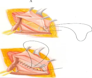
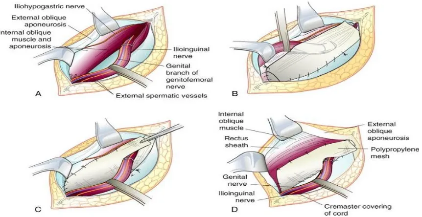
[image:58.612.144.457.108.374.2]Figure-19.LichtensteinTensionfreeMeshRepair
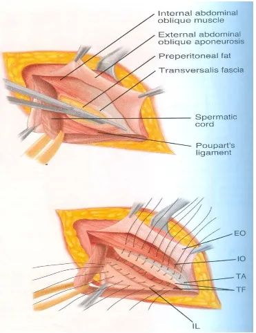
[image:59.612.139.464.405.599.2]Tension –FreeMeshHernioplasty (LichtensteinRepair)14 ,46
In 1984, Lichtenstein described a "tension free" onlay of polypropylene mesh for
inguinal hernia repairs. In this technique Lichtenstein repairs all primary direct and
indirect hernias without closure of defect.
Operative technique: A transverse skin crease incision is deepened down to the
external oblique aponeurosis. The spermatic cord is mobilized in the usual way.Direct
sacs are inverted and imbricated using a non absorbable suture to flatten the
posterior wall. Indirect sacs are dissected from the cord up to extra peritoneal fat and
then either excised or inverted. High dissection, rather than high ligation, is the
important feature of this stage.If deep ring is widened (Gilbert classification 2 or 3), a
cone of mesh is inserted and anchored,usually superolaterally and sometimes
inferiorly to the inguinal ligament by two or three non absorbable sutures.
Inguinoscrotal sacs are transected in the canal and the proximal portion closed and
dealt as mentioned earlier, where as the mouth of distal portion is left undissected, but
wide open.
Onlay mesh:47 ,48 A polypropylene mesh is sutured along its lower border to the
pubic tubercle,the lacunar ligament and the inguinal ligament to beyond the inte rnal
ring with a continuous suture of monofilament 3-0 polypropylene. The medial edge is
sutured to the rectus sheath, also with continuous suture. The superior edge is tacked