A STUDY ON BACTERIAL AND FUNGAL
INFECTIONS IN PATIENTS WITH DACRYOCYSTITIS
IN A TERTIARY CARE HOSPITAL
Dissertation submitted to
THE TAMILNADU DR.M.G.R.MEDICAL UNIVERSITY
CHENNAI
In partial fulfilment of the regulations for the award of
degree of
M.D. MICROBIOLOGY
(BRANCH – IV)
MADRAS MEDICAL COLLEGE
CHENNAI
CERTIFICATE
This is to certify that this dissertation titled “A STUDY ON BACTERIAL AND FUNGAL INFECTIONS IN PATIENTS WITH
DACRYOCYSTITIS IN A TERTIARY CARE HOSPITAL” is a bonafide
record of work done by DR.S.SHANMUGA SUNDARI during the period of her post graduate study from 2013-2016 under guidance and supervision in the Institute of Microbiology, Madras Medical College and Rajiv Gandhi Government General Hospital Chennai – 600003, in partial fulfilment of the requirement of M.D. MICROBIOLOGY degree examination of THE
TAMILNADU DR.MGR MEDICAL UNIVERSITY, CHENNAI, to be held
in April 2016.
Dr. R. VIMALA, M.D., Dr. MANGALA ADISESH, M.D.,
DEAN, DIRECTOR I/C,
Madras Medical College & Institute of Microbiology, Rajiv Gandhi Madras Medical College & Government General Hospital, Rajiv Gandhi
DECLARATION
I declare that the dissertation entitled “A STUDY ON BACTERIAL
AND FUNGAL INFECTIONS IN PATIENTS WITH
DACRYOCYSTITIS IN A TERTIARY CARE HOSPITAL” submitted by
me for the degree of M.D. MICROBIOLOGY is the record work carried out by me during the period of October 2014 – August 2015 under the Guidance of Prof. Dr.Mangala Adisesh, M.D., Institute of Microbiology, Madras Medical College, Chennai. This dissertation is submitted to The Tamilnadu Dr.M.G.R. Medical University, Chennai in partial fulfilment of the University regulations for the award of degree of M.D. Microbiology (Branch IV) examination to be held in April 2016.
Place: Chennai Signature of the Candidate
Date : (Dr.S.Shanmuga sundari)
Signature of the guide
Dr. Mangala Adisesh, M.D.,
Professor
Institute of Microbiology Madras Medical College,
ACKNOWLEDGEMENT
I humbly submit this work to the Almighty who has given the health and ability to pass through all the difficulties in the compilation and proclamation of this blue print.
I wish to express my sincere thanks to our Dean, Dr.R.Vimala, M.D.,
for permitting me to use the resources of this institution for my study.
I owe special thanks to Prof. Dr.Mangala Adisesh, M.D., Director I/C, Institute of Microbiology for her support, valuable suggestions, erudite guidance in my study and for being a source of inspiration in my endeavours.
I feel fortunate to work under her guidance and am thankful for her valuable suggestions and great support throughout my study.
My sincere thanks to Dr.K.Namitha Bhuvaneswari, M.S.D.O.,
Director & Superintendent, Regional Institute of Ophthalmology and Government Ophthalmic Hospital, Chennai, for permitting me to carry out my study.
I express my gratitude to our former Director, Prof. Dr.G.Jayalakshmi, M.D.,DTCD and former Professors Dr.T.Sheila Doris, M.D., and
Dr.K.Muthulakshmi, M.D., for their guidance and support.
I extend my whole hearted gratitude and special thanks to my Assistant Professor Dr.C.S.Sripriya,M.D.,for her valuable guidance and constant support in my study.
I also express my thanks to our Assistant professors Dr.R.Deepa, M.D., Dr.N.Rathnapriya, M.D., Dr.T.Usha Krishnan M.D., Dr.David Agatha
M.D., Dr.N.Lakshmipriya M.D.,DCH., Dr.K.G.Venkatesh M.D, and
Dr.B.Natesan M.D.,DLO., for their immense support in my study.
I hereby express my gratitude to all the technical staff for their help throughout my study.
I would like to thank my department colleagues and friends for their constant support and co-operation.
I would like to thank the Institutional Ethics Committee for approving my study.
TABLE OF CONTENTS
S. NO TITLE PAGE NO
1 INTRODUCTION 1
2 AIMS OF THE STUDY 4
3 REVIEW OF LITERATURE 5
4 MATERIALS AND METHODS 28
5 RESULTS 45
6 DISCUSSION 67
7 SUMMARY 77
8 CONCLUSION 80
9 COLOUR PLATES
10 APPENDIX-I ABBREVIATIONS
11 APPENDIX-II STAINS, REAGENTS AND MEDIA
12 ANNEXURE-I CERTIFICATE OF APPROVAL
13 ANNEXURE-II PROFORMA
14 ANNEXURE-III PATIENTS CONSENT FORM
15 ANNEXURE-IV MASTER CHART
ABSTRACT
Introduction:
Inflammation of the lacrimal sac is known as dacryocystitis which usually occurs due to obstruction in the nasolacrimal duct. It is a significant cause of ocular morbidity in children and adults. It can be congenital or acquired. Acquired dacryocystitis can occur as acute and chronic dacryocystitis. Dacryocystitis is mostly caused by bacteria and rarely by fungi. There is a change in etiological agents causing dacryocystitis over the time. Hence this study was undertaken to know the etiological agents of congenital, acute and chronic dacryocystitis.
Aims of the study:
To isolate and identify the bacteria and fungi from clinically diagnosed cases of dacryocystitis. To determine the antimicrobial susceptibility pattern of the bacterial and fungal isolates.
Materials and methods:
A total of 100 clinically diagnosed cases of dacryocystitis of all age groups attending the outpatient department at the Regional Institute of Ophthalmology and Government Ophthalmic Hospital were included in the study. Samples were collected from these patients and processed by standard microbiological techniques.
Results:
(34%). Left eye (63%) was commonly affected than the right eye (35%)and 2% of cases were bilateral. Overall culture positivity was 51.9%. Among the culture positive samples, 90.5% yielded single organism and 9.4% yielded mixed organisms. Totally 58 organisms were isolated, of which 58.6% were Gram positive bacteria, 37.9% were Gram negative bacteria, and 3.4% were fungal isolates.Streptococcus pneumoniae was the common pathogen in congenital dacryocystitis and Staphylococcus aureus was the common pathogen in acute and chronic dacryocystitis. The incidence of Gram negative pathogens were more in chronic dacryocystitis.Gram positive bacteria were highly sensitive to Gatifloxacin and least sensitive to Ciprofloxacin. Gram negative bacteria were highly sensitive to Tobramycin and least sensitive to Ciprofloxacin and Gentamicin. Candida albicans was isolated from two cases of chronic dacryocystitis and both were sensitive to Amphotericin B, Fluconazole, Voriconazole and Itraconazole.
Conclusion:
Microbial culture and sensitivity should be performed in all dacryocystitis cases. This would contribute to the choice of appropriate and effective antimicrobial agents.
1
INTRODUCTION
Inflammation of the lacrimal sac is known as dacryocystitis which usually occurs due to obstruction in the nasolacrimal duct. It has bimodal distribution affecting children less than 1 year and adults over 40 years of age[1]. It is a significant cause of ocular morbidity in children and adults. This disease is more common in patients with poor personal hygiene[2].
Dacryocystitis is an unpleasant disease, as it causes constant watering and discharge. Dacryocystitis is also a threat to the integrity of the eye by becoming the source of infection to orbital cellulitis and panophthalmitis[3,4].
Dacryocystitis can be classified as congenital and acquired dacryocystitis. Acquired dacryocystitis can occur as acute and chronic dacryocystitis. Chronic dacryocystitis is more common[1].
Nasolacrimal duct obstruction(NLD) can occur from different aetiologies, such as primary idiopathic obstruction and secondary obstruction which finally results in stasis of tears, desquamated cells and mucoid secretions in the lacrimal sac, this creates favourable environment for inflammation and infection[5,6].
2
Dacryocystitis is mostly caused by bacteria and rarely by fungi. The organisms causing dacryocystitis may be different in acute and chronic infections. In chronic dacryocystitis mixed infections are more common[5].
Congenital dacryocystitis is usually presents with epiphora in newborn. Later purulent discharge may develop resulting in matting of eyelashes. When not treated early, complications such as recurrent conjunctivitis, acute on chronic dacryocystitis, lacrimal abscess and fistula formation can occur.
Acute dacryocystitis usually presents with pain and tenderness over the lacrimal sac area. It may present with lacrimal abscess. Complications include acute conjunctivitis, lid abscess, orbital cellulitis, acute ethmoiditis and very rarely cavernous sinus thrombosis.
Chronic dacryocystitis is common than the acute one. It usually presents with persistent watering and discharge from the eye. Complications like chronic conjunctivitis, ectropion of the lower eye lid can occur. Because of prolonged watering, eczema and maceration of lower eye lid skin can occur. It is an important contributory factor for corneal ulcer development and panophthalmitis[7].
3
Untreated dacryocystitis will not undergo spontaneous resolution[1].
There are several bacteria known to have been implicated as etiological agents of dacryocystitis. There is a change in etiological agents causing dacryocystitis over the time. So knowing the range of the microorganisms causing dacryocystitis and their antibiotic sensitivity pattern in recent times may help in choosing the appropriate antimicrobial therapy.
There are relatively few studies conducted regarding microbiological characteristics of dacryocystitis. Most of them have studied a specific type of dacryocystitis.
4
AIMS OF THE STUDY
1. To isolate and identify the bacteria and fungi from clinically diagnosed
cases of dacryocystitis.
2. To determine the antibiotic susceptibility pattern of the bacterial isolates.
3. To determine the antifungal susceptibility pattern of fungal isolates obtained.
4. To determine and compare the incidence of the various bacterial agents
5
REVIEW OF LITERATURE
BACKGROUND:
Dacryocystitis occurs usually due to lacrimal outflow obstruction. It results in stasis of tears in the lacrimal sac. Lacrimal sac infection is usually due to bacterial infections. Only in 1.2% of cases dacryocystitis is caused by fungal organisms (CodenDj[8]).
HISTORICAL PERSPECTIVE:
The disease dacryocystitis has been known from the very earliest times due to its manifestations such as abscesses and fistula on the face. It has been known as Aegilops in the earliest times. The term, Aegilops include all inner canthus swellings. In 1702, George and Sehl of Helle described about Aegilops that, it was not the soft tissue affection but a consequence of obstruction to the lacrimal outflow which results in inflammation[9].
Hirschberg noted that the school of Hippocrates recognized the relationship between epiphora and aging[10]. Lacrimal sac abscess and fistula formation were reported by the ancient Greeks[11].
Platner(1724) observed the presence of nasal disease as a source of infection to dacryocystitis. Later this fact was stressed by Schirmer(1877) and also by Kuhnt(1891-95).
6
The disease commonly occurs in the middle age group, with high incidence in the fifth decade and it is rare in adolescents and children. In adults it is more common in females than males due to the narrow bony canal in females. In the newborn this disease affects both sexes equally. This fact was described by Meller(1929), Ruiz Barrabco, Martinez Romain(1966) and others.
Importance of racial and geographical incidence was described by Santos-Femandez, 1903-21. According to them, this disease is rare in Negroes. It is because of the presence of shorter and wider canal in Negroes.
Kofler (1919), Stenger(1920) and Bockstein(1926) observed that, septal deflection as a cause of nasolacrimal duct obstruction at the lower end. Schaeffer(1920) found that, mucosal abnormalities can produce stasis and also obstruction to the lacrimal outflow.
Dacryocystitis caused by Aspergillosis has been reported by Wright(1930) and Rosen Vold(1942).
Dacryocystitis caused by Candida albicans was reported by Fine and Waring(1947). Lacrimal sac abscess due to Candida albicans has been reported by Janokta (1970).
7
EPIDIMIOLOGY OF DACRYOCYSTITIS:
FREQUENCY:
Individuals having brachycephalic heads will have a higher incidence of dacryocystitis than those having dolichocephalic and mesocephalic heads. This is because; brachycephalic skulls have a narrow diameter of inlet in to the nasolacrimal duct. Individuals having flat nose and narrow face are more prone for development of dacryocystitis. This may be due to the presence of the narrow osseous canal in these individuals[14].
MORBIDITY AND MORTALITY:
In acute dacryocystitis patients will have severe morbidity and rarely mortality. Morbidity is primarily due to lacrimal abscess and spreading of infection.
The morbidity in chronic dacryocystitis is due to chronic watering, matting of eyelashes and inflammation, infection of conjunctiva.
Congenital dacryocystitis is associated with both morbidity and mortality. If not treated properly on time, orbital cellulitis can occur in newborns as orbital septum is poorly formed in them. Other complications like, brain abscess, meningitis and sepsis can occur in congenital dacryocystitis[14, 15]
8
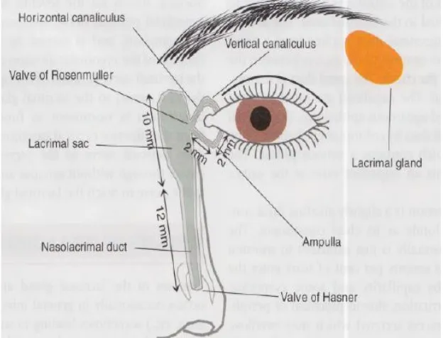
ANATOMY OF THE LACRIMAL DRAINAGE SYSTEM
The lacrimal apparatus consists of the following structures[16]:
1. Lacrimal gland- secretes tears,
2. Lacrimal punctum,
3. Lacrimal canaliculi,
4. Lacrimal sac,
[image:17.595.156.468.318.556.2]5. Nasolacrimal duct- carries tears in to nasal cavity.
Fig 1: Anatomy of the lacrimal apparatus.
Lacrimal gland:
9
surface between the fornix and tarsus edge. The ducts of acini, by forming a large duct open in to the fornix.
Lacrimal punctum:
Each eye lid has one punctum. It is situated near the posterior border of the eye lid, 6 mm from the medial canthus.
Lacrimal canaliculi:
Each eye lid has one canaliculi. It starts at the punctum. First it is directed vertically for 2 mm and it turns at the ampulla and then it runs horizontally for 6 to 7 mm. A common canaliculi is formed by the union of upper and lower canaliculi and opens in to the lacrimal sac. A fold of mucosa at this area forms the valve of Rosenmuller. This valve prevents the reflux of tears. The walls of canaliculi are lined by stratified squamous epithelium.
Lacrimal sac:
10 Nasolacrimal duct:
It is 18 mm long. It connects the lower end of lacrimal sac with the inferior turbinate of the nose. In the middle part it is narrower than the both ends. The nasolacrimal duct is lodged in the nasolacrimal bony canal, which lies between the nasal cavity and the maxillary sinus. It runs downwards, slightly outwards and backwards. It opens at the anterior part of inferior meatus of the nose. A mucosal flap forms the valve of Hasner close this opening when necessary. The nasolacrimal duct is lined by columnar cells.
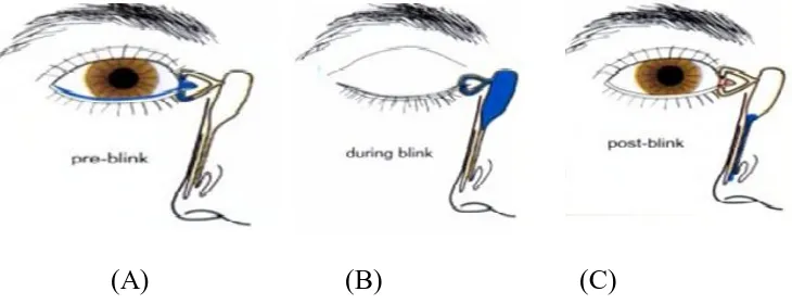
PHYSIOLOGY OF THE LACRIMAL DRAINAGE SYSTEM[1, 17]
Tears secreted by the lacrimal glands pass across the ocular surface, a variable amount of which is lost by evaporation. This is related to blink rate, ambient temperature, humidity and the size of the palpebral aperture. The remaining tears drain as follows(Fig 2):
Tears flow along the upper and lower marginal strips and it enters by
capillary action and suction into the upper and lower canaliculi(Fig 2 A).
With each blink of eye, the pretarsal orbicularis oculi compresses the
11
When the eyes open, the muscles relax; the lacrimal sac and canaliculi
expand and creates a negative pressure which draws the tears from the eye in to the empty lacrimal sac.
[image:20.595.129.494.190.330.2](A) (B) (C)
Fig 2: Physiology of the lacrimal drainage system.
12 DACRYOCYSTITIS
Dacryocystitis is defined as inflammation of the lacrimal sac. It occurs due to obstruction in the nasolacrimal duct. It is an unpleasant disease, because it can cause conspicuous watering and has minimum tendency to resolve. Nasolacrimal duct obstruction can occur from different aetiologies, such as primary idiopathic obstruction or secondary obstruction.
The reasons for secondary obstruction include stricture of the duct caused by chronic atrophic rhinitis, obstruction due to lacrimal sac tumours and maxillary sinusitis; obstruction of the distal end of the duct due to nasal polyp or inferior turbinate hypertrophy. Tuberculosis, syphilis and leprosy-originating from the surrounding bones or nose can also cause secondary obstruction [18]
.All the above conditions results in stasis of tears, desquamated cells and mucoid secretions in the lacrimal sac, which creates favourable environment for infection. The infection is mostly due to bacteria and rarely due to fungi. Dacryocystitis is treated with topical, systemic antibiotics, probing and silicone stent intubation. Surgery can be done to re-establish the duct patency[19].
Dacryocystitis can occur in two forms, congenital dacryocystitis and acquired dacryocystitis.
CONGENITAL DACRYOCYSTITIS
Etiology:
13
nasolacrimal duct especially at the valve of Hasner. It is also known as dacryocystitis neonatorum [20]. The common bacteria associated with this condition are Staphylococci, Pneumococci and Streptococci[21].
Clinical presentation:
Epiphora- develops seven days after birth,
Positive regurgitation test - when pressure is applied over the lacrimal sac area, there may be a mucoid or mucopurulent regurgitation through the puncta,
Swelling over the sac area may appear.
Complications:
If it is not treated early, complications like,
Recurrent conjunctivitis,
Acute on chronic dacryocystitis,
Lacrimal abscess and formation of fistula can occur[21].
ACQUIRED DACRYOCYSTITIS:
Acquired dacryocystitis can occur both as acute and chronic dacryocystitis. Chronic dacryocystitis is more common.
ACUTE DACRYOCYSTITIS:
14 Etiology:
It may develop due to direct involvement from infected para nasal sinuses, dental abscess or caries teeth or, as an acute exacerbation of chronic dacryocystitis. The most common organisms responsible are Staphylococcus
aureus and Streptococcus species[22].
Clinical presentation:
It can be divided in to three stages.
1. Stage of cellulitis:
It presents with,
Painful swelling over the lacrimal sac area,
Epiphora,
Fever and malaise,
Spread of edema to eyelids and cheeks.
2. Stage of lacrimal abscess:
Continuation of inflammation results in occlusion of canaliculi. The lacrimal sac is filled with pus. It presents with large fluctuant swelling pointing below and outer part of the sac.
3. Stage of fistula formation:
15 Complications:
It includes,
Mucocele,
Fistula formation,
Chronic conjunctivitis,
Orbital cellulitis.
CHRONIC DACRYOCYSTITIS
Chronic dacryocystitis is more common than the acute one.
Etiology:
The etiological factors are grouped as,
I.Predisposing factors
Age: It is more common over 40 years of age.
Sex: It is more common in females because of comparatively narrow bony canal lumen. As it coincides with menopausal age, an endocrine basis can be suggested. Due to endocrine changes, hypertrophy of the mucous membrane can occur, which will be infected easily and leads to obstruction of nasolacrimal duct[18].
Heredity: It may play a role by affecting the facial configuration with respect to the length and width of the NLD.
16
II. Factors result in stasis of tears in lacrimal sac
Anatomical factor: Those who are having narrow bony canal are at risk of developing this condition.
Excessive lacrimation: Primary and reflux lacrimation can cause stagnation of tears in the lacrimal sac.
Mild inflammation of sac: It can occur due to recurrent conjunctivitis. The epithelial debris,mucus plugs will block the NLD.
Lower end NLD obstruction: This can occur due to nasal diseases such as, nasal polyps, inferior turbinate hypertrophy, deviated nasal septum, stenosis caused by atrophic rhinitis[21, 23].
III. Source of infection:
Infection can spread from conjunctiva, nasal cavity, paranasal sinuses.
IV. Causative microorganisms:
Staphylococcus species, Pneumococcus species, Pseudomonas species
are common infective organisms[21].
Clinical presentation:
It is divided in to four stages.
1.Stage of catarrhal dacryocystitis: The only symptom at this stage is watering from the eye.
17
pressing the swelling there will be mucoid discharge regurgitates from the lower punctum.
3.Stage of chronic suppurative dacryocystitis: At this stage, because of pyogenic infections, the mucoid discharge will become purulent. The mucocele is converted to pyocele. Patients present with, epiphora, recurrent conjunctivitis and swelling at the medial canthus. On pressing the swelling there will be frank purulent discharge which regurgitates from the lower punctum.
4.Stage of chronic fibrotic sac: Due to repeated infections, a small fibrotic sac will develop. Patients present with persistent epiphora and discharge.
Complications:
Chronic conjunctivitis
Acute on chronic dacryocystitis
Ectropion of the lower eye lid
Corneal abrasions, which can be infected and leads to hypopyon
corneal ulcer,
Risk of development of endophthalmitis, if any intraocular
18
CLINICAL EVALUATION OF THE NASOLACRIMAL SYSTEM
Examination by diffuse illumination using magnification:
This test is done to rule out causes of reflex hypersecretion located in eye lids, conjunctiva and cornea.
Regurgitation test:
In this test, steady pressure is applied over the lacrimal sac area. Reflux of watery, mucoid or mucopurulent discharge through the punctum indicates obstruction in the nasolacrimal duct.
Syringing[24]:
In this test, after instillation of topical anaesthetic agent, with the help of Nettleship punctum dilator, the lower punctum is dilated. Then saline is injected through a smooth tipped cannula passed into the lacrimal canaliculi. If saline passes into the nose or throat freely, it indicates a patent nasolacrimal duct. In the presence of nasolacrimal duct obstruction, the fluid will regurgitate through the upper punctum.
Fluorescein dye disappearance test[25]:
19
Nasal endoscopy:
This is helpful in evaluation of diseases of nasal septum and turbinate[14].
Dacryocystography[26]:
This is used to know the level of obstruction. To perform the procedure, a radiopaque material such as lipiodol is pushed into the lacrimal sac by using a lacrimal cannula. Then X rays are taken after 5 and 30 minutes for visualisation of the entire passage.
CT scan:
This is useful in craniofacial injuries, congenital craniofacial deformities. It also helps in evaluation of concomitant sinus or nasal diseases[27].
MANAGEMENT OF CONGENITAL DACRYOCYSTITIS[21]:
Up to 6-8 weeks of age:
Massage over the lacrimal sac area should be done at least 4 times per day. This should be followed by installation of antibiotic drops. This method can cure 90% of infants.
Up to 2 months of age:
20
antibiotic installation. Syringing should be carried out once or twice a week.
Up to 3-4 months of age:
If the condition is not cured by syringing, Probing of nasolacrimal duct with Bowman’s probe can be done. It is performed under general anaesthesia. If it is failed the same procedure can be repeated after one month.
Up to 4 years:
Conservative treatment like, massaging, topical antibiotics and intermittent syringing should be done up to 4 years of age.
After 4 years of age:
Dacryocystorhinostomy surgery can be performed.
MANAGEMENT OF ACUTE DACRYOCYSTITIS:
At the Stage of cellulitis:
At this stage, treatment consists of systemic antibiotics and anti-inflammatory drugs with hot fomentation.
At the Stage of lacrimal abscess:
21
At the Stage of fistula formation:
Fistulectomy along with Dacryocystectomy or Dacryocystorhinostomy can be done.
MANAGEMENT OF CHRONIC DACRYOCYSTITIS:
Conservative treatment:
In the early period, repeated lacrimal syringing can be done. Antibiotics should be given.
Surgical procedures:
1. Dacryocystorhinostomy (DCR):
It is the preferred surgery because the lacrimal drainage is reestablished. The infection should be controlled before doing the surgery. DCR can be performed either by conventional external approach or by endo nasal DCR.
2.Dacryocystectomy (DCT):
When DCR is contraindicated, this surgery can be performed. Indications are,
Patients more than 60 years of age,
Presence of shrunken and fibrosed sac,
Infections with tuberculosis, leprosy and mycotic infections
Lacrimal sac tumours
Nasal diseases particularly atrophic rhinitis
3.Conjunctivodacryocystorhinostomy:
22
MICROBIOLOGY OF DACRYOCYSTITIS
Normal flora of the conjunctiva:
The predominant organisms of the conjunctiva include, diphtheroids
mainly Corynebacterium xerosis, Moraxella species, Staphylococcus
epidermidis and nonhemolytic Streptococci. Normally the conjunctival flora is
held in check by the flow of tears and the antibacterial lysozyme it contain[28,29].
Bacterial and fungal profile of dacryocystitis:
Because of the contiguity of mucous lined tract of the lacrimal drainage system with conjunctival and nasal mucosal surfaces, organisms from both ends of lacrimal passage can infect the lacrimal sac. Infection from para nasal sinuses and oral cavity can also spread to the sac.
The common bacteria causing dacryocystitis are Streptococcus
pneumoniae, Staphylococcus aureus, Streptococcus pyogenes and Haemophilus
influenzae. The common fungi causing dacryocystitis are Candida albicans,
Aspergillus species[30].
23
isolated in acute cases. In chronic cases also the predominant organism was
Streptococcus pneumoniae(35.7%) followed by Staphylococcus
aureus(28.5%)and Pseudomonas aeruginosa(25%).
JithendraKandati et al[5] in January 2015 studied 298 patients of dacryocystitis. It included 126 acute cases and 172 chronic cases. In both acute and chronic cases, females were more affected than males. Bacterial and fungal culture was performed in all the cases. Culture positivity was 85.57%. In two cases of chronic dacryocystitis, Candida albicans was isolated. Polymicrobial growth was exclusively present in chronic dacryocystitis. Overall,
Staphylococcus aureus(24.5%) was the common gram positive bacteria
isolated, followed by Staphylococcus epidermidis(20.3%) and Streptococcus
pneumoniae(3.8%). Pseudomonas aeruginosa(21.3%) was the common gram
negative bacteria isolated, followed by Escherichia coli(16.8%), Klebsiella
pneumoniae (7%).
IndrajitSarkar et al[32] in July 2015 studied 90 patients of dacryocystitis. It included 3 cases of congenital cases and 87 cases of chronic cases. Culture positivity was 83.33%. Single organism was isolated in 73.3% of samples, mixed organisms were isolated in 10% of samples.78.57%of isolates were Gram-positive bacteria and 21.43% of isolates were Gram-negative bacteria. The predominant organism isolated was Staphylococcus aureus(57.1%) followed by Streptococcus pneumoniae(14.2%), Pseudomonas
24
Shwetha B A et al[3] in 2014 studied 200 patients of chronic dacryocystitis. The percentage of culture positivity was 57.5%. The predominant organism isolated was Staphylococcus aureus(32.17%) followed
by Klebsiella pneumoniae (14.78%),Streptococcus pneumoniae(11.3%),
Staphylococcus epidermidis(8.69%) and mixed growth (6.08%).
Bahram Eshraghi et al [33] in 2014 studied 100 patients of dacryocystitis which included 60 acute cases and 40 chronic cases. Overall culture positivity was 85%. Staphylococcus aureus was the predominant organism isolated from acute cases (35%) followed by Streptococcus viridans (17%), Klebsiella
species(8%), Citrobacter species(8%), Pseudomonas aeruginosa(4%),
Escherichia coli(4%),Staphylococcus epidermidis(4%) and Aspergillus
species(2%).In chronic cases Staphylococcus epidermidis (38%) was the
predominant organism isolated, followed by Streptococcus viridans(20%),
Klebsiella species(10%) and Staphylococcus aureus(10%).
Khevna Patel et al [2] in 2014 studied 100 patients of chronic dacryocystitis which included 52 female patients and 48 male patients. Culture positivity was seen in 83% of samples. The predominant organism isolated was
Staphylococcus aureus(41%) followed by Escherichia coli(17%),
Pseudomonas aeruginosa(12%), Streptococcus pneumoniae(9%) and
Klebsiella pneumoniae(3%).
25
were isolated from 44 specimens. The predominant isolate was Coagulase
negative Staphylococci(71%), followed by Staphylococcus aureus (14%),
Escherichia coli(4.7%) and Citrobacter freundii(4.7%).
Prakash R et al[35]in 2012 studied 80 patients of dacryocystitis which included both congenital and acquired cases. Females (70%) were more affected than males(30%). Staphylococcus aureus (29.76%) was the common organism isolated in acquired cases, followed by Streptococcus
pneumoniae(20.23%), Pseudomonas aeuginosa(14.28%), Klebsiella
pneumoniae(8.33%) and Staphylococcus epidermidis(8.33%). Streptococcus
pneumoniae(50%) was the common organism isolated in congenital cases,
followed by Pseudomonas aeruginosa(20%) and Staphylococcus aureus(10%).
Shah C P et al[36] in 2011 studied 100 cases of dacryocystitis, which included 45 acute cases and 55 chronic cases. Culture positivity was 100%. Mixed cultures were isolated in 9.1% of samples. Escherichia coli was the predominant organism isolated from acute cases (22.9%) followed by
Staphylococcus aureus(18.75%), Pseudomonas aeruginosa(18.75%),
Coagulase negative Staphylococci(16.6%), Streptococcus species(12.5%) and
Klebsiella species(10.4%). Staphylococcus aureus was the predominant
organism isolated from chronic cases (27%) followed by, Pseudomonas
aeruginosa(21.6%), Klebsiella species(14.8%), Coagulase negative
Staphylococci(12.1%), Streptococci species(12.1%) and Escherichia
26
Chaudhary M et al [37] in 2010 studied 120 patients of chronic dacryocystitis. Culture positivity was 76.66%. Single organism was isolated in 85.86% and mixed growth was present in 14.13% of culture positive cases. The predominant organism isolated was Coagulase Negative
Staphylococci(33.96%) followed by, Staphylococcus aureus(25.46%) and
Streptococcus pneumoniae(19.81%). Escherichia coli(5.66%) was the common
gram negative organism isolated.
Bharathi M J et al[38] in 2008 studied 1891 patients of dacryocystitis, which included 566 patients of acute dacryocystitis, and 1325 patients of chronic dacryocystitis. Culture was positive from 80.3% of the samples. The percentage of culture positivity was 90% in chronic dacryocystitis and 57.4% in acute dacryocystitis. In acute dacryocystitis the predominant organism was
Staphylococcus aureus(22.3%) followed by Pseudomonas aeruginosa(21.1%).
In chronic dacryocystitis the predominant organism was Coagulase negative
Staphylococci(44.2%) followed by Staphylococcus aureus(10.8%) and
Streptococcus pneumoniae(10%).
Mandal R et al [39] in 2008 studied 56 patients of chronic dacryocystitis who underwent either dacryocystectomy or dacryocystorhinostomy. Part of the sac was collected as samples. Culture positivity was 53.6%. Staphylococcus
aureus(40%) was the common gram positive organism isolated, followed by
Staphylococcus epidermidis(10%) and Streptococcus pneumoniae(10%).
27
isolated, followed by Klebsiella pneumoniae(6.6%) and Haemophilus
influenzae (6.6%).
BhavnaRaina et al [40] in 2010 studied 30 cases of congenital dacryocystitis. Culture positivity was 72.9%. Majority of bacterial isolates were Gram positive cocci (56.7%). Streptococcus pneumoniae (27.9%) was the predominant organism isolated, followed by Staphylococcus aureus(16.2%)
and Staphylococcus albus(10.8%).
Andreas Kuchar et al [41] in 2000 studied 47 cases of congenital dacryocystitis, which included 3 bilateral cases. Totally 50 samples were studied. Culture positivity was 72.64%. 20 samples had mixed growth. The most common organism isolated was Streptococcus pneumoniae(35.4%), followed by Haemophillus influenzae (19.6%), Pseudomonas
28
MATERIALS AND METHODS
This study was carried out in the Institute of Microbiology, Madras Medical College, Chennai in association with Regional Institute of Ophthalmology and Government Ophthalmic Hospital, Chennai. A total of 100 clinically diagnosed cases of dacryocystitis of all age groups attending outpatient department at the Regional Institute of Ophthalmology and Government Ophthalmic Hospital were included in the study. The patients were categorized as acute, chronic and congenital dacryocystitis based on the age of onset, symptoms and signs.
ETHICAL CLEARANCE:
Before the commencement of the study, approval was obtained from the Institutional Ethics Committee. Informed consent was obtained from all the patients who satisfied the inclusion criteria.
STUDY DESIGN:
Cross sectional study.
STUDY PERIOD:
October 2014 to August 2015
INCLUSION CRITERIA:
29
Patients presenting with pain, redness and swelling in the region of
lacrimal sac were included as acute dacryocystitis.
Adult patients presenting with persistent epiphora for more than 3 weeks and regurgitation of serous, mucoid or mucopurulent material on pressure over the lacrimal sac area or on lacrimal syringing were included as chronic dacryocystitis.
Children presenting with persistent epiphora from the first week of birth
and regurgitation of serous, mucoid or mucopurulent material on pressure over the lacrimal sac area or on lacrimal syringing were included as congenital dacryocystitis.
EXCLUSION CRITERIA:
The patients with the above symptoms who had received either topical or systemic antibiotics for the past one week during their visit to the hospital were excluded from the study.
All cases of pseudo epiphora and epiphora caused by conditions other than nasolacrimal duct obstruction were excluded from the study.
The diagnosis of dacryocystitis when given by Ophthalmologist, then samples were collected with their help.
SAMPLE COLLECTION:
30
SAMPLES:
Pus or mucopurulent discharge drained by incision and drainage was
collected in cases of acute dacryocystitis.
Serous, mucoid or mucopurulent discharge obtained by syringing of the lacrimal sac was collected in cases of chronic dacryocystitis. Lacrimal sac contents obtained during DCT or DCR were also collected in few cases.
Serous, mucoid or mucopurulent discharge obtained by syringing of the lacrimal sac or obtained on pressure over the lacrimal sac area was collected in cases of congenital dacryocystitis. Lacrimal sac contents obtained during DCR were also collected in some cases.
TRANSPORTATION AND PROCESSING OF SAMPLES:
The samples were transported to the laboratory immediately and processed. Of the three swabs collected, one swab was used for direct gram staining and KOH mount. The other swab was used for inoculation of bacterial culture. The last swab was used for inoculation of fungal culture.
DIRECT EXAMINATION:
Direct gram staining:
31
arrangement, morphology and presence of gram positive budding yeast cells if any.
10% KOH wet mount:
The sample was added to a drop of 10% KOH on a clean glass slide. A coverslip was placed over the drop and allowed to stand at room temperature for 10 minutes and examined under microscope using low power and high power objectives for the presence of fungal elements.
BACTERIAL CULTURE:
The next swab was inoculated on to MacConkey agar plate, Blood agar plate and were incubated at 37ºC and on to chocolate agar plate and incubated at 5-10% CO2 atmosphere at 37
º
C for 24 hours.
After 24 hours of incubation the culture plates were observed for growth, morphology of colonies and were subjected to Gram staining. If Gram staining shows Gram positive cocci, catalase test, coagulase test and other standard biochemical reactions and tests[42, 43] were done. If Gram staining shows Gram negative bacilli, catalase test, oxidase test, motility by hanging drop method and other standard biochemical reactions and tests were done.
If there was no growth at 24 hours, the plates were further incubated for another 24 hours. If no growth was observed after 48 hours of incubation the culture was considered as negative for aerobic bacterial growth.
Bacterial cultures were considered significant, if
32
Presence of confluent growth at the inoculation site,
Same growth of organism was demonstrated on all the inoculated
culture media plates.
IDENTIFICATION OF ISOLATES:
Beta hemolytic colonies and golden yellow pigment on blood agar, Gram positive cocci in clusters in Gram stain, catalase test positive, slide coagulase test positive, tube coagulase test positive, urease test positive, mannitol fermenting, phosphatase producing, MR test positive and VP test positive isolates were identified as Staphylococcus aureus.
White opaque colonies on blood agar, Gram positive cocci in clusters in Gram stain, catalase test positive, slide coagulase test negative, tube coagulase test negative, phosphatase producing, Novobiocin sensitive and Polymyxin B resistant isolates were identified as Staphylococcus epidermidis.
Alpha haemolytic colonies on blood agar, Gram positive flame shaped
diplococci in Gram stain, catalase test negative, optochin sensitive, bile solubility test positive and inulin fermenting isolates were identified as
Streptococcus pneumoniae.
33
Lactose fermenting mucoid colonies on MacConkey agar, short Gram
negative bacilli in Gram stain, non-motile bacilli detected by hanging drop method, catalase test positive, oxidase test negative, nitrate reduction test positive, indole test negative, MR test negative, VP test positive, citrate utilization test positive, acid butt and acid slant with gas on TSI, lysine decarboxylase test positive, ornithine decarboxylase test negative, urease test positive isolates were identified as Klebsiella
pneumoniae.
Lactose fermenting colonies on MacConkey agar, Gram negative bacilli
in Gram stain, motile bacilli detected by hanging drop method, catalase test positive, oxidase test negative, nitrate reduction test positive, indole test positive, MR test positive, VP test negative, citrate utilization test negative, acid butt and acid slant with gas on TSI, lysine decarboxylase test positive,urease test negative isolates were identified as Escherichia coli.
34
ANTIMICROBIAL SUSCEPTIBILITY TESTING:[44,46]
All the bacterial isolates obtained were subjected to antimicrobial susceptibility testing by using Kirby-Bauer disc diffusion method. To know the efficacy, antibiotic discs were tested against standard American Type Culture Collection (ATCC) control strains, Staphylococcus aureus ATCC 25923,
Pseudomonas aeruginosa ATCC 27853, Escherichia coli ATCC 25922 as a
quality control laboratory procedure.
Antimicrobial susceptibility testing by Kirby – Bauer Disc Diffusion method:
3- 5 well isolated colonies from an agar plate culture were touched by a
sterile bacteriological loop and emulsified in 3-4ml of sterile peptone water. The bacterial suspension was matched to a 0.5 McFarland standards.
By using a sterile cotton swab, the suspension was streaked in three directions on to the surface of a Mueller Hinton Agar plate. Mueller Hinton Agar with 5% sheep blood was used for antibiotic sensitivity testing of Streptococcus pneumoniae.
With the lid in place, the inoculated agar surface was allowed to dry for 3 to 5 minutes before placing the antibiotic discs.
By using sterile forceps, appropriate antibiotic discs were placed on the surface of the agar. Five discs were placed per 90 mm plate.
35
Panel of antibiotics used in antimicrobial susceptibility testing of Gram positive cocci.
Antibiotic Disc content
Gram positive cocci
Diameter of zone of inhibition in mm[44]
Sensitive Inter
-mediate Resistant
Penicillin 10 units Staphylococcus
species ≥ 29 - ≤ 28
Cefoxitin 30 µg
Staphylococcus
aureus ≥ 22 - ≤ 21
CoNS ≥ 25 - ≤ 24
Oxacillin 1 µg S.pneumoniae ≥20 Amikacin 30 µg Staphylococcus
species ≥ 17 15-16 ≤ 14
Tobramycin 10 µg Staphylococcus
species ≥15 13-14 ≤12
Erythromycin 15 µg
Staphylococcus species& Enterococcus species
≥ 23 14-22 ≤13
S.pneumoniae ≥ 21 16-20 ≤15
Ciprofloxacin 5 µg
Staphylococcus species& Enterococcus species
≥ 21 16-20 ≤15
Gatifloxacin 5 µg
Staphylococcus
species ≥ 23 20-22 ≤19
S.pneumoniae ≥ 21 18-20 ≤17
Trimethoprim- Sulfamethoxazole
1.25/
23.75µg S.pneumoniae ≥ 19 16-18 ≤15 Ampicillin 10 µg Enterococcus
species ≥17 - ≤16
Vancomycin 30 µg Enterococcus
species ≥17 15-16 ≤14
Tetracycline 30 µg
Enterococcus species& Staphylococcus species
≥ 19 15-18 ≤14
S.pneumoniae ≥ 28 25-27 ≤24
Chloramphenicol 30 µg
Staphylococcus species& Enterococcus species
≥ 18 13-17 ≤12
36
Panel of antibiotics used in antimicrobial susceptibility testing of Gram negative bacilli.
Antibiotic Disc content
Gram negative bacilli
Diameter of zone of inhibition in mm[44]
Sensitive Inter
-mediate Resistant
Amikacin 30 µg
Enterobacteriaceae &Pseudomonas aeruginosa
≥ 17 15-16 ≤ 14
Gentamicin 10 µg
Enterobacteriaceae &Pseudomonas aeruginosa
≥15 13-14 ≤12
Cefotaxime 30µg Enterobacteriaceae ≥26 23-25 ≤22
Ceftazidime 30µg
Enterobacteriaceae
≥21 18-20 ≤17
Pseudomonas
aeruginosa ≥18 15-17 ≤14
Ciprofloxacin 5 µg
Enterobacteriaceae &Pseudomonas aeruginosa
≥ 21 16-20 ≤15
Tobramycin 10 µg
Enterobacteriaceae &Pseudomonas aeruginosa
≥15 13-14 ≤12
Gatifloxacin 5 µg Enterobacteriaceae ≥18 15-17 ≤14
Chloramphenicol 30 µg Enterobacteriaceae ≥ 18 13-17 ≤12
Piperacillin-tazobactam
100µg/ 10 µg
Pseudomonas
aeruginosa ≥21 15-20 ≤14
37
Detection of methicillin resistance in Staphylococcus aureus and Coagulase negative Staphylococci (CoNS) isolates by cefoxitin disc diffusion test [44]:
All Staphylococcus aureus and Coagulase negative Staphylococci
(CoNS) isolates were subjected to cefoxitin disc diffusion test. Cefoxitin is used
as a surrogate marker for detection of mecA-mediated oxacillin resistance [44]
.The bacterial suspension of test isolates were matched to a 0.5 McFarland standards and lawn cultured on Mueller Hinton Agar plates separately. Cefoxitin (30µg) disc were placed on the surface of lawn culture of the isolates. The plates were incubated in ambient air at 35°C for 24 hours.
Interpretation as per CLSI guidelines:
For Staphylococcus aureus and Staphylococcus lugdunensis
Zone of inhibition ≥22mm - Sensitive
Zone of inhibition ≤21mm - Resistant
For Coagulase negative Staphylococci except Staphylococcus lugdunensis
Zone of inhibition ≥25mm - Sensitive
Zone of inhibition ≤24mm - Resistant
38
Screening Test for Extended -Spectrum β-Lactamases (ESBLs) in
Klebsiella pneumoniae and Escherichia coli isolates[44,45].
All the Klebsiella pneumoniae and Escherichia coli isolates were subjected to initial screening test for Extended -Spectrum β-Lactamases by using Cefotaxime and Ceftazidime discs as per CLSI guidelines. The use of more than one antimicrobial agent for screening improves the sensitivity of ESBL detection [44].
Test method - Disk diffusion
Procedure:
The bacterial suspension of test isolates were matched to a 0.5 McFarland standards and lawn cultured on Mueller Hinton Agar plates separately. Ceftazidime (30 µg) and Cefotaxime (30 µg) discs were placed on the surface of lawn culture of the isolates. The plates were incubated in ambient air at 37°C for 16-18 hours.
Results:
39 FUNGAL CULTURE:
The last swab was inoculated on to two Sabourauds Dextrose Agar (SDA) slopes and incubated at 250C and 370C. The culture was examined daily for one week and then three times per week for the next three weeks. If there is no growth at 4 weeks of incubation, the culture was considered as negative for fungal growth.
IDENTIFICATION OF FUNGAL ISOLATES[47]:
If growth was seen on SDA slope, the morphology of colonies was observed. If cream coloured pasty colonies were seen, Gram staining was performed. Presence of Gram positive ovoid budding yeast cells in Gram staining was suggestive of Candida species. The following tests were done.
Germ tube test:
0.5 ml of sterile serum was taken in a sterile test tube. With a sterile bacteriological loop, a colony of yeast was touched and emulsified in the serum. The tube was incubated at 37oC for 2 hours. After 2 hours, a drop of serum was transferred to a clean glass slide. A coverslip was put and examined under microscope using low power and high power objectives[47,48].
Interpretation:
40 Candida CHROM Agar:
The yeast colonies were inoculated on to Candida CHROM Agar and incubated at 37oC. The plates were read after 48 hours and the colour of the colony was observed.
Observation & Interpretation:
Candida albicans - Light green
Candida dubliniensis - Dark green
Candida glabrata - Pink to Purple
Candida krusei - Pink
Candida parapsilosis - Cream to pale pink
Candida tropicalis - Blue with pink hallow[49]
Corn Meal Tween 80 Agar (Dalmau Plate Culture Technique)[48,49]
By using a straight wire an isolated colony from the primary culture
media was taken and inoculated on to cornmeal agar plate by making three parallel lines.
A sterile cover slip was placed over the surface of the agar, covering the
inoculated streaks in such a way that the streak project beyond the coverslip.
41
After 48 hours, the edge of the coverslip was examined under low power
objective and high power objective for the presence of pseudo hyphae, blastoconidia and chlamydospores.
ANTIFUNGAL SUSCEPTIBILITY TESTING OF YEAST:
Disc Diffusion method
Broth Micro dilution Method
Disc diffusion method[50]:
This method was carried out following the M 44-A CLSI guidelines using fluconazole and voriconazole discs.
Inoculum preparation and application of discs:
With a sterile bacteriological loop, 3- 5 yeast colonies were taken from the culture grown on SDA and emulsified in 5ml of sterile saline.
The yeast suspension was matched to a 0.5 McFarland standards.
By using a sterile cotton swab, the suspension was streaked in three
directions on to the surface of a Mueller Hinton Agar plate supplemented with 2% glucose and 0.5 µg/ml methylene blue.
By using sterile forceps, fluconazole and voriconazole discs were placed
42
The plates were incubated at 37oC. After 24 hours of incubation, the diameter of zone of inhibition was measured and interpreted as sensitive or resistant according to the CLSI guidelines44A. Quality control strain used-ATCC Candida albicans 90028.
Antifungal disc Disc content
Diameter of zone of inhibition in mm
Sensitive
Susceptible Dose Dependent
Resistant
Fluconazole 25 µg ≥17 14-16 ≤13
Voriconazole 1 µg ≥17 14-16 ≤13
Broth micro dilution method[51]
The test was performed as per the CLSI guidelines. Minimal Inhibitory Concentration of Fluconazole, Amphotericin B and Itraconazole were determined. Dimethyl sulfoxide (DMSO) was used as solvent. Media used was RPMI 1640.Preparation of inoculum and antifungal stock solution was done according to the CLSI guidelines.
The concentrations of the drugs tested were,
Amphotericin B 16 to 0.0313μg/ml
Fluconazole 64 to 0.125 μg/ml
Itraconazole 16 to 0.0313 μg/ml
43
Procedure:
The broth micro dilution test was performed by using sterile,
disposable, multi well micro dilution plates (96U-shaped wells).
100μl of varying drug concentrations was dispensed in each row
from 1 to 10. Then 100μl of test inoculum was dispensed from
row 1 to 10.
Row 11 was the growth control well contains 100 μl of sterile,
drug-free medium and was inoculated with 100 μl of the corresponding inoculum suspension. Row 12 was the drug control well contains 100 μl of drug and 100 μl of sterile, drug-free medium.
The micro dilution plates were incubated at 350C for 48 hours. After 48 hours the micro dilution wells were observed with the aid of reading mirror. The growth in each well was compared with that of the growth control well.
For Amphotericin B, MIC was interpreted as the lowest concentration in which the well was clear.
44
Interpretive Guidelines for In Vitro Susceptibility Testing of Candida Species:
Break points (μg/ml) for Candida species.
Antifungal
agent Susceptible
Susceptible Dose
Dependent Resistant
Fluconazole ≤ 8 16-32 ≥64
Itraconazole ≤ 0.125 0.25-0.5 ≥1
Amphotericin B ≤ 1 - >1
Statistical analysis:
45
RESULTS & ANALYSIS
This study was carried out at the Institute of Microbiology, Madras Medical College, Chennai, in association with Regional Institute of Ophthalmology and Government Ophthalmic Hospital, Chennai. 100 clinically diagnosed patients of dacryocystitis of all age groups were studied from October 2014 to August 2015.
Out of 100 patients, 98 patients had unilateral dacryocystitis and 2 patients had bilateral dacryocystitis. Totally 102 samples were collected from 100 patients.
TABLE NO: 1. ANALYSIS OF AGE DISTRIBUTION OF
DACRYOCYSTITIS CASES
Age group in years
Male Female
Total
No Percentage
Below 10 7 5 12 12 %
11-20 0 1 1 1 %
21-30 0 2 2 2 %
31-40 3 6 9 9 %
41-50 4 23 27 27 %
51-60 8 14 22 22 %
61-70 7 11 18 18 %
46
12%
1% 2%
9%
27% 22%
18%
9%
Below 10
11-20
21-30
31-40
41-50
51-60
61-70
71-80
This study shows highest number of acquired dacryocystitis cases among people in the age group of 41-50 years(27%) followed by 51-60 years (22%)and 61-70 years(18%). Overall the mean age is 47.97 years. The highest number of females were in the age group of 41-50 years and of males were in the age group of 51-60 years.
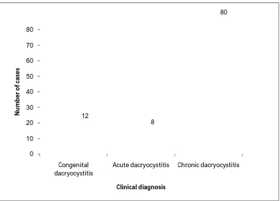
47 0 10 20 30 40 50 60 70 80 Congenital dacryocystitis
Acute dacryocystitis Chronic dacryocystitis 12 8 80 N u m b e r o f ca se s Clinical diagnosis
TABLE NO: 2. CLINICAL DIAGNOSIS OF DACRYOCYSTITIS AND THEIR DISTRIBUTION
Clinical diagnosis of dacryocystitis
Total no of
cases Percentage
P value<0.05 Congenital dacryocystitis 12 12%
Acute dacryocystitis 8 8%
Chronic dacryocystitis 80 80%
From the above table it is observed that, in our study chronic dacryocystitis is more common than the congenital and acute dacryocystitis and it is statistically significant.
[image:56.595.110.514.426.717.2]48
34%
66%
Male
Female
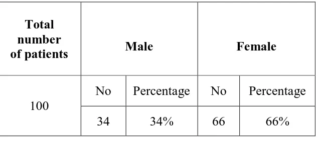
TABLE NO: 3. GENDER DISTRIBUTION OF DACRYOCYSTITIS CASES
Total number
of patients Male Female
100
No Percentage No Percentage
34 34% 66 66%
Gender distribution of dacryocystitis cases.
[image:57.595.143.460.120.262.2]49 0 10 20 30 40 50 60 Congenital Dacryocystitis Acute Dacryocystitis Chronic Dacryocystitis 7 3 24 5 5 56 N u m b er o f ca se s Diagnosis Male Female
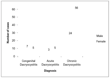
TABLE NO: 4. DISTRIBUTION OF DACRYOCYSTITIS CASES WITH RESPECT TO GENDER AND CLINICAL DIAGNOSIS.
Gender
No of cases
Total P value >0.05 Congenital dacryocystitis Acute dacryocystitis Chronic dacryocystitis
Male 7 3 24 34
Female 5 5 56 66
Total 12 8 80 100
Distribution of dacryocystitis cases with respect to gender and clinical diagnosis.
50
63% 35%
2%
Left eye
Right eye
Bilateral
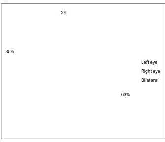
TABLE NO:5. DISTRIBUTION OF DACRYOCYSTITIS CASES ACCORDING TO THE EYE AFFECTED
Side affected No of cases Percentage
Left eye 63 63
Right eye 35 35
Bilateral 2 2
Total 100 100
Distribution of dacryocystitis cases according to the eye affected
From these findings it is observed that in this study, left eye (63%) is more affected than the right eye (35%).
[image:59.595.140.475.319.608.2]51 0 5 10 15 20 25 30 35 40 45
Left Right Bilateral
[image:60.595.141.505.328.601.2]21 12 1 42 23 1 N u m b e r o f ca se s Eye affected Male Female
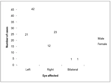
TABLENO:6. ANALYSIS OF DISTRIBUTION OF DACRYOCYSTITIS CASES ACCORDING TO GENDER AND EYE AFFECTED
Gender Eye affected( No of cases) Total
P value >0.05
Left Right Bilateral
Male 21 12 1 34
Female 42 23 1 66
Total 63 35 2 100
Distribution of dacryocystitis cases according to gender and eye affected
52
51.90% 48.03%
Culture positive
[image:61.595.130.499.274.548.2]Culture negative
TABLE NO: 7. CULTURE POSITIVITY IN DACRYOCYSTITIS CASES
Total no of cases
Total no of
samples Culture positive Culture negative
100 102
Number Percentage Number Percentage
53 51.9% 49 48.03%
Percentage of culture positivity in dacryocystitis cases
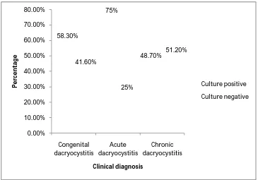
53 58.30% 75% 48.70% 41.60% 25% 51.20% 0.00% 10.00% 20.00% 30.00% 40.00% 50.00% 60.00% 70.00% 80.00% Congenital dacryocystitis Acute dacryocystitis Chronic dacryocystitis P e rc e n ta ge Clinical diagnosis Culture positive Culture negative
TABLE NO: 8. ANALYSIS OF CULTURE POSITIVITY AMONG CONGENITAL, ACUTE AND CHRONIC DACRYOCYSTITIS CASES
Clinical type Total no of cases Total no of samples
Culture positive Culture negative Number Percentage Number Percentage
Congenital
dacryocystitis 12 12 7 58.3% 5 41.6%
Acute
dacryocystitis 8 8 6 75% 2 25%
Chronic
dacryocystitis 80 82 40 48.7% 42 51.2%
Analysis of culture positivity among congenital, acute and chronic dacryocystitis cases
[image:62.595.136.501.379.634.2]54
90.50% 9.40%
Single organism
Mixed organisms
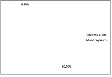
TABLE NO: 9. DISTRIBUTION OF SINGLE AND MIXED GROWTH ISOLATED FROM DACRYOCYSTITIS CASES
Total no of culture positive
samples
Single/Mixed growth
No. of
samples Percentage
P value <0.05
53
Single organism 48 90.5%
Mixed organisms 5 9.4%
Distribution of single and mixed growth isolated from dacryocystitis cases
[image:63.595.128.495.322.575.2]55 0 5 10 15 20 25 30 35 Congenital dacryocystitis Acute dacryocystitis Chronic dacryocystitis 7 6 35 0 0 5 N u m b e r o f sa m p le s Clinical diagnosis Single growth Mixed growth
TABLE NO: 10. DISTRIBUTION OF SINGLE AND MIXED GROWTH AMONG CONGENITAL, ACUTE AND CHRONIC DACRYOCYSTITIS CASES
Single/Mixed growth
No of culture positive samples
Total Congenital dacryocystitis Acute dacryocystitis Chronic dacryocystitis
Single growth 7 6 35 48
Mixed growth - - 5 5
Total 7 6 40 53
Distribution of single and mixed growth among congenital, acute and chronic dacryocystitis cases
56
TABLE NO: 11. DISTRIBUTION OF MIXED ISOLATES
Organisms No of cases
Staphylococcus aureus + Candida albicans 1
Escherichia coli + Staphylococcus epidermidis 1
Klebsiella pneumoniae + Staphylococcus epidermidis 1
Klebsiella pneumoniae + Staphylococcus aureus 1
Pseudomonas aeruginosa + Staphylococcus epidermidis 1
57 0.00% 10.00% 20.00% 30.00% 40.00% 50.00% 60.00% 70.00% 80.00% 90.00% 100.00%
Bacterial growth Fungal growth
96.55% 3.44% P e rc en ta ge
TABLE NO: 12. DISTRIBUTION OF BACTERIAL AND FUNGAL ISOLATES AMONG DACRYOCYSTITIS CASES.
Type of growth
No of isolates
Total no of isolates Percentage P value<0.05 Single growth Mixed growth
Bacterial 47 9 56 96.5%
Fungal 1 1 2 3.4%
Total 48 10 58 100%
Distribution of bacterial and fungal isolates among dacryocystitis cases.
[image:66.595.124.491.353.648.2]58 0 5 10 15 20 25 6 5 23 1 1 20
0 0 2
N u m b e r o f is o la te s Clinical diagnosis
Gram positive bacteria
Gram negative bacteria
Fungal isolates
TAB NO: 13. DISTRIBUTION OF GRAM POSITIVE & GRAM
NEGATIVE BACTERIA AND FUNGAL ISOLATES AMONG
CONGENITAL, ACUTE AND CHRONIC DACRYOCYSTITIS CASES
Microorganism
Number of isolates
Total Percentage Congenital dacryocystitis Acute dacryocystitis Chronic dacryocystitis Gram positive
bacteria 6 5 23 34 58.6%
Gram negative
bacteria 1 1 20 22 37.9%
Fungal
isolates - - 2 2 3.4%
Total 7 6 45 58 100%
Distribution of Gram positive &Gram negative bacteria and fungal isolates among congenital, acute and chronic dacryocystitis cases
59
28.50%
14.20% 42.80%
14.20%
Staphylococcus aureus
Staphylococcus epidermidis
Streptococcus pneumoniae
Pseudomonas aeruginosa
TABLE NO:14. DISTRIBUTION OF BACTERIAL ISOLATES IN CONGENITAL DACRYOCYSTITIS CASES.
Total no of cases/samples
No of organisms
isolated
Organism No of
isolates Percentage
12 7
Staphylococcus
aureus 2 28.5%
Staphylococcus
epidermidis 1 14.2%
Streptococcus
pneumoniae 3 42.8%
Pseudomonas
aeruginosa 1 14.2%
Distribution of bacterial isolates in congenital dacryocystitis cases.
From the above findings it is observed that in this study, the predominant organism isolated from congenital dacryocystitis cases is
[image:68.595.140.485.352.622.2]60
50%
33.30% 16.60%
Staphylococcus aureus
Staphylococcus epidermidis
Escherichia coli
TABLE NO: 15. DISTRIBUTION OF BACTERIAL ISOLATES IN ACUTE DACRYOCYSTITIS CASES.
Total no of cases/ samples
No of organisms
isolated Organism No of
isolates Percentage
8 6
Staphylococcus
aureus 3 50%
Staphylococcus
epidermidis 2 33.3%
Escherichia coli 1 16.6%
Distribution of bacterial isolates in acute dacryocystitis cases.
From the above findings it is observed that in this study, the predominant organism isolated from acute dacryocystitis cases is
[image:69.595.127.479.340.609.2]