[1]
A STUDY OF THE CLINICAL PROFILE OF PATIENTS
WITH MYOCARDIAL INFARCTION
Dissertation submitted to
THE TAMILNADU DR.MGR MEDICAL UNIVERSITY
CHENNAI- TAMILNADU
In partial fulfillment for the Degree of
DOCTOR OF MEDICINE
BRANCH I –M.D., (General Medicine)
APRIL-2015
DEPARTMENT OF MEDICINE
TIRUNELVELI MEDICAL COLLEGE
TIRUNELVELI- 627011
[2]
CERTIFICATE
This is to certify that the Dissertation entitled ―A STUDY OF THE CLINICAL
PROFILE OF PATIENTS WITH MYOCARDIAL INFARCTION” submitted
by Dr.E.SARAVANAN to The Tamilnadu Dr.M.G.R. Medical University,
Chennai, in partial fulfillment of the requirement for M . D. ( B r a n c h - I ) Ge n e r a l
M e d i c i n e Ex a mi n a t i o n to b e h e l d on Ap r i l 2 0 15 is a bonafide work
carried out by him under my guidance and supervision. This dissertation partially
or fully has not been submitted for any other degree or diploma of this university
or other.
Prof.Dr.NAZAR. MD., Prof.Dr.VAIRAMUTHURAJU,MD.,
Unit Chief, UNIT V Professor and HOD, Department of Medicine, Department of Medicine Tirunelveli Medical College , Tirunelveli Medical College, Tirunelveli – 627011. Tirunelveli – 627011.
The Dean,
[5]
DECLARATION
I, Dr.E.SARAVANAN, solemnly declare that the I carried out this work on
“A STUDY OF THE CLINICAL PROFILE OF PATIENTS WITH
MYOCARDIAL INFARCTION” at Department of General Medicine,
Tirunelveli Medical College and Hospital during the period of August 2013 to
August 2014.
This is submitted to the Tamilnadu Dr.M.G.R. Medical University,
Chennai, in partial fulfillment of the rules and regulations for the MD Degree
Branch I (General Medicine) Examination.
It was not submitted to the award of any degree/diploma to any
University either in part or in full previously.
Place: TIRUNELVELI
Date:
DR.E.SARAVANAN,
POST GRADUATE,
M.D. GENERAL MEDICINE
[6]
ACKNOWLEDGEMENT
At the outset I wish to thank our Dean Dr.THULASIRAM, MS, for
permitting me to carry out this study in our hospital.
I e x p r e s s my sincere thanks to my Professor and H.O.D
DR.VAIRAMUTHURAJU for his support and encouragement throughout the
study. I am deeply indebted to my Chief Prof .DR.NAZAR, who was the main
motivator behind the study. I would like to thank Prof.DR.RAJAGOPALA
MARTHANDAM for his valuable advice and guidance.
I would like to thank Prof DR.RAVICHANDRAN EDWIN HOD,
Department of Cardiology for his constant support and encouragement. I thank
the Department of Cardiology for the unparalleled help and cooperation.
I am thankful to my assistant professors DR.BARATH, DR.SHAVANA,
DR.MADHAVAN, DR.GOVINDARAJAN for their valuable suggestions.
I also thank the Departments of Microbiology and Biochemistry for the
laboratory support to this study.
No words of gratitude will be enough to thank my parents for their
unconditional support and encouragement at each step in my way.
Last but not the least, I sincerely thank all the patients who cooperated
[7]
CONTENTS
S.NO TITLE PAGE NO
1. INTRODUCTION 8
2 AIM OF THE STUDY 12 3 REVIEW OF LITERATURE 14 4 MATERIALS AND METHODS 81 5 OBSERVATIONS AND RESULTS 87 6 DISCUSSION 110 7 SUMMARY 114 8 CONCLUSIONS 116 9 BIBLIOGRAPHY 118 ANNEXURES i. PROFORMA 129
ii. MASTER CHART 132
iii. KEY TO MASTER CHART 133
A STUDY OF CLINICAL PROFILE OF PATIENTS WITH
MYOCARDIAL INFARCTION
ABSTRACT:
BACKGROUND:
Cardiovascular Diseases (CVD) is a leading cause of non communicable morbidity
and mortality in India. By the year 2030 CVD is projected to be the leading cause
of death worldwide. One of the most important advances in cardiovascular
research was the identification of risk factors associated with cardiovascular
diseases with subsequent treatments developed and rigorously tested to modify the
risk factors with the goal of preventing CVD. Although 80% of global burden of
cardiovascular diseases occurs in low and middle income countries, knowledge of
importance of risk factors is largely derived from developed countries. The effect
of factors on risk of coronary artery disease in most regions of the world including
developing countries like India remains largely unknown. Various studies have
AIM:
To study the symptoms, clinical profile, risk factors, complications and outcome in
predominantly rural Indian population.
MATERIALS AND METHODS:
An observational cross sectional study involving 200 patients admitted with a
diagnosis of ST Elevation Myocardial Infarction in Tirunelveli Medical College
between August 2013 and August 2014. Data was collected based on Inclusion and
Exclusion criteria.
INCLUSION CRITERIA:
All patients admitted to our hospital with a diagnosis of MI. The final diagnosis of
MI will be based on the following criteria:
1. Ischemic chest pain for atleast 30 minutes
2. ECG evidence of myocardial injury:
0.1 mv or more ST segment elevation in 2 contiguous limb leads or
Development of pathological Q waves
EXCLUSION CRITERIA:
Patients admitted with MI who refused to give consent for the study. Patients who
are known cases of CKD, OLD MI, post angioplasty, post CABG patients.
The outcome of the patients at the end of one week was studied using TIMI
(Thrombolysis In Myocardial Infarction) Risk Score calculated at the time of
admission. The information collected regarding all the selected cases were
recorded in a Master Chart. Data analysis was done with the help of computer
using Epidemiological Information Package (EPI 2010) developed by Centre for
Disease Control, Atlanta, USA.
RESULTS:
Among the 200 patients, 91% were discharged alive and 9% expired. The most
common cause of death was cardiogenic shock. Among the 18 patients who
expired, 14 patients had a Thrombolysis in Myocardial Infarction (TIMI) risk score
of 9 and above and the remaining four had a TIMI score of 5 to 8. Mortality was
not seen in those with a TIMI risk score of below five. The association was
statistically significant ( p<0.0001). In a subgroup analysis, In Young MI
(<45 years), 91.7% were smokers, 88.9% consumed alcohol, around 50% had
100% had low HDL. The percentage of smoking and alcoholism were higher in
young MI group compared to overall study group and all the patients in the young
MI group were male.
CONCLUSION:
Cardiovascular Diseases affect mainly economically productive age group in our
country and has become a major public health burden. Smoking, Alcoholism,
Diabetes, Hypertension and Dyslipidemia remain the most important modifiable
risk factors. Patients with a higher TIMI Risk Score have a greater mortality.
KEYWORDS: Cardiovascular diseases, Myocardial Infarction, Thrombolysis In
[9]
INTRODUCTION
Cardiovascular Diseases is an important cause of morbidity and mortality in the
developed as well as the developing world. By 2030, WHO predicts that 33%of the
deaths occurring worldwide will be caused by Cardiovascular diseases. With the
epidemiologic transition well and truly taking place humans have travelled a long
way from the stage of pestilence and famine to the stage of obesity and inactivity.
Due to these factors we face a host of challenges and the morbidity due to
cardiovascular diseases is accelerating with each passing day. In India alone
Cardiovascular diseases account for 25% of the total deaths.
In our country the CVD risk factors among the rural as well as the urban poor
and middle class are on the rise. The demon of cardiovascular diseases is very
much at our door step. This is a frightening scenario considering that India is home
to almost 17% of the world‘s population. With a bulging population not to
mention the bulging waistline of the populace the challenges that lie ahead of us
are monumental to say the least. Like many other non-communicable diseases ,
cardiovascular diseases have a long latency and have numerous modifiable risk
[10]
One of the important advances in cardiovascular research has been with regard to
the identification of risk factors associated with cardiovascular diseases. Based
on these risk factors treatment plans have been drawn and meticulously tested with
the goal of altering the outcome.
The major chunk of the global burden of these diseases is from the low and
middle-income countries like India. But the data regarding the importance of risk
factors has been derived mainly from developed countries.
In a multifaceted country like ours, with various ethnicities, cultural and
food patterns and above all with varied political outlooks on the aspects of health
coupled with significant infrastructure limitations has effectively prevented us
from having completely representative comprehensive surveys with regard to
demography. The disease and death registration systems is in a state of shambles.
This has been a big bane of our health care system and has prevented us from
taking pre-emptive measures. With a health care system as frail as ours with
numerous lacunae it becomes even more imperative to initiate cost-effective
[11]
Numerous studies have been conducted to highlight the importance of
risk factors associated with Cardiovascular diseases. This study is based on risks
[12]
[13]
AIMS OF THE STUDY
To study:
1.
The type of presentation(symptoms)
2.
The type of MI
3.
The relative incidence of risk factors
4.
The Thrombolysis in Myocardial Infarction(TIMI) score
[14]
[15]
REVIEW OF LITERATURE
:
CORONARY ARTERY ANATOMY:
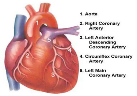
[1]LEFT MAIN CORONARY ARTERY:
It arises from the superior portion of Left aortic sinus, just below the sino tubular
ridge of aorta. The sino tubular ridge separates the Sinus of Valsalva and the
[image:18.612.78.549.301.632.2]smooth tubular portion of aorta.
[16]
LMCA has a variable length ranging from 10to 15 mm and an internal
diameter that varies from 3 to 6mm. It runs behind Right Ventricular
outflow tract and divides into Left Anterior Descending Artery (LAD) and Left
Circumflex Artery. In a few cases, LMCA may be absent and there may be
separateostia for LAD and LCX arteries. In some cases LMCA trifurcates
into LAD , LCX and Ramus Intermedius.
LEFT ANTERIOR DESCENDING ARTERY:
It arises from the Left main Coronary Artery. This artery runs in the anterior
interventricular groove along with the Great Cardiac vein towards the apex of the
heart. It divides into septal and diagonal branches. Wide variation exists regarding
the size and the number of septal perforators and diagonals. The Left Anterior
Descending Artery supplies the anterior part of the septum via the septal
perforators and the anterior, apical and the lateral walls of the left ventricle via the
diagonal branches.
Commonly implicated in the majority of deaths due to Acute coronary Syndromes,
[17]
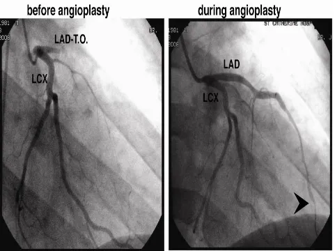
FIG. 2 TOTAL OCCLUSION OF LAD- ANGIOGRAPHY
LEFT CIRCUMFLEX ARTERY:
This artery originates from Left Main Coronary Artery and runs along posterior
atrio ventricular groove toward inferior interventricular groove. It gives off large
obtuse marginal branches. These Branches supply lateral free wall of left ventricle.
[18]
Descending Artery (Left-Dominance). It also gives Left Atrial Circumflex
[image:21.612.75.499.146.441.2]branches that Supply posterior and lateral part of the left atrium.
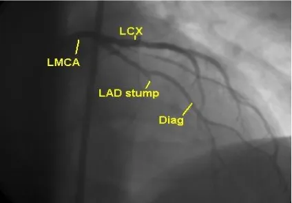
FIG.3 ANGIOGRAPHY- CORONARY ARTERIES
RIGHT CORONARY ARTERY:
This artery originates from right anterior aortic Sinus inferior to the origin of
LMCA and runs along the posterior right atrioventricular groove. Its first branch is
the conus artery,then it gives the Sinoatrial nodal artery in around 60% of the
individuals. In the remaining the sinoatrial nodal artery arises from the Left
[19]
coming from both the arteries. The Acute Marginal Branches arise from the mid
portion of the vessel and supplies the anterior wall of the right ventricle. The RCA
then gives posterolateral branches and continues as the posterior descending artery
which supplies the lower part of the interventricular septum. Right dominance is
seen in 85% of the individuals; that is RCA gives rise to the Posterior Descending
artery and atleast one posterolateral branch. In the remaining 15%, in one half,
Left Circumflex artery gives rise to the PDA and the posterolateral branches. In the
other half ,PDA arises from the Right Coronary artery and the posterolateral
branches from the Left circumflex Artery(co-dominant or balanced circulation).
The Dominant artery usually gives rise to the Atrioventricular nodal artery which
supplies the AV node.
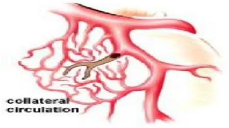
CORONARY COLLATERAL CIRCULATION:
When a coronary vessel becomes totally occluded blood flow persists through
collateral vessels. These channels develop when intracoronary pressure gradient
occurs between source and recipient vessel. The extent and the function of
coronary collaterals vary from patient to patient. In some, collaterals are well
developed so that they are able to maintain perfusion at rest and also at sub
maximal cardiac workloads[2]. Improved survival and a lower risk of
cardiovascular events have been shown in patients with elevated distal coronary
[20]
coronary ischemia cause proliferation of coronary collaterals, a process called
arteriogenesis[4]. When stenosis of the coronary vessel exceeds 70% the increase in
endothelial shear stress in the pre-existing collateral vessels cause a Nitric Oxide
[image:23.612.72.521.218.469.2]dependent vasodilatation.
FIG.4 COLLATERAL CIRCULATION
But human randomized clinical trials to improve angiogenesis in mature coronary
vessels has been highly disappointing[5]. But this may be because of the fact that
improvements in functioning of the myocardium may be due to altered cell
growth and repair rather than angiogenesis.[6] When Nitric Oxide synthesis is
deficient, collaterals constrict and this can relieved by nitroglycerin.[7] This is
[21]
CORONARY MICROCIRCULATION:
Individual coronary resistance arteries are a longitudinally distributed network and
the mechanisms controlling the resistance in these arteries is variable. [8] These
vessels dilate in a co-ordinated manner so that they are able to meet the demands
of the distal vascular bed . This is done by shear stress mediated control of the
blood vessels or through myogenic control.
Epicardial arteries with a diameter greater than 400 micron are mainly regulated by
shear stress. The resistance vessels are divided into resistance arteries and
arterioles. The arteries are regulated by shear stress and myogenic response
whereas the arterioles are mainly controlled by local metabolic factors and they
regulate the perfusion of coronary capillary bed. Average myocardial capillary
density is around 3500/mm.square. The capillary density is considerably higher in
the sub endocardium than the epicardium.
MYOGENIC CONTROL:
This is a mechanism by which vessels dilate in response to decrease in pressure
and constrict when the pressure increases. It is due to the tone of smooth muscle [9].
[22]
mainly in arterioles<100 micron diameter and is an important mechanism in
coronary auto regulation[10] .
Kuo and colleagues have shown flow-induced dilatation of coronary vessels [8,11].
This is mediated by NO and hence endothelium dependent. Endothelium
Dependent Hyperpolarising factor ,an arachidonic acid metabolite primarily
regulates epicardial conductance vessels[12]
FIG.5 CORONARY MICROCIRCULATION
The resistance vessels are controlled by Nitric oxide[11]. EDHF may be
upregulated during acquired disease states when Nitric oxide is deficient and serve
[23]
VASCULAR BIOLOGY OF ATHEROSCLEROSIS:
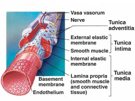
THE NORMAL ARTERY:
The Normal Artery has three layers an innermost layer of tunica intima, middle
[image:26.612.75.541.310.659.2]layer of tunica media and the outer layer of tunica adventitia.
[24]
TUNICA INTIMA:
The tunica intima is composed of a single layer of endothelial cells which rests on
a basal lamina. The internal elastic lamina separates tunica intima and tunica
media. The endothelial cells arise from precursor cells called Endothelial
Progenitor Cells.
These cells help to repair areas of endothelial desquamation [14,15]. Older
individuals have lowered levels of endothelial progenitor cells and hence less
ability to repair any damage that occurs in the tunica intima [16]. New evidence has
challenged the belief that Endothelial Progenitor cells populate murine
atherosclerotic plaques [17].There is a variable expression of endothelial genes in
different types of blood vessels. This is mainly regulated by the local
environment[18]
TUNICA MEDIA:
The middle layer tunica media is composed of numerous layers of smooth muscle
layers embedded in an extracellular matrix . The extracellular matrix is rich in
elastin. This gives the tensile strength to the arteries. The external elastic lamina
[25]
TUNICA ADVENTITIA:
This layer consists of loosely arranged collagen fibrils along with vasa vasorum
and free nerve endings. The two most common types of cells seen are the
fibroblasts and mast cells. Experimental evidence in animal models have shown
that mast cells may well have an important in atheroma formation and also a role in
pathogenesis of aneurysms[19].
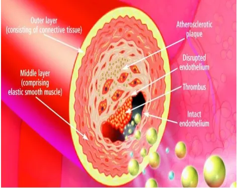
ATHEROMA FORMATION:
The first step is accumulation of lipoprotein particles in the intimal layer followed
by their aggregation[20].Oxidative stress, including products found in modified
lipoproteins, results in the elaboration of various cytokines locally. The oxidative
stress occurs in the form of oxidation, glycation, etc. The cytokines formed,
increase expression of adhesion molecules for leukocytes that cause their
attachment and chemoattractant molecules that direct their migration into the
intima. The leukocytes thus move between endothelial cell junctions or sometimes
penetrate through endothelial cells, a process called transcytosis. The leukocytes,
on entering the artery wall in response to chemoattractant cytokines such as
monocyte chemoattractant protein 1 (MCP-1) encounter stimuli such as
[26]
in increased expression of scavenger receptors. Scavenger receptors mediate the
uptake of modified lipoprotein particles and promote the development of foam
cells. T cells also tend to adhere to the endothelium and this effect is also mediated
[image:29.612.72.545.211.585.2]by leukocyte adhesion molecules.
FIG.7 THROMBUS FORMATION
The commonly implicated leukocyte adhesion molecules are Vascular Cell
[27]
Molecule 1(ICAM-1), E selectin, P selectin, etc. Macrophage foam cells are a
source of mediators, such as further cytokines and effector molecules like
hypochlorous acid, superoxide anion (O2 −
), and matrix metalloproteinases. These
influences result in the migration of smooth muscle cells from the tunica media to
the tunica intima. There is considerable variation between the smooth muscle cells
found in the normal tunica media and those found in the intima of an evolving
atheroma [23,24]. Emerging evidence has challenged the concept of migration of
blood borne smooth muscle cells into the plaques [25].Further division of smooth
muscle cells occur coupled with elaboration of extracellular matrix.
But an important point to note is that smooth muscle death also occurs hand in
hand along with smooth muscle cell proliferation and migration. Interactions
between the Fas ligand and the Fas expressed on the surface of smooth muscle
cells coupled with the action of various pro inflammatory cytokines results in the
death of smooth muscle cells. Thus the smooth muscle accumulation in the
atheromatous plaque can be said as a finely done balancing act between smooth
muscle cell replication and cell death. The extracellular matrix makes up the
major volume of the plaque rather than the cells. It is composed of mainly collagen
(types 1 and 3), proteoglycans like biglycan, aggrecan, decorin and versican. As
the atherosclerotic plaques grow they develop their own blood supply. This
[28]
growth factor (PIGF), certain forms of fibroblast growth factor and oncostatin M.
This helps the plaque to overcome limitations in oxygen and nutrient supply and
maintain its growth a mechanism similar to tumour angiogenesis[26].In later stages,
calcification can occur and fibrosis continues, sometimes accompanied by smooth
muscle cell death (including programmed cell death or apoptosis), yielding a
relatively acellular fibrous capsule surrounding a lipid-rich core that may also
contain dying or dead cells.
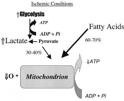
PATHOPHYSIOLOGY OF MYOCARDIAL ISCHEMIA:
The normal function of the heart muscle is supported by high rates of myocardial
blood flow, oxygen consumption, and combustion of fat and carbohydrates
(glucose and lactate). Under normal aerobic conditions, cardiac energy is mainly
derived from fatty acids, which contribute around 60 to 90 percent for the synthesis
of adenosine triphosphate (ATP). The rest of the energy (10 to 40 percent) comes
from oxidation of pyruvate, formed from glycolysis and lactate oxidation. Almost
all of the ATP formed comes from oxidative phosphorylation in the mitochondria;
only a small amount of ATP (<2 percent) is synthesized by glycolysis.
[29]
shortening; the remaining one-third is used by sarcoplasmic reticulum Ca2+
[image:32.612.72.481.143.475.2]ATPase and other ion pumps.
FIG. 8 PATHOPHYSIOLOGY OF MYOCARDIAL ISCHEMIA
When a major artery is suddenly occluded , a change in the metabolism occurs
from aerobic or mitochondrial phosphorylation to anerobic glycolysis. This change
occurs within a matter of seconds. This results in increased production of lactate
and a fall in ph. This fall in ph leads to a greater ATP requirement in order to
[30]
between oxygen supply and demand. Biochemical abnormalities begin at the onset
of ischemia, loss of myocardial contractility occurs within 60 seconds and the loss
of viability occurs around 20 to 40 minutes following total coronary occlusion.
Two zones of myocardial damage are seen: a central zone with no or very low flow
and a zone of collateral vessels in a surrounding marginal zone. The survival of the
marginal zone is dependent on two variables , one being the level of ischemia and
the other being the duration of ischemia.
The extent of coronary collateral flow is one of the principal determinants of
infarct size. Patients with well developed collaterals have low risk of developing an
acute MI on total occlusion of coronary artery [28]. These collaterals seem to be
well developed in younger individuals and also those who have experienced
previous episodes of angina [29].
For the clinical diagnosis of acute myocardial infarction, the World Health
Organization requires that at least two of the following three criteria be present:
(1) a history of chest pain or discomfort;
(2) a rise and subsequent fall in serum cardiac enzymes; and
(3) the development of electrocardiogram (ECG) abnormalities (new Q waves or
[31]
Since the ECG lacks sufficient sensitivity and specificity to detect myocardial
necrosis, the presence of myocardial injury is often dependent upon the release of
cardiac-specific serum markers such as troponin T, troponin I, and CK-MB. It has
been shown that infarct size generally correlates with the peak rise in serum
[image:34.612.72.541.240.592.2]CK-MB level.[31]
[32]
WAVEFRONT PHENOMENON:
The area of myocardium that is necrosed, usually depends on the duration of
coronary artery occlusion. This was first proved by Reimer and Jennings in an
experiment using canine coronary artery which was occluded for variable duration.
After only 15 min of occlusion, no infarct occurred. At 40 min, the infarct was sub
endocardial, involving only the papillary muscle, placing 28 percent of the
myocardium at risk. At 3 h following coronary artery occlusion and reperfusion,
the infarct was significantly smaller compared with non reperfused permanently
occluded infarct (62 percent of area at risk). The infarct size was greatest in
permanent occlusion, becoming transmural and involving 75 percent of the area at
risk[32] . In the dog model, it is impossible to achieve 100 percent infarction of area
at risk because of species-related native collaterals. In humans, it has been shown
that approximately 40 percent of patients with acute MI have a well-developed
collateral circulation [29].
GROSS PATHOLOGICAL CHANGES:
The earliest change that usually occurs in the evolution of an acute MI is the pallor
of the myocardium; this change usually occurs after 12 hours or later after the
onset of irreversible ischemia. The detection of infarction can be enhanced by the
[33]
gross section of fresh heart tissue in the presence of dehydrogenase-mediated
activity. The tetrazolium salts [nitrobluetetrazolium (NBT) and
2,3,5-triphenyltetrazolium chloride (TTC)] are dyes that are sensitive to the presence of
tissue dehydrogenase enzyme activity, which is depleted in the infarcted
myocardium. It has been shown that myocardial infarct can be detected by NBT as
early as 2 to 3 h in the dog and a little less in the pig because of poor collaterals. [33]
Red (TTC) or blue color (NBT) will form only in the normal noninfarcted
myocardium, thus revealing the pale, unstained infarcted region. In humans, the
necrotic myocardium can be detected within 2 to 3 h after infarct by immersion of
the fresh heart slices in a solution of TTC or NBT. TTC staining demonstrated a
diagnostic sensitivity of 77 percent and a specificity of 93 percent compared with
routine histology, with predictive values of positive and negative test of 81 and 91
percent, respectively.
Enhancement of pallor is seen 24 hours after the onset of ischemia. In this era of
thrombolytic therapy, most in-hospital patients will have received tissue
plasminogen activator, streptokinase, or IIb/IIIa inhibitors, which lyse the
thrombus and restore blood flow into the area of infarction. Therefore, in a
reperfused infarct, the infarcted region will appear red from trapping of the red
[34]
However, if there has been no reperfusion, the area of the infarct is better defined
at 2 to 3 days, with a central area of yellow discoloration surrounded by a thin rim
of highly vascularized hyperemia. At 5 to 7 days, the regions are much more
distinct, with a central soft area and a depressed hyperemic border. At 1 to 2 weeks
healing begins, with infiltration by macrophages as well as early fibroblasts at the
margins. At the same time, the infarct begins to be more depressed, especially at
the margins, where organization takes place, and there is a white border . The time
taken for the infarct to completely heal may vary from as early as 4 to 6 weeks in
small infarcts to as long as 2 to 3 months when the area of infarction is large.
Healed infarcts are white from the scarring, and the ventricular wall may or may
not be thinned (aneurysmal). In general, infarcts that are transmural and confluent
are likely to result in thinning. But this is not the case with nonconfluent and sub
endocardial infarcts which usually don‘t result in thinning.
LIGHT MICROSCOPIC CHANGES:
The earliest morphologic characteristic of MI that occurs is the hypereosinophilic
myocyte.This change is usually seen between 12 to 24 h after onset of chest pain.
The hypereosinophilia of the cytoplasm is best visualized by routine hematoxylin
and eosin staining. The myocyte striations appear normal and some chromatin
condensation may be seen in the nucleus. The area of infarction may show
[35]
hearts.This finding is better appreciated in animal models.The earliest
characteristic change, the appearance of ‗wavy fibers‘ is best seen in experimental
animal models and they are believed to be the the result of stretching of the
ischemic noncontractile fibers by the adjoining viable contracting myocytes.[34]
Wavy fiber change is, however, nonspecific and occurs in the absence of ischemia,
especially in the right ventricle. Neutrophil infiltration is present by 24 h at the
border areas. As the infarct progresses between 24 and 48 h, coagulation necrosis
is established, with various degrees of nuclear pyknosis, early karyorrhexis, and
karyolysis. The myocyte striations are preserved and the sarcomeres elongate. The
border areas show prominent neutrophil infiltration by 48 h.
After 3 to 5 days, the central portion of the infarct shows loss of myocyte nuclei
and striations; in smaller infarcts, neutrophils invade within the infarct and
fragment, resulting in more severe karyorrhexis (nuclear dust). Loss of myocyte
striations is best visualised by Mallory's trichrome stain. Another stain that has
been used to detect early areas of infarction is hematoxylin–basic fuchsin–picric
acid. But this technique is not very reliable for the early detection (6 to 8 hours) of
infarction in humans and hence not commonly used.
Immuno histochemical staining has also been used to study early changes of
[36]
kinase, ceruloplasmin, myoglobin, C-reactive protein, complement complex
(C5b-9), fibronectin, and others—have also not been found to be useful. In the border
areas of the infarct macrophage and fibroblast proliferation can be seen. By 1
week, neutrophils decline and granulation tissue is established, with neocapillary
invasion as well as lymphocytic and plasma cell infiltration. Although lymphocytes
may be seen as early as 2 to 3 days, they are not prominent at any stage of infarct
evolution. Eosinophils may be seen within the inflammatory infiltrate but are
present in only 24 percent of infarcts. There is phagocytic removal of the necrotic
myocytes by macrophages, and pigment is seen within macrophages.
By the second week, fibroblasts become the most dominant cell type, but their
presence can be seen as early as day 4 at the periphery of the infarct. Necrotic
myocytes are continuously removed and the fibroblasts actively produce collagen
and angiogenesis tends to occur in the area of healing. The healing continues to
occur. Depending on the amount of cardiac myocytes necrosed, the healing may
be complete as early as 4 weeks, or sometimes take 8 weeks or longer to complete .
The central area of infarction may remain unhealed, showing mummified myocytes
for extended periods, despite the fact that the infarct borders are completely healed.
For this reason, it is important to evaluate the age of the infarct by examining the
[37]
The magnitude of repair and healing is dependent not only on infarct size but also
on local and systemic factors. If there is good collateral blood flow locally, healing
will be relatively rapid, especially at the lateral borders, where viable myocardium
interdigitates with necrotic myocardium. There may be various levels of healing
within an infarct, because of differences in blood flow in adjoining vascular beds
caused by variable extents of coronary narrowing. The border areas may show
hemorrhage and contraction-band necrosis, depending on regional variations in
blood flow.
In humans, if reperfusion occurs within 4 to 6 h following the onset of chest pain
or ECG changes; there is myocardial salvage and the infarct is likely to be
subendocardial without transmural extension. There will be a nearly confluent area
of hemorrhage within the infarcted myocardium, with extensive contraction-band
necrosis. The extent of hemorrhage is dependent on the extent of reperfusion of the
infarct as well as the extent of capillary necrosis. The larger the infarct and the
longer the duration of the infarct, the greater the hemorrhage. The degree of
hemorrhage may be variable and non uniform, as blood flow is dependent upon the
residual area of coronary narrowing and the amount of thrombolysis. Within a few
hours of reperfusion, neutrophils are evident within the area of necrosis, but they
are usually sparse . In contrast to nonreperfused infarcts, neutrophils do not show
[38]
of necrosis at the periphery, with interdigitation with noninfarcted myocardium.
Macrophages begin to appear by day 2 to 3 and stromal cells show enlarged nuclei
and nucleoli by days 3 and . Neutrophil debris, which may be concentrated at the
border areas in cases of incomplete reperfusion, is seen by 3 to 5 days. By days 3
to 5, fibroblasts appear, with an accelerated rate of healing as compared to
nonreperfused infarcts. By 1 week, there is collagen deposition, with disappearance
of neutrophils; there is also prominence of macrophages containing pigment
derived from ingested myocytes. Angiogenesis is prominent and lymphocytes are
often seen. Infarcts at 5 to 10 days are more cellular, and there is prominent
myocytolysis (loss of myofibrils). As early as 2 to 3 weeks, subendocardial infarcts
may be fully healed. Some 5 to 10 layers of subendocardial myocytes are spared
without necrosis. However, myofibrillar loss, which is a result of ischemia not
severe enough to cause cell death, is prominent in this subendocardial zone. Larger
infarcts and those reperfused after 6 h take longer to heal. Infarcts reperfused after
6 h show larger areas of hemorrhage as compared to occlusions with more
immediate reperfusion. However, myocytes maintain their striations, become
stretched and elongated, and—as they do not respond to calcium influx—do not
show significant contraction-band necrosis. Despite the fact that reperfusion should
occur within 6 h of occlusion for maximal myocyte salvage, there appears to be
[39]
The No-Reflow Phenomenon
This phenomenon was first described by Kloner and Jennings in 1974.They used
an experimental canine model of myocardial infarction [35] and demonstrated
homogenous distribution of thioflavin S dye after 40 min of ischemia and
reperfusion; however, after 90 min of ischemia, areas of no reflow were identified
mainly in the subendocardial regions as zones not staining with thioflavin S.
Electron microscopic examination revealed swollen endothelial protrusions and
membrane-bound intraluminal bodies, which obstructed the capillary lumen and
resulted in plugging of the capillaries by red cells, neutrophils, platelets, and fibrin
thrombi. The areas not stained by thioflavin S were characterized by low regional
myocardial blood flow.
Myocardial dysfunction associated with reperfusion of the ischemic myocardium
[40]
TABLE-1 PATHOLOGICAL CHANGES IN MI
PERMANENT OCCLUSION/NO REPERFUSION REPERFUSION FOLLOWING OCCLUSION Time of Occlusion
Gross Histologic Gross Histologic
12 h No
change/pallor
Wavy fibers Mottled, prominent hemorrhage
CBN 24–48 h Pallor—
yellow, soft
Hypereosinophilic fibers, PMNs at borders
Prominent hemorrhage
Hypereosinophilic fibers + CBN + PMNs +
hemorrhage throughout 3–5 days Yellow center,
hyperemic borders
Large number of PMNs at border, coagulation necrosis, loss of nuclei
Prominent hemorrhage Aggressive phagocytosis profuse fibroblast infiltration + collagen77[None] 6–10 days Yellow,
depressed central infarct, tan-red
margins
Mummified fibers in center, macrophage phagocytosis + granulation tissue at borders Depressed red-brown infarct with gray-white intermingled Aggressive healing with greater collagen 10–14 days Gray red borders, infiltrating central tan-yellow infarct if large Marked granulation tissue, collagen deposition, subendocardialmyocyte sparing Gray-white intermingled with brown Aggressive healing with greater collagen
2–8 weeks Gelatinous to gray-white scar, greater healing at border zone
[41]
This is assessed by contractile performance, lowering of the arrhythmogenic
threshold, conversion of reversible to irreversible myocyte injury, and micro vessel
dysfunction. Recent studies have shown that the angiographic no-reflow
phenomenon is a strong predictor of major cardiac events, such as congestive heart
failure, malignant arrhythmias, and cardiac death after MI.
GLOBAL BURDEN OF CARDIOVASCULAR DISEASES:
Cardiovascular diseases is the number one cause of deaths in all the continents of
the world the solitary exception being Africa. Even in sub Saharan Africa
cardiovascular diseases will be the major cause of mortality in the coming decade.
According to a study the major burden of cardiovascular diseases in terms of
mortality and disability occur in middle and low income countries [36]. Among all
cause of deaths occurring world cardiovascular diseases alone account for around
30% of the deaths [37]. The patterns of diseases in humans have undergone major
changes as a result of changes in food patterns, economy and demography. This
change in the pattern of diseases is called epidemiologic transition. There is a shift
from diseases related to infections to those associated with lifestyle changes[38].
This transition has occurred very rapidly in the developing countries which are
[42]
TABLE-2 EPIDEMIOLOGICAL TRANSITION
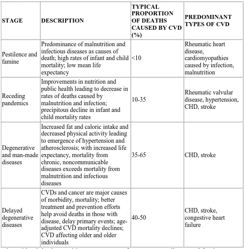
The epidemiologic transition encompasses four stages pestilence and famine,
receding pandemics, degenerative and man-made diseases and delayed
degenerative diseases[39].In addition to this a fifth stage of obesity and inactivity STAGE DESCRIPTION
TYPICAL PROPORTION OF DEATHS CAUSED BY CVD (%)
PREDOMINANT TYPES OF CVD
Pestilence and famine
Predominance of malnutrition and infectious diseases as causes of death; high rates of infant and child mortality; low mean life
expectancy
<10
Rheumatic heart disease,
cardiomyopathies caused by infection, malnutrition
Receding pandemics
Improvements in nutrition and public health leading to decrease in rates of deaths caused by
malnutrition and infection; precipitous decline in infant and child mortality rates
10-35 Rheumatic valvular disease, hypertension, CHD, stroke Degenerative and man-made diseases
Increased fat and caloric intake and decreased physical activity leading to emergence of hypertension and atherosclerosis; with increased life expectancy, mortality from
chronic, noncommunicable diseases exceeds mortality from malnutrition and infectious diseases
35-65 CHD, stroke
Delayed degenerative diseases
CVDs and cancer are major causes of morbidity, mortality; better treatment and prevention efforts help avoid deaths in those with disease, delay primary events; age-adjusted CVD mortality declines; CVD affecting older and older individuals
40-50
[43]
has also been described[40].Here obesity, diabetes and lipid abnormalities are
increasing in children.20% of Chinese people are overweight or obese [41]
The dynamics of health transition are very different in the developing countries
compared to the developed countries. Compression of the time course of the
cardiovascular epidemic along with inadequate public health infrastructure coupled
with low levels of education and awareness about the disease, co-existing burden
of communicable diseases are all major obstacles that prevent us from taking
effective measures.
In India alone around 32 million people are believed to suffering from
cardiovascular diseases [42]. Approximately 2.03 million deaths have been believed
to have occurred due to cardiovascular diseases by 2010. In the 30 year period
from 1990 to 2020, 111% increase in deaths due to cardiovascular diseases is
expected[42]. Some studies indicate women are more likely to have cardiovascular
diseases than men in India[43]. More than half of the Cardiovascular deaths in India
occurred in people less than 70 years.
A study conducted in south India in rural Andhra Pradesh has shown that
prevalence may well be higher in rural areas[44]. Developing countries are affected
[44]
bread- winnerof the family imposing a significant burden on the family as well as
the community.
RISK FACTORS:
SMOKING:
Smoking is the single most important risk factor for coronary artery disease.
Cigarette consumption is the most important cause of preventable death in the
world [45]. The use of tobacco is rising among women, adolescents and young
adults[46] . Around 930,000 adult deaths have been estimated to have occurred in
India due to smoking related causes [47]. Smoking causes acceleration of
atherosclerosis, impairs vasodilatory capacity of coronary arteries, increases levels
of stress hormones, increases bronchial secretions, impairs mucociliary clearance
and aggravates associated pulmonary dysfunction. Passive smoking is equally
hazardous. Smoking also causes :
1.increased levels of oxidized LDL
2.lowers levels of cardio protective HDL
3.nicotine and carbon monoxide in smoke produce endothelial damage
4.increases vascular reactivity
[45]
6.increases fibrinogen levels and platelet aggregability.
Cessation of cigarette consumption remains the single most important intervention
in preventive cardiology. A study demonstrated, smoking cessation reduced
cardiovascular mortality by 36% as compared with mortality in subjects who
continued smoking, an effect that did not vary by age, sex, or country of origin[48].
Reductions in smoking from any mechanism improve health outcomes,
especiallywhen coupled along with lifestyle changes, including exercise and
dietary control. Trials of nicotine replacement therapy using transdermal nicotine
or nicotine chewing gum increase abstention rates after cessation. Such
pharmacologic programs, as well as physician-guided counseling, are
cost-effective and should be provided as standard prevention services. Low-yield
cigarettes do not appear to reduce the risks of myocardial infarction. Although the
elevated cardiovascular risks associated with smoking decrease significantly after
cessation, the risks of cancer of the lungs, pancreas, and stomach persist for more
than a decade, as do the risks of developing chronic obstructive pulmonary disease.
Smoking cessation has clear benefit, but smoking reduction alone appears to have
only a marginal effect. Patients often do not understand the importance of smoking
cessation. This is common especially in the developing world where low levels of
[46]
Studies of social networking indicate that smoking cessation by a spouse decreases
a person's chance of smoking by 67%.Smoking cessation by a brother or sister,
friend, or neighbour, people who work in the same environment decreases a
person's chance of smoking by 25% to 36%. Smoking cessation programs that have
provided direct financial benefits to those who have stopped smoking have found
to be effective. But the most important component of any effective smoking
reduction strategy is health education to the community and physician based
primary prevention program[49].
HYPERTENSION:
The prevalence of hypertension is steadily increasing in the Indian population. The
total number of adults with hypertension in the world is anticipated to exceed 1.5
billion[50].Worldwide, hypertension causes 7.6 million premature deaths annually,
with 80% of this burden occurring in low- and middle-income countries[51].
Isolated systolic hypertension, in particular, has at least as much importance as
diastolic blood pressure for the outcomes of total cardiovascular mortality and
stroke. There is strong evidence favouring the treatment of systolic hypertension
even in the older adults .Pulse pressure, generally reflecting vascular wall stiffness,
[47]
the difference between systolic and diastolic blood pressures. Pulse pressure
appears to predict cardiovascular events independently, particularly heart failure.
Home-based monitoring of blood pressure is a better indicator for vascular events
than office-based evaluation [52].Nocturnal hypertension diagnosed by continuous
ambulatory blood pressure monitoring is associated with an increased risk of
congestive cardiac failure .Self-measurement helped in the detection of those with
white coat hypertension but did not improve overall management.It also did not
alter the objective measures of compliance, like left ventricular mass. Blood
pressure reductions as miniscule as 4 to 5 mm Hg result in large and clinically
significant reductions in risk for stroke, vascular mortality, congestive heart failure,
and total CHD in people of all age groups irrespective of race such including those
with diabetes and peripheral arterial disease.
Though non pharmacological measures like diet and life-style management
have an important role in control of hypertension when used alone the results have
been disappointing. But Diet and lifestyle changes have an important role in the
prevention of hypertension and they can reduce the burden of high blood pressure
in the community. Multiple drug regimens involving anti hypertensives at low
doses capable of reducing systolic blood pressure by 20mm Hg and diastolic blood
pressure by 11mm Hg have reduced the risk of stroke and CHD by 63% and 46%
[48]
CARDIOVASCULAR RISK IN DIABETES) showed that in diabetic patients
aiming for systolic BP less than 120mm Hg as compared to 140mm Hg did not
reduce the rate of cardiovascular events[54]. In order to maintain clinical benefits
blood pressure control must be maintained life-long. Another important aspect is
that hypertension co exists with glucose intolerance, insulin resistance,
hyperinsulinemia ,obesity, left ventricular hypertrophy and renal failure making its
treatment complicated.
Hypertension increases risk of coronary events by
1.impairing function of the endothelium
2.increased endothelial permeability to lipoproteins
3.increased oxidative stress
4.increased adherence to leukocytes
5.acute plaque rupture due to haemodynamic stress
6.increasing oxygen demand of myocardium
7.decreasing myocardial compliance and impaired myocardial relaxation
[49]
DYSLIPIDEMIA:
Cross-sectional population studies have consistently revealed a relationship
between serum cholesterol levels and CHD death. The validity of such studies are
however limited by the presence of multiple confounding factors. The emergence
of data from prospective cohort studies, such as that begun in Framingham in the
1950s, bolstered the relationship between cholesterol and CHD. This study, as well
as others performed with different populations around the world, established the
concept of cholesterol more firmly as a culprit in CHD.
Although based on experimental and clinical observation, doubts persisted
regarding the role of cholesterol in atherosclerosis until very recently. Through the
beginning of the 1990s, controversy enveloped the role of cholesterol-lowering
therapy in CHD risk reduction. Despite evidence that high cholesterol levels
correlated with coronary death, the proposition that cholesterol-lowering therapy
could reduce CHD morbidity remained unproven. Critics pointed to the J-shaped
curve, which apparently described the relationship of serum cholesterol to
mortality. Advocates of the cholesterol hypothesis countered that the heightened
risk for all-cause death in individuals with low levels of cholesterol might reflect
co morbidities such as cancer, inanition, or liver disease. The goal of reducing
[50]
cholesterol-lowering medications appeared to cause an increase in the incidence of
some events, including non coronary death. In the pioneering coronary drug
project, estrogen treatment led to excess mortality in the cohort of men studied.
The World Health Organization study of clofibrate showed excess non coronary
death. Dietary interventions to lower cholesterol often proved ineffective and,
together, such results seemed to challenge the validity of cholesterol as a
therapeutic target.
Conclusive evidence regarding the cholesterol hypothesis awaited clinical trials of
cholesterol lowering using the hydroxyl methyl glutaryl coenzyme A (HMG-CoA)
reductase inhibitors (statins), agents that lowered LDL cholesterol much more
efficiently than previously available drugs. Unassailable clinical trial evidence now
shows that lowering LDL cholesterol levels reduces coronary events in broad
segments of the population. The ensemble of large clinical trials of statins has
substantiated a decrease in total mortality in almost all major study populations. As
a group, placebo-controlled trials of statins lowered LDL cholesterol levels by 20%
to 60% and reduced coronary events by up to one third over a 5-year period, with
[51]
The Heart Protection Study showed that statins can reduce stroke and coronary
events in those with preexisting vascular disease. Several head to head trials
comparing different statin regimens have shown that even more aggressive LDL
reduction is associated with greater clinical improvements .
The case for LDL cholesterol as a CHD risk factor thus meets most of the criteria
established by Robert Koch in the 19th century to establish a causative agent in a
disease. High cholesterol levels consistently predict risk of future cardiovascular
events in human populations. Animal studies in multiple species have shown a
causal relationship between hypercholesterolemia and atherosclerosis. Knowledge
of the LDL receptor pathway plus emerging understanding of the vascular biology
of atherosclerosis provides biologic plausibility for the involvement of LDL in
atherogenesis. The human mutations in the LDL receptor produce
hypercholesterolemia on a monogenic basis that causes accelerated atherosclerosis
as early as the first decade of life in individuals with homozygous familial
hypercholesterolemia. Finally, interventions in large clinical trials to lower LDL
cholesterol levels by various approaches (e.g., bile acid–binding resins, intestinal
bypass surgery, statins) have shown a reduction in cardiovascular events. Thus,
LDL cholesterol fulfills the criteria of modified Koch's postulates as one causative
[52]
Several independent lines of evidence have suggested that what is regarded as
―normal‖ cholesterol levels in Western society exceed levels that good health
requires.[55] In particular, certain rural agrarian societies with very low rates of
atherothrombosis have total cholesterol levels well below those accepted as normal
in Western societies. Another line of evidence derives from phylogeny.
Contemporary humans have much higher total cholesterol levels than those of
many other species of higher organisms that thrive nonetheless. Clinical trials have
shown that LDL cholesterol levels as low as 50 mg/dL provide optimal outcomes,
even in primary prevention.
Cholesterol levels measured early in life influence long-term cardiovascular risk.
Substantial evidence suggests that the burden of risk for cardiovascular disease
begins in young adulthood. Autopsy studies from the Korean and Vietnam
conflicts, and recent explorations of coronary anatomy by intravascular
ultrasonography, indicate that atherosclerosis affects adolescents in our society. In
very high-risk children, clinical trial evidence has demonstrated the efficacy of
statin therapy for lipid-lowering in familial hypercholesterolemia. Statin trials have
now demonstrated convincing benefits on hard clinical endpoints, even among
those without established cardiovascular disease. Not all patient subgroups,
however, have shown such clear benefits with statin therapy. In those with severe
[53]
event rates in statin-treated patients, suggesting that the use of these agents late in
the disease process may yield less benefit.
As is the case with LDL cholesterol, abundant prospective cohort studies have
demonstrated a strong inverse relationship between high-density lipoprotein (HDL)
cholesterol and vascular risk. In general, each increase of HDL cholesterol by
1 mg/dl is associated with a 2% to 3% decrease in risk of total cardiovascular
disease. Patients with angiographically proven coronary artery disease more often
have low levels of HDL than high levels of LDL, as defined by current criteria.
The process of reverse cholesterol transport may explain in part the apparent
protective role of HDL against coronary death. According to this concept, HDL
could ferry cholesterol from the vessel wall, augmenting peripheral catabolism of
cholesterol . HDL can also carry antioxidant enzymes that may reduce the levels of
oxidized phospholipids in atheromatous lesions, which could enhance
atherogenesis. Furthermore, overexpressing the apolipoprotein A-I gene in
transgenic mice and infusing complexes of apolipoprotein A-I and phospholipids
into hyperlipidemic rabbits not only increases HDL cholesterol levels (HDL-C) but
also decreases atherosclerotic development. Evidence is also accruing of
[54]
In contrast to LDL, however, we currently lack evidence that increasing HDL-C
levels reduces risk. The Investigation of Lipid Level Management to Understand
Its Impact in Atherosclerotic Events (ILLUMINATE) trial, in which the
cholesteryl ester transfer protein inhibitor torcetrapib was given to patients at high
vascular risk, showed an unanticipated increase in all-cause mortality,[56] findings
that have led to considerable controversy as to whether HDL-C remains a viable
therapeutic target.
Nonetheless, the consistency of the observational data, both cross-sectional
and prospective, strongly supports the HDL level as a negative risk factor, as
incorporated in the Adult Treatment Panel (ATP-III) guidelines, and supports the
continued careful evaluation of agents that can directly increase HDL levels. This
recognition by the ATP-III—that a biomarker can be useful without fulfilling
Koch's postulates—has importance for clinical practice and has implications for
several other emerging risk factors (see later). Optimism that HDL-C remains in
the causal pathway for atherosclerotic events also comes from recent genetic
studies evaluating the relationships of polymorphisms in HDL-related loci as
predictors of incident vascular events.
Several investigators have suggested that the measurement of
[55]
than HDL and LDL cholesterol in clinical practice. Two recent
prospective cohort studies have shown this to be the case for men[57] and
women.[58] However, both of these studies also found that non-HDL
cholesterol (defined as total cholesterol minus HDL cholesterol)
provided clinical risk information at least as strong as that of
apolipoprotein B100; this was an unsurprising observation, because non–
HDL-C correlates very closely with apolipoprotein B100 levels.
Furthermore, these studies found that the total cholesterol–to–HDL-C
ratio remained a very strong predictor of risk, superior even to the ratio
of apolipoprotein B100 to apolipoprotein A-I. Thus, despite evidence
favoring apolipoproteins A-I and B100 in univariate analyses as
replacements for HDL and LDL cholesterol, there remain little clinical
data that use of these measures improves overall risk prediction
compared with standard lipid testing.
Beyond standard chemical measures of total, LDL, and HDL cholesterol,
which appropriately form the basis of current lipid screening and
reduction guidelines, the amount of cholesterol carried by different
classes of lipoprotein particles may influence function and vary widely
[56]
lipoprotein particle size have been hypothesized to provide better
measures for risk prediction. Several lines of evidence have indicated
that small LDL particles may be more atherogenic than large particles
and contribute particularly to the dyslipidemia of diabetes. Currently, a
number of technologies are available for the evaluation of LDL
subclasses and particle size. Studies using density gradient
ultracentrifugation and gradient gel electrophoresis have generally found
that lipoprotein subclass identifies individuals at higher risk for coronary
disease and have successfully shown a preferential benefit of
lipid-lowering therapy for patients with small, dense LDL particles compared
with large LDL particles. LDL particle concentration, as measured by
nuclear magnetic resonance (NMR) studies, correlates well with
coronary arterial lumen diameter after statin therapy and may offer
predictive value for future vascular events. In both the Women's Health
Study and the Multi-Ethnic Study of Atherosclerosis, LDL particle
concentration measured by NMR predicted incident vascular events
better than standard chemical measurement of LDL cholesterol.[41,42]
However, in these studies, lipoprotein profiles evaluated by NMR were
not superior to those of other standard measures, such as the total
[57]
pathophysiologic information regarding lipid reduction with statins and
gemfibrozil has come from NMR studies, as well as density gradient
studies, it remains unclear whether these novel methods of lipid
evaluation add importantly to standard lipid screening in routine practice
or should remain specialized tools for research and lipid clinics.
Triglyceride-Rich Lipoproteins and Cardiovascular Risk
In contrast to compelling evidence favoring a causal role for LDL-C in
atherogenesis, the role of triglycerides remains controversial. Part of this
controversy reflects the inverse correlation of triglyceride levels with HDL-C.
Adjustment for HDL-C attenuates the relationship between triglycerides and
cardiovascular disease. A recent meta-analysis has suggested that the adjusted risk
ratio for coronary disease among those in the top third of triglyceride levels,
compared with the bottom third, decreases from approximately 2.0 to 1.5 after
accounting for HDL-C.[45] A second controversy reflects the continued
recommendation in guidelines that triglycerides be measured in the fasting state,
whereas much of the prognostic value of plasma triglyceride levels depends on
postprandial levels. Two major cohort studies have recently reported that
[58]
factors, but that fasting triglyceride levels show little independent
relationship.[46,47]On this basis, prediction of vascular risk may need to be based on
an oral triglyceride tolerance test, analogous to a glucose tolerance test used to
diagnose diabetes.[48]Nonfasting triglycerides have also recently been found to
predict incident stroke, again in contrast to fasting levels.[49]
For these reasons, among others, current guidelines do not establish a target value
of triglycerides. However, in view of the tight link of triglyceride levels with
known risk factors for atherosclerosis (e.g., low HDL-C level, uncontrolled
diabetes, hypothyroidism), the finding of marked and persistently elevated
triglyceride levels should enter into overall risk assessment for an individual and
stimulate consideration of the reason for triglyceride level elevation, including
careful exclusion of secondary causes . A cautious approach to triglyceride
reduction seems prudent, because randomized trials using fenofibrate in diabetic
patients have not shown significant reductions in risk when added to statin therapy.
DIABETES:
The demon of diabetes is taking a meteoric toll on the India population. India and
China house a very large number of world‘s diabetic population. China is home to
around 90 million diabetics and India the number hovers around 61 million
[59]
countries are in a neck and neck race to earn the dubious distinction of ‗Diabetic
Capital‘ of the world.
Indians in particular are affected by diabetes at a younger age, they tend to be
leaner, exibhit higher degrees of insulin resistance and have abdominal obesity.
According to a study, if a patient develops diabetes, the disease conferred a risk
equivalent to 15 years aging [59]. This is much higher than that conferred by
smoking. Diabetes causes acceleration of atherosclerosis in the major arteries in
addition to increasing the atherosclerotic burden in the microvasular tone. Even
before diabetes becomes evident clinically, cardiovascular risk seems to
increase[54]. Insulin resistance commonly a twin of obesity, by itself increases the
rate of atherosclerosis. It also increases the risk of heart failure. Diabetes leads to
widespread endothelial dysfunction, increased oxidative stress, impaired
fibrinolysis coupled with increased platelet aggregability. This is done through
various mechanisms. Alterations in the coagulation pathway due to increased
levels of tissue thromboplastin, factor vii, increased amounts of von Willebrand
factor coupled with decreased levels of antithrombin iii, proteinC, proteinS, etc
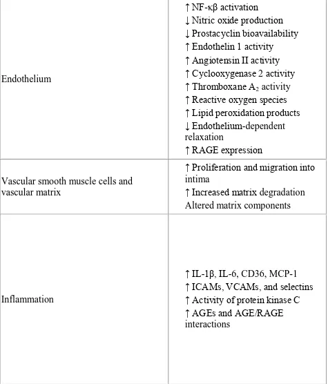
leads to a prothrombotic state.The accumulation of advanced glycation end
[60]
TABLE -3 MECHANISMS OF DIABETIC VASCULAR DISEASE
Endothelium
↑ NF-κβ activation
↓ Nitric oxide production ↓ Prostacyclin bioavailability ↑ Endothelin 1 activity
↑ Angiotensin II activity ↑ Cyclooxygenase 2 activity ↑ Thromboxane A2 activity
↑ Reactive oxygen species ↑ Lipid peroxidation products
↓ Endothelium-dependent relaxation
↑ RAGE expression
Vascular smooth muscle cells and vascular matrix
↑ Proliferation and migration into intima
↑ Increased matrix degradation Altered matrix components
Inflammation
↑ IL-1β, IL-6, CD36, MCP-1 ↑ ICAMs, VCAMs, and selectins ↑ Activity of protein kinase C
↑ AGEs and AGE/RAGE
[61]
The insulin resistance syndrome or metabolic syndrome comprises abdominal
obesity as seen by increased waist circumference ,elevated triglycerides, low HDL,
elevated blood pressure, increased blood sugar values. Its definition and diagnostic
criteria have varied depending on study groups and national guidelines and is
mired in ambiguities. But what is not in doubt is that the metabolic syndrome leads
to increased cardiovascular and all cause mortality[60]. Though central obesity
,which is calculated by measuring waist circumference,is an integral component
of metabolic syndrome, there are not many studies which have measured visceral
fat deposition. Visceral fat deposition is believed to be the main driver of
dysmetabolism[61]. Nowadays the definition of metabolic syndrome includes a pro
inflammatory state as evidenced by elevated hs CRP levels in these patients[62].
EXERCISE:
Regular exercise reduces the risk of cardiovascular diseases. Earlier it was believed
for exercise to be beneficial, it must be vigorous. But recent evidence indicates that
exercising in moderation confers significant cardiovascular benefits. Exercise
improves insulin sensitivity, lowers LDL and total cholesterol levels, improves
HDL levels and lowers triglyceride levels. These benefits tend to occur even in the
[62]
Studies have shown that if children increase the time spent in physical activity by
more than one hour daily occurrence of cardiovascular risk factors at an early age
can be prevented[63].Exercise further improves insulin sensitivity and glycemic
control, with major benefits for diabetic patients, including reductions in glycated
hemoglobin and reduced requirements for therapy. Various studies have shown
that even moderate intensity exercise has significantly reduced the incidence of
diabetes. Regular exercise lowers CRP levels, improves coronary endothelial
function, and appears to benefit hemostatic variables, including tissue-type
plasminogen activator, fibrinogen, von Willebrand factor, fibrin, D-dimer and
plasma viscosity.
Chronic exercise training appears to have a considerably favorable influence on
endothelial function in the peripheral arteries and the coronary arteries. This effect
is due to the increased production of vasodilating nitric oxide which leads to a
decrease in oxidative stress. Regular exercise also tends to increase levels of
endothelial progenitor cells. Such improvements have been linked to a reduction
in adverse cardiovascular events. Finally, exercise training appears to modulate the
balance between sympathetic and parasympathetic tone favorably, an effect