Copyright © 2002, American Society for Microbiology. All Rights Reserved.
Adenovirus E3-6.7K Maintains Calcium Homeostasis and Prevents
Apoptosis and Arachidonic Acid Release
Alexander R. Moise,
1,2Jason R. Grant,
1,2Timothy Z. Vitalis,
3and Wilfred A. Jefferies
1,2*
Biotechnology Laboratory,1Biomedical Research Centre, and Departments of Medical Genetics, Microbiology and Immunology,
and Zoology, University of British Columbia, Vancouver, British Columbia V6T 1Z3,2and Pulmonary Research Laboratory,
University of British Columbia, St. Paul’s Hospital, Vancouver, British Columbia V6Z 1Y6,3Canada
Received 5 June 2001/Accepted 9 November 2001
E3-6.7K is a small and hydrophobic membrane glycoprotein encoded by the E3 region of subgroup C adenovirus. Recently, E3-6.7K has been shown to be required for the downregulation of tumor necrosis factor (TNF)-related apoptosis-inducing ligand (TRAIL) receptors by the adenovirus E3/10.4K and E3/14.5K complex of proteins. We demonstrate here that E3-6.7K has additional protective roles, independent of other virus proteins. In transfected Jurkat T-cell lymphoma cells, E3-6.7K was found to maintain endoplasmic reticulum-Ca2ⴙhomeostasis and inhibit the induction of apoptosis by thapsigargin. The presence of E3-6.7K also lead
to a reduction in the TNF-induced release of arachidonic acid from transfected U937 human histiocytic lymphoma cells. In addition, E3-6.7K protected cells against apoptosis induced through Fas, TNF receptor, and TRAIL receptors. Therefore, E3-6.7K confers a wide range of protective effects against both Ca2ⴙ
flux-induced and death receptor-mediated apoptosis.
Adenovirus can lead to persistent infections of the eye, ear, and respiratory and digestive tracts in humans (23). Neverthe-less, during the early stages of adenovirus infection, events related to cellular transformation and viral gene expression are responsible for triggering apoptosis of the host cell. Primarily, the expression of the adenoviral genes E1A and E4orf4 induces apoptosis in both a dependent (59) and a p53-independent manner (53, 91). E1A is also implicated in the increased susceptibility of infected cells to death receptor-induced apoptosis (15). Therefore, other virus proteins must preserve the integrity of the cell and allow viral replication to be completed. Adenovirus antiapoptotic proteins include the Bcl-2 homologue E1B 19K (8, 32) and the inhibitors of p53-induced apoptosis E1B 55K (104) and E4orf6 (20). Meanwhile, the E3 region encodes inhibitors of death receptor-induced apoptosis, such as the 6.7K (6) and 14.7K (27, 54) proteins and the complex formed by the 10.4K and 14.5K proteins, also known as receptor internalization and degradation (RID) pro-teins␣and, respectively (6, 22, 83, 93).
Most viral survival factors can be classified according to which host survival protein they mimic. For example, there are viral Bcl-2-like proteins, such as BHRF1 (33) and A179L (1), and others that mimic the Fadd-like interleukin-converting enzyme-inhibitory protein, such as K13, BORFE2, and MC159 (92). There are also homologues of the cellular inhibitor of apoptosis proteins (17) and serpin homologues (19), both of which inhibit caspases and/or caspase activation. Ultimately, there are viral proteins that mimic death receptors and prevent their ligation, like MT-2 (79), SFV-T2 (85), CrmB (37), CrmC, and CrmD (58).
Notable exceptions to this classification are antiapoptotic
viral proteins for which no cellular homologue or mode of action has been identified. Interestingly, some of these proteins are localized in the endoplasmic reticulum (ER). For instance, a subset of the M-T2 protein, the myxoma virus homologue of the tumor necrosis factor (TNF) receptor, is ER retained, yet it is able to block apoptosis (80). MT-4, another ER-localized myxoma virus protein, has also been shown to prevent apopto-sis, through an unknown mechanism (4). Similar to M-T2, UL144 is an intracellular membrane protein homologous to the TNF receptor superfamily of proteins (5). Even ER-local-ized cellular proteins, shown to have an effect on apoptosis, have a poorly defined mechanism. Some of these cellular fac-tors are protective, such as DAD1 (also known as Ost2p) (88), PDI (90), Grp94 (56), calreticulin (57), and Grp78 (87), while others are proapoptotic, such as BAP31 (70) and mutant forms of PS-1 (45) and PS-2 (40), associated with familial Alzhei-mer’s disease.
The adenovirus type 2 (Ad2) E3-6.7K protein is localized primarily in the ER, as evidenced by cellular immunofluores-cence staining and by the presence of high-mannose N-linked glycan modifications in the mature protein (103). There is evidence that a small subset of this protein is found on the plasma membrane where it associates with RIDand allows the RID complex to downregulate the DR4 and DR5 TNF-related apoptosis-inducing ligand (TRAIL) receptors (6). However, a recent study using different experimental condi-tions suggests that E3-6.7K is not required for the downregu-lation of DR4 by RID (95), yet it may still be required by RID in downregulating DR5. The RID complex also downregulates Fas (6, 22, 83, 93) and epidermal growth factor receptor (94) and prevents TNF-induced apoptosis (28), arachidonic acid release (48), and translocation of cytosolic phospholipase A2
(cPLA2) to the membrane (18). E3-6.7K, however, was not
shown to be required for any of the latter effects but may play a role in augmenting RID function. We focused our studies on
* Corresponding author. Mailing address: The Biomedical Research Centre, 2222 Health Sciences Mall, University of British Columbia, Vancouver, BC V6T 1Z3, Canada. Phone: (604) 822-6961. Fax: (604) 822-6780. E-mail: wilf@unixg.ubc.ca.
1578
on November 8, 2019 by guest
http://jvi.asm.org/
broad survival mechanism by preventing death receptor- or Ca2⫹efflux-induced apoptosis and by reducing the release of
inflammatory mediators.
MATERIALS AND METHODS
Plasmid constructs.To amplify the cDNA for E3-6.7K, the forward primer ACCACCATGAGCAATTCAAGTAACTC, the reverse primer CCTTATCTT
GGATGTTGCCCCCAG, and template DNA consisting of theEcoRI D
frag-ment of the E3 region of Ad2 (a kind gift from W. S. M. Wold) were used. The PCR product was cloned into the bovine papillomavirus-based episomal expres-sion vector pBCMGSneo for transfection into U937 cells. The cDNA was also subcloned into pIRESpuro2 (Clontech) for transfection into Jurkat cells. Both strands of the inserted E3-6.7K coding region were sequenced by LoneStar Labs (Austin, Tex.) to ensure accuracy.
Generation of U937 and Jurkat cell lines stably expressing E3-6. 7K.U937 human histiocytic lymphoma (89) and Jurkat E6-1 T-cell lymphoma (101) cells, obtained from the American Type Culture Collection, were maintained in RPMI
1640 containing 10% fetal calf serum, 2 mML-glutamine, 10 mM HEPES, 100 U
of penicillin per ml, and 100g of streptomycin per ml in an atmosphere of 5%
CO2and 100% humidity.
U937 cells were transfected with the vector pBCMGSneo (44) alone or with pBCMGSneo carrying E3-6.7K by using the DMRIE-C cationic lipid reagent
(Life Technologies) and following the manufacturer’s protocol. Briefly, 4g of
DNA was mixed and incubated for 30 min with 0.5 ml of Opti-MEM medium
(Life Technologies) and 12l of DMRIE-C. The liposome-DNA mix was then
mixed and incubated with 0.5 ml of 5⫻105cells in Opti-MEM medium for an
additional 4 h. Stably transfected cells were selected with 1 mg of G-418 sulfate per ml 48 h later.
Jurkat cells were transfected by electroporation. Cells (2.5⫻107) were washed
twice in Opti-MEM medium, mixed with 20g of the appropriate plasmid, and
electroporated with a Bio-Rad Electroporator at 250 V and 950F. Transfected
cells were selected in medium containing 0.5g of puromycin per ml. The
pIRESpuro2 vector (Clontech) carries the encephalomyocarditis virus internal ribosome entry site (IRES), allowing cocistronic expression of E3-6.7K and the puromycin resistance gene.
Several subclones of the transfected cell lines were generated by serial dilution and examined for expression of E3-6.7K transcript by Northern blotting. The expression of E3-6.7K was very similar in all the clones examined. However, all the G-418- and puromycin-resistant U937 and Jurkat cells that survived the selection procedure were pooled and used in our experiments, in order to avoid clonal variation.
Ratiometric intracellular [Ca2ⴙ] determination.Jurkat-neo and Jurkat-6.7K
cells were washed and resuspended at a concentration of 107cells/ml in
Opti-MEM (Life Technologies). Aliquots of 100l of the cell suspension were
incu-bated for 90 min at 37°C with 6.0g of Indo-1 AM ester per ml. Prior to analysis,
1.9 ml of Opti-MEM was added to the cell suspension. Intracellular Ca2⫹levels
were measured with the ratiometric Ca2⫹indicator Indo-1; its emission
maxi-mum shifts from⬃485 nm in Ca2⫹-free medium to⬃400 nm when the dye is
saturated with Ca2⫹. The ratio of the emission signals at 405 nm and 485 nm
represents the ratio of Ca2⫹-bound to Ca2⫹-free Indo-1. The kinetic analysis
examined approximately 2⫻104cells/min with a FACS-Vantage sorter equipped
with an UV laser and appropriate filters for the 405- and 485-nm wavelengths.
minute) of incorporated [3H]arachidonic acid. The amount of [3H]arachidonic
acid released in experimental samples was expressed as a percentage of the total
incorporated [3H]arachidonic acid.
Thapsigargin-induced apoptosis assays in transfected Jurkat cells.Following
induction of apoptosis in Jurkat-neo and Jurkat-6.7K cells with 1 or 10M
thapsigargin for 24 h, we used the DNA-intercalating dye YO-PRO-1 (Molecular Probes) as an assay of membrane permeability changes. Cells were harvested,
washed in PBS, and resuspended at a concentration of 106cells/ml. Following
staining with 0.1M YO-PRO-1 and 1g of propidium iodide (PI) per ml, the
cells were analyzed by flow cytometry with FL1⬃530 nm- and FL2⬃575
nm-compensated emission readings on a FACScan (Becton Dickson). Viable, apo-ptotic cells showed green fluorescence, necrotic cells showed red and green fluorescence, and live, nonapoptotic cells showed very little fluorescence. The apoptotic index (AI) excluded the necrotic cell population and was calculated as
(number of viable apoptotic cells/total number of viable cells)⫻100.
Death receptor-induced apoptosis assays in transfected U937 and Jurkat cells.Fluorescein isothiocyanate (FITC)-conjugated annexin V (Pharmingen) was used to determine the binding of annexin V to externalized phosphatidyl serine. The protocol was based on the manufacturer’s annexin V-FITC staining
protocol. For each sample, 5⫻106U937-neo or U937-6.7K cells were harvested,
washed twice in PBS, and treated for 7 h with medium alone, 100 ng of TNF per
ml, 10g of CHX per ml, or a combination of 100 ng of TNF and 10g of CHX
per ml. The cells were stained with 5l of annexin V-FITC in a mixture
containing 10 mM HEPES–NaOH (pH 7.4), 140 mM NaCl, and 2.5 mM CaCl2
and analyzed with a fluorescence-activated cell sorter (FACS). Viable cells were gated based on their side scatter characteristics, and apoptotic cells were distin-guished from nonapoptotic cells based on their green fluorescence.
Alternatively, in the experiments examining FasL and TRAIL-induced
apo-ptosis, 5⫻106Jurkat-6.7K or Jurkat–neo cells each were harvested, washed
twice in PBS, and treated with agonist for 12 h. The treatment consisted of either
1g of DX2 (anti-human Fas monoclonal antibody) (Pharmingen) per ml and 2
g of goat anti-mouse secondary antibody per ml or a recombinant fusion
protein containing the extracellular domain of TRAIL (10 ng/ml; Upstate
Bio-tech.) and a potentiating reagent (5g/ml; Upstate Biotechnology) which
con-sists of a monoclonal antibody against the tag present in purified TRAIL. In these experiments, we used both the annexin V–Alexa-488 and the previously described YO-PRO-1 apoptosis assays (Molecular Probes). Like annexin-FITC, annexin V–Alexa-488 (Molecular Probes) labels cells that externalize phosphati-dyl serine. The same staining protocol was followed, with the exception that a
highly fluorescent non-cell-permeating DNA-intercalating dye, 50 M
Cy-toxGreen (Molecular Probes), was used to label necrotic cells. We examined the green fluorescence of stained cells with a FACS (Becton Dickinson). Viable, nonapoptotic cells showed little or no green fluorescence, while viable, apoptotic cells showed intermediate green fluorescence. Necrotic cells, on the other hand, showed 10 times greater fluorescence due to CytoxGreen staining than the apoptotic cells labeled with annexin V–Alexa-488 alone.
Activation of caspase-3 and cleavage of PARP by Western blotting.To analyze the cleavage of poly(ADP-ribose) polymerase (PARP), cell lysate equivalent to
105cells was denatured in sodium dodecyl sulfate (SDS) sample buffer (0.1 M
Tris [pH 6.8], 24% glycerol, 8% SDS, 0.2 M dithiothreitol, 0.02% bromophenol blue). The protein concentration was determined with a bicinchoninic acid assay kit (Pierce). Equivalent amounts of protein from each sample were resolved on a 10% glycine–SDS–polyacrylamide gel electrophoresis (PAGE) Laemmli gel
system, blotted with the Towbin vertical-transfer wet system onto a 0.45-
on November 8, 2019 by guest
pore-size Immobilon-P polyvinylidene difluoride (PVDF) membrane (Milli-pore), and incubated with anti-PARP mouse monoclonal antibody (1:5,000 di-lution; Pharmingen). After four washes in TBS containing 0.1% Tween 20 detergent (TBS-T), the blot was incubated with horseradish peroxidase-conju-gated goat anti-mouse antiserum (1:50,000 dilution) and visualized by chemilu-minescence with a SuperSignal West Pico kit (Pierce Chemical). Alternatively, to detect caspase-3 activation, the cell lysate was resolved on a 10% T, 3% C Tricine-SDS-PAGE gel without spacer, blotted with a Towbin vertical-transfer
wet system on a 0.2-m-pore-size Immobilon-P PVDF membrane (Millipore),
and incubated with anti-caspase-3 rabbit polyclonal antibody (1:5,000 dilution; Pharmingen). After four washes in TBS-T, the blot was incubated with horse-radish peroxidase-conjugated goat anti-rabbit antiserum (1:50,000 dilution) and visualized by chemiluminescence with a SuperSignal West Pico kit (Pierce Chem-ical).
RESULTS
The expression of E3-6.7K is sufficient to protect transfected cells against death receptor and thapsigargin-induced apopto-sis.It has been previously shown that E3-6.7K cooperates with RID␣andto downregulate the level of TRAIL receptors 1 and 2 from the surfaces of cells (6). While this identifies a role for the subset of the E3-6.7K protein expressed at the cell surface and possibly the endosomal compartment, it does not directly indicate a role for the large subset of the protein which is retained in the early secretory compartment. We began our studies by examining the effect of E3-6.7K on death receptor-induced apoptosis in transfected cells, in the absence of other viral proteins.
We chose to study the effects of E3-6.7K in cells of lymphoid and monocytic origin. Both cell types have been implicated as a possible reservoir for persistent adenovirus infections in vivo (2, 3, 13, 35, 84, 99). Immune evasion and the establishment of viral persistence may be more appropriately studied in cells resembling the in vivo reservoir of adenovirus. Subgroup C Ad5 can establish persistent infection in the human monocytic cell line U937, which maintains large copy numbers of uninte-grated viral genomes following infection (13). Infected cells continue to grow and produce minute amounts of mature virus
a year after infection. The Ad5-infected U937 cells have been proposed to provide a model of persistent adenovirus infection in humans. The human Jurkat T-cell line, on the other hand, has been used to study the efficacy of the immunoevasive protein E3/19K in downregulating major histocompatibility complex class I in lymphoid cells (47). We examined the re-sponses of E3-6.7K-transfected Jurkat or U937 cells to medi-ators of apoptosis and inflammation.
We transfected E3-6.7K cDNA amplified by PCR from a plasmid bearing the E3 region into the TNF-sensitive U937 human histiocytic lymphoma cell line. We established a stable population of U937 cells transfected with the episomal vector pBCMGSneo containing the cDNA for E3-6.7K (U937-6.7K) and another population transfected with vector alone (U937-neo). All surviving neomycin-resistant cells (in excess of 103
clones) were pooled and used in the following experiments to avoid the clonal variation known to arise in U937 cells. Ex-pression of E3-6.7K was analyzed by Northern blotting and by immunoprecipitation. We confirmed that the E3-6.7K protein was properly synthesized by immunoprecipitating it with a polyclonal rabbit antiserum raised against an E3-6.7K C-ter-minus-derived peptide and examined it by SDS-PAGE (results not shown).
We assayed the response of U937-neo and U937-6.7K cells to TNF-induced apoptosis by measuring the externalization of phosphatidyl serine by apoptotic cells (61) stained with an-nexin V-FITC. The presence of E3-6.7K yielded a 2.2-fold reduction in the proportion of apoptotic cells in U937-6.7K compared to U937-neo, following stimulation with TNF (Fig. 1). The U937-6.7K cells showed a 2.8-fold reduction in apo-ptosis compared to U937-neo cells following augmented stim-ulation with a combination of TNF and CHX, a protein syn-thesis inhibitor synergistic with TNF. The presence of E3-6.7K significantly decreased the apoptotic response in U937 cells upon stimulation with TNF or a combination of TNF and CHX.
FIG. 1. E3-6.7K protects against apoptosis induced by TNF in U937 cells. U937-neo (a to d) and U937-6.7K (e to h) cells were stimulated for 7 h with medium alone, 100 ng of TNF per ml, 10g of CHX per ml, or a combination of 100 ng of TNF and 10g of CHX per ml. For each cell population, a sample treatment was stained with annexin V-FITC and analyzed with a FACS. A second sample was analyzed with a FACS in the absence of annexin V-FITC in order to determine the background fluorescence, which corresponds to the fluorescence associated with the annexin V-negative cell population from each sample. The AI was calculated as (number of annexin V-positive viable cells/total number of viable cells)⫻100. The results are representative of three repeat experiments.
on November 8, 2019 by guest
http://jvi.asm.org/
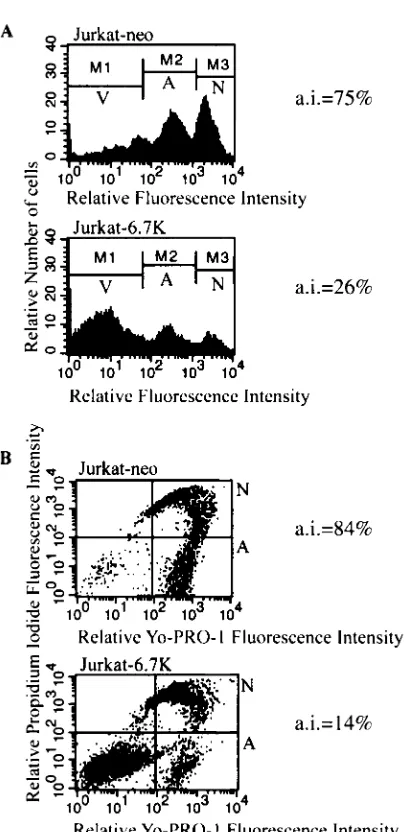
(Jurkat-6.7K) cells were less sensitive to apoptosis induced through Fas (Fig. 2) or TRAIL (Fig. 3) receptors than Jurkat cells transfected with vector alone (Jurkat-neo) cells. Follow-ing stimulation with FasL or TRAIL, the apoptotic response was assayed by two independent methods. In addition to an-nexin V labeling of externalized phosphatidyl serine, we also measured the exclusion of the dye YO-PRO-1 by viable, non-apoptotic cells. In both assays we used a viability dye to stain the necrotic and late-apoptotic cell population, commonly ex-cluded from the analysis of induction of apoptosis. Analysis of the apoptotic response was performed in accordance with manufacturer’s instructions and in accordance with published methods used in similar studies (11, 42, 78, 81, 97). Both assays demonstrated that E3-6.7K was effective in reducing TRAIL-and FasL-induced apoptosis in Jurkat cells. It is important to note that the antiapoptotic effect of E3-6.7K against TNF- and TRAIL-induced apoptosis was more pronounced than that against FasL-induced apoptosis, as observed by measuring the externalization of phosphatidyl serine (Fig. 1, 2A, and 3A).
In addition to death receptor-induced apoptosis, E3-6.7K con-fers a similar degree of protection against thapsigargin, a media-tor of apoptosis that acts intracellularly by mimicking a sustained Ca2⫹flux (43). Thapsigargin, a sesquiterpene lactone, isolated
from the umbelliferous plantThapsa garganica, selectively in-hibits the ER Ca2⫹-ATPase that directs Ca2⫹uptake into the
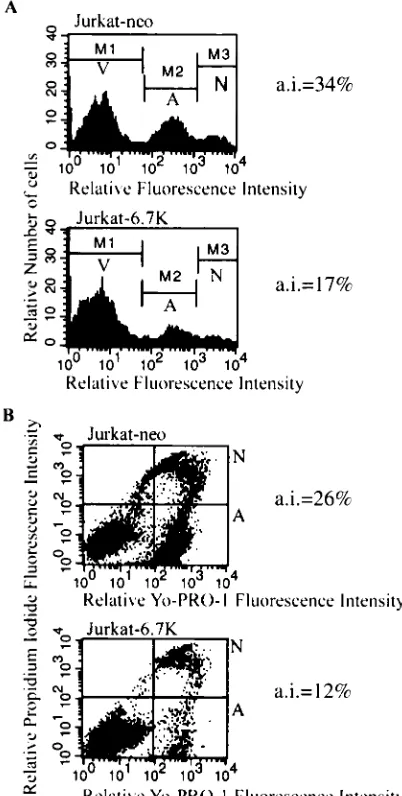
ER. This agent has been shown to induce apoptosis in Jurkat cells at high doses (86). Following induction of apoptosis in both Jur-kat-6.7K and in control Jurkat-neo cells, we measured the increase in membrane permeability of apoptotic cells to the DNA-intercalating dye YO-PRO-1 (38). We found that E3-6.7K pre-vented thapsigargin-induced apoptosis, which suggests an effect on ER Ca2⫹homeostasis (Fig 4A). The effects of
E3-6.7K on TRAIL- and 10M thapsigargin-induced apoptosis were comparable, resulting in dramatic improvement in the survival of transfected cells, as assayed by measuring the increase in membrane permeability during apoptosis (Fig. 3B and 4A ).
Efflux of Ca2ⴙfrom the ER in response to thapsigargin is
reduced in the presence of E3-6.7K.Thapsigargin induces ap-optosis through the increase in cytosolic Ca2⫹followed by the
activation of mediators of apoptosis. To better understand the mechanism of action of E3-6.7K, we assayed whether it has any direct effect on the thapsigargin-induced rise in cytosolic Ca2⫹
or whether it simply acts downstream of the Ca2⫹ flux by
affecting apoptotic mediators. Both neo and Jurkat-6.7K cells were loaded with the Ca2⫹-sensitive fluorophore
Indo-1. Following treatment with thapsigargin, the Ca2⫹flux
was assayed by FACS and represented using a ratiometric value of the amount of Ca2⫹-bound Indo-1 to the amount of
Ca2⫹-free Indo-1 per cell. The graph shown in Fig. 4B
[image:4.587.320.521.77.475.2]repre-sents the kinetic analysis of the mean of the ratiometric values of approximately 20,000 cells/min.
FIG. 2. E3-6.7K protects against apoptosis induced by Fas in Jur-kat-neo and Jurkat-6.7K cells. To study Fas-induced apoptosis, cells were stimulated for 12 h with 1g of the anti-human Fas monoclonal antibody DX2 (Pharmingen) per ml and 2 g of goat anti-mouse polyclonal antibody per ml. (A) Apoptotic cells were stained with annexin V–Alexa-488, indicating externalization of phosphatidyl serine; AI ⫽ (number of annexin V-positive, CytoxGreen-negative cells/total number of CytoxGreen negative cells)⫻100. The V sector indicates the nonapoptotic, viable cell population, the A sector indi-cates the apoptotic, viable cell population, and the N sector indiindi-cates the necrotic cell population. (B) Alternatively, cells were stained with YO-PRO-1, indicating an increase in membrane permeability; AI⫽
(number of YO-PRO-1-positive, negative cells/total number of PI-negative cells)⫻100. The lower left quadrant is the nonapoptotic, viable cell population, the lower right quadrant (A) represents the apoptotic, viable cell population, and the upper right quadrant (N) represents the necrotic cell population.
on November 8, 2019 by guest
Thapsigargin-induced Ca2⫹flux in Jurkat-neo cells resulted
in an 80% increase in the level of intracellular calcium. In Jurkat-6.7K cells, however, the observed increase was re-stricted to 34% (Fig. 4B). The presence of E3-6.7K reduced
the efflux of Ca2⫹in response to thapsigargin. The observed
decrease in Ca2⫹ homeostasis correlates with the protective
[image:5.587.61.263.66.482.2]role conferred by E3-6.7K against thapsigargin-induced apo-ptosis. Both apoptosis and Ca2⫹flux assays were conducted in FIG. 3. E3-6.7K protects against apoptosis induced by TRAIL in
Jurkat-neo and Jurkat-6.7K cells. To study TRAIL-induced apoptosis, cells were stimulated for 12 h with 10 ng of a recombinant fusion protein containing the extracellular domain of TRAIL (Upstate Bio-technology) per ml and a potentiating reagent (5g/ml; Upstate Bio-technology), which consists of a monoclonal antibody against the tag present in purified TRAIL. (A) The apoptotic cells were stained with annexin V–Alexa-488, indicating externalization of phosphatidyl serine; AI ⫽ (number of annexin V-positive, CytoxGreen-negative cells/total number of CytoxGreen-negative cells)⫻100. The V sector indicates the nonapoptotic, viable cell population, the A sector indi-cates the apoptotic, viable cell population, and the N sector indiindi-cates the necrotic cell population. (B) Alternatively, cells were stained with YO-PRO-1, indicating an increase in membrane permeability; AI⫽
(number of YO-PRO-1-positive, negative cells/total number of PI-negative cells)⫻100. The lower left quadrant is the nonapoptotic, viable cell population, the lower right quadrant (A) represents the apoptotic, viable cell population, and the upper right quadrant (N) represents the necrotic cell population.
FIG. 4. The presence of E3-6.7K results in the reduction of the thapsigargin-induced Ca2⫹flux and apoptosis. (A) Effect of E3-6.7K
on thapsigargin-induced apoptosis in transfected Jurkat cells. Jurkat-6.7K and Jurkat-neo cells were stimulated with medium containing 1% dimethyl sulfoxide (top),or 1 or 10M thapsigargin (TG) for 24 h, and then they were stained with YO-PRO-1 to examine the increase in the membrane permeability of apoptotic cells. The AI excluded the ne-crotic cell population and was calculated as (number of YO-PRO-1-positive, PI-negative apoptotic cells/total number of PI-negative, viable cells)⫻100. The A region represents the apoptotic, viable population, and V represents the nonapoptotic, viable population; AI⫽A/(A⫹V). The experiment was repeated with similar results: 23.2%⫾1.4% and 47.8%⫾4.7% for Jurkat-neo cells stimulated with 1 and 10M TG, respectively, and 10.3%⫾1.3% and 21.9%⫾6.6% for Jurkat-6.7K cells stimulated with 1 and 10M TG, respectively (P⬍0.05). (B) Ki-netic analysis of the effect of E3-6.7K on the release of Ca2⫹from the
ER in response to TG. Jurkat-6.7K and Jurkat-neo cells were loaded for 90 min at 37°C with 0.5M Indo-1 AM ester and examined on a UV-equipped FACS-Vantage flow cytometer (Becton Dickinson) for 5 min to establish the baseline fluorescence, equivalent to the resting level of intracellular calcium. At 5 min, cells were treated with 5 nM TG and analyzed for 25 min for a total of 5 ⫻ 105 events. The experiment was repeated three times with similar results.
on November 8, 2019 by guest
http://jvi.asm.org/
media containing extracellular Ca2⫹, which is essential for the
induction of apoptosis in Jurkat cells by thapsigargin (31). As a result, the initial ER Ca2⫹flux is amplified by capacitative
Ca2⫹entry through Ca2⫹release-activated Ca2⫹channels.
The presence of E3-6. 7K delays TNF-induced activation of caspase-3 and cleavage of PARP.Given the protective role of E3-6.7K against both death receptor- and Ca2⫹-induced
apo-ptosis, we examined its effect on the so-called “executioner” phase of apoptosis. More specifically, we assayed its effect on the TNF-induced cleavage of procaspase-3 and of the DNA repair enzyme PARP (Fig. 5). The 17- and 11-kDa proteolytic products of procaspase-3 are subunits of the heterodimeric, active form of caspase-3. The appearance of the active forms of caspase-3 was significantly delayed in 6.7K versus U937-neo cells (Fig. 5A). In addition, there was a significant reduc-tion in the amount of active caspase-3 in U937-6.7K cells versus U937-neo cells. The 85-kDa inactive form of PARP appeared in both U937-neo and U937-6.7K cell lines at 2 h after stimulation with TNF-CHX (Fig. 5B). There was, how-ever, a noticeable difference with regard to the kinetics of PARP inactivation: the amount of inactive PARP present in the U937-6.7K cells was considerably reduced compared to that in U937-neo cells. Therefore, the presence of E3-6.7K
delayed and reduced the activation of caspase-3 and inhibited PARP inactivation during TNF-induced apoptosis.
TNF-mediated arachidonic acid release is reduced in the presence of E3-6. 7K.As E3-6.7K affects Ca2⫹homeostasis in
transfected cells, it became imperative to test its effect on the TNF-induced release of arachidonic acid, which is a Ca2⫹
-dependent process. One possible candidate for the TNF-induc-ible release of arachidonic acid is the calcium-dependent cPLA2, which is activated by the dual signal of Ca2⫹release
from the ER stores and phosphorylation (68). In the presence of a sustained Ca2⫹flux, cPLA
2translocates preferentially to
the ER and nuclear envelope (72, 77). There, it releases ara-chidonic acid from the sn-2 position of various phospholipids, leading to the formation of eicosanoids, such as leukotrienes and prostaglandins, important mediators of inflammation.
We found that in the presence of E3-6.7K there was a re-duction in the release of [3H]arachidonic acid from U937-6.7K
cells by 50% compared to that from U937-neo cells following stimulation with TNF (Fig. 6). When the stimulus was in-creased by the addition of TNF and CHX, U937-6.7K cells released 60% less [3H]arachidonic acid than U937-neo cells
(Fig. 6). This effect can be explained by the fact that E3-6.7K affects Ca2⫹homeostasis, which indirectly affects the activity of
cPLA2. Our assay of [3H]arachidonic acid release was
con-ducted for periods ranging from 12 to 48 h after stimulation (results not shown). The amount of soluble [3H]arachidonic
acid is an equilibrium determined by cycles of deacylation and reacylation. We found that in our system, the equilibrium fa-vored deacylation of incorporated [3H]arachidonic acid for the
[image:6.587.45.282.72.321.2]first 24 h, reaching the peak of release of [3H]arachidonic acid
FIG. 5. Effect of E3-6.7K on the induction of procaspase-3 process-ing and PARP cleavage durprocess-ing TNF-induced apoptosis in vivo. Cell extracts were obtained from U937-neo and U937-6.7K cells that had been treated with 10 ng of TNF and 0.5g of CHX per ml for various lengths of time. Lysates containing equivalent amounts of protein based on the bicinchoninic acid (Pierce) protein concentration assay were loaded in each lane. After electrophoresis and transfer to PVDF membranes, blots were incubated with anti-caspase-3 rabbit antiserum that recognizes the 17- and 11-kDa subunits of the active, processed protein (A) and anti-PARP mouse monoclonal antibody that recog-nizes both the active 116-kDa and inactive 85-kDa forms of the protein (B). The blots were developed with a secondary antibody and visual-ized by chemiluminescence (Pierce Chemical). Similar results were obtained in two repeat experiments.
FIG. 6. Analysis of the effect of E3-6.7K on the inducible release of radiolabeled arachidonic acid. U937-neo and U937-6.7K cells were stimulated with medium alone, 90 ng of TNF per ml, 2g of CHX per ml, or a combination of 90 ng of TNF and 2g of CHX per ml. After 20 h, the amount (in counts per minute) of [3H]arachidonic acid released in the medium was expressed as a percentage of the total incorporated [3H]arachidonic acid. The assay was set up in triplicate; however, due to their relatively small values, the standard deviations of the values for the samples from U937-6.7K cells treated with medium alone (6.72%⫾0.037%) and U937-6.7K treated with CHX (5.74%⫾
0.027%) are difficult to observe on this graph. These results are rep-resentative of six repeat experiments.
on November 8, 2019 by guest
[image:6.587.349.488.74.260.2]at 20 h poststimulation. The release of incorporated [3
H]ara-chidonic acid over the next 24 h was minimal as intracellular stores of [3H]arachidonic acid became depleted. As a result, we
studied the effect of E3-6.7K at 20 h poststimulation. The effect of E3-6.7K on the release of arachidonic acid is consistent with its protective role against apoptosis and correlates with its effect on Ca2⫹homeostasis.
DISCUSSION
Adenovirus has been isolated from adenoids and tonsils of patients with acute respiratory diseases. A large percentage of these tissues are composed of lymphoid cells. Adenovirus DNA and infectious particles has been detected in cells of lymphoid origin, which may form a reservoir for persistent adenovirus infection in humans (2, 3, 13, 35, 84, 99). In spite of this, adenovirus has been propagated and studied in cells of nonlymphoid origin, such as HeLa, A549, and HEK-293 cells, because infection and replication of adenovirus in lymphoid cells are not as efficient as those in nonlymphoid cells. Lym-phoid-derived cell lines and peripheral blood lymphocytes are not permissive to Ad2 infection (36) and can be infected by adenovirus only at a high multiplicity of infection (84, 96). Cultured lymphocytes isolated from adenoids were found to support viral replication very poorly (96). This may be due, in part, to the fact that lymphocytes appear to lack the coxsack-ievirus and adenovirus receptor protein (76). Nevertheless, it is possible that the delayed kinetics of infection and replication of adenovirus in lymphoid cells are related to the establish-ment of viral persistence in a small fraction of lymphoid cells (52). As a result, in vitro models of persistent infection of lymphoid and monocytic cells have been established in tissue culture of Burkitt’s lymphoma cell line, Raji, the acute lym-phoblastic-T-cell-leukemia-derived cell line MOLT-3 (84, 96), and the histiocytic lymphoma cell line U937 (13).
The promoter that controls the E3 region is active in cells of lymphoid origin in the absence of E1A (102) and is responsive to TNF (16). It is possible that the role of the E3 region proteins in lymphoid cells is to aid in the establishment of persistent infections. Our studies examine the role of E3-6.7K in protecting cells of lymphoid and monocytic origin from apoptosis, with implications for the persistent viral infection seen in vivo in tonsillar or adenoidal lymphoid cells or in cells of monocytic origin. The effect of E3-6.7K on apoptosis in these cells was examined by transfection of the cDNA coding for E3-6.7K and creation of stable cell lines. The alternate approach would have been to study the response of lymphoid cells to apoptosis following infection of these cells with wild-type virus or virus with a deletion of E3-6.7K. We favored the single-gene-transfection approach for a number of reasons. Firstly, infection of lymphoid cells with subgroup C virus is extremely inefficient (84, 96), though there is a report of pro-ductive infection of Jurkat cells by Ad5 (51). Secondly, the only published deletion virus considered to have a single deletion in the E3-6.7K gene, dl739, was recently observed to express reduced levels of RID␣ protein (W. S. M. Wold, personal communication). This may be due to a secondary deletion but is more likely due to the fact that any deletion in the very complex transcription unit, E3, has the potential to alter the splicing pattern and 3'-end formation of E3 transcripts. Such
cis-acting sequences have been found to lead to the aberrant expression of genes not directly affected by the deletion (7, 9). This therefore precludes the assumption that viruses with de-letions of various E3 proteins have a normal pattern of expres-sion of the remaining genes and affects the concluexpres-sions derived from studies that employ such viruses.
Our results indicate that the E3-6.7K protein acted in the absence of other adenovirus proteins to protect transfected cells against TNF-, FasL-, and TRAIL-induced apoptosis and against TNF-induced arachidonic acid release. More impor-tantly, thapsigargin-induced Ca2⫹ efflux and apoptosis were
dramatically reduced in the presence of E3-6.7K, which sug-gests a possible effect of E3-6.7K on events that regulate Ca2⫹
homeostasis. The three proteins coded by the E3 region, E3/ 10.4K, E3/14.5K, and E3/14.7K, that prevent death receptor-induced apoptosis have also been shown to prevent the release of arachidonic acid following stimulation with TNF (48). Sim-ilarly, E3-6.7K is effective in reducing the release of arachi-donic acid. This newest addition to the E3 region proteins that prevent both TNF-induced apoptosis and release of tory mediators reflects the importance of TNF and inflamma-tion in the immune responses against adenovirus (21, 25).
The effect of E3-6.7K on TRAIL- or TNF-induced apoptosis was comparable with its effect on thapsigargin-induced apo-ptosis. This suggests that its effect on death receptor-induced apoptosis, in the absence of RID ␣ and , could in fact be related to its effect on downstream events such as Ca2⫹release.
Currently, the role of Ca2⫹efflux from the ER during death
receptor-induced apoptosis is not very well understood. Some studies describe an effect of Ca2⫹efflux on the apoptotic
re-sponse of cells to FasL (41) or TNF (46), while others disagree (62). Still other studies attribute a role to Ca2⫹efflux in the late
and not early events following FasL-induced apoptosis (82). Through the discovery of proteins that specifically affect Ca2⫹
release, through well-defined mechanisms, it will be possible to explore the role of Ca2⫹flux in death receptor-induced
apo-ptosis.
A previous study indicated that E3-6.7K is required by RID ␣andto reduce the surface level of TRAIL R1 and R2 and prevent TRAIL-induced apoptosis (6). Interestingly, we noted that many of the studies that examined the effect of RID␣and  made use of viral deletion mutants that were deficient in RID␣andor both but retained the expression of E3-6.7K. It is then plausible that E3-6.7K acts to augment the effects of RID ␣ and  in death receptor-induced apoptosis by acting both alone and in combination with them. We propose, there-fore, that E3-6.7K is an antiapoptotic protein with two possible mechanisms. First, as previously shown, cooperating with other viral proteins E3-6.7K results in the downregulation of TRAIL receptors. Secondly, E3-6.7K acts alone to prevent apoptosis against a variety of death receptors as well as intracellular mediators of apoptosis. Its effect, in the absence of other viral proteins, appears to involve the maintenance of cytosolic Ca2⫹
homeostasis.
It is common for viral proteins to have more than one mech-anism or function, a reflection, possibly, of the large number of functions that need to be performed by a successful virus. Some viral antiapoptotic proteins can use two distinct mecha-nisms to achieve their goal, as previously described for the TNF receptor homologue M-T2 (80) coded by myxoma virus.
on November 8, 2019 by guest
http://jvi.asm.org/
Mitochondrion-localized Bcl-2 exerts its antiapoptotic effects by blocking the release of cytochromecfrom the mitochondria (29). A subset of Bcl-2 proteins localizes to the cytoplasmic face of the ER membrane (39). Both wild-type Bcl-2 and an ER-restricted Bcl-2-cb5 fusion protein block the apoptotic cross talk between the ER and the mitochondria (30), and similarly to E3-6.7K, Bcl-2 has been shown to block thapsigar-gin-induced Ca2⫹efflux from the ER (49). Recently, Bcl-2 was
shown to decrease the free Ca2⫹concentration within the ER
(24, 73) by increasing its permeability to Ca2⫹, which results in
the depletion of the mobilizable Ca2⫹ reserves used during
agonist- or apoptosis-induced efflux. It is possible that the mechanism of E3-6.7K is related to the current model of the mechanism of action of ER localized Bcl-2. In this case, ex-pression of E3-6.7K should be able to induce a Ca2⫹“leak” in
the ER membrane, draining the mobilizable Ca2⫹reserves. It
could achieve this by regulating an existing channel or by forming a channel of its own.
The effect of E3-6.7K on thapsigargin-induced Ca2⫹ flux
places it upstream of many apoptotic effectors that are acti-vated by of Ca2⫹release from the ER. Some of these effectors
are the death-associated protein kinase (14), protein kinase C (98), the MEF2 transcription factor (105), and calcineurin. Calcineurin, a Ca2⫹-regulated serine-threonine phosphatase,
has been implicated in a variety of processes that result in the induction of apoptosis by activating nuclear factor of activated T-cells, which results in the expression of FasL (50), or by activating the proapoptotic Bcl-2 family member Bad (100). Recently, Ca2⫹-induced proteases, such as the calpain class of
calcium-regulated cysteine proteases (74) and caspase-12, have been implicated in apoptosis. The recently discovered caspase-12 is activated by inducers of the ER stress response and by calpains (65) but not by other inducers of apoptosis, such as TNF (66).
Recently, the luminal environment of the ER and, more particularly, ER chaperones has been shown to play an impor-tant role in determining the sensitivity of cells to apoptosis (67). Several ER-resident chaperone proteins, such as calreti-culin (10) and BiP/Grp78 (57), appear to increase the Ca2⫹
storage capacity of the ER (55, 67) and may regulate capaci-tative Ca2⫹ entry (64). Chaperone induction has also been
associated with viral infection, as many viruses induce an ER overload response through the abundant expression of se-creted viral proteins, as in the case of E3-6.7K’s cocistronic partner E3/19K (71). The link between viral infection, ER
ER localized pro-and antiapoptotic proteins will bridge the gap between the ER stress response and apoptosis. We propose that like E3-6.7K, other ER-localized viral survival factors are good candidates for the functional dissection of these common pathways.
ACKNOWLEDGMENTS
This work was supported by a grant from the National Cancer Institute and a Proof of Principle Program grant from the Canadian Institute of Health Research of Canada to W.A.J.
We thank W. S. Wold for the generous gift of polyclonal antisera recognizing E3-6.7K and the plasmid bearing the Ad2 E3 region. We thank E. White, D. R. Green, and R. Lippe for helpful discussions and G. Osborne from the Multi User Flow Cytometry Core Facility for technical assistance in measuring apoptosis and calcium fluxes.
REFERENCES
1.Afonso, C. L., J. G. Neilan, G. F. Kutish, and D. L. Rock.1996. An African swine fever virus Bc1–2 homolog, 5-HL, suppresses apoptotic cell death.
J. Virol.70:4858–4863.
2.Andiman, W. A., R. I. Jacobson, and G. Tucker.1977. Leukocyte-associated viremia with adenovirus type 2 in an infant with lower-respiratory-tract
disease. N. Engl. J. Med.297:100–101.
3.Andiman, W. A., and G. Miller.1982. Persistent infection with adenovirus types 5 and 6 in lymphoid cells from humans and woolly monkeys. J. Infect.
Dis.145:83–88.
4.Barry, M., S. Hnatiuk, K. Mossman, S. F. Lee, L. Boshkov, and G. Mc-Fadden.1997. The myxoma virus M-T4 gene encodes a novel RDEL-containing protein that is retained within the endoplasmic reticulum and is
important for the productive infection of lymphocytes. Virology239:360–
377.
5.Benedict, C. A., K. D. Butrovich, N. S. Lurain, J. Corbeil, I. Rooney, P. Schneider, J. Tschopp, and C. F. Ware.1999. Cutting edge: a novel viral TNF receptor superfamily member in virulent strains of human
cytomeg-alovirus. J. Immunol.162:6967–6970.
6.Benedict, C. A., P. S. Norris, T. I. Prigozy, J. L. Bodmer, J. A. Mahr, C. T. Garnett, F. Martinon, J. Tschopp, L. R. Gooding, and C. F. Ware.2001. Three adenovirus e3 proteins cooperate to evade apoptosis by tumor ne-crosis factor-related apoptosis-inducing ligand receptor-1 and -2. J. Biol.
Chem.276:3270–3278.
7.Bhat, B. M., and W. S. Wold.1987. A small deletion distant from a splice or polyadenylation site dramatically alters pre-mRNA processing in region
E3 of adenovirus. J. Virol.61:3938–3945.
8.Boyd, J. M., S. Malstrom, T. Subramanian, L. K. Venkatesh, U. Schaeper, B. Elangovan, C. D’Sa-Eipper, and G. Chinnadurai.1994. Adenovirus E1B 19 kDa and Bcl-2 proteins interact with a common set of cellular proteins.
Cell79:341–351. (Erratum,79, following p. 1120.)
9.Brady, H. A., A. Scaria, and W. S. Wold.1992. Map of cis-acting sequences that determine alternative pre-mRNA processing in the E3 complex
tran-scription unit of adenovirus. J. Virol.66:5914–5923.
10.Camacho, P., and J. D. Lechleiter.1995. Calreticulin inhibits repetitive
intracellular Ca2⫹waves. Cell82:765–771.
11.Casella, C. R., E. L. Rapaport, and T. H. Finkel. 1999. Vpu increases susceptibility of human immunodeficiency virus type 1-infected cells to fas
killing. J. Virol.73:92–100.
12.Chiocca, S., A. Baker, and M. Cotten.1997. Identification of a novel anti-apoptotic protein, GAM-1, encoded by the CELO adenovirus. J. Virol.
71:3168–3177.
on November 8, 2019 by guest
13.Chu, Y., K. Sperber, L. Mayer, and M. T. Hsu.1992. Persistent infection of
human adenovirus type 5 in human monocyte cell lines. Virology188:793–
800.
14.Cohen, O., B. Inbal, J. L. Kissil, T. Raveh, H. Berissi, T. Spivak-Kroiza-man, E. Feinstein, and A. Kimchi.1999. DAP-kinase participates in TNF-alpha- and Fas-induced apoptosis and its function requires the death
do-main. J. Cell Biol.146:141–148.
15.Cook, J. L., T. A. Walker, A. M. Lewis, Jr., H. E. Ruley, F. L. Graham, and S. H. Pilder.1986. Expression of the adenovirus E1A oncogene during cell transformation is sufficient to induce susceptibility to lysis by host
inflam-matory cells. Proc. Natl. Acad. Sci. USA83:6965–6969.
16.Deryckere, F., and H. G. Burgert.1996. Tumor necrosis factor alpha
in-duces the adenovirus early 3 promoter by activation of NF-B. J. Biol.
Chem.271:30249–30255.
17.Deveraux, Q. L., and J. C. Reed.1999. IAP family proteins–-suppressors of
apoptosis. Genes Dev.13:239–252.
18.Dimitrov, T., P. Krajcsi, T. W. Hermiston, A. E. Tollefson, M. Hannink, and W. S. Wold.1997. Adenovirus E3–10.4K/14.5K protein complex inhibits tumor necrosis factor-induced translocation of cytosolic phospholipase A2
to membranes. J. Virol.71:2830–2837.
19.Dobbelstein, M., and T. Shenk.1996. Protection against apoptosis by the
vaccinia virus SPI-2 (B13R) gene product. J. Virol.70:6479–6485.
20.Dobner, T., N. Horikoshi, S. Rubenwolf, and T. Shenk.1996. Blockage by adenovirus E4orf6 of transcriptional activation by the p53 tumor
suppres-sor. Science272:1470–1473.
21.Elkon, K. B., C. C. Liu, J. G. Gall, J. Trevejo, M. W. Marino, K. A. Abrahamsen, X. Song, J. L. Zhou, L. J. Old, R. G. Crystal, and E. Falck-Pedersen.1997. Tumor necrosis factor alpha plays a central role in im-mune-mediated clearance of adenoviral vectors. Proc. Natl. Acad. Sci. USA
94:9814–9819.
22.Elsing, A., and H. G. Burgert.1998. The adenovirus E3/10.4K-14.5K pro-teins down-modulate the apoptosis receptor Fas/Apo-1 by inducing its
internalization. Proc. Natl. Acad. Sci. USA95:10072–10077.
23.Fox, J. P., C. E. Hall, and M. K. Cooney.1977. The Seattle Virus Watch.
VII. Observations of adenovirus infections. Am. J. Epidemiol.105:362–386.
24.Foyouzi-Youssefi, R., S. Arnaudeau, C. Borner, W. L. Kelley, J. Tschopp, D. P. Lew, N. Demaurex, and K. H. Krause.2000. Bcl-2 decreases the free
Ca2⫹concentration within the endoplasmic reticulum. Proc. Natl. Acad.
Sci. USA97:5723–5728.
25.Ginsberg, H. S., L. L. Moldawer, P. B. Sehgal, M. Redington, P. L. Kilian, R. M. Chanock, and G. A. Prince.1991. A mouse model for investigating the molecular pathogenesis of adenovirus pneumonia. Proc. Natl. Acad.
Sci. USA88:1651–1655.
26.Glotzer, J. B., M. Saltik, S. Chiocca, A. I. Michou, P. Moseley, and M. Cotten.2000. Activation of heat-shock response by an adenovirus is
essen-tial for virus replication. Nature407:207–211.
27.Gooding, L. R., L. W. Elmore, A. E. Tollefson, H. A. Brady, and W. S. Wold.
1988. A 14,700 MW protein from the E3 region of adenovirus inhibits
cytolysis by tumor necrosis factor. Cell53:341–346.
28.Gooding, L. R., T. S. Ranheim, A. E. Tollefson, L. Aquino, P. Duerksen-Hughes, T. M. Horton, and W. S. Wold.1991. The 10,400- and 14,500-dalton proteins encoded by region E3 of adenovirus function together to protect many but not all mouse cell lines against lysis by tumor necrosis
factor. J. Virol.65:4114–4123.
29.Green, D. R., and J. C. Reed.1998. Mitochondria and apoptosis. Science
281:1309–1312.
30.Hacki, J., L. Egger, L. Monney, S. Conus, T. Rosse, I. Fellay, and C. Borner.
2000. Apoptotic crosstalk between the endoplasmic reticulum and
mito-chondria controlled by Bcl-2. Oncogene19:2286–2295.
31.Hampton, M. B., D. M. Vanags, M. I. Porn-Ares, and S. Orrenius.1996. Involvement of extracellular calcium in phosphatidylserine exposure during
apoptosis. FEBS Lett.399:277–282.
32.Han, J., P. Sabbatini, D. Perez, L. Rao, D. Modha, and E. White.1996. The E1B 19K protein blocks apoptosis by interacting with and inhibiting the
p53-inducible and death-promoting Bax protein. Genes Dev.10:461–477.
33.Henderson, S., D. Huen, M. Rowe, C. Dawson, G. Johnson, and A. Rick-inson.1993. Epstein-Barr virus-coded BHRF1 protein, a viral homologue of Bcl-2, protects human B cells from programmed cell death. Proc. Natl.
Acad. Sci. USA90:8479–8483.
34.Higashino, F., J. M. Pipas, and T. Shenk.1998. Adenovirus E4orf6 onco-protein modulates the function of the p53-related onco-protein, p73. Proc. Natl.
Acad. Sci. USA95:15683–15687.
35.Horvath, J., L. Palkonyay, and J. Weber.1986. Group C adenovirus DNA
sequences in human lymphoid cells. J. Virol.59:189–192.
36.Horvath, J., and J. M. Weber.1988. Nonpermissivity of human peripheral
blood lymphocytes to adenovirus type 2 infection. J. Virol.62:341–345.
37.Hu, F. Q., C. A. Smith, and D. J. Pickup.1994. Cowpox virus contains two copies of an early gene encoding a soluble secreted form of the type II TNF
receptor. Virology204:343–356.
38.Idziorek, T., J. Estaquier, F. De Bels, and J. C. Ameisen.1995. YOPRO-1 permits cytofluorometric analysis of programmed cell death (apoptosis)
without interfering with cell viability. J. Immunol. Methods185:249–258.
39.Janiak, F., B. Leber, and D. W. Andrews.1994. Assembly of Bcl-2 into
microsomal and outer mitochondrial membranes. J. Biol. Chem.269:9842–
9849.
40.Janicki, S., and M. J. Monteiro.1997. Increased apoptosis arising from increased expression of the Alzheimer’s disease-associated presenilin-2
mu-tation (N141I). J. Cell Biol.139:485–495.
41.Jayaraman, T., and A. R. Marks.1997. T cells deficient in inositol
1,4,5-trisphosphate receptor are resistant to apoptosis. Mol. Cell. Biol.17:3005–
3012.
42.Jerome, K. R., J. F. Tait, D. M. Koelle, and L. Corey.1998. Herpes simplex virus type 1 renders infected cells resistant to cytotoxic
T-lymphocyte-induced apoptosis. J. Virol.72:436–441.
43.Jiang, S., S. C. Chow, P. Nicotera, and S. Orrenius.1994. Intracellular Ca2⫹
signals activate apoptosis in thymocytes: studies using the Ca2⫹-ATPase
inhibitor thapsigargin. Exp. Cell Res.212:84–92.
44.Karasuyama, H., and F. Melchers.1988. Establishment of mouse cell lines which constitutively secrete large quantities of interleukin 2, 3, 4 or 5, using
modified cDNA expression vectors. Eur. J. Immunol.18:97–104.
45.Katayama, T., K. Imaizumi, N. Sato, K. Miyoshi, T. Kudo, J. Hitomi, T. Morihara, T. Yoneda, F. Gomi, Y. Mori, Y. Nakano, J. Takeda, T. Tsuda, Y. Itoyama, O. Murayama, A. Takashima, P. St George-Hyslop, M. Takeda, and M. Tohyama.1999. Presenilin-1 mutations downregulate the signalling
pathway of the unfolded-protein response. Nat. Cell Biol.1:479–485.
46.Ko, S., T. T. Kwok, K. P. Fung, Y. M. Choy, C. Y. Lee, and S. K. Kong.2000.
Slow rise of Ca2⫹and slow release of reactive oxygen species are two
cross-talked events important in tumour necrosis factor-alpha-mediated
apoptosis. Free Radic. Res.33:295–304.
47.Korner, H., and H. G. Burgert.1994. Down-regulation of HLA antigens by the adenovirus type 2 E3/19K protein in a T-lymphoma cell line. J. Virol.
68:1442–1448.
48.Krajcsi, P., T. Dimitrov, T. W. Hermiston, A. E. Tollefson, T. S. Ranheim, S. B. Vande Pol, A. H. Stephenson, and W. S. Wold.1996. The adenovirus E3–14.7K protein and the E3–10.4K/14.5K complex of proteins, which independently inhibit tumor necrosis factor (TNF)-induced apoptosis, also independently inhibit TNF-induced release of arachidonic acid. J. Virol.
70:4904–4913.
49.Lam, M., G. Dubyak, L. Chen, G. Nunez, R. L. Miesfeld, and C. W. Distelhorst.1994. Evidence that BCL-2 represses apoptosis by regulating
endoplasmic reticulum-associated Ca2⫹fluxes. Proc. Natl. Acad. Sci. USA
91:6569–6573.
50.Latinis, K. M., L. L. Carr, E. J. Peterson, L. A. Norian, S. L. Eliason, and G. A. Koretzky.1997. Regulation of CD95 (Fas) ligand expression by
TCR-mediated signaling events. J. Immunol.158:4602–4611.
51.Lavery, D., S. M. Fu, T. Lufkin, and S. Chen-Kiang. 1987. Productive
infection of cultured human lymphoid cells by adenovirus. J. Virol.61:
1466–1472.
52.Lavery, D. J., and S. Chen-Kiang.1990. Adenovirus E1A and E1B genes
are regulated posttranscriptionally in human lymphoid cells. J. Virol.64:
5349–5359.
53.Lavoie, J. N., M. Nguyen, R. C. Marcellus, P. E. Branton, and G. C. Shore.
1998. E4orf4, a novel adenovirus death factor that induces p53-independent apoptosis by a pathway that is not inhibited by zVAD-fmk. J. Cell Biol.
140:637–645.
54.Li, Y., J. Kang, J. Friedman, L. Tarassishin, J. Ye, A. Kovalenko, D. Wallach, and M. S. Horwitz.1999. Identification of a cell protein (FIP-3) as
a modulator of NF-B activity and as a target of an adenovirus inhibitor of
tumor necrosis factor alpha-induced apoptosis. Proc. Natl. Acad. Sci. USA
96:1042–1047.
55.Lievremont, J. P., R. Rizzuto, L. Hendershot, and J. Meldolesi.1997. BiP, a major chaperone protein of the endoplasmic reticulum lumen, plays a direct and important role in the storage of the rapidly exchanging pool of
Ca2⫹. J. Biol. Chem.272:30873–30879.
56.Little, E., and A. S. Lee.1995. Generation of a mammalian cell line defi-cient in glucose-regulated protein stress induction through targeted
ri-bozyme driven by a stress-inducible promoter. J. Biol. Chem.270:9526–
9534.
57.Liu, H., R. C. Bowes III, B. van de Water, C. Sillence, J. F. Nagelkerke, and J. L. Stevens.1997. Endoplasmic reticulum chaperones GRP78 and
calre-ticulin prevent oxidative stress, Ca2⫹disturbances, and cell death in renal
epithelial cells. J. Biol. Chem.272:21751–21759.
58.Loparev, V. N., J. M. Parsons, J. C. Knight, J. F. Panus, C. A. Ray, R. M. Buller, D. J. Pickup, and J. J. Esposito. 1998. A third distinct tumor necrosis factor receptor of orthopoxviruses. Proc. Natl. Acad. Sci. USA
95:3786–3791.
59.Lowe, S. W., and H. E. Ruley.1993. Stabilization of the p53 tumor sup-pressor is induced by adenovirus 5 E1A and accompanies apoptosis. Genes
Dev.7:535–545.
60.Marcellus, R. C., J. N. Lavoie, D. Boivin, G. C. Shore, G. Ketner, and P. E. Branton.1998. The early region 4 orf4 protein of human adenovirus type 5
induces p53-independent cell death by apoptosis. J. Virol.72:7144–7153.
61.Martin, S. J., C. P. Reutelingsperger, A. J. McGahon, J. A. Rader, R. C. van Schie, D. M. LaFace, and D. R. Green.1995. Early redistribution of plasma
on November 8, 2019 by guest
http://jvi.asm.org/
Goping, M. Opas, R. C. Bleackley, D. R. Green, and M. Michalak.2000. Changes in endoplasmic reticulum luminal environment affect cell
sensitiv-ity to apoptosis. J. Cell Biol.150:731–740.
68.Nalefski, E. A., L. A. Sultzman, D. M. Martin, R. W. Kriz, P. S. Towler, J. L. Knopf, and J. D. Clark. 1994. Delineation of two functionally distinct
domains of cytosolic phospholipase A2, a regulatory Ca2⫹-dependent
lipid-binding domain and a Ca2⫹-independent catalytic domain. J. Biol. Chem.
269:18239–18249.
69.Nevins, J. R.1982. Induction of the synthesis of a 70,000 dalton mammalian
heat shock protein by the adenovirus E1A gene product. Cell29:913–919.
70.Ng, F. W., M. Nguyen, T. Kwan, P. E. Branton, D. W. Nicholson, J. A. Cromlish, and G. C. Shore.1997. p28 Bap31, a Bcl-2/Bcl-XL- and pro-caspase-8-associated protein in the endoplasmic reticulum. J. Cell Biol.
139:327–338.
71.Pahl, H. L., M. Sester, H. G. Burgert, and P. A. Baeuerle.1996. Activation
of transcription factor NF-B by the adenovirus E3/19K protein requires its
ER retention. J. Cell Biol.132:511–522.
72.Peters-Golden, M., K. Song, T. Marshall, and T. Brock.1996. Translocation of cytosolic phospholipase A2 to the nuclear envelope elicits
topographi-cally localized phospholipid hydrolysis. Biochem. J.318:797–803.
73.Pinton, P., D. Ferrari, P. Magalhaes, K. Schulze-Osthoff, F. Di Virgilio, T. Pozzan, and R. Rizzuto.2000. Reduced loading of intracellular Ca2⫹stores
and downregulation of capacitative Ca2⫹ influx in Bcl-2-overexpressing
cells. J. Cell Biol.148:857–862.
74.Porn-Ares, M. I., A. Samali, and S. Orrenius.1998. Cleavage of the calpain
inhibitor, calpastatin, during apoptosis. Cell Death Differ.5:1028–1033.
75.Qi, X. M., H. He, H. Zhong, and C. W. Distelhorst.1997. Baculovirus p35 and Z-VAD-fmk inhibit thapsigargin-induced apoptosis of breast cancer
cells. Oncogene15:1207–1212.
76.Rebel, V. I., S. Hartnett, J. Denham, M. Chan, R. Finberg, and C. A. Sieff.
2000. Maturation and lineage-specific expression of the coxsackie and
ad-enovirus receptor in hematopoietic cells. Stem Cells18:176–182.
77.Schievella, A. R., M. K. Regier, W. L. Smith, and L. L. Lin.1995. Calcium-mediated translocation of cytosolic phospholipase A2 to the nuclear
enve-lope and endoplasmic reticulum. J. Biol. Chem.270:30749–30754.
78.Schmidt, M., S. Afione, and R. M. Kotin.2000. Adeno-associated virus type 2 Rep78 induces apoptosis through caspase activation independently of
p53. J. Virol.74:9441–9450.
79.Schreiber, M., and G. McFadden.1996. Mutational analysis of the ligand-binding domain of M-T2 protein, the tumor necrosis factor receptor
ho-mologue of myxoma virus. J. Immunol.157:4486–4495.
80.Schreiber, M., L. Sedger, and G. McFadden. 1997. Distinct domains of M-T2, the myxoma virus tumor necrosis factor (TNF) receptor homolog, mediate extracellular TNF binding and intracellular apoptosis inhibition.
J. Virol.71:2171–2181.
81.Schuler, M., E. Bossy-Wetzel, J. C. Goldstein, P. Fitzgerald, and D. R. Green.2000. p53 induces apoptosis by caspase activation through
mito-chondrial cytochrome c release. J. Biol. Chem.275:7337–7342.
82.Scoltock, A. B., C. D. Bortner, G. S. J. Bird, J. W. Putney, Jr., and J. A. Cidlowski. 2000. A selective requirement for elevated calcium in DNA degradation, but not early events in anti-Fas-induced apoptosis. J. Biol.
Chem.275:30586–30596.
83.Shisler, J., C. Yang, B. Walter, C. F. Ware, and L. R. Gooding.1997. The
disulfide isomerase in response to hypoxia/brain ischemia and its protective
effect against apoptotic cell death. J. Biol. Chem.275:10388–10393.
91.Teodoro, J. G., G. C. Shore, and P. E. Branton.1995. Adenovirus E1A proteins induce apoptosis by both p53-dependent and p53-independent
mechanisms. Oncogene11:467–474.
92.Thome, M., P. Schneider, K. Hofmann, H. Fickenscher, E. Meinl, F. Neipel, C. Mattmann, K. Burns, J. L. Bodmer, M. Schroter, C. Scaffidi, P. H. Krammer, M. E. Peter, and J. Tschopp. 1997. Viral FLICE-inhibitory proteins (FLIPs) prevent apoptosis induced by death receptors. Nature
386:517–521.
93.Tollefson, A. E., T. W. Hermiston, D. L. Lichtenstein, C. F. Colle, R. A. Tripp, T. Dimitrov, K. Toth, C. E. Wells, P. C. Doherty, and W. S. Wold.
1998. Forced degradation of Fas inhibits apoptosis in adenovirus-infected
cells. Nature392:726–730.
94.Tollefson, A. E., A. R. Stewart, S. P. Yei, S. K. Saha, and W. S. Wold.1991. The 10,400- and 14,500-dalton proteins encoded by region E3 of adenovirus form a complex and function together to down-regulate the epidermal
growth factor receptor. J. Virol.65:3095–3105.
95.Tollefson, A. E., K. Toth, K. Doronin, M. Kuppuswamy, O. A. Doronina, D. L. Lichtenstein, T. W. Hermiston, C. A. Smith, and W. S. Wold.2001. Inhibition of TRAIL-induced apoptosis and forced internalization of
TRAIL receptor 1 by adenovirus proteins. J. Virol.75:8875–8887.
96.van der Veen, J., and M. Lambriex.1973. Relationship of adenovirus to lymphocytes in naturally infected human tonsils and adenoids. Infect.
Im-mun.7:604–609.
97.Vermes, I., C. Haanen, H. Steffens-Nakken, and C. Reutelingsperger.1995. A novel assay for apoptosis. Flow cytometric detection of phosphatidylser-ine expression on early apoptotic cells using fluorescein labelled annexin
V. J. Immunol. Methods184:39–51.
98.Villalba, M., S. Kasibhatla, L. Genestier, A. Mahboubi, D. R. Green, and A. Altman.1999. Protein kinase ccooperates with calcineurin to induce Fas
ligand expression during activation-induced T cell death. J. Immunol.163:
5813–5819.
99.Wallace, R. E.1969. Susceptibility of human lymphoblasts (RPMI 7466) to
viral infections in vitro. Proc. Soc. Exp. Biol. Med.130:702–710.
100.Wang, H. G., N. Pathan, I. M. Ethell, S. Krajewski, Y. Yamaguchi, F. Shibasaki, F. McKeon, T. Bobo, T. F. Franke, and J. C. Reed.1999. Ca2⫹
-induced apoptosis through calcineurin dephosphorylation of BAD. Science
284:339–343.
101.Weiss, A., J. Imboden, D. Shoback, and J. Stobo.1984. Role of T3 surface molecules in human T-cell activation: T3-dependent activation results in an
increase in cytoplasmic free calcium. Proc. Natl. Acad. Sci. USA81:4169–
4173.
102.Williams, J. L., J. Garcia, D. Harrich, L. Pearson, F. Wu, and R. Gaynor.
1990. Lymphoid specific gene expression of the adenovirus early region 3
promoter is mediated by NF-B binding motifs. EMBO J.9:4435–4442.
103.Wilson-Rawls, J., and W. S. Wold.1993. The E3–6.7K protein of adenovirus is an Asn-linked integral membrane glycoprotein localized in the
endoplas-mic reticulum. Virology195:6–15.
104.Yew, P. R., and A. J. Berk.1992. Inhibition of p53 transactivation required
for transformation by adenovirus early 1B protein. Nature357:82–85.
105.Youn, H. D., L. Sun, R. Prywes, and J. O. Liu.1999. Apoptosis of T cells
mediated by Ca2⫹-induced release of the transcription factor MEF2.
Sci-ence286:790–793.