TARGETING
DECAPRENYLPHOSPHORYL-BETA-D-RIBOSE 2’EPIMERASE-1” A dissertation submitted to
THE TAMILNADU DR.M.G.R MEDICAL UNIVERSITY CHENNAI-600032.
In the partial fulfilment of the requirements For the award of the degree of
MASTER OF PHARMACY IN
PHARMACEUTICAL CHEMISTRY
Submitted by
Reg. No. 261415716
Under the guidance of
Dr. A. Jerad Suresh, M.Pharm., Ph.D., M.B.A.,
DEPARTMENT OF PHARMACEUTICAL CHEMISTRY COLLEGE OF PHARMACY
MADRAS MEDICAL COLLEGE CHENNAI-600 003
S.NO
TITLE
PAGE NO
1
INTRODUCTION
1
2
REVIEW OF LITERATURE
27
3
AIM AND OBJECTIVE
32
4
MATERIALS AND METHODS
Drug Design
Reactant profile
Synthetic methodology
Characterization studies
34
37
39
41
5
RESULTS AND DISCUSSION
Drug design
Drug profile
Characterization
In silico toxicity screening
In vitro antitubercular screening
44
46
49
60
62
6
SUMMARY AND CONCLUSION
64
7
REFERENCE
66
First and foremos00t I thank the Almighty for all the love, grace and blessings
he has bestowed upon me and for strengthening me throughout, to successfully
complete this dissertation.
I take this opportunity to express my gratitude to the people who were
instrumental in the successful completion of this project.
I express my immense gratitude to Government of Tamilnadu for providing me the
monthly scholarship.
At this moment of accomplishment, first of all I express my deep sense of
gratitude to Mrs. Vimala M.D., Dean, Madras Medical College, Chennai-3 for
providing all facilities during the period of study.
It is my privilege and great pleasure to express my gratitude to my esteemed
Principal and Guide Dr. A. Jerad Suresh, M.Pharm., Ph.D., M.B.A., College of
Pharmacy, Madras Medical College, Chennai-3. This work would not have been
possible without his able guidance, support and encouragement. Under his guidance I
successfully overcame many difficulties and learned a lot. His passion for perfection
combined with eagerness and enthusiasm to impart knowledge to students is what I
am in awe of. His knowledge and commitment to the highest standards inspired and
motivated me. I thank him immensely for his guidance.
I submit my thanks to Mrs. T. Saraswathy M.Pharm.,
Mrs. R. Priyadarshini M.Pharm., Ph.D., Mrs. P.G Sunitha M.Pharm., Ph.D, and
Mr. Sathish M.Pharm., Ph.D, Tutors, Department of Pharmaceutical Chemistry,
College of Pharmacy, Madras Medical College for their timely help and cooperation
Mr.K.N.Umapathi, Lab Technicians Department of Pharmaceutical Chemistry,
Madras Medical College for helping me to do this project.
I sincerely thank Mrs.Devi, Mr.K.M.Noorulla M.pharm , Ms.P.R.Suriya,
Scholars for their support, valuable suggestions and comments during my project.
I convey my thanks to Dr. Kishore Bhatt, Professor and Head, Department of
Microbiology, Maratha Mandal’s Institute of Dental Science and Research Institute,
Belgaum, Karnadaka and SASTRA University & IIT for helping me to carry out the
spectral studies in due time.
My thanks are also due to my dear friends N. Shantha sheela, S. Navaneetha,
S. C. Suruthi, S. Nandhini, P. Vijayalakshmi, W. Nivetha, K. Madhesh,
R. Kalaiselvi, M .Neelakandan, R. Sathyavani, for their constant help and
motivation. Without their support this project would not have materialized.
I heartily thanks to my dear friends P.Karunya, P.Elamathi for their care,
support and encouragement throughout.
I take immense pleasure in thanking all my seniors, juniors and friends from
all other departments.
Finally yet importantly, I Wish to express my heartfelt thanks to my beloved
Father, Mother, sister and my Husband for their blessings, encouragement and
TB Tubercule Bacillus
HIV Human Immuno Deficiency Syndrome AIDS Acquired Immuno Deficiency Syndrome BCG Bacilli Chalmette Guerin
DOTS Directly Observed Treatment Short-Course MDR-TB Multi Drug Resistant
XRD-TB Extensively Drug Resistant-TB LTBI Latent Tuberculosis Infection
DprE1 Decaprenylphosphoryl-beta-d-ribose 2’ Epimerase 1 CADD Computer Aided Drug Design
QSAR Quantitative Structural Activity Relationship PSA Polar Surface Area
OSIRIS Optical, Spectroscopic and Infrared Remote Imaging System
OPLS Optimized Potential For Liquid Simulation TPSA Total Polar Surface Area
SBDD Structure Based Drug Design LBDD Ligand Based Drug Design Log P Partition Co-Efficient WHO World Health Organization
MIC Minimum Inhibitory Concentration PDB Protein Data Bank
TLC World Health Organization IR Infrared Spectroscopy
NMR Nuclear Magnetic Resonance
GC-MS Gas Chromatography-Mass spectroscopy REMA Resazurin Micro Plate Assay
DEDICATED TO MY
DEDICATED TO MY
DEDICATED TO MY
DEDICATED TO MY
FAMILY AND
FAMILY AND
FAMILY AND
FAMILY AND
FRIENDS
FRIENDS
FRIENDS
INTRODUCTION
INTRODUCTION
INTRODUCTION
INTRODUCTION
Department of pharmaceutical chemistry. Page 1 1. INTRODUCTION
BACKGROUND
Tuberculosis is a major disease causing death every year 1.8 million worldwide and represents the leading cause of mortality resulting from a bacterial infection. Introduction in the 60’sof first-line drug regimen resulted in the control of the disease and TB was perceived as defeated.
In 2011, tuberculosis (TB) remained the second cause of death from infectious disease worldwide. It mainly affects the poorest countries of Africa and Southeast Asia. In 2010, according to the world health organization (WHO), TB incidence and prevalence were estimated at 8.8 and 12 million cases respectively about 1.1 million among HIV-negative people and 0.35 million among HIV-positive people died from TB. Most importantly, one third of the world population is infected with latent infection and 10% of those infected people will develop active TB in their life.
Department of pharmaceutical chemistry. Page 2
TB IN INDIA
India is the country with the highest burden of TB, with World Health Organization (WHO) statistics for 2014 giving an estimated incidence 2.2 million cases of TB for India out of a global incidence of 9 million. The estimated TB prevalence figure for 2014 is given as 2.5 million. It is estimated that about 40% of the Indian population is infected with TB bacteria, the vast majority of whom have latent rather than active TB.2
CURRENT THERAPIES
Tuberculosis (TB) is caused by bacteria mycobacterium tuberculosis that most often affect the lungs. Tuberculosis is curable and preventable. In 1882, The German physician Robert Koch isolated the bacterium. Tuberculosis is contagious and airborne disease.
In 1944, streptomycin was to treat tuberculosis (TB).This amino glycoside interferes with protein biosynthesis through an interaction with the small 30S subunit of the ribosome. The discovery of Para amino salicylic acid in 1946 was quickly followed by the important discovery of Isoniazide (INH), as one of the most active anti-TB drugs used today. Inhibition of mycolic acids biosynthesis, one of the essential components of the mycobacterium cell wall was determined as the mechanism of action. Pyrazinamide (PZA) appeared as a potential Anti-TB drug in 1952.
Department of pharmaceutical chemistry. Page 3 60’s .Ethambutol is an ethylenediamine discovered in 1961, which affects the cell wall by specifically targeting the polymerization of arabinogalactan and lipoarabinomannan. Finally, Rifampin appeared as a drug of choice for TB treatment around 1970, by acting on replicating and non-replicating mycobacteria.This derivative belongs to the rifampicin family and inhibits bacterial RNA synthesis by binding to the b-subunit of the DNA-dependent polymerase.
Department of pharmaceutical chemistry. Page 4
BACTERIOLOGICAL PROFILE OF MYCOBACTERIUM TUBERCULOSIS
SPECIES
The mycobacterium tuberculosis complex consists of Mycobacterium bovis, Mycobacterium africanoum and Mycobacterium canettii, Mycobacterium capre, Mycobacterium microti and Mycobacterium pinnipedi.3
Department of pharmaceutical chemistry. Page 5
CELL STRUCTURE
Mycobacterium tuberculosis is a slow-growing aerobic rod-shaped bacterium. The cell wall is composed of two segments, upper and lower. Beyond the membrane is Peptidoglycon (PG) in covalent attachment to arabinogalactan (AG), which in turn is attached to mycolic acids with their long meromycolate and short α
chain. This cell wall core complies of the mycolyl arabinogalactan-petidoglycon (mAGP) complex. The upper segements is composed of free lipids, some with longer fatty acids complementing the longer chains. Interspersed with the cell-wall proteins are the phosphatidylinositol mannosides (PIMs), the phthiocerol containing lipids, lipomannan (LM), and lipoarabinomannan (LAM).When cell walls are disrupted, for instance when extracted with various solvents, free lipids,proteins,LAM and PIMs are solubilised and the mycolic acid-arabinogalactan-peptidoglycon complex remains as the insoluble residue.5
Department of pharmaceutical chemistry. Page 6
GENOME STRUCTURE
Mycobacterium tuberculosis H37RV was first isolated in 1905. It has remained pathogenic and is the most widely used strain in tuberculosis research. The complete genome sequence and annotation of the strain was published in 1998.Approximately 9%of the M.tuberculosis genome consists of two related families of genes that have been named the PE and PPE families. These names derive from the presence of conserved proline glutamate(PE) or proline-proline-glutamate(PPE) residues near the N terminus of the predicted proteins of the family.These predicted protein share a conserved N terminal domain of 110 amino acids (PE) or 180 aminoacids (PPE)with divergent C-terminal sequence. A Subfamily of the PE family has been designated the PGRS proteins based on the C-terminal extension rich in a repetitive glycine/alanine (GA) motif. A large portion of the coding capacity of the M.tuberculosis genome is putatively involved with lipid biosynthesis or lipid degradation.4
CHOLESTROL CATABOLISM
Department of pharmaceutical chemistry. Page 7
PATHOPHYSILOGY OF M.TUBERCULOSIS
Department of pharmaceutical chemistry. Page 8
Department of pharmaceutical chemistry. Page 9
MEDICINAL CHEMISTRY
In the so called pre-scientific period, natural products having a history as folk remedies were in use, but little of the drug therapy of today is based on these remedies. Some of natural products currently used either themselves or as derivatives, were often used originally for other purpose, such as arrow poisons, part of religious or other rituals, or even cosmetics. Examples of such products include opium, belladonna, cinchona bark, ergot, curare, nutmeg, Calabar bean, foxglove and squill.
Development of drug therapy could not progress until knowledge of anatomy and physiology had reached the status of science. The empiric observation of Harvey and Sydenham were of great importance to this development in the seventeenth century. The work of magendie (1783-1855), an instructor of anatomy in Paris, probably represents the earliest application of the experimental medicine.
Following the French revolution, the study and classification of disease made considerable progress. Ineffective remedies were recognized and discarded. In Germany, much of the drug discovery in the nineteenth century resulted from the investigation in the chemical industries mainly concerned with dyes. It was not until the twentieth century, that the search for new drugs entities or classes took place in university laboratories.
Department of pharmaceutical chemistry. Page 10 The approaches to practice of Medicinal Chemistry has developed from an empiric one involving organic synthesis of new compounds, based largely on modification of structure of known activity, to one that is more logical and less intuitive is mostly because of advancement in Molecular Biology, Pharmacology, and Enzymology.8
Department of pharmaceutical chemistry. Page 11
ENZYME PROFILE
Resistance against currently used Antitubercular therapeutics increasingly undermines efforts to contain the worldwide tuberculosis (TB) epidemic. Recently, benzothiazinone (BTZ) inhibitors have shown nanomolar potency against both drug-susceptible and multidrug-resistant strains of the tubercle bacillus. However, their proposed mode of action is lacking structural evidence. The crystal structure of the BTZ target, FAD-containing oxido-reductase Mycobacterium tuberculosis DprE1, which is essential for viability.
Different crystal forms of ligand-free DprE1 reveal considerable levels of structural flexibility of two surface loops that seem to govern accessibility of the active site. Structures of complexes with the BTZ-derived nitroso derivative CT325 reveal the mode of inhibitor binding.
More recently nitro-benzothiazinone (BTZs) have emerged as a promising class of inhibitors, effective against both drug-susceptible and MDR/XDR strains of Mycobacterium tuberculosis at significantly lower minimum inhibitory concentrations (MICs) than either isoniazid or Rifampicin, in combination with reduced toxicity.
Biochemical studies showed that rv3790 and the neighboring gene rv3791 code for proteins that act in concert to catalyze the epimerization of decaprenylphosphoryl ribose (DPR) to decaprenylphosphoryl arabinose (DPA) a precursor for arabinan synthesis without which a complete mycobacterial cell wall cannot be produced.
Department of pharmaceutical chemistry. Page 12 intersection of cell wall biosynthesis. A motion confirmed by transposon mutagenesis. This situation has led DprE1as a magic drug target.10
[image:21.595.192.451.248.575.2]
Department of pharmaceutical chemistry. Page 13 General annotation11
[image:22.595.99.535.82.712.2]Table. 1 Gene name dprE1
Rv number Rv3790
Type CDS
Function
Together dprE2(Rv3791,catalyzes epimerization of decarprenyl phosphoryl ribose
(DPA) to decaprenyl phosphoryl arabinose (DPA) in arabinan synthesis.
Product Decaprenylphosphoryl-beta-D-ribose 2’-epimerase-1 Molecular mass 50163.2
Isoelecric point 7.769 Gene length(bp) 1386 Protein length 461 Location(kb) 4235.78
Functional category Lipid metabolism
Proteomics
Identified in the membrane fraction of M.tuberculosis H37Rv using ID-SDS-PAGE and uLC-MS/MS(see Gu et al.,2003). Identified in the membrane fraction of M.tuberculosis H37Rv using 2DLC/MS (see Mawuenyega et al., 2005).
Identified by mass spectroscopy in triton X-114 extracts of M.tuberculosis H37Rv(see Malen et al.,2010).Identified by mass spectroscopy in the membrane protein fraction and whole cell lysates of M.tuberculosis H37Rv but not the culture filtrate(See de souza et al.,2011).Translation start site
supported by proteomics data(See kelkar et al.,2011)
Mutation
Department of pharmaceutical chemistry. Page 14
DRUG DESIGN PROFILE
The phrase "drug design" is to some extent a misnomer.12 what is really meant by drug design is ligand design (i.e., design of a small molecule that will bind tightly to its target). Drug design involves the design of small molecules that are complementary in shape and charge to the bimolecular target with which they interact and therefore will bind to it. Drug design is sometimes referred to as rational drug design or more simply rational design is the inventive process of finding new medications based on the knowledge of a biological target. The drug is most commonly an organic small molecule that activates or inhibits the function of a biomolecule such as a protein, which in turn results in a therapeutic benefit.
There are two major types of drug design. The first is referred to as ligand-based drug design and the second, structure-based drug design.12
TYPES OF DRUG DESIGN
LIGAND BASED DRUG DESIGN
Department of pharmaceutical chemistry. Page 15 Fig. 4
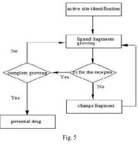
STRUCTURE BASED DRUG DESIGN
Department of pharmaceutical chemistry. Page 16 Fig. 5
ACTIVE SITE IDENTIFICATION
Active site identification is the first step. It analyzes the protein to find the binding pocket, derives key interaction sites within the binding pocket, and then prepares the necessary data for ligand fragment link. Both ligand and protein atoms need to be classified and their atomic properties should be defined, basically, into four atomic types;
Hydrophobic atom: All carbons in hydrocarbon chains or in aromatic groups. H-bond donor: Oxygen and nitrogen atoms bonded to hydrogen atom [s]. H-bond acceptor: Oxygen and sp2 or sp hybridized nitrogen atoms with lone electron pair [s].
Department of pharmaceutical chemistry. Page 17
SCORING METHOD
Structure-based drug design attempts to use the structure of proteins as a basis for designing new ligands by applying accepted principles of molecular recognition. The basic assumption underlying structure-based drug design is that a good ligand molecule should bind tightly to its target.12
SCREENING AND DESIGN
The process of finding a new small molecule (ligand) against a chosen target for a particular disease usually involves high-throughput screening (HTS).
The structure-activity relationship (SAR) is to improve certain features of the lead compound:
Increase activity against the chosen target Reduce activity against unrelated targets
Improve the drug likeness or ADME properties of the molecule.
Department of pharmaceutical chemistry. Page 18 Another important method for drug discovery is FBLD drug design, whereby the biological and physical properties of the target are studied, and a prediction is made on the sorts of small molecules that might (e.g.) fit into an active site. The fragment-based lead discovery (FBLD), novel pharmacophore can emerge very rapidly from these exercises. In general, computer-aided drug design is often used to try to improve the potency and properties of new drug leads.12
COMPUTER-AIDED DRUG DESIGN
Computer-aided drug design uses computational chemistry to discover, enhance, or study drugs and related biologically active molecules. Molecular mechanics or molecular dynamics are most often used to predict the conformation of the small molecule and to model conformational changes in the biological target that may occur when the small molecule binds to it.
Molecular mechanics methods may also be used to provide semi-quantitative prediction of the binding affinity. Also, knowledge-based scoring function may be used to provide binding affinity estimates.
Drug design with the help of computers may be used at any of the following stages of drug discovery: 12
Hit identification using virtual screening (structure- or ligand-based design) Hit-to-lead optimization of affinity and selectivity (structure-based design, QSAR, etc.)
Lead optimization: optimization of other pharmacokinetic properties while maintaining affinity.
Department of pharmaceutical chemistry. Page 19
MOLECULAR PROPERTY PREDICTION
All the data set molecules were subjected to the toxicity risk assessment by using Osiris program, which is available online. The OSIRIS property Explorer shown in this page is an integral part of Actelion's in house substance registration system. It allows drawing chemical structures and also calculates various drug relevant properties whenever a structure is valid. Prediction results are color coded in which the red colour shows high risks with undesired effects like mutagenicity or a poor intestinal absorption and green colour indicates drug-conform behave
Molecular property prediction includes Toxicity risk assessment Clog prediction
Solubility prediction Molecular weight Drug linkers prediction Drug linkers
TOXICITY RISK ASSESSMENT
Department of pharmaceutical chemistry. Page 20
Clog P PREDICTION
The logP value of a compound, which is the logarithm of its partition coefficient between n- octanol and water log (octanol/water), is a well-established measure of the compound's hydrophilicity. Low hydrophilicities and therefore high logP values cause poor absorption or permeation. clogP value must not be greater than 5.0 for permeability. (13)
SOLUBILITY PREDICTION
The aqueous solubility of a compound significantly affects its absorption and distribution characteristics. Typically, a low solubility goes along with a bad absorption and therefore the general aim is to avoid poorly soluble compounds.(13)
MOLECULAR WEIGHT
Optimizing compounds for high activity on a biological target almost often goes along with increased molecular weights. However, compounds with higher weights are less likely to be absorbed and therefore to ever reach the place of action. Thus, trying to keep molecular weights as low as possible should be the desire of every drug forger. (13)
DRUG LIKENESS
Department of pharmaceutical chemistry. Page 21 known as LogP, is used to predict the solubility of a potential oral drug. This coefficient can be experimentally measured or predicted computationally, in which case it is sometimes called "clogP". (13)
The logP value of a compound, which is the logarithm of its partition coefficient between n- octanol and water log (octanol/water), is a well-established measure of the compound's hydrophilicity. Low hydrophilicities and therefore high logP values cause poor absorption or permeation. clogP value must not be greater than 5.0 for permeability. (13)
SOLUBILITY PREDICTION
The aqueous solubility of a compound significantly affects its absorption and distribution characteristics. Typically, a low solubility goes along with a bad absorption and therefore the general aim is to avoid poorly soluble compounds. (13)
MOLECULAR WEIGHT
Optimizing compounds for high activity on a biological target almost often goes along with increased molecular weights. However, compounds with higher weights are less likely to be absorbed and therefore to ever reach the place of action. Thus, trying to keep molecular weights as low as possible should be the desire of every drug forger. (13)
DRUG LIKENESS
Department of pharmaceutical chemistry. Page 22 of a cell. A model compound for the lipophilic cellular membrane is octanol (a lipophilic hydrocarbon), so the logarithm of the octanol/water partition coefficient, known as LogP, is used to predict the solubility of a potential oral drug. This coefficient can be experimentally measured or predicted computationally, in which case it is sometimes called "clogP". (13)
LIPINSKI’S RULE OF FIVE(14-15)
Lipinski's rule of five also known as the Pfizer's rule of five or simply the Rule of five (RO5) is to evaluate drug likeness or determine if a chemical compound with a certain pharmacological or biological activity has properties that would make it a likely orally active drug in humans. The rule was formulated by Christopher A. Lipinski in 1997.
The rule describes molecular properties important for a drug’s pharmacokinetics in the human body, including their absorption, distribution, metabolism, and excretion ("ADME"). However, the rule does not predict if a compound is pharmacologically active.
Lipinski's rule states that, in general, an orally active drug has no more than one violation of the following criteria:
Not more than 5 hydrogen bond donors (nitrogen or oxygen atoms with one or more hydrogen atoms).
Not more than 10 hydrogen bond acceptors (nitrogen or oxygen atoms) A molecular mass less than 500 Daltons.
Department of pharmaceutical chemistry. Page 23
BIOLOGICAL ACTIVITY OF BENZIMIDAZOLE
N
H
N
Benzimidazole is the heterocyclic compound formed from benzene and imidazole ring wide interest because of their diverse biological activity and clinical applications, they are remarkably effective compounds both with respect to their inhibitory activity and their favorable selectivity ratio. Reported nucleus is a constituent of vitamin-B12. It occurs as white crystals. It is used as muscle relaxant.(16)
SYNONYMS(17)
Benziminazole, 3-benzodiazole, Azindole,
Benzoglyoxaline, 3-azaindole,
1,3-diazaindene
Meting point 170-17200C
BIOLOGICAL ACTIVITY(18)
Department of pharmaceutical chemistry.
Anti-ulcer drugs
These are the drugs which inhibits both basal and stimulated gastric acid secretion. Some drugs containing benzimidazole nucleus are Pantoprazole, Rabeprazole, Lansoprazole, Omeprazole etc
Anti-psychotic agents
In psychosis thinking of patient becomes illogical, bizarre and loosely organized. Patient has difficulty in understanding reality and their own conditions.
Some drugs containing benzimidazole nucleus are droperidol, pimozide, and benperidol.
Ex. Droperidol
Department of pharmaceutical chemistry.
These are the drugs which inhibits both basal and stimulated gastric acid Some drugs containing benzimidazole nucleus are Pantoprazole,
, Lansoprazole, Omeprazole etc Ex. Omeprazole
psychotic agents
In psychosis thinking of patient becomes illogical, bizarre and loosely organized. Patient has difficulty in understanding reality and their own conditions.
Some drugs containing benzimidazole nucleus are droperidol, pimozide, and
Page 24 These are the drugs which inhibits both basal and stimulated gastric acid
Some drugs containing benzimidazole nucleus are Pantoprazole,
In psychosis thinking of patient becomes illogical, bizarre and loosely organized. Patient has difficulty in understanding reality and their own conditions.
Department of pharmaceutical chemistry.
Anthelmintic
These are the drugs that either kill or expel infesting helminthes. Some drugs containing benzimidazole nucleus are Thibendazole, Mebendazole, and Albendazole etc. Ex. Mebendazole
Anti-protozoal agents
These are the drugs which are used to treat the amoebiasis caused by E.histolytica. They exert cytotoxicity by damaging DNA and result in DNA helix destabilization strand breakage. The antiprotozoal drugs containing imi
are metronidazole, benznidazole.
Department of pharmaceutical chemistry.
These are the drugs that either kill or expel infesting helminthes. Some drugs containing benzimidazole nucleus are Thibendazole, Mebendazole, and Albendazole
protozoal agents
These are the drugs which are used to treat the amoebiasis caused by . They exert cytotoxicity by damaging DNA and result in DNA helix destabilization strand breakage. The antiprotozoal drugs containing imi
are metronidazole, benznidazole. Ex. Metronidazole
Page 25 These are the drugs that either kill or expel infesting helminthes. Some drugs containing benzimidazole nucleus are Thibendazole, Mebendazole, and Albendazole
Department of pharmaceutical chemistry.
Antifungal
These are the drugs used for superficial and deep fungal infections. Fungal infections are termed mycoses and in divided in to superficial infections (skin, nails, and scalp) and systemic infections (deeper tissues and organs) some conditions are blastomycosis, histoplasmosis, candidiasis, coccibiomycosis etc. superficial fungal infections can be classified in to the
antifungal agents containing imidazole nucleus are Clotriamazole, Miconazole, Ketoconazole.(18). Ex.
Department of pharmaceutical chemistry.
These are the drugs used for superficial and deep fungal infections. Fungal infections are termed mycoses and in divided in to superficial infections (skin, nails, and scalp) and systemic infections (deeper tissues and organs) some conditions are osis, histoplasmosis, candidiasis, coccibiomycosis etc. superficial fungal infections can be classified in to the dermatomycoses and candidiasis
antifungal agents containing imidazole nucleus are Clotriamazole, Miconazole, x. Ketaconazole
REVIEW
REVIEW
REVIEW
REVIEW
OF
OF
OF
OF
LITERATURE
LITERATURE
LITERATURE
LITERATURE
Department of Pharmaceutical Chemistry Page 27 2. REVIEW OF LITERATURE
The Purpose of Literature Review is to:
Establish a theoretical frame work for a topic /subject area Define key terms and terminology
Identify studies, models, case studies etc supporting a topic Define /establish an area of study
The review on following works provided basic information about the target
enzyme, DprE1 and its function
Sarah M. Batt., et al., (2012) Structural basis of inhibition of Mycobacterium tuberculosis DprE1 by benzothiazinone inhibitors .(10)
Maria Loreto Incandela., et.al.,(2013) reported that DprE1, a new taxonomic marker in mycobacteria.(19)
The following works throws a light upon the various genomic aspects of
M.tuberculosis and also various targets intended for drug action:
Donald R Ronning., et al., (2012) Targeting the mycobacterial envelope for tuberculosis drug development.(20)
Sarala Menon., et al., (2012), studied the Drug resistance profiles of Mycobacterium tuberculosis isolates to first line anti-tuberculous drugs.(21)
Department of Pharmaceutical Chemistry Page 28
Christian Lienhardt., et al., (2010) Had a review on New drugs and new regimens for the treatment of tuberculosis: review of the drug development pipeline and implications for national programmes.(23)
Sarah L. Kinnings., et al., (2010), reviewed the Mycobacterium tuberculosis Drugome and Its Polypharmacological Implications.(24)
The review on following works provided ideas for synthesis of the desired
chemical entities:
Elina R. Marinho.,et al The reaction of o-phenylenediamine with ethoxymethylene compounds and aromatic aldehyde,( 2009).(25)
A. M. Soliman, Synthesis and biological activity of dihydroimidazole and 3,4-dihydrobenzo[4,5]imidazo[1,2-a][1,3,5]triazins, (2012) . (26)
Shaaban K Mohamed, Eco-Friendly Synthesis of Pyrimidine and Dihydropyrimidinone Derivatives under Solvent Free Condition and their Anti-microbial Activity, (2013) (27)
Babu K,Synthesis of novel benzimidazole derivatives.(28)
N
H
N
NH
N
H
Department of Pharmaceutical Chemistry Page 29 SHIFF BASE:
Zhaoqi Yang, Pinhua Sun, Compare of three ways of synthesis of simple Schiff base, 2006.(29)
Marcus Vinı´cius Nora de Souza ,carried out Synthesis and antimycobacterial activity of (E)-N0-(monosubstituted-benzylidene) isonicotino
hydrazide derivatives,2008.(30)
A.Mobinikhaled, et al.,An efficient synthesis of Schiff bases containing
Benzimidazole moiety catalyzed by transition metal nitrates,(2010).(31)
Mostafa M. H. Khalil,et al., Synthesis and characterization of a novel schiff base metal complexes and their application in determination of iron in different types of natural water, 2012.(32)
Sandeep Miglani, et al., The rapid synthesis of schiff-bases without solvent under microwave irradiation and their antimicrobial activity, (2012).(33)
R
O NH2
R
+
N
R
Department of Pharmaceutical Chemistry Page 30
K. Brodowska,et al., Schiff bases – interesting range of applications in various fields of science,(2014).(34)
Panneer Selvam .T et al., synthesized a novel series of 2-substituted benzimidazole derivatives were synthesized and characterized.(35)
The following literatures were surveyed in depth to provide supporting data for
the drug design study
LaurieAT.,et.al.,(2005) Q site finder”on energy based method for the prediction of protein –ligand binding site” Bioinformatics,(36)
http ; // www.modelling leads.ac.uk /q site finder / (37)
Department of Pharmaceutical Chemistry Page 31 The review on following works revealed the basics of Alamar blue assay for
evaluating the anti-mycobacterial action.
David A. J. Moore., et al., (2008), Inter- and Intra-Assay Reproducibility of Micro plate Alamar Blue Assay Results for Isoniazid, Rifampicin, Ethambutol, Streptomycin, Ciprofloxacin, and Capreomycin Drug Susceptibility Testing ocobacterium tuberculosis. (39)
Todd P. Primm., et al., (2007), Recent Advances in Methodologies for the Discovery of Antimycobacterial Drugs.(40)
AIM
AIM
AIM
AIM
AND
AND
AND
AND
OBJECTIVES
OBJECTIVES
OBJECTIVES
OBJECTIVES
Department of Pharmaceutical Chemistry Page 32
3. AIM AND OBJECTIVES
With the ongoing progress in protein crystallography and NMR, structure-based drug design is gaining increasing importance in the search for new drugs. Modeling starts from the 3D structure of a target protein in order to construct molecules which are complementary to a binding site, in their geometry as well as in the pattern of their physicochemical properties around the molecules The present study relates to the synthesis of various aryl carboxylic acid derivatives and subsequent screening for their anti-tubercular activity. Due to several toxic effects of isoniazid, attempts were made to eliminate the toxicophore and substituting with a group contributing to the anti-tubercular action. This work also aims the same motive and the compounds were synthesized according to the developed and valid synthetic route.
DOCKING
A chemical data base consisting 500 compounds of various scaffolds had been sketched and docked against the protein target of DPrE1. Based upon the docking score the 50 compounds were selected. For further modifications /derivatisation in order to achieve improved binding affinity and then once again docked against the same target.
Based upon the docking score the first 20 compounds with diverse scaffolds were selected. From these 20 compounds 5 top ranked compounds were selected
Department of Pharmaceutical Chemistry Page 33 The compounds are as follows:
COMPOUND1:
N-1H-benzimidazol-2-yl-N'-{(Z)-[4-(dimethylamino)phenyl]methylidene} guanidine
COMPOUND2:
N-1H-benzimidazol-2-yl-N'-[(Z)-(4-hydroxyphenyl)methylidene]guanidine
COMPOUND3:
N-1H-benzimidazol-2-yl-N'-[(Z)-(2,4-dichlorophenyl)methylidene]guanidine
CHARACTERIZATION
The above compounds will be characterized by using infrared spectroscopy, nuclear magnetic spectroscopy and mass spectroscopy.
BIOLOGICAL EVALUATION
MATERIALS
MATERIALS
MATERIALS
MATERIALS
AND METHODS
AND METHODS
AND METHODS
AND METHODS
Department of Pharmaceutical Chemistry Page 34
4.METERIALS AND METHODS
I) DOCKING
1) MOLECULAR DOCKING BY ARGUS LAB SOFTWARE
Docking analysis of synthesized compounds i.e. ligands will be carried out by
Argus labR docking software. Docking allows the medicinal chemist to virtually screen a set of compounds and predict the strongest binding capacity based on various
scoring function. It explores ways in which two molecules such as ligand and receptor
(protein) fit together and docks to each other well. The molecule binding to a receptor
inhibits its function and thus acts as drug.
Argus lab 4.0 distributed freely for windows platforms by planaria software.
It is an introductory molecular modeling package with academics. Argus docking
engine implementry in Argus lab approximates an exhaustive search method which is
similar to DOCK and GLIDE. Flexible ligand docking is possible with Argus lab,
where the ligand is described as torsion tree and grids are constructed that overlay the
binding site. The accuracy of the Argus lab docking algorithm takes into account, the
key features such as the nature of the binding site and the number of rotatable bonds
to the ligand.
Molegro molecular viewer
Molegro molecular viewer is an application which helps in analyzing the
energies and interaction of the binding site.
Q-site finder
Q-site finder is an energy-based method for protein-ligand binding site
prediction. During prediction we use the crystal structures of macromolecules
(receptor) with small substrates (pdb ID). Identifying the location of binding sites on a
protein is of fundamental importance for a range of applications including molecular
docking. It uses the interaction energy between the protein and a simple vanderwaals
Department of Pharmaceutical Chemistry Page 35 A. PREPARATION OF PROTEIN:
STEP 1
Enter protein pdb ID (3VAE) in the protein data bank in the protein data bank.
Go to download files and select pdb as text file.
Save the downloaded pdb text file to desktop.
STEP 2
Open Argus lab file → open → Import pdb file from the desktop.
3D structure of the protein will appear in the workspace of Argus lab.
Left side of the screen shows molecular tree view.
Open pdb → open ‘Residues’ → open ‘Misc’.
From ‘Misc’ delete the inhibitor and hetero residues, do not delete cofactor
Open water press shift, select all water molecules and delete.
Add hydrogen atoms.
Go to calculation on toolbar → energy by UFF method → start.
Save the prepared protein as *.agl file format in the desktop.
B. IDENTIFICATION/ SELECTION OF ACTIVE SITE
STEP 1
Q-site finder was opened through online.
The pdb format of the protein was imported.
Found all the active site and make a list out of the common amino acid
residues.
STEP 2
residues → open amino acids was opened.
Control was selected and select the amino acids which were listed from the
Q-site finder.
Department of Pharmaceutical Chemistry Page 36 Right click on the mouse → make a group from the selected residues → give
name → Binding site → OK.
C. PREPARATION OF LIGANDS
Drawn the structure from chem. Sketch and save as MDL mol format.
The ligand wae imported into workspace of Argus lab.
Clean geometry → clean hybridization. Select the ligand; right click on the
mouse → make a group from the residues → give name→ ligand→ OK.
The ligand was selected; right click on the mouse → make a group from the
residues → give name→ ligand→ OK.
D. DOCKING PARAMETER :
Calculation was selected from the toolbar → Dock a ligand.
‘Argus Dock’ as the Docking engine.
‘Dock’ was selected as calculation type.
‘Flexible’ for the ligand.
Ascore as the scoring function.
Calculation size.
Docking was started.
Save the Docked protein Ligand complex as Brookhaven pdb files (*.pdb)
E. VISUALIZATION/INTERPRETATION OF DOCKING :
Molegro molecular viewer will help in analyzing the energies and interaction
Department of Pharmaceutical Chemistry Page 37
II) REACTANT PROFILE(41)
O-PHENYLENE DIAMINE:
NH2
NH2
Molecular Formula :C6H 8N2
Molecular Weight :108.14
Description : brown in colour
Melting point :102-1040C
CYANOQUANIDINE:
N H2
NH
NH N
Molecular Formula :C2H 4N2
Molecular Weight :84.08
Description : White crystals
Melting point :2090C
4-HYDROXYL BENZALDEHYDE:
OH
O
Molecular Formula :C7H 6O2
Molecular Weight :121.12
Description : Light yellowish to light brown
Department of Pharmaceutical Chemistry Page 38
N,N’ DIMETHYL BENZALDEHYDE:
N
O CH3 C
H3
Molecular Formula :C 9H 11NO
Molecular Weight :149.19
Description : White crystalline powder
Melting point :72-750C
2,4 DICHLORO BENZALDEHYDE:
O Cl Cl
Molecular Formula :C7H 4Cl 2O
Molecular Weight :175.01
Description : White crystalline powder
Department of Pharmaceutical Chemistry Page 39
3.SYNTHETIC METHODOLOGY
STEP1
Synthesis of 2-benzimidazolylguanidine(26)
A mixture of o-phenylenediamine (100 mmol), cyanoguanidine (100 mmol)
and concentrated Hydrochloric acid (20ml) in water (200 ml) is heated under reflux
for 3 hrs. The reaction mixture is cooled at 0°C and KOH (10%; 50 ml) was added
slowly. The precipitates of 2-guanidinobenzimidazole is collected by filtration,
washed with water, dried and recrystallized by ethanol.
NH2 NH2
+
N H N NH NH2 N H NH NH2 NH NHCl, reflux 3 h
10 %KOH
STEP 2
Synthesis of Schiff base(30)
An equimolar quantity of 2-benzimidazoylguanidine and substituted aromatic
aldehyde is refluxed for 10-15 hours in 20ml ethanol. Completion of reaction is
monitored by TLC. After completion of the reaction the content is poured into ice
cold water. The precipitate collected by filtration, washed with water and dried.
Recrystallization is carried out by ethanol.
N H N NH NH NH2
+
O RReflux for 10-15 hrs
Department of Pharmaceutical Chemistry Page 40 SYNTHESIS OF COMPOUNDS:
2-benzimidazolylguanidine reacts with
1. N,N’ dimethylaminobenzaldehyde
2. p-hydroxybenzaldehyde
Department of Pharmaceutical Chemistry Page 41
JUSTIFICATION OF PURITY:
A.THIN LAYER CHROMATOGRAPHY
Precoated aluminium TLC plates are used. Solutions of the reactants and
products are prepared by dissolving them in methanol. Stationary Phase: Precoated
Silica Gel Plates. Mobile Phase: Chloroform: Methanol (3:2) Visualization: Iodine
Vapors and UV chamber A single spot not corresponding to the parent compound is
indicative. Absence of other spots justifies the purity of the product.
B. MELTING POINT
The melting points of synthesized compounds are determined by open tube
capillary method with an aid of a melting point apparatus. The melting points were
Sharp melting point are indicate the purity of the product.
CHARACTERISATION STUDIES
A. INFRA RED (IR) SPECTROSCOPY
The recrystallised compounds are subjected to IR spectral analysis for
functional group identification using KBr pellet method. Stretching and bending
vibrations for the new functional are indicated. Absence of the vibrational bands for
the parent functional group is ensured.
B. NUCLEAR MAGNETIC RESONANCE (NMR) SPECTROSCOPY
1H NMR spectra are recorded on samples are prepared by dissolving a minute
quantity of pure compounds in DMSO. Chemical shifts are reported in parts per
million (ppm)
C. MASS SPECTROSCOPY:
Mass Spectra is recorded on Shimadzu HPLC-MS using Electron Spray
Ionization Technique and was quantified using La Solutions Software 7.0, Samples
are prepared by dissolving a minute quantity of pure compounds in methanol. The
Department of Pharmaceutical Chemistry Page 42
MICROBIOLOGICAL ASSAY:
Microbial assays or microbiological assays is a type of bioassay and are
designed to analyze the compounds or substances which have effect on
micro-organisms. Microbiological assay is defined as the determination or estimation of
concentration or potency of an antibiotic by means of measuring and comparing the
area of zone of inhibition or turbidity produced by test substance with that of standard
over a suitable microbe under standard conditions. So as definition says the
hypothesis is that when an antibiotic is administered, there is inhibition in the growth
of microbe as indicated by decrease in area of zone of microbial colony on nutrition
media or decrease in turbidity due to decrease in microbial concentration.(43,44)
TYPES OF MICROBIOLOGICAL ASSAY:
REDOX BASED METHODS:
Micro plate Alamar blue assay. Resazurin Microtiter Assay, REMA, or Micro
dilution Resazurin Assay, MRA. Tetrazolium Dyes, Tetrazolium Microplate Assay,
TEMA.
REPORTER GENE-BASED METHODS:
Green Fluorescent Protein Micro plate Assay, GFPMA; Luciferase Assays;
Beta-Galactosidase Assays.
OTHER METHODS: BACTEC 460 TB
Nitrate Reductase Assay, NRA; Disk Diffusion; Visual Micro broth, or Broth
Micro dilution; Malachite Green; STC Agar; Flow Cytometr
MICROPLATE ALAMAR BLUE ASSAY:
MABA is clearly the standard in the field for HTS of compounds against
mycobacteria, and is the most widely cited. The primary reference for the method is
Collins and Franzblau in 1997. In that study, MABA was effective with MTB H37Rv,
H37Ra and M. avium strain ATCC 25291, a relatively virulent isolate. MABA is
reliable with clinical isolates of MTB and MIC. MABA has also been applied to M.
kansasii and M. malmoense, as well as M. leprae. In addition to screening and
sensitivity applications, MABA has also been used in patient treatment follow-up.
Department of Pharmaceutical Chemistry Page 43 change indicates viability,with the MIC recorded as the lowest compound
concentration in wells which remain blue). Newly synthesized product are assayed in
vitro for anti-tubercular activity. Evaluation of the products for their in vitro
antitubercular activity against Mycobacterium Tuberculosis H37Rv using
MicroplateAlamar Blue Assay (MABA) biological test is done. This methodology is
nontoxic, uses a thermally-stable reagent and shows good correlation with
proportional and BACTEC radiometric methods.(40,42)
Anti-TB activity using Alamar Blue Dye:
1. The anti-mycobacterial activity of compound are assessed against M.
tuberculosis using micro plate Alamar Blue assay (MABA).
2. This methodology is non-toxic, uses a thermally stable reagent and shows
good correlation with proportional and BACTEC radiometric method.
3. Briefly, 200µl of sterile deionized water is added to all outer perimeter wells
of sterile 96 wells plate to minimized evaporation of medium in the test wells
during incubation.
4. The 96 wells plate receive 100 µl of the Middle brook 7H9 broth and serial
dilutions of compounds were made directly on plate.
5. The final drug concentrations tested are 100 to 0.8 µg/ml.
6. Plates were covered and sealed with par film and incubated at 37ºC for five
days.
7. After this time, 25µl of freshly prepared 1:1 mixture of Alamar Blue reagent
and 10% tween 80 was added to the plate and incubated for 24 hrs.
8. A blue color in the well was interpreted as no bacterial growth, and pink color
was scored as growth.
9. The MIC was defined as lowest drug concentration which prevented the color
RESULTS
RESULTS
RESULTS
RESULTS
AND
AND
AND
AND
DISCUSSION
DISCUSSION
DISCUSSION
DISCUSSION
Department of Pharmaceutical Sciences Page 44
RESULTS AND DISCUSSION
Results of drug design
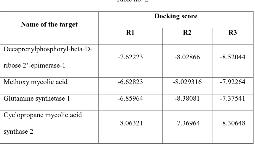
Two hundred molecules, which are sketched using chem sketch were docked against the MTB enzyme Decaprenylphosphoryl-beta-D-ribose 2’-epimerase-1by using Argus lab 4.0.1 software. The molecules with best docking score and good interactions were selected and synthesized.
The molecules were also docked against the following targets 1. Methoxy mycolic acid
2. Glutamine synthetase 1
[image:57.595.102.532.373.617.2]3. Cyclopropane mycolic acid synthase 2 Table no: 2
Name of the target
Docking score
R1 R2 R3
Decaprenylphosphoryl-beta-D-ribose 2’-epimerase-1
-7.62223 -8.02866 -8.52044
Methoxy mycolic acid -6.62823 -8.029316 -7.92264 Glutamine synthetase 1 -6.85964 -8.38081 -7.37541 Cyclopropane mycolic acid
synthase 2
Department of Pharmaceutical Sciences Page 45
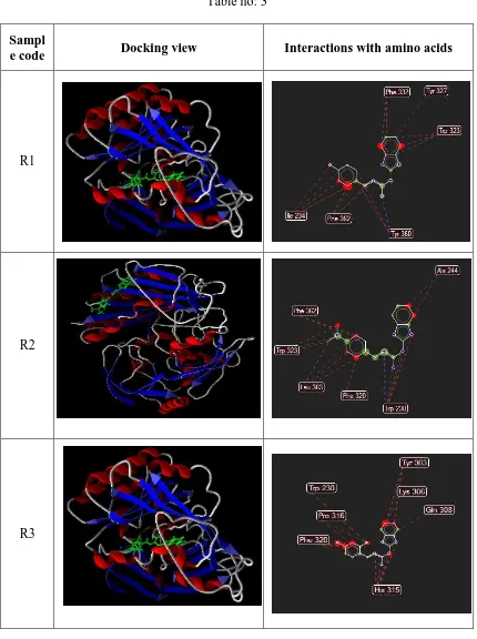
Interactions of the docked molecules with the enzyme
[image:58.595.102.532.140.716.2]Decaprenylphosphoryl-beta-D-ribose 2’-epimerase-1.
Table no: 3
Sampl
e code Docking view Interactions with amino acids
R1
R2
Department of Pharmaceutical Sciences Page 46
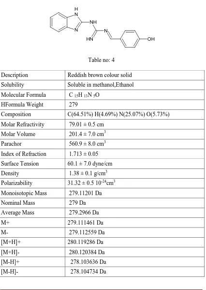
Product profile
Compound R1
Physicochemical Properties of N-1H-Benzimidazol-2-Yl-N'-[(E)-(4 hydroxy phenyl) methylidene] Guanidine
[image:59.595.106.532.176.775.2]N H N NH N H N OH
Table no: 4
Description Reddish brown colour solid Solubility Soluble in methanol,Ethanol Molecular Formula C 15H 13N 5O
HFormula Weight 279
Composition C(64.51%) H(4.69%) N(25.07%) O(5.73%) Molar Refractivity 79.01 ± 0.5 cm
Molar Volume 201.4 ± 7.0 cm3 Parachor 560.9 ± 8.0 cm3 Index of Refraction 1.713 ± 0.05 Surface Tension 60.1 ± 7.0 dyne/cm Density 1.38 ± 0.1 g/cm3 Polarizability 31.32 ± 0.5 10-24cm3 Monoisotopic Mass 279.11201 Da Nominal Mass 279 Da Average Mass 279.2966 Da
M+ 279.111461 Da
Department of Pharmaceutical Sciences Page 47
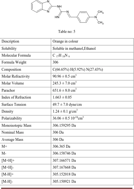
Compound R2
Physicochemical Properties of N-1H-Benzimidazol-2-Yl-N'-[(Z)-(4 dimethyl amino) phenyl] methylidene}Guanidine
N N H N NH N H
N CH3
[image:60.595.103.530.174.778.2]CH3
Table no: 5
Description Orange in colour
Solubility Soluble in methanol,Ethanol Molecular Formula C 17H 18N 6
Formula Weight 306
Composition C(66.65%) H(5.92%) N(27.43%) Molar Refractivity 90.96 ± 0.5 cm3
Molar Volume 245.3 ± 7.0 cm3 Parachor 651.6 ± 8.0 cm3 Index of Refraction 1.663 ± 0.05 Surface Tension 49.7 ± 7.0 dyne/cm Density 1.24 ± 0.1 g/cm3 Polarizability 36.06 ± 0.5 10-24cm3 Monoisotopic Mass 306.159295 Da Nominal Mass 306 Da
Average Mass 306 Da
M+ 306.365 Da
M- 306.158746 Da
Department of Pharmaceutical Sciences Page 48
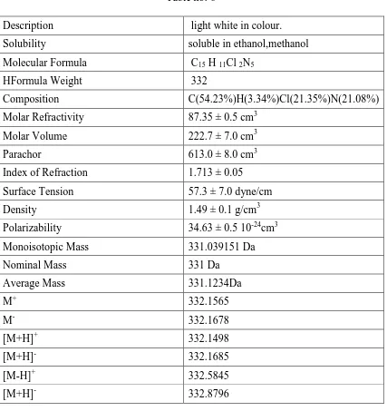
Compound R3
[image:61.595.105.532.314.767.2]Physicochemical properties of N-1H-benzimidazole-2-yl N’{(z)[2,4(dichloro)phenyl]methylidine} guanidine. N H N NH N NH Cl Cl
Table no: 6
Description light white in colour. Solubility soluble in ethanol,methanol Molecular Formula C15 H 11Cl 2N5
HFormula Weight 332
Composition C(54.23%)H(3.34%)Cl(21.35%)N(21.08%) Molar Refractivity 87.35 ± 0.5 cm3
Molar Volume 222.7 ± 7.0 cm3 Parachor 613.0 ± 8.0 cm3 Index of Refraction 1.713 ± 0.05 Surface Tension 57.3 ± 7.0 dyne/cm Density 1.49 ± 0.1 g/cm3 Polarizability 34.63 ± 0.5 10-24cm3 Monoisotopic Mass 331.039151 Da Nominal Mass 331 Da
Average Mass 331.1234Da
M+ 332.1565
M- 332.1678
[M+H]+ 332.1498
[M+H]- 332.1685
[M-H]+ 332.5845
Department of Pharmaceutical Sciences Page 49
CHARACTERIZATION:
Newly synthesized compounds were characterized by
• IR
• NMR
• GC-MS
IR SPECTROSCOPY
The samples were prepared by the KBr pellet technique and spectrum obtained from ABB(MB 3000) spectrophotometer
The spectra were examined for the absence of the functional group region of parent compound and examined for presence of the vibrational absorption band for the new functional group.
Our reaction involves reaction between Aldehyde and amine to yield of Schiff bases.
• The absorption band for aldehydes are
2800-2700cm-1
1700-1750cm-1 • The absorption band for amine 3400-3600 cm-1
[image:62.595.102.533.610.704.2]• The absorption of newly synthesized –C=N- is 1650-1600cm-1
Table no: 7
IR absorption band R1 R2 R3
Aldehyde - - -
Amine - - -
-C=N- + + +
Department of Pharmaceutical Sciences Page 50 IR SPECTRUM OF R1:
Department of Pharmaceutical Sciences Page 51 IR SPECTRUM OF R2:
4000.0 3600 3200 2800 2400 2000 1800 1600 1400 1200 1000 800 600 400.0
0.0 5 10 15 20 25 30 35 40 45 50 55 60 65 70 75 80 85 90 95 100.0 cm-1 %T R3 3297 2953 2931 2872 2314 1656 1542 1435 1402 1315 1060 645 522 464 448 N N H N NH N H
N CH3
CH3
Department of Pharmaceutical Sciences Page 52 IR SPECTRUM OF R3:
4000.0 3600 3200 2800 2400 2000 1800 1600 1400 1200 1000 800 600 400.0
Department of Pharmaceutical Sciences Page 53
NMR
1H NMR spectra are recorded on Bruker Advance III 500 MHz NMR spectrophotometer
COMPOUND R1
Table no: 8
TYPE OF PROTON NO OF PROTON DELTA(δ) NATURE OF PEAK
-NH 3 3.4-4.1 Multiplate
-OH 1 5.4 Singlet
Ar-H 8 6.3-7.1 Multiplate
N H
N NH
N H
N
Department of Pharmaceutical Sciences Page 54 COMPOUND R2
Table no:9
TYPE OF PROTON NO OF PROTON DELTA(δ) NATURE OF PEAK
-NH 3 3.2-3.7 Multiplate
-N(CH3)2 4 2.8-3.1 Singlet
Ar-H 8 6.4 Singlet
N N
H
N NH
N H
N CH3
Department of Pharmaceutical Sciences Page 55 COMPOUND R3
Table no:10
TYPE OF PROTON NO OF PROTON DELTA(δ) NATURE OF PEAK
-NH 1 2.2-2.4 Singlet
Ar-H 7 7.8-8 Multiplate
=CH- 1 8.2 Singlet
N H
N NH
N NH
Cl
Department of Pharmaceutical Sciences Page 56
GC-MS:
The molecular weight of the synthesized compounds were compared by GC-MS
[image:69.595.102.531.206.377.2]analysis.
Table no: 11
Compound Calculated mass Actual Mass
R1 279 279
R2 306 306
Department of Pharmaceutical Sciences Page 57 Compound R1:
N H
N NH
N H
N
Department of Pharmaceutical Sciences Page 58 Compound R2:
Compound R3:
N N
H
N NH
N H
N CH3
Department of Pharmaceutical Sciences Page 59
N H
N NH
N NH
Cl
Department of Pharmaceutical Sciences Page 60
TOXICITY RISK ASSESSMENT
All the data set molecules were subjected to the toxicity risk assessment by using Osiris program, which is available online free of cost. The OSIRIS property Explorer shown in this page is an integral part of Actelion's in house substance registration system. It allows drawing chemical structures and also calculates various drug relevant properties whenever a structure is valid. Prediction results are color coded in which the red colour shows high risks with undesired effects like mutagenicity or a poor intestinal absorption and green colour indicates drug-conform behavior.
[image:73.595.102.529.513.645.2]Molecular property prediction includes Toxicity risk assessment Clog P predicition Solubility prediction Molecular weight
Table no: 12
Samples R1 R2 R3
Mutagenic + + +
Tumorigenic + - +
Irritant + + +
Reproductive effective + + +
(+) indicates absence of toxicity
Department of Pharmaceutical Sciences
[image:74.595.131.503.107.282.2]Department of Pharmaceutical Sciences
Fig. R1
[image:74.595.127.503.110.491.2]Fig. R2
Fig. R3
Department of Pharmaceutical Sciences Page 62
BIOLOGICAL SCREENING
[image:75.595.103.531.545.736.2]The synthesized compounds were screened for their in-vitro anti mycobacterial activity by means of alamar blue assay. The compounds were tested in the concentration range of 100 to 0.8 µg/ml against M.tuberculosis H37Rv strain grown in Middlebrook 7H9 broth in 96 well titre plate. Pyrazinamide- 3.125µg/ml and Streptomycin- 6.25µg/ml were used as standards for comparison. A blue color in the well was interpreted as no bacterial growth so it is termed as sensitive, and pink color was scored as growth and is referred as resistant. The MIC was defined as lowest drug concentration which prevented the color change from blue to pink.
Table no: 13
Compound Docking score MIC value
R1 -7.62223 50 µg/ml
R2 -8.02866 25 µg/ml
R3 -8.52044 50 µg/ml
MABA REPORT OF THE SYNTHESISED COMPOUNDS
Table no: 14
S.
No Sample
100 µg/ml 50 µg/ml 25 µg/ml 12.5 µg/ml 6.25 µg/ml 3.12 µg/ml 1.6 µg/ml 0.8 µg/ml
1 R1 S S R R R R R R
2 R2 S S S R R R R R
Department of Pharmaceutical Sciences Page 63 Fig. Standard drug photograph
Fig. synthesized compound photograph
S.
N
o
Sampl
e
100
µg/ml 50
µg/ml 25
µg/ml
12.5
µg/ml
6.25
µg/ml
3.12
µg/ml
1.6
µg/m
l
0.8
µg/m
l
1 R1
2 R2
[image:76.595.105.531.436.672.2]SUMMARY
SUMMARY
SUMMARY
SUMMARY
AND
AND
AND
AND
CONCLUSION
CONCLUSION
CONCLUSION
CONCLUSION
Department of Pharmaceutical Sciences Page 64
SUMMARY
Decaprenylphosphoryl-b-d-ribose 2’-Epimerase 1(DprE1) a enzyme of OxidoReductase family is a critical enzyme for the growth of Mycobacterium tuberculosis H37Rv.
From the review of literature DprE1 was chosen for our study for drug design.
A database of 100 molecules with high potential of inhibiting the target possessing PDB ID of 4FDO were carefully chosen by making changes to the lead molecule aryl carboxylic acid derivatives.
The 3D structure of the molecules were docked against the 3D structure of DprE1 using the docking platform argus lab.
Three compounds with good Docking score (lower Binding energy) were selected for laboratory synthesis. The reaction conditions were optimized.
The Compounds were labeled as R1, R2, R3 were synthesized with satisfactory yield
The purity of the synthesized compounds was ensured by repeated recrystallization and column chromatography.Further the compounds were evaluated by melting point and TLC.
The characterizations of the synthesized compounds were done by Infrared, Nuclear magnetic resonance and Mass spectroscopic methods.
Department of Pharmaceutical Sciences Page 65 The synthesized compounds were active at 25mcg/ml to <100mcg/ml, which are compared to that of the known anti-tubercular agents at 50mcg/ml against the MIC of known TB drugs. The synthesized compounds were lesser active than that of the standard TB drugs. Pyrazinamide: 3.125mcg/ml, Ciprefloxacin: 3.125mcg/ml and Streptomycin 6.25mcg/ml.
The synthesized compounds were subjected to toxicity prediction assessments by OSIRS software. The results are coded as a green colour which confirms the drug likeness.
CONCLUSION
Our work concludes that our synthesized molecules are effective in inhibiting Decaprenylphosphoryl-beta-D-ribose 2’-Epimerase 1(DprE1) which is important for the growth of Mycobacterium tuberculosis.
REFERENCES
REFERENCES
REFERENCES
Department of Pharmaceutical sciences Page 66
7. REFERENCES
1. Baptise villemagne, Celine crauste, Marion Flipo, Alian R. Baulard et.al.,Tuberculosis The drug development pipeline at a glance. European Journal of Medicinal Chemistry 2012; 51: P.1-16.
2. www.tbfacts.org
3. Issar smith. Mycobacterium tuberculosis pathogenesis and Molecular Determination of Virulence Clin. Microbiol. 2003; 16(3): P.463. DOI: 10.1128/CMR.16.3.
4. Michael S. Glickman and William R.Jacobs. Microbial pathogenesis review of Mycobacterium tuberculosis. Dawn of a Discipline cell. 2001; 2: P. 104, 447-485.
5. P.J.Brennan. Structure, function, and biogenesis of cell wall of Mycobacterium Tuberculosis. 2003; P. 83, 91-9.
6. http://tuberculist.epfl.ch/
7. Nancy A.Knechel. Tuberculosis Pathophysiology, Clinical features, and Diagnosis, Crit Care Nurse. 2009; P. 29:34-43 doi: 4037//ccn2009968.
8. William O.Foye, Thomas L.Lemke, David A.Williams. Principles of Medicinal Chemistry. B.I Waverly Pvt Ltd,New Dehli,4th ed. 1999; P. 1.
Department of Pharmaceutical sciences Page 67 10. Sarah M.Batta, Talat Jabeena, Veemal Bhowrutha, Lee Quilla et.al.,structural basis of inhibition of Mycobacterium Tuberculosis DprE1 by benzothiazinone inhibitors.
11. http://tuberculist.epfl.ch/
12. Mickey Sahu, Sitesh Kumar Sinha, Krishna Kumar Pandey. Computer Aided Drug Design: The Most Fundamental Goal is to Predict Whether a Given Molecule will Bind to a Target and if so How Strongly Computer Engineering and Intelligent Systems. 2013; 4.
13. http://www.organic-chemistry.org/prog/peo/.
14. Lipinski CA, Lombardo F, Dominy BW, Feeney PJ (March 2001). "Experimental And computational approaches to estimate solubility and permeability in drug discovery and development settings". Adv. Drug Deliv. 2001; P. 3–26. doi: 10.1016/S0169 409X (00)00129-0. PMID 11259830.
15. Lipinski CA (December 2004). "Lead- and drug-like compounds: the rule-of-five revolution". Drug Discovery Today: Technologies 2004; 1 (4): 337–341. doi:10.1016/j.ddtec.2004.11.007.
16. Namrata Singh, Annamalai Pandurangan, Kavita Rana, Preeti Anand. Benzimidazole: A short review of their antimicrobial activities, International Current Pharmaceutical Journal. 2012, 1(5): P. 119-127.
Department of Pharmaceutical sciences Page 68 18. Ingle R.G. Magar D.D. Heterocyclic chemistry of Benzimidazole and potential activities of derivatives. International Journal of Drug Research and Technology. 2011; 1 (1): P. 26-32.
19. Maria Loreto Incandela, Elena Perrin2, Marco Fondi, Ana Luisa de Jesus Lopes Ribeiro, Giorgia Mori, Alessia Moiana, Maurizio Gramegna, Renato Fani2, Giovanna Riccardi & Giorgia. International Journal of Drug Research and Technology. 2013; 1 (1): P. 26-30.
20. Lorenz a Favrot and Donald R Ronning Targeting the mycobacterial envelope for tuberculosis drug development, Expert Rev Anti Infect There. 2012; 10(9): P. 1023.
21. Sarala Menon, Sujata Dharmshale, Chhaya Chande, ArunaGohil. Drug resistance profiles of Mycobacterium tuberculosis isolates to first line anti-tuberculosis drugs. Lung India. 2012; 29(3): 227–231.
22. A.G.Nikalje and P.Mudassar, Multi drug resistant Mycobacterium tuberculosis: A brief review, Asian Journal of Biological sciences. 2011; 4 (2): P. 101-115.
Department of Pharmaceutical sciences Page 69 24. Sarah L. Kinnings, Li Xie, Kingston H. Fung, Richard M. Jackson. The My