A Dissertation on
A COMPARATIVE STUDY OF BENIGN VOCAL CORD MASS LESIONS USING VIDEOSTROBOSCOPY,VOICE
ANALYSIS AND VOICE HANDICAP INDEX BEFORE AND AFTER MICROLARYNGEAL SURGERY
Submitted to the
THE TAMILNADU DR. M.G.R.MEDICAL UNIVERSITY In Partial fulfillment of the requirements
For the award of the degree of
M.S.BRANCH IV
(OTORHINOLARYNGOLOGY)
GOVERNMENT STANLEY MEDICAL COLLEGE & HOSPITAL
THE TAMILNADU DR. M.G.R.MEDICAL UNIVERSITY, CHENNAI, TAMILNADU
DECLARATION
I, Dr. RAVIKUMAR.V, solemnly declare that the dissertation, titled “A COMPARATIVE STUDY OF BENIGN VOCAL CORD MASS LESIONS USING VIDEOSTROBOSCOPY,VOICE ANALYSIS AND VOICE HANDICAP INDEX BEFORE AND AFTER MICROLARYNGEAL SURGERY” is bonafidework done by me during the period of Dec 2014 to September 2015 at Government Stanley
Medical College and Hospital, Chennai under the expert supervision of
Prof.Dr.F.ANTHONY IRUDHAYARAJAN, MS., D.L.O., Department of Otorhinolaryngology, Government Stanley Medical College and hospitals,
Chennai.
This dissertation is submitted to The Tamil Nadu Dr. M.G.R.
Medical University, in partial fulfillment of the rules and regulations for
the M.S. degree examinations in Otorhinolaryngology to be held in April
2016.
Place:Chennai-1
CERTIFICATE
This is to certify that the dissertation - “A COMPARATIVE STUDY OF BENIGN VOCAL CORD MASS LESIONS USING VIDEOSTROBOSCOPY, VOICE ANALYSIS AND VOICE HANDICAP INDEX BEFORE AND AFTER MICROLARYNGEAL SURGERY” presented by Dr. RAVIKUMAR.V, is an original work done in the department of otorhinolarymgology, Government Stanley
Medical College and hospital, Chennai in partial fulfilment of regulations
of the The Tamil Nadu Dr. M.G.R. Medical University, for the award of
degree of M.S. (Otorhinolaryngology) Branch IV, under my supervision
during the academic period 2013-2016.
Prof.Dr.ISSAC CHRISTIAN MOSES M.D FICP FACP
THE DEAN
Govt. Stanley Medical College, and Hospital Chennai-1
Prof..Dr.T.BALASUBRAMANIYAN, M.S.,D.L.O.,
Professor and Head of the Department Govt. Stanley Medical College and Hospital
Chennai-1.
Prof.Dr.F.ANTHONY IRUDHAYARAJAN, MS., D.L.O.,
PROFESSOR OF OTORHINOLARYNGOLOGY, Govt.Stanley Medical College and Hospital,Chennai -1.
Place : Chennai
ACKNOWLEDGEMENTS
I wish to express my sincere thanks to Prof. Dr. ISAAC CHRISTIAN MOSES MD, FIC, FACP. DEAN, Government Stanley Medical College and Hospital for having permitted me to utilize the
facilities of the hospital for conducting this study.
My heartfelt gratitude to Prof. Dr. T. BALASUBRAMANIAN, M.S., D.L.O., Professor and Head of the Department, Department of Otorhinolaryngology, Government Stanley Medical College and Hospital
for his constant motivation, valuable suggestions, and expert supervision
during the course of this study.
I express my whole hearted gratitude to Prof. Dr. F.ANTHONY IRUDHAYARAJAN, MS.,D.L.O., Professor of Otorhinolaryngology, and Chief of ENT UNIT II, and Prof. Dr. N. SEETHALAKSHMI, M.S., D.L.O., D.N.B., Professor of Otorhinolaryngology for supporting, guiding and encouraging me in this study.
I also thank Mrs.RADHAKALAISELVAN, Audiologist and Speech Pathologist of ENT Department Government Stanley Hospital for
her expert assistance.
I am grateful to all the other post-graduates who most willingly
helped me during this study period.
I also thank the staff nurse and theatre personnel, Government
Stanley Hospital for their co-operation and assistance in the conduct of
this study.
Last but not least, I am indebted and grateful to all the Patients who constitute the backbone of this study, who most willingly and
selflessly subjected themselves to this study for the sake of the benefit
of their community and without whom this study would not have been
CONTENTS
S.No Topics Page No.
1 ABSTRACT I
2 INTRODUCTION 1
3 AIM AND OBJECTIVE 2
4 REVIEW OF LITERATURE 4
5 MATERIALS AND METHODS 43
6 RESULTS AND OBSERVATION 59
7 DISCUSSION 78
8 CONCLUSION 81
9 ANNEXURES
A. BIBLIOGRAPHY
B. PROFORMA
C. ETHICAL COMMITTEE APPROVAL
LETTER
D. PATIENT INFORMATION SHEET
E. INFORMED CONSENT FORM
F. PLAGIARISM
LIST OF FIGURES
Fig -1 Shows Various Stages of Development of larynx
Fig-2 Shows Larynx – Intrinsic muscles
Fig-3A,3B Shows Various layers in the Body Cover Complex
Fig-4 Shows Cover Body Complex
Fig-5A Shows vocal cord cross sectional study
Fig-5B Shows Basement membrane zone of Vocal Cord
Fig-6 Shows Still and moving images explain the Talbot’s Law the basic principles of stroboscopy
Fig-7 Shows schematic diagram of Vocal Nodule formation
Fig-8 Shows Stroboscopic picture of B/L Vocal Cord Nodule
Fig-9 Shows schematic diagram of Vocal Cyst formation
Fig-9A Shows Stroboscopic picture of Rt Vocal Cord Cyst Fig-9B Shows Stroboscopic picture of Lt Vocal Cord Cyst
Fig-10 Shows schematic diagram of Vocal Polyp formation
Fig-10A Shows Stroboscopic picture of Rt Vocal Cord Polyp Fig-10B Shows Stroboscopic picture of Lt Vocal Cord Polyp
Fig-11 Shows schematic diagram of Rienke’s Edema Formation
Fig-12 Shows STROBOSCOPY UNIT
Fig-12A Monitor
Fig-12B CAMERA UNIT
Fig-12C LIGHT SOURCE
Fig-12D DIGITAL PRINTER
Fig-12E FOOT PEDAL
Fig-12F UPS
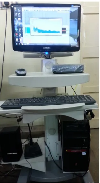
Fig 13 PHONOLAB-ECLERIS VERSON 03.02
Fig-13A MONITOR
Fig-13B PRINCIPAL UNIT
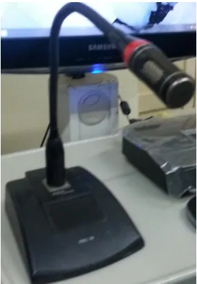
Fig-13C MICROPHONE
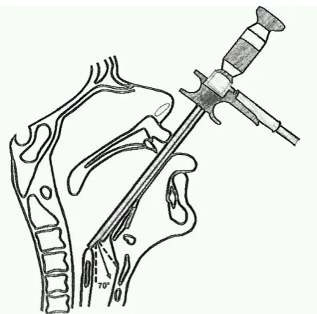
Fig-14 Shows Procedure of Using Endoscope
Fig-15 Shows NORMAL MUCOSAL WAVE PATTERN
SEQUENCE by video stroboscope
Fig-16,16A,16B,16C, Shows Pre-OP Vocal Nodule, Intra-OP Rt Vocal Nodule Removal, Intra Op Lt Vocal Cord Nodule Removal, Post-OP Vocal Cord Status
Fig-17,17A,17B Shows PRE-OP Vocal Polyp Rt, INTRA OP Rt Vocal Polyp Removal, POST OP Vocal Cord Status
Fig-18,18A Shows PRE OP Vocal Cyst, POST OP Vocal Cord Status Fig-19,19A,19B Shows PRE-OP Vocal Cord Polyp, INTRA P(COBLATION)
LIST OF TABLES
Tab-1 Shows Dimension Of Larynx
Tab-2 Shows Ligaments Of Larynx
Tab-3 Shows Functions of Laryngeal Muscles in Vocal Cord
Tab-4 Shows FUNCTIONAL TWO-LAYERED STRUCTURE OF
THE VOCAL FOLDS
Tab-5 Shows Pre Operative Stroboscopic analysis
Tab-6 Shows Fundamental Frequency
Tab-7 Shows Standard Deviation of Pitch
Tab-8 Shows Jitter
Tab-9 Shows Shimmer
Tab-10 Shows Harmonic Noise Ratio
Tab-11 Shows Maximum Phonation Time
Tab-12 Shows Voice Handicap Index
Tab-13 Shows Sub group analysis Male and Female
ABSTRACT
Vocal cords are very delicated and intricate structure that help a
human being to Breath, Speak as well as to sings. It is a micro structure
and their functions are very accurate, even a small change in it by a lesion
can produce enormous change in its function (Voice and Singing). Video
Laryngo Stroboscopy is the process in which the normal and
pathological vibrating pattern of vocal cord is made visible during
phonation. Voice is analysed using Voice analyser and Voice Handicap
Index.
AIM
The present study is aimed at examining the objective and
subjective parameters using Stroboscopy voice analysis and Voice
Handicap Index(VHI) of patients with Benign Vocal Cord Lesions
before and after Microlaryngeal surgery.
METHOD
This study was conducted in the Department of
Otorhinolaryngology of Government Stanley Medical College and
Hospital during the period of December 2014 to September 2015. Based
on the inclusion and exclusion criteria,38 patients with voice disorders
were selected and sub categorized into Vocal Cord Nodule, Vocal cord
Polyp, Vocal cord Cyst.
Among 38 Patients 16 are presented as Vocal cord Nodule,16
Presented as Vocal cord Cyst and 6 Presented as Vocal Cord Polyp which
includes both male and females. After getting valid consent from the
patients, pre operative and Postoperative subjective and objective
parameters using Stroboscopy voice analysis and Voice Handicap
Index(VHI) were evaluated and analysed.
RESULTS
Pre and Post operative assessment of patients with benign vocal
cord mass lesions using stroboscopy, voice analyses and Voice Handicap
index is a useful way to assess the degree of improvement following
surgery. Both the patients and surgeon were provided with definitive
evaluation with respect to the benefit following surgery and speech
INTRODUCTION
The voice is an integral part of the unique human ability to
communicate by speech. The larynx is the major source of sound used
during speaking. Phonation is the generation of sound by vibration of vocal
cords. Benign vocal cord mass lesions such as polyps, nodules and cysts
affect voice production.
The objective measurement of the degree of pathology in the vocal
folds is performed by using various parameters of videostrobosocopic
examination.
A comparison of pre operative and post operative changes in the
vocal folds can show the degree of improvement. Videostrobosocopic
examination may also be combined with analysis of voice parameters to
assess the improvement or worsening of voice after Micro Laryngeal
surgery.
The present study aimed at examining the subjective and
objective parameters using stroboscopic voice analysis and VHI Of
patients undergoing Microlaryngeal Surgery for Benign Vocal Cord
AIM AND OBJECTIVES
AIM OF THE STUDY
A Comparative Study Of Benign Vocal Cord Mass Lesions
Using Videostroboscopy, Voice Analysis And Voice Handicap Index
Before And After Microlaryngeal Surgery.
OBJECTIVE OF THE STUDY
1. Comparing pre and post operative vocal cord vibratory changes in a
patient with benign vocal cord lesions using stroboscopy.
2. Comparing pre and post operative voice changes in a patient with
benign vocal cord mass lesions using voice analysis.
3. Assessing the subjective improvement of patients voice using voice
handicap index before and after Microlaryngeal Surgery.
4. To assess the predictive value of the tests.
INCLUSION CRITERIA
1. Patients with benign looking vocal cord lesion
2. Age more than 12 years
3. On Rigid fiberoptic endoscopy / indirect Laryngoscopy vocal cords
EXCLUSION CRITERIA
1. Patient with malignant looking lesion on endoscopy.
2. Patient in respiratory distress
3. Patient with ischemic heart disease.
REVIEW
REVIEW OF LITERATURE
EMBRYOLOGY OF LARYNX (Fig 1)
1. Development of larynx begins at 4th week of intrauterine life in the form of laryngotracheal groove which is nothing but the midline ventral respiratory diverticulum of the foregut. This groove gradually deepens and fused to form a septum which separates the laryngotracheal tube from the larynx and oesophagus.
2. This fusion starts caudally and extends cranially. Cranial end forms the larynx and trachea, Caudal end divides into two from which two main bronchi develop.
3. Epiglottis develop from the posterior part of hypobranchial eminence.
4. DEVELOPMENT OF LARYNGEAL CARTILAGE
· Thyroid cartilage – Ventral ends of 4th arch cartilage
· Arytenoids – 6th arch
· Corniculate – 6th arch
· Cueniform-6th arch
· Epiglottis – Hypobranchial eminence
· Cricoid &Tracheal Cartilage – 6thbranchial arch
FRAMEWORK OF LARYNX
Definition:
Larynx composed of articulated cartilages interconnected by
Ligaments, internally invested by mucosa, acted upon by muscles
supplied by blood vessels and nerves and drained by lymphatics.
Situated from C3 to C6 Vertebra in men, Somewhat higher in women
and children.
Dimension of Larynx 2A
SEXES LENGTH TRANSVERSE DIAMETER
ANTEROPOSTERIOR
DIAMETER CIRCUMFERENCE
MALES 44mm 43mm 36mm 136mm
FEMALES 36mm 41mm 26mm 112mm
Table:-1 Dimension Of Larynx
CARTILAGES OF LARYNX
The Cartilage of larynx divided into Paired and unpaired. Paired
cartilages are three in numbers named 1.Corniculate 2. Cuneiform
3.Aretynoids. Unpaired cartilages are also three in number named
THYROID CARTILAGE
Shield in Shape, Consists of two pentagonal plates which meets
anteriorly in midline to form an angle of 90 degree in men and 120
degree in women. It is largest of all laryngeal cartilages. The fused
anterior border in men produce a prominence which is easily
palpable and known as “ADAMS APPLE” Lamina of thyroid cartilage
deviated posteriorly and prolonged to form a slender process called
superior and inferior cornua. Superior cornua is curves upward and
backward and medially and ends in a conical projection where
thyrohyoid ligament attached. The short and thicker inferior cornua
arches downward medially and articulate with the small facet present
in the cricoid cartilage.
In the Outer surface of lamina there is a oblique line extending
from the superior thyroid tubercle to inferior thyroid tubercle.
Following structures attached to the oblique line, they are
1.Thyrohyoid muscle 2.Sternohyoid muscle 3.Inferior Constrictor
Muscle. Inner part of lamina covered by mucous membrane.
CRICOID CARTILAGE
It is the only cartilage having a complete cartilaginous ring in the
airway system. It has narrow arch anteriorly and broad lamina
posteriorly. Lamina has facets for arytenoids near the junction of arch
of joint, its vertical line gives attachment to oesophageal longitudinal
fibres. Superior margin gives attachment to the cricothyroid ligament.
Lateral gives attachment to the crico arytenoid muscle.
ARYTENOID CARTILAGES
It positioned at upper and lateral border of cricoid lamina.
Shape looks like irregular three sided pyramid. Its forward projection
is called as Vocal Process, Lateral projection is called as Muscular
process – to which posterior and lateral cricoarytenoids attached. Medial
surface gives attachment to vestibular ligaments upper part and lower
part gives attachment to crico arytenoid muscle.
CORNICULATE (SANTORINI) AND CUNEIFORM (WRISBERG) CARTILAGES
Small conical nodule like structure two in number which is a
type of fibroelasticcartilage and articulate as a synovial joint. In front
of corniculate, the elongated cuneiform cartilage present. They are
enclosed in aryepiglottic fold.
EPIGLOTTIS
Type of elastofibro cartilage thin leaf like sheet projecting
behind the tongue and hyoid bone body. Below the thyroid notch, a
narrow stalk is attached by the thyroepiglottic ligament to the angle
posterior surface is concave, smooth and with a central
tubercle.Median and lateral glossoepiglottic folds forming vallecula
presents at anterior surface. Hyoepiglottic ligament is connected to
hyoid.
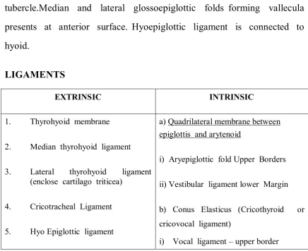
LIGAMENTS
EXTRINSIC INTRINSIC
1. Thyrohyoid membrane
2. Median thyrohyoid ligament
3. Lateral thyrohyoid ligament (enclose cartilago triticea)
4. Cricotracheal Ligament
5. Hyo Epiglottic ligament
a) Quadrilateral membrane between epiglottis and arytenoid
i) Aryepiglottic fold Upper Borders
ii) Vestibular ligament lower Margin
b) Conus Elasticus (Cricothyroid or cricovocal ligament)
[image:22.595.94.531.104.460.2]i) Vocal ligament – upper border
Table-2-Ligaments
MUCOUS MEMBRANE
The Mucous Membrane continuous with lining of pharynx above and
below with trachea. It is rich in mucous glands especially in the region of
ventricle of morgagni (Laryngeal ventricle). This mucous membrane
closely adherent to epiglottis, aryepiglottic fold and vocal cords . The
Lining epithelium of larynx are squamous , ciliated columnar or
LARYNGEAL MUSCLES
The Laryngeal muscles are subdivided into intrinsic, extrinsic and
accessory.
INTRINSIC LARYNGEAL MUSCLES
Based on the effect on the shape of glottis and vibratory behavior of
vocal cord they are classified into addcutors, abductors, relaxers and tensor
muscles.
THE POSTERIOR CRICOARYTENOID MUSCLE
The Posterior cricoarytenoid muscle originate behind the cricoid
cartilage and get inserted into the muscular processes of arytenoids. In
humans the PCA composed of vertical and horizontal comportments insert
into the lateral and medial aspect of muscular process of arytenoids. Each
of these comportments receive its own nerve from recurrent laryngeal nerve
and have different function.
The functions of PCA during phonations is unclear but widely
accepted that it pulls the vocal fold away during voice.
INTERARYTENOID MUSCLE
It is an unpaired muscle get originated from the posterior aspect of
arytenoid cartilage there by it has major role in the phonatory and
sphincteric mechanism of larynx.
LATERAL CRICOARYTENOID MUSCLE
This muscle originates from the cricoids arch and inserted into the
muscular processes of arytenoids cartilage. Its main function is to adducts
the vocal folds.
THE THYROARYTENOID MUSCLE
Through isometric contraction the muscle can influence the tension
of vocal folds and can affect vibration ability. The M,Vocalis rises the
medical section of the M. thyroarytaenoideus from the inner surface of the
anterior surface of the thyroid cartilage and appears in the form of thin
projection on the front surface of the processus vocalis of the artyenoid
cartilage.
They serve as an elastic receptor and are responsible for a control
system in the measurement of the tension and the length of the muscle
Table 33
Function of Laryngeal Muscles in vocal cord
CT VOC LCA IA PCA
Position Paramed ian
Adduct(Membra nous portion)
Adduct(Enti re Fold)
Adduct
(Cartilaginous portion)
Abduct
Level Lower Lower Lower -- Elevate
Length Elongate Shorten Elongate (Shorten) Elongate
Thickness Thin Thicken Thin (Thicken) Thin
Edge Sharpen Round Sharpen -- Round
Cover Stiffen Slacken Stiffen Slacken Stiffen
Transition Stiffen Slacken Stiffen Slacken Stiffen
[image:25.595.114.516.447.694.2]Body Stiffen Stiffen Stiffen Slacken Stiffen
Table-3-Functions of Laryngeal Muscles in Vocal Cord.
FUNCTIONAL ANATOMY
Many Factors are involved in the production of the human voice.
The vibration of vocal folds requires a highly differentiated and precisely
co-ordinated series of many individual movements of the larynx, It is
important to note the energy is reduced to a minimum while speaking,
whereas during singing energy increases. The micro structure of the vocal
folds is responsible for a precise series of movements. The vocal tract and
neuro-anatomy of voice production plays a fundamental role in voice
production and biochemical and biophysical factors are also influential.
MICROSTRUCTURE OF VOCAL FOLDS
This consists of
1. The Mucosa(Epithelium and parts of the lamina propria)
2. The Vocalis muscle complex.
LAMINA PROPRIA
The tissue of lamina propria connects to the epithelial layer of the
vocal fold. This can be differentiated into three layers.
1. The Supericial layer :This consists of a light connective tissue rich in
the subepithelial connective tissue-is analogous to the Reinke’s
space.
2. The intermediate layer : This consists much elastic tissue
3. The Deep Layer : It Consists mainly of collagen fibres, but also
contains elastic fibres.
VOCAL CORD (VOCAL LIGAMENT) AND CONUS ELASTICUS
These are formed out of the intermediate and deep layer of the
lamina propria. The vocal ligament alone forms the upper free edge of the
conus elasticus.
M. THYROARYTAENOIDEUS(M.VOCALIS)
Through isometric contraction the muscle can influence the tension
of vocal folds and can affect vibration ability. The M,Vocalis rises the
medical section of the M. thyroarytaenoideus from the inner surface of the
anterior surface of the thyroid cartilage and appears in the form of thin on
the front surface of the processus vocalis of the artyenoid cartilage.
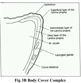
Fig 3A Body Cover Complex7
Fig 3B Body Cover Complex
[image:28.595.179.449.412.690.2]Table-4-FUNCTIONAL TWO-LAYERED STRUCTURE OF THE VOCAL FOLDS
Fig 4-Cover Body Complex
THE BASEMENT MEMBRANE ZONE
This is an area between the epithelium and the superficial layer of the
superficial layer of lamina propria. The basal cells of epidermis is attached
anchoring filaments(AFL) through the lamina densa of the BMZ to the
anchoring fibers to the superficial layer of the lamina propria. Many of the
structures are composed of one or more proteins. One such protein,
epidermolysis bullosa acquisita (EBA) is involved in the actual attachments
of the epithelium to the underlying tissue.5
The lamina densa area contains proteins that add strength to the
BMZ. Type IV collagen is also found in the lamina densa region.The BMZ
is very susceptible to injury due to vibration and shearing forces.
The fibers that anchor the BMZ loop from the lamina densa into the
superficial layer of the lamina propria and then back to the lamina densa.
Type III collagen fibers also appear to pass through these loops creating an
arrangement that resembles a chain link fence. The lamina densa area
contains proteins that add strength to the BMZ.
Type IV collagen is also found in the lamina densa region. The BMZ
is very susceptible to injury due to vibration and shearing forces. The fibers
that anchor the BMZ loop from the lamina densa into the superficial layer
of the lamina propria and then back to the lamina densa. Type III collagen
fibers also appear to pass through these loops creating an arrangement that
resembles a chain link fence.
Disease and trauma may damage these fragile connection links
between the epithelium and the BMZ. It is conjectured that some
lesions. Once laid down, it tends to stay and had been implicated in scar
formation. It has been found in human vocal nodules. But neither
fibronectin nor collagen type II is found in human polyps5 .
Fig 5A-vocal cord cross sectional study. (SE- squamous epithelium, LPlamina propria, RS- Reinke’s space, VL- vocal ligament, VM- vocalis muscle,
[image:31.595.167.458.192.406.2]BM- body of thyroarytenoid muscle, V- ventricle, FF- false cord)
PHYSIOLOGY OF PHONATION
The voice is an integral part of the unique human ability to
communicate by speech. The larynx is the major source of sound used
during speaking. Phonation is the generation of sound by vibration of vocal
cords.
THEORIES OF PHONATION6
It is based on three theories.
a) Aerodynamic or myoelastic theory: (Van den Berg - 1958) this theory postulates that vocal cords are subject to well established
aerodynamic and physical forces. There is a building up of infraglottic air
column, and its pressure act on the vocal folds which are kept tensed by the
tonic contraction of the laryngeal muscles. This increased infraglottic
pressure forces the vocal cords apart and it is set in vibration, once again
the pressure falls, vocal cords recoil following which the subglottic
pressure raises. The mode and frequency of vibration is dependent on
properties of the cord and interplay of the intrinsic muscles of the larynx.
b) Neuromuscular or clonic theory (Husson): This is not accepted now. This states that each new vibratory cycles are initiated by nerve
impulses transmitted from brain to the vocalis muscle by way of the vagus
on rate of impulses delivered. There was very little conclusive evidence to
support this theory.
c) Cavity tone or transient theory (Wills) this states that larynx functions simply to supply puffs of air that might excite the supraglottal
resonating cavity. This explains sound production based on the resonation
chambers alone.
MECHANISM OF PHONATION
Phonation requires co-ordinated interaction of the mouth pharynx,
larynx, diaphragm and neck muscles. Normal phonation requires five
conditions8:
1) Adequate breath support
2) Approximation of vocal cords
3) Favorable vibrating properties
4) Favorable vocal cord shape
5) Control of length and tension.
The upward movement of diaphragm pushes air from the lungs
through the vocal folds, producing a train of air pulses. This pulse train is
shaped by the resonances of the vocal tract. Basic resonances called vocal
formants, can be changed by the action of the articulators to produce
PHONATORY PHYSIOLOGY
GLOTTAL TONE INITIATION9
The process of phonation begins with inhalation of air. The vocal
folds are approximated in the midline or near the midline (phonatory
postion) and the glottis space is obliterated. The subglottic pressure builds
up to about 7cm of water, for conversational speech. The subglottic
pressure then pushes the vocal cord progressively apart from the bottom up
until a space develops. Bernoulli’s effect of air flow along with the elastic
forces of the cords begin to close the glottis almost immediately even while
the upper edges are still separating. The upper portion of vocal cords has
strong elastic properties which make the vocal cords to snap back to the
midline completing the glottic cycle. Subglottic pressure then builds up
again and events are repeated.
Bernoulli’s effect is an important aerodynamic event responsible for
closing the vocal cords. Bernoulli’s law states that the sum of the static
pressures at the kinetic pressures in a gas system is always equal to a
constant. In the larynx, the vocal cords cause a partial obstruction of
airflow. The molecules travelling along the sides of the trachea, when
meeting the vocal cords, must travel a greater distance to meet the
molecules in the centre of trachea. The molecules along the surface of vocal
folds must increase their velocity and kinetic pressure. Thus the static
pliable and movable vocal folds will begin to move towards the centre of
the trachea because of this pressure differential. Eventually, the two cords
will meet in the midline, and airflow will cease.
When the cords close there is sudden decrease of airflow and when it
opens there is a momentary delay in starting the flow of air due to the
inertia.
This gives the characteristic shape of the airflow pulse through the
glottis where the rising airflow phase is slower than the opening of the
vocal folds. Intra glottis pressure is solely dependent on particle velocity.
It is necessary to properly tense and elongate the vocal cords prior to
actually producing sounds, which is regulated by the laryngeal muscles. It
is also important that in the myoelastic – aerodynalmic mechanism of
phonation the vocal cords emit pulses of air and also there is a vertical
phase difference; that is: the lower portion of vocal cord begins to open and
close before the upper thus producing a rippling displacement of the vocal
cord cover. Thus mucosal wave can be examined by stroboscopic light.
The vocal cord length, mass and tension determines the fundamental
frequency. Fundamental frequency which corresponds to pitch can be
altered by changing the air pressure or the mechanical properties of the
Contraction of cricothyroid muscle along with the thyroarytenoid
muscle increases the length and tension of the vocal cords, resulting in
raising the pitch.
In the lower vocal range, contraction of thyroarytenoid alone results
in lower pitch because it decreases the tension in the vocal cover. Vocal
frequency decrease as the mass of the cord increases. The vocal intensity
corresponds to loudness.
The sound generated by vibration of the vocal cords is then
modulated by the resonating chambers. Resonance is controlled by altering
the shape and volume of pharynx, by raising and lowering the larynx, by
moving tongue or jaw position, or by the nasopharynx and nose. Voice
training for singing, acting and public speaking concentrates heavily on
refining and maximizing resonance. The goal is to produce the most loudest
and pleasing sound with minimal strain or pressure on the larynx.
COMMON VOICE COMPLAINTS
Hoarseness means change in voice. This term is used by patients to
describe changes in their voice quality. Dysphonia means abnormal voice,
but the degree of dysphonia does not correlate with any particular specific
The symptoms of dysphonia may be further subclassified, as
Diplophonia (double-tone), Dysresonance (change in the resonance of the
voice). Voice breaks exemplify pitch-specific dysphonias. Odynophonia
implies uncomfortable or painful speaking. Vocal fatigue is a common
symptom among voice disorder patients and implies the development of
symptoms (dysphonia or odynophonia) sometime after the initiation of
vocalization10 .
Aphonia is used to describe the loss of voice; such patients may still
be able to communicate in a quiet environment using the airstream for
articulation, but the glottis does not participate in phonation. The sound of
aphonia, then, is characteristically no voice or extreme breathiness.
EVALUATION
Evaluation of the voice includes examination of the laryngeal
structures by stroboscopy voice analysis. Additionally, administration of
the Voice Handicap Index (VHI) provides an assessment of the level of
handicap experienced by the patient with the voice disorder.
STROBOSCOPY
It is a technique for observation of laryngeal vibration which was
BASIC PRINCIPLE OF STROBOSCOPY
The vocal folds vibrate at a frequency of approximately 250 times
per second the human eye cannot discern necessary detail during rapid
motion. The vocal folds vibrates at such a high frequency that their
movements cannot be recorded during an indirect laryngoscopy or an
endoscopy via rigid endoscopylaryngoscopy without additional apparatus.
However with the aid of stroboscopy vibration can be observed.
TALBOT'S LAW & STILL AND MOVING IMAGES
Stroboscopy is based on Talbolt’s Law. It states that the images on
the human retina linger for 0.2 seconds after exposure. Therefore the
sequential images produced at intervals of less than 0.2 seconds(more than
5 images per sec ) produce the illusion of a continuous image. The
stroboscopy actually illuminate different point on successive wave of
vibratory cycles, each of which is retained on the retina for 0.2 sec. The
light source of the machine emit intermittent flash at a rate that can be set
by examiner or controlled by fundamental frequency of voice. When vocal
cord vibrations and the stroboscopy light are synchronized the vocal cord
will appear still rather than moves in slow motion. The frequency of the
examinee sustained voice is picked up by the microphone and trigger the
the vocal cord vibration, that is, at an identical phase point in successive
vibratory cycle a sharp and clear images of the vocal cord is observed.
When the frequency of the flashes is less or greater than vocal vibration,
and there is a delay in the portion of the vibrating cycle illuminated and the
illusion of slow motion is obtained.
The parameters studied by stroboscopy include symmetry,
amplitude, periodicity, completeness of vibratory closure, and the mucosal
Fig : 6 Still and moving above. Above : a moving image is obtained through the illumination of consecutive phases. Below: a still image obtained through
THE PARAMETERS STUDIED BY STROBOSCOPY ARE DESCRIBED BELOW
1) FUNDAMENTAL FREQUENCY
The rate at which the vocal cord vibrate for 1 second is called
fundamental frequency. It varies from person to person. Usually lower in
males and compare to females.
2) PERIODICITY
It refers to the regularity of successive vocal movement. A
Periodicity occurs when the vocal folds vibrations follows each other at the
same interval which mainly depends on balanced control of expiratory
force and the mechanical characteristics of the vocal folds.
Aperiodic movements is caused either by inability to maintain a
steady expiration stream of air or inability to sustain steady laryngeal
muscle contraction or difference in the mass or stiffness of one of the vocal
cords.
3) AMPLITUDE
This refers to the medio - lateral deflection of each individual vocal
folds usually it is one third of the total width of vocal card .when the voice
of pathological increase of stiffness in the vocal fold, amplitude will
decrease.
4) MUCOSAL WAVE
A Mucosal wave involves wave shaped shift of the mucosa
(Epithelium and lamina propria) in contrasts to the tonalise M.Vocalis. Its
decreased by musosal stiffness, masses, scarring, dryness. Loud phonation
may increase the mucosal wave.
5) GLOTTIC CLOSURE
In normal individual complete closure of the membranous portion of
the vocal cord occurred, however in some individual the posterior
cartilaginous glottis may remain open. Glottal closure may be consistent or
intermittent, complete or incomplete. Adynamic (nonvibrating ) should be
specified. The failure of glottic glossure may be due to local pathological or
mural paralysis.
ACOUSTIC ANALYSIS
The various parameters of voice production that are analyzed in
voice analysis are fundamental frequency, maximum phonation time, jitter,
1) FUNDAMENTAL FREQUENCY
The rate of vibration of vocal cord during the production of sustained
vowels and while reading the passage. It is used for comparing intra and
inter subject pitch levels. It depending upon the type of speech material
used it can vary. Variability is less for sustained vowels when compare to
reading passages.
2) MAXIMUM PHONATION TIME
It refers to the maximum duration that a particular vowel sounds can
be sustained.
3) JITTER / FREQUENCY PERTURBATION
It is the measure of instability of the vocal cord during sustained
Phonation otherwise, to the variation of fundamental frequency that present
in all speakers to some degree and detected when the subject is attempting
to produce a steady, sustained vowel. The frequency variations are the
result of instability of the vocal fold cause frequency vibration. Normal
speakers have a little amount of frequency perturbations, which varies
according to age, sex and physical condition.
4) SHIMMER / AMPLITUDE PERTURBATION
The vocal cords exhibit slight variation of amplitude from one cycle
or “shimmer”. A small amount of shimmer in a Normal speakers which
depends on sex and vowel used .
5) MAXIMUM PHONATION RANGE
It defined as range of lower frequency to higher frequency
individual can produce. The person asked to sustain the tone for one full
second without control the intensity and if he asked to sustained further on
the production of sound then we can expect may be altered magnitude of
the range obtained.
6) VOCAL INTENSITY
Vocal intensity of phonation during speech is depends on Various
factors which includes intensity produced at glottis, the amount of lip
opening , shape of vocal tract mean distance of microphone from the lips of
the subject and physical and emotional factors.
7) HARMONIC TO NOISE RATIO
Voice is mixture of periodic and aperiodic noise. Whenever the noise
component of voice increases and replaces the harmonic structure the
quality of hoarseness is perceived and this is measured as harmonic to
VOICE HANDICAP INDEX:
It is the measure of the impairment of voice stated by a patient
developed to determine the outcome pre and post operatively. Several type
of questionnaires are available to self evaluate voice disability. Some of
then are Llewellyn Thomas (1984) developed a linear analogue scale for
self assessment of voice quality in laryngeal cancer.12
Smith et al (1994) developed a questionnaire regarding functional
impact of voice disorders in various aspects of like, employment, risk
factors, symptoms, and family histories. The other questionnaire are Voiss
(Voice Symptom Scale) and VHI10.
The Voice handicap index used in this study was developed by
Jacobson and Jhonson in 1997 which consists of set of questionnaire with
three domains namely Functional, Emotional and Physical .Each Part has
10 questions each subset has 0 to 4 scores to the maximum of 120 and the
minimum of 0. The minimum value for a normal person is less than 10.13
BENIGN VOCAL CORD LESIONS: PATHOGENESIS, PATHOLOGY AND CLINICAL FEATURES
Benign vocal mass lesions are lesions which has a different
pathology. They include pathologies like vocal nodules, vocal polyps, vocal
cysts and Reinke’s oedema. The lesions having similar pathogenesis. The
Tobacco smoking, laryngoparyngeal reflux (LPR), environmental factors,
hypothyroidism, allergy. The mechanism by which these factors produce
lesions include, microtrauma; Excessive force causes damage of capillaries
and changes in micro vascular circulation.
Temporary ischemia leading to increased permeability,lamina
propria odema and hematoma. Organization followed by fibrosis and a
nodule results at the edges.
Hammering effect of each vocal fold during Phonation causes direct
compression.16 Maximum trauma occurs at junction of anterior one third and posterior two third because this part having maximum vibration.
Roughly it is the midpoint of membranous cord.
MICROSCOPIC PATHOLOGY
With the use of Heamotoxylin and Eosin stains the nodules and
polyps look alike.Hemorrhagic event or increased vascular permeability
leads to vocal polyp. Whereas Nodules produced by thickening of the
epithelium or lamina propria.
Trauma of the vocal cord respond in Two Ways
TYPE ONE:
Trauma leads to lamina propria damage, associated with severe
marked by fibronectin deposition. The extent and aggravation of basement
membrane zone injury determined by thickness of the collagen type IV
band. Repetitive injury, leads to aberrant healing and a fibroblastic
response involving increased fibronectin deposition. Its excess presence
probably does not contribute to efficient tissue vibration.
TYPE TWO
Absence of the structural glycoproteins and fibrous proteins would
lead to a vocal fold with excessive propensity for deformability. The injury
often seems to be confined to the lamina propria and apparently not
contain fibroblastic response of tissue repair. This type seen more often in
Reinkes oedema and some polyps17.
INDIVIDUAL BENIGN VOCAL CORD LESIONS
NODULE OF VOCAL CORD
Most common vocal cord lesion in humans and commonly seen in
people who speaks in high pitched voice like school teachers, Church
preachers, and childrens, hence known as singer’s nodules, teachers
nodule and screamer’s nodules in children. Hoarseness is the common
symptom. It occurs at the junction of anterior one third and posterior two
third free edge of the vocal cord. Nodules of Vocal Cord seen bilaterally
and looks white, small, sessile, and often symmetric. Long standing polyps
Oedema and vasodilatation are the initial event in the formation of
nodule and this progress into hyalinization of the nodule and hyperplasia
of epithelium. Histology shows, hyperkeratotic epithelium with
parakeratosis. The subepithelial layer shows infiltration of irregular
collagen fibers and fibroblasts.
Stroboscopy visualize pronounced vibration of the anterior segment
of vocal cord with asymmetry and aperiodicity. Voice analysis pictures
breathiness of voice with reduced loudness and increased jitter, shimmer
and harmonic to noise ratio19.
Fig 8:-B/L Vocal Nodule
VOCAL CORD CYSTS
Cysts of Vocal Cord mostly affect the adult population. Two
types of cysts, mucous retentions cysts and epidermoid cysts. They are
mainly unilocular and unilateral. Multilocular and bilateral, multiple vocal
cord cysts of various sizes also occurred20 Main differential diagnosis of
Small sized vocal cord cysts are vocal nodules.
Mucous retention cysts are formed by mucous glands blockage seen
at vocal cord under surface.They are lined by cuboidal or coloumnar
epithelium looks like respiratory epithelium. Epidermoid cysts are formed
by the epithelial ingrowth into the cord, by microtrauma or as a congenital
anomely.The cavity is lined by keratinizing squamous epithelium and
Content of the cyst of mucous retention cysts are watery thin and
mucoid and in epidermoid cysts it is inspissated debris. Stroboscopy of
cysts pictures hyperkinetic pattern of mucosal wave in regions other than
the area of cyst. Usually normal or reduced mucosal wave seen in cysts.
Voice analysis shows the same picture as vocal nodule.
Fig 9-Vocal Cyst
Fig 9B-Lt Vocal Cyst VOCAL CORD POLYP
These are arise only from the vocal cord membranous part, usually
close to the anterior commisure. Vocal polyps develop when there is an
initial high subglottic pressure followed by abrupt reduction. This results in
trauma to the basement membrane zone, leads to hyperemia and edema.
Accumulation of oedema is focused at a particular point that part is
ballooned and polyp is formed.22 Use of anti platelet drugs or
anticoagulants can predispose to hemorrhagic polyps by Phonotrauma.
Histopathologically there are three types of polyps namely;
gelatinous, telangiectatic and mixed type. Gelatinous polyps having very
loose edematous stroma and sparse collagen, minimal fibrocytes,
histiocytes and mast cells.
Telangiectatic polyps consists homogenous eosinophillic deposits
and fibrin collections in the stroma. Labrynthine type of vascular channels
are seen in stroma. The mixed type of polyp having features of both the
gelatinous and telangiectatic type. These are the most common type of
polyp seen.
Fig 10A- Rt Vocal Polyp
Stroboscopy Shows adynamic segments, with aperiodicity and
asymmetry. Voice analysis will pictures breathiness of voice with reduced
loudness and with increased jitter, shimmer and harmonic to noise ratio.
REINKE’S OEDEMA
Also known as polypoidal degeneration, chronic polypoidal corditis,
chronic edematous hypertrophy or chronic hypertrophic laryngitis19.
Smoking is the major etiological factor. Fritzel in his retrospective study
(1986) found that more than 95% of affected patients were smokers and
Kleinsasser 98% were smokers20. Others include LPR and voice abuse.
Abnormal increase of the glottic pressure with caustic exposure leads to
increased vascular permeability and oedema of the vocal cord.
The association between thyroid dysfunction and Reinke’s oedema
has also been mentioned. Accumulation of fluid at the superficial lamina
propria.
Histologically Reinke’s oedema is divided to pale and livid types23.
In pale type, limited, fusiform glazed swelling noted. The epithelium is thin
and clear watery fluid collection is visible. Stromal cells and numerous
intracytoplasmic granules are seen. In the livid type, the colour changes to
yellow gray and fluid will be glue like consistency. Additionally multiple
Hoarseness main presenting symptom. Stroboscopy pictures that the
body of the cord is not affected. Glottic closure is usually complete and the
cord shows a fusiform, ballooned out appearance. Stroboscopy also shows
loss of mucosal wave. Variable Loss of symmetry and the closure can be
complete. Voice analysis pictures low fundamental frequency and high
jitter and shimmer. Harmonic to noise ratio is increased.
MATERIAL AND
MATERIALS AND METHODS
MATERIALS STUDY DESIGN
A prospective cohort study of 38 patients with benign vocal cord
lesions, were included in the study.
SETTINGS :
Department of ENT and Head and Neck Surgery, Govt Stanley
Medical College and Hospital, Multi Speciality Teaching Institute
Chennai-1
PATIENTS :
INCLUSION CRITERIA :
• Benign looking vocal cord lesion patients
• Age 12 years above
• On Rigid fiberoptic endoscopy / indirect laryngoscopic examination
the vocal cords will be Freely mobile.
EXCLUSION CRITERIA:
• Patients with malignant looking lesions.
• Patients suffering from Respiratory distress
• Patient with ischemic/Coronary heart diseases.
METHODOLOGY :
Patients included in this study after fulfilling the inclusion and
exclusion criteria defined in this study and after getting valid consent. .
Each patient with benign vocal cord mass lesion underwent pre and post
operative three procedures like, Video laryngoscopy, stroboscopy and voice
analysis.
VIDEO LARYNGOSTROBOSCOPY:
Fiberoptic laryngoscopy demonstrating laryngeal pathology and
provides an excellent view of the larynx, particularly suited for patients
who are unco-operative for indirect laryngoscopy.
PROCEDURE:
The patient sat and must facing the examiner. Before starting this
procedure Each patient was sprayed with a mixture of 4% or 10%
lignocaine applied at oropharyngeal mucosa. A 4 mm rigid Endoscope was
passed per orally under direct control and vision.
The visualized areas were the, oropharynx and the larynx. The larynx
examined in detail regarding the size, shape of vocal cord lesion, presence
of hemorrhage and pedunculated or sessile lesion. The contralateral vocal
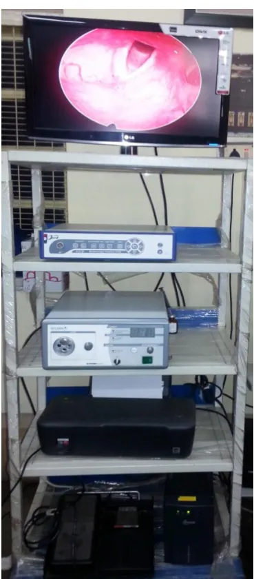
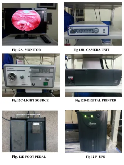
VIDEO STROBOSCOPIC UNIT Components:-
1. Stroboscopic Unit a) Light Source b) Microphone 2. Video Camera 3. Endoscope 4. Videorecorder
[image:59.595.222.407.304.725.2]STROBOSCOPY
Fig 12A- MONITOR Fig 12B- CAMERA UNIT
Fig 12C-LIGHT SOURCE Fig 12D-DIGITAL PRINTER
[image:60.595.96.519.133.695.2]Fig 13A-MONITOR Fig. 13B-PRINCIPAL UNIT
Stroboscopy is a procedure which allows routine slow motion
examination using a laryngeal stroboscope, a 70 degree Hopkins telescope.
The procedure was performed after rigid video laryngoscopy. The
procedure was explained to the patient in detail regarding the need to keep
the mouth open and avoid swallowing during the test. Local anesthetic was
provided to the larynx with 10% lignocaine spray. After few minutes the
patient was asked to open the mouth wide and protrude the tongue. The
tongue was held by the examiner with gauze piece, simultaneously
supporting the jaw at its upper part. The telescope was introduced into the
oropharynx to visualize the glottis after using antifogging solution. The
patient was asked to phonate long “eee” to see the cords in motion. While
the patient was phonating the stroboscopy pedal was pressed so as to
produce pulsed light in accordance to the patient’s fundamental frequency.
The patient underwent for stroboscopy which was done by using a
Karl Storz stroboscope. The output picture and sound was captured into a
computer monitor using a capture card without compression and analyzed
Fig 14-Endoscopic Procedure
The stroboscopy picture was analyzed to note:
1. Symmetry (symmetry of movement and approximation of vocal
cords)
2. Mucosal wave (pattern of light traveling on the vocal cord)
NORMAL MUCOSAL WAVE PATTERN SEQUENCE
VOICE HANDICAP INDEX
Voice handicap questionnaire have been designed for assessment of
patients with voice disorders. Here questionnaire developed by “Jacobson
et al13” was used. All patients administered the questionnaire and a
measure called the voice handicap index was calculated. The measurer
refers to the total sum obtained (minimum of ‘0’ and maximum of ‘120’)
when the voice handicap score is administered.
VOICE ANALYSIS
The next step is voice analysis. Each patient was taken to a sound
proof room to record voice using a low impedance commercial microphone
and asked to phonate in low and comfortable voices. Each of these vowel
sounds were voiced for at least of 15 to 20 seconds for low and comfortable
intensity. Speech is recorded in intensity comfortable for the patient.
Only a good quality continuous signal was selected and used for
analysis. Speech sound was recorded by asking the patient to count
numbers slowly and clearly. The recorded voices are stored in computer for
analysis. Voice analysis was performed on PHONOLAB software version
03.02.08 ECLERIS.
“PHONOLAB” is an open source freeware developed specifically
keeping in mind scientific analysis of the sound signals recorded in “wav”
opens source code; which means that the program can be altered to fit to
individual necessity.
The parameters that were observed on voice analysis were
1. Fundamental frequency (vocal cord vibrating frequency)
2. Standard deviation of fundamental frequency.
3. Jitter (fundamental frequency variation)
4. Shimmer (one cycle to cycle amplititude variation )
5. Harmonics to noise ratio (amount of noise in voice)
SURGERY
After the complete preoperative workup, under general anesthesia all
patients underwent micro laryngeal surgery using a suspension
laryngoscope. The surgeon was not the same for all the patients. In surgery,
the lesions were completely excised to the satisfaction of the surgeon and
an independent observer.
Post operatively, patients were advised strict voice rest for a period
of fourteen days. Advice regarding the usage of voice and the do’s and
don’ts following surgery were given. Contact numbers of all patients were
VOCALNODULE
Fig 16-Pre-OP Vocal Nodule
[image:68.595.146.487.414.691.2]Fig 16B-Intra Op Lt Vocal Cord Nodule Removal
[image:69.595.134.498.422.710.2]VOCAL POLYP
Fig 17-PRE-OP Vocal Polyp Rt
Fig 17 A-INTRA OP Rt Vocal Polyp Removal
VOCALCYST
[image:71.595.128.502.439.736.2]Fig 18-PRE OP Vocal Cyst
VOCAL POLYP(ANGIOMATOUS)
Fig 19-PRE-OP Vocal Cord Polyp
Fig 19 A-INTRA OP(COBLATION) Polyp Removal
POST OPERATIVE REVIEW
All the operated patients were asked to come for review after 6
weeks(42 days) following surgery. On review,
1. Stroboscopy
2. Voice analysis
3. Voice handicap index questionnaire
The methodology used for each patient was similar to that used
preoperatively.
RESULTS
DEMOGRAPHIC CHARACTERISTICS
AGE DISTRIBUTION:
In the group of 38 patients the mean age was 36.47 . The youngest
RESULTS AND
SEX DISTRIBUTION:
A total of 38 patients were enrolled in this study. A Female
preponderance 22 was seen.
GEOGRAPHIC DISTRIBUTION:
The majority of my patients came from Chennai , while the outside
to Chennai city consisted of 0.
DURATION OF COMPLAINT:
Among the 38 patients; a mean duration was 12.44 months. The
range of duration of complaint was 02 to 48 months.
Female 58% Male
42%
ASSOCIATED HABITS AND HISTORY
VOICE DEMAND AND VOICE ABUSE
In the group of 38 patients, there was one professional voice (grade
I) (n=1) user as preacher. On categorizing them according to the voice
demands, there were professional voice users (grade II) (n=03) non
professional voice users (grade III)(n=07) and non professional non voice
users (grade IV) (n=27).
Voice abuse is one of the leading known etiological factors for
benign vocal cord lesions. In this study, we found that almost all patients
with history of voice abuse.
SMOKING
Among the 38 patients 74 % were non smokers and 26 % were
STROBOSCOPIC PARAMETERS
Pre operative analysis ( Table )
SYMMETRY
Almost All patients with vocal cord lesions had Asymmetry .
1 26%
2 74%
SMOKING
0 5 10 15 20 25 30 35 40
Pre Post
SYMMETRY
MUCOSAL WAVE
Right and left patten were analyzed Individually.
MUCOSAL WAVE (RIGHT)
Nearly 55.26 % of patients was found with absent mucosal wave on
right vocal cord.
MUCOSAL WAVE (LEFT)
Among 38 patients only 76.31 % of patients sures absence mucosal
wave on the left.
0 5 10 15 20 25 30 35 40
Pre Post Pre Post
RIGHT LEFT
MUCOSAL WAVE
GLOTTAL CLOSURE
Incomplete closure of the glottal chink were noted in all the
patients who were examined.
Analysis of GLOTTIC CLOSURE(Vocal Nodule)
Complete Hourglass Irregular Posterior chink
Present 0 14 (95%) 0 2 (5%)
Absent 16 (100%) 2 (5%) 0 14 (95%)
0 5 10 15 20 25 30 35 40
Pre Post
GLOTTAL CLOSURE
TABLE
Pre operative stroboscopic analysis (n=38 )
` Present Absent
Symmetry 0 38
Mucosal wave (right) 17 21
Mucosal wave (left) 9 29
Glottic closure 0 38
Table 5-Pre Operative Stroboscopic analysis
VOCAL CORD LESIONS FOR SIDE AND SITE
Among 38 patients 28.9 % involved right cord and 31.6 % add
lesion on both vocal cords.Left side involvement were found in 39.5 %.
Glottic closure pattern were different in concern with size of polyps.
Majority were hour glass type of closure.
0 10 20 30 40
Pre Post
SIDE OF LESION
POSITION OF LESION
The majority of them had left cord lesions (39.5 %) at the junction
of anterior and middle third of cord ( 42.10 %) and Middle third lesions
(42.10%) were more frequent than the anterior third lesions (15.78 %)
TYPES OF LESIONS
The majority of patients had vocal nodules and cysts at equal
number and percentage 16 (42.1%)Vocal polyp for 6 (15.8%) Patients.
0 5 10 15 20 25 30 35 40
Pre Post
POSTION OF LESION
POST OPERATIVE ANALYSIS:
SYMMETRY:
Glottics post operative symmetry have risen from 0 to 100 in the
patient.
MUCOSAL WAVE RIGHT:
The Mucosal wave right pattern considerably increases 55 % to 95 %.
MUCOSAL WAVE LEFT
The Mucosal wave on the left vocal cord were increase from
76 % to 96 %.
0 5 10 15 20 25 30 35 40
Pre Post
LESIONS
GLOTTAL CLOSURE
Remarkable improvement in complete glottal closure were noted
post operatively which source and increase of 96% in this parameters .
ADDITIONAL FINDINGS
Post operatively, the prevalence of additional findings was clearly
ACOUSTIC ANALYSSIS Fundamental Frequency:-
Among 38 Patients the mean fundamental frequency recorded for
comfortable voice is 197.1.While the speech frequency is Ranging from
128.3 to 254.2.
F0 is governed by this equation
F0 =
F0 = fundamental frequency, 2L = Length of Vocal folds
s = Longitudinal stress r = Tissue density
Fundamental Frequency
Mean S.D P-Value
LOW/A/ MEAN PIT PRE 178.68 41.84 0.001
POST 184.56 43.38
LOW/E/ MEAN PIT PRE 179.94 45.70 0.074
POST 185.23 37.19
LOW/I/ MEAN PIT PRE 175.32 41.72 0.051
POST 179.61 43.56
COM/A/ MEAN PIT PRE 195.27 38.06 0.141
POST 202.52 49.94
COM/E/ MEAN PIT PRE 207.21 42.41 0.333
POST 210.26 43.56
COM/I/ MEAN PIT PRE 191.75 35.33 0.0005
POST 206.71 33.78
COM/SP/ MEAN PIT PRE 194.55 40.62 0.378
[image:84.595.112.517.343.670.2]POST 197.12 42.05
Table-6-Fundamental Frequency
Post Operative analysis reveals significant deviation from the pre
STANDARD DEVIATION OF PITCH
Pre-OP mean value is 28.3 and Post-Op Mean Value is 18.7 for this
parameter.
STANDARD DEVIATION OF PITCH
Mean S.D P-Value
Std Devi 1
Pre OP 33.00 17.93
0.0005
Post OP 15.24 11.66
Std Devi 2
Pre OP 25.89 14.08
0.005
Post OP 16.40 15.66
Std Devi 3
Pre OP 31.03 23.00
0.0005
Post OP 17.32 18.30
Std Devi 4
Pre OP 26.42 30.92
0.06
Post OP 16.98 15.28
Std Devi 5
Pre OP 27.03 23.79
0.0005
Post OP 13.90 11.66
Std Devi 6
Pre OP 28.33 18.74
0.725
Post OP 26.59 17.38
Std Devi 7
Pre OP 26.50 10.14
0.408
[image:85.595.129.500.191.583.2]Post OP 24.35 11.77
Table 7 - Standard Deviation of Pitch
JITTER
Pre Operative Mean Value of jitter is 1.91 and Post Operative mean is 1.30.
JITTER
Mean S.D P-Value
Jitter 1
Pre OP 1.740 0.68
0.0005
Post OP 1.191 0.57
Jitter 2
Pre OP 3.060 3.85
0.026
Post OP 1.515 1.19
Jitter3
Pre OP 1.783 0.89
0.071
Post OP 1.526 0.98
Jitter 4
Pre OP 1.291 0.64
0.009
Post OP 0.824 0.66
Jitter 5
Pre OP 1.951 1.39
0.208
Post OP 1.475 1.64
Jitter 6
Pre OP 1.847 0.83
0.0005
Post OP 1.028 0.76
Jitter 7
Pre OP 1.761 0.42
0.007
[image:86.595.127.500.142.554.2]Post OP 1.551 0.20
Table 8 - Jitter
SHIMMER
The mean of this parameter Pre Operatively is 7.9 and Post
Operatively is 4.5.
SHIMMER
Mean S.D P-Value
Shimmer 1 Pre OP 9.527 4.48 0.002
Post OP 6.118 3.85
Shimmer 2 Pre OP 10.184 8.78 0.009
Post OP 6.413 3.04
Shimmer 3 Pre OP 7.924 2.26 0.007
Post OP 6.593 2.38
Shuimmer 4 Pre OP 7.654 4.65 0.0005
Post OP 1.492 1.16
Shimmer 5 Pre OP 6.141 2.91 0.0005
Post OP 1.947 1.16
Shimmer 6 Pre OP 5.868 2.66 0.0005
Post OP 2.737 1.89
Shimmer 7 Pre OP 8.526 2.87 0.001
[image:87.595.135.492.169.506.2]Post OP 6.443 1.75
Table-9-Shimmer
There is improvement in Shimmer shows When compared to post
HARMONIC NOISE RATIO (HNR)
Mean of this parameter pre operatively 0.0104 and Post Operatively
0.119.
Harmonic Noise Ratio(HNR)
Mean S.D P-Value
HNR 1
Pre OP .1500 .069
0.004
Post OP .1104 .045
HNR 2 Pre OP .0619 .024 0.01
Post OP .0828 .038
HNR 3 Pre OP .0848 .037 0.603
Post OP .0879 .019
HNR 4
Pre OP .1605 .079
0.033
Post OP .1942 .084
HNR 5
Pre OP .0942 .058
0.0005
Post OP .1375 .062
HNR 6 Pre OP .0912 .063 0.0005
Post OP .1242 .031
HNR 7
Pre OP .0907 .036
0.052
[image:88.595.122.508.223.548.2]Post OP .1018 .032
Table-10-Harmonic Noise Ratio
Post Operative data analysis shows improvement in Harmonic Noise
MAXIMUM PHONATION TIME
The Mean Value of this parameter Pre Operatively 6.45 and
Post-Operatively is 10.05
Maximum Phonation Time
Mean S.D P-Value
MPT Pre OP 6.447 2.617 0.0005
[image:89.595.121.509.366.653.2]Post OP 10.053 2.026
Table-11-Maximum Phonation Time
Postoperatively the maximum phonation time shows increase trend
with statistically significant p value (p=0.0005)
0.00 2.00 4.00 6.00 8.00 10.00 12.00
Pre Post
VOICE HANDICAP INDEX (VHI)
The Mean of this parameter Preoperatively is 23.7 and
post operatively is 6.9.
VOICE HANDICAP INDEX
Mean S.D P-Value
VHI 1
Pre OP 22.26 4.14
0.0005
Post OP 7.39 1.15
VHI 2
Pre OP 23.55 4.88
0.0005
Post OP 6.13 1.51
VHI 3
Pre OP 25.50 4.40
0.0005
Post OP 7.45 1.62
VHI 4 Pre OP TOTAL 52.53 16.44 0.0005
[image:90.595.123.508.242.512.2]Post OP TOTAL 20.97 3.01
Table-12-Voice Handicap Index
Analysis was done by using RATHER PAIRED T test with
statistically significant p value(p=0.0005) which shows overall