ON THE SHEAR BOND STRENGTH OF COMPOSITE
TO GLASS IONOMER – AN INVITRO STUDY
Dissertation submitted to
The Tamil Nadu Dr M.G.R. Medical University
In partial fulfillment of the degree of
MASTER OF DENTAL SURGERY
Branch IV
CONSERVATIVE DENTISTRY AND ENDODONTICS
This is to certify that this dissertation titled “The effect of four different
conditioners on the shear bond strength of composite to glass ionomer – an in
vitro study”, is a bonafide record of the work done Dr. Rahul S under our guidance
during his / her post graduate study during the period of 2012-2015 under
THE TAMIL NADU DR. MGR MEDICAL UNIVERSITY, CHENNAI, in partial
fulfillment for the degree of MASTER OF DENTAL SURGERY IN
CONSERVATIVE DENTISTRY AND ENDODONTICS, BRANCH IV. It has
not been submitted (Partial or full) for the award of any other degree or diploma.
Dr. Rajesh S MDS
GUIDE
PROFESSOR & HOD
Department of
Conservative Dentistry &
Endodontics
Dr. Mano Christaine Angelo MDS
CO-GUIDE
PROFESSOR
Department of
Conservative Dentistry &
ENDORSEMENT BY THE PRINCIPAL / HEAD OF THE INSTITUTION
This is to certify that the dissertation entitled “THE EFFECT OF FOUR
DIFFERENT CONDITIONERS ON THE SHEAR BOND STRENGTH OF
COMPOSITE TO GLASS IONOMER – AN IN-VITRO STUDY” is a bonafide
research work done by Dr. RAHUL S under the guidance of Dr RAJESH S, MDS,
Professor & HOD, Department of Conservative Dentistry and Endodontics, Sree
Mookambika Institute of Dental Sciences, Kulasekharam.
Date: Dr. ELIZABETH KOSHI; MDS
PRINCIPAL
SREE MOOKAMBIKA INSTITUTE OF
DENTAL SCIENCES,
VPM HOSPITAL COMPLEX,
PADANILAM, KULASEKHARAM,
KANYAKUMARI DISTRICT,
I avail this opportunity to express my profound sense of sincere and deep
gratitude to many people who are responsible for the knowledge and experience I
have gained during the study.
I take this opportunity to express my sincere thanks to chairman
Dr.C.K.Velayuthan Nair, MBBS,MS and Director Dr. Rema V Nair, MBBS, MD,
DGO for their constant support and for providing facilities and infrastructure which
was essential for me to complete my post graduation.
I express my deep sense of gratitude and sincere thanks to my teacher and
guide Dr. Rajesh S MDS, Professor for his continuous support, boundless patience,
timely help, constant advice and immense knowledge all throughout my study and
during the entire period of my post graduation.
Words are inadequate to express my sincere and deep gratitude to
Dr.Mano Christaine Angelo MDS, Professor and co - guide for his brilliant advices,
constructive criticisms, incessant encouragement, meticulous supervision and
valuable suggestions right from the start of this study.
I take this opportunity to thank my teacher Dr.Vijay Mathai MDS, Professor,
Department of conservative dentistry &Endodontics for his immense support,
encouragement, and everlasting inspiration during the entire period of my post
MDS. Their high teaching standards has been a source of encouragement to me and
beneficial to my professional improvement as a post graduate student.
My heartfelt thanks to Dr.Vineeth MDS, Dr. Manu Unnikrishnan, MDS,
Dr. Vineesh MDS and Dr.T.S.Manoj Kumar BDS for their valuable help and
words of encouragement during the prepraration of the study.
It is my previlege to acknowledge the debt I owe to Dr. Roy Joseph, Head of
the Department of Polymer processing laboratory, BMT wing, SCTIMST,
Trivandrum for their technical help, constant encouragement and liberty they
provided me during the course of this study.
I am thankful to Mr. Sarath Babu for providing me with his timely statistical
analysis involved in this study.
I would like to thank Mr. Satheesh, Good morning Xerox for DTP and photo
copying works.
I would like to thank my batch mate Dr. Sarah Christopher and my fellow
post graduates Dr.Betty Babu, Dr.Eeshan Mushtaq, Dr.Aswathy Prasad and
Dr.RejoyJohn for their motivation and help.
I would like to thank my wife, Seetha. S and my family for the continuous
love and support they provided in taking decisions in my life.
SL NO: INDEX PAGE NO:
1. List of Abbreviations i
2. List of Tables ii
3. List of Figures iii
4. Abstract iv-v
5. Introduction 1-3
6. Aims & Objectives 4
7. Review of Literature 5-22
8. Materials & Methods 23-29
9. Results & Observations 30-32
10. Tables 33-36
11. Discussion 37-49
12. Summary & Conclusion 50-52
13. Figures vi-xii
i
GIC - Glass ionomer Cement
RMGIC - Resin Modified Glass Ionomer Cement
MPa - Mega Pascals
SBS - Shear Bond Strength
PAA - Polyacrylic acid
TCA - Trichloro Acetic Acid
SEM - Scanning Electron Microscope
ANOVA - Analysis of Variance
SPSS - Statistical package for Social Sciences
kg/cm2 - Kilogram per Centimeter Square
mm - milli meter
µm - micro meter
[image:9.595.109.461.121.504.2]Er,cr:YSGG - Erbium, Chromium doped Yttrium Scandium Gallium Garnet
ii
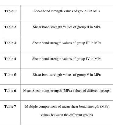
[image:10.595.131.503.107.523.2]LIST OF TABLES
Table 1 Shear bond strength values of group I in MPa
Table 2 Shear bond strength values of group II in MPa
Table 3 Shear bond strength values of group III in MPa
Table 4 Shear bond strength values of group IV in MPa
Table 5 Shear bond strength values of group V in MPa
Table 6 Mean Shear bong strength (MPa) values of different groups
Table 7 Multiple comparisons of mean shear bond strength (MPa)
iii
Figure 1 Light Cured Resin Modified Glass ionomer Capsule
Figure 2 GC Applier with Glass ionomer capsule
Figure 3 Glass ionomer discs prepared from stainless steel molds
Figure 4 Specimens embedded in acrylic resin blocks
Figure 5 Materials used in the study
Figure 6 Shear bond strength testing
Figure 7 Gold sputtering machine
Figure 8 Scanning Electron Microscope
Figure 9 Photomicrograph of Group II a (35%Phosphoric acid treated specimen)
Figure 10 Photomicrograph of Group III a ( 10%Poly acrylic acid treated specimen )
Figure 11 Photomicrograph of Group IV a (10%Citric acid treated specimen)
Figure 12 Photomicrograph of Group V a (35%trichloro acetic acid treated specimen)
[image:11.595.103.511.113.718.2]iv
Introduction
The use of esthetic resin composites has increased over the past decades
mainly due to the patient’s esthetic concerns and improvements in the technology of
products. With the advent of sandwich restorations the problems related to composites
such as recurrent caries and post- operative sensitivity have drastically reduced.
Studies have shown improvement in bond strength of composite to glass ionomer
when conditioners were used on glass ionomer surface.
Aims and Objectives
This study was done to evaluate the effect of four different conditioners on the
bond strength of composite to glass ionomer and to examine the resulting etched glass
ionomer surface under Scanning Electron Microscope.
Methodology
Fifty glass ionomer discs were prepared in stainless steel moulds. It was
divided into five groups of 10 moulds each out of which seven samples were tested
for bond strength and remaining three samples were evaluated for scanning electron
microscopic analysis. Group I is control without any surface treatment. Group II, III,
IV and V were conditioned with 35% phosphoric acid, 10% polyacrylic acid, 10%
citric acid and 35% trichloroacetic acid respectively. Adper single bond 2 was applied
to all the specimens. Filtek Z350 XT light cure composite were place on the glass
ionomer surface and light cured. All specimens were stored in deionized water for 24
v conditioned with four different conditioners, gold sputtered and examined under
scanning electron microscope.
Results and Observations
The values obtained were tabulated and statistically analysed using ANOVA
and Dunnett’s test and it revealed significant differences in bond strength among
groups. Significantly higher bond strength values were observed in group treated with
35% Phosporic acid followed by 10 % Polyacrylic acid, 10% Citric acid and 35%
trichloroacetic acid when compared with group without any surface treatment. SEM
observations revealed least salt crump formation in specimens treated with 35%
phosphoric acid. Salt crump formation was greater in specimens treated with 35%
trichloroacetic acid.
Conclusion
Under the limitations of this study it was found that the shear bond strength of
composite reins to resin modified glass ionomer was increased following surface
treatment.
Clinical significance
The use of conditioners will effectively improve the bonding between
1 The use of direct posterior resin-based composite has dramatically increased
over the past two decades primarily due to patients esthetic desires and product
improvements. Other factors contributing to increased use of resin-based composite
are environmental and health concerns with dental amalgam. The ability of resin
composite to mimic natural tooth structure gave it a distinct advantage for patients
and dental professionals over other materials. Resin-based composite consists mainly
of a resin matrix surrounded by inorganic filler particles. Polymerization occurs
through a free radical addition reaction. The double-bonded carbons of the
methacrylate groups at each end of the active site on the monomer cross-links during
the polymerization process, producing initially a linear polymer; then by reacting with
the second site, a highly cross-linked polymer is produced.1
In contrast to the superior esthetics of resin-based composites stands their
great constraint, which is their shrinkage associated with polymerization resulting in
microleakage. Microleakage is defined as the clinically detectable passage of bacteria,
fluids, molecules or ions between a cavity wall and the restorative materials applied to
it and are a major problem in clinical dentistry. Shrinkage of the composite resin
transfers stress to the cavity walls. Polymerization shrinkage can split the adhesive
bond to the tooth or pull the opposing cusps together by deforming the tooth, resulting
in fracture of marginal tooth structure, leading to microleakage, postoperative
2 In order to overcome this drawback sandwich technique was proposed by
Mclean and Wilson in which glass ionomer cements are placed on dentin prior to the
application of resin composite. Glass ionomer is an ideal dentin replacement material,
because its coefficient of thermal expansion is very close to that of dentin. No other
commonly used restorative material possesses this advantageous characteristic. In
addition, the hydrophilic property of glass ionomers makes it well suited to bond and
adapt to the dentin surfaces.3
Glass ionomer cements provide better retention and seal due to chemical
bonding to the tooth structure reducing microleakage and marginal gap in non enamel
margins.4 Glass ionomer cements also has the ability to release fluoride ions, thereby
decreasing the possibility of recurrent caries. Glass ionomers also adhere directly to,
even humid, dental hard tissues.
The main drawback of conventional glass ionomer cement is its limited bond
with the resin composite due to the low cohesive strength of the material and minimal
chemical bonding between the two materials due to different chemical reaction. To
overcome this drawback resin modified glass ionomer was used. The resin-modified
glass ionomer cement show better aesthetic properties and is less technique sensitive
and soluble compared to the conventional glass ionomer cement because of the resin
3 In sandwich restorations the bond between glass ionomer and composite resin
is one of the main factors in retention, durability and sealing of the restoration.6 The
bond between glass ionomer and composite resin is micromechanical bond.
The use of conditioners has shown to greatly improve the bond strength
between composite and glass ionomer. Acid treatment of glass ionomer improves its
bond to composite by producing a rough surface in which glass particles stand out
above the matrix. Hence, the study hypothesis is that the application of conditioners
on glass ionomer surface will enhance the bond strength of composite to glass
4
AIM
To evaluate the effect of four different conditioners on the bond strength of
composite to glass ionomer and to examine the resulting etched glass ionomer
surface under Scanning Electron Microscope.
OBJECTIVE
To evaluate the bond strength of composite to glass ionomer following the use
of four different conditioners.
To compare the resulting etched glass ionomer surfaces using scanning
5
Sneed et al 19857 in an in vitro study evaluated the shear bond strength of a composite resin bonded to an etched glass ionomer. Results indicate that shear bond
strength between the two materials is greater than the cohesive strength of the glass
ionomer itself. This property combined with other beneficial properties of glass
ionomer materials may lead to their use as bases for composite resin restorations.
Hinoura et al 19878 in a study determined that etching or roughening the surface of glass ionomer cement before use of composite resins and bond agents
produced bond strengths comparable to the bond strength between glass ionomers and
dentin. Bond failure at such surfaces occurs within the glass ionomer. Adequate
washing with water after acid etching the glass ionomer is essential to obtain optimal
bond strength. Apparently, some combinations of ionomer cements and resins are
more effective than are others in providing a good bond in the "sandwich technique".
Chin et al 19889 in an in vitro study determined the tensile bond strengths of three glass ionomer cements to dentine and the tensile bond strengths of composite to
the three glass ionomers after etching. The tensile bond strength to untreated dentine
was in the range of 4.47–5.52 MPa, being approximately twice that of a glass ionomer
restorative material. After etching the glass ionomer, the bond strength to composite
resin ranged from 1.83 MPa to 6.17 MPa, depending on the ionomer and on the time
6 a composite restoration via etched glass ionomer cement would probably need to be
supplemented by additional mechanical retention.
Wexler et al 198810 in a study evaluated the tensile bond strengths of a composite restorative material to phosphoric acid etched restorative glass ionomer
cement were examined as a function of cement brand, time after mixing at which
etching was performed, thickness of film on enamel and dentine and extent of
exposure to water during cement maturation. Optimum bond strengths were obtained
with mature cements isolated from water during maturation. Bond strengths were
similar to or greater than tensile strengths of cements and many currently available
dentine bonding agents for composites. Bonding to etched cement occurs by
micromechanical interlocking and the zone of bond failure in tension was in the
surface layer of the cement.
Subrata et al 198911 studied the effect of various surface treatments on the shear bond strength of composite resin to a glass-ionomer cement. Acid etching with
phosphoric acid and polyacrylic acid, roughening by way of grinding or air drying,
the use of a dentine bonding system and a silane coupling agent were the variables.
Acid etching, grinding or air drying the surface of the cement had a significant effect
on the bond strength. The use of a dentine bonding system led to a significant
improvement in the resulting bonding. Silane coupling agent did not improve the
7 application of the dentine bonding system on such a surface produced a moderate
bond.
Peutzfeldt et al 198912 assesed the tensile bond strength between etched glass-ionomer cement and composite resin, and also gap formation as assessed by
wall-to-wall polymerization contraction and by microleakage with a silver nitrate technique.
The influence of the following variables was examined: type of glass-ionomer cement
and composite resin, duration of acid etching, irradiation time of unfilled and
composite resin, preparation of bevel, conditioning with polyacrylic acid, and storing
time in water before gap measurement. Glass-ionomer cement lining reduced
wall-to-wall contraction and penetration of silver nitrate. A positive correlation was found
between wall-to-wall contraction and silver nitrate penetration.
GJ Mount 198913 in a review paper discussed the result of testing a broad variety of combinations of different glass ionomer cements and composite resins that
have been reported on previously, and suggests that a number of factors need to be
taken into account if the optimum physical properties are to be achieved from the
union. There would appear to be four main factors which dictate the final strength of
the union. The tensile strength of the cement itself is of primary importance and it
seems the wettability of the resin bonding agent is also significant. When using some
of the less heavily filled composite resins, the stresses set up by the setting contraction
of the resin may be too great and, finally, the more heavily filled composite resins for
8
Joynt et al 198914 in an invitro study examined the effects of etching time on surface morphology and adhesion of posterior composite resin to glass-ionomer
cement. Three glass-ionomer cements and four etch times were studied. Bond shear
strength results revealed significant differences by both cement and etch time.
Glass-ionomer surfaces etched for 30 seconds produced the strongest bond to resin.
Ketac-Silver cement provided greater shear resistance than either Ketac-Bond cement or GC
lining cement. Scanning electron microscopy examination showed greater surface
roughness for etched versus unetched glass ionomer. However, no subsurface
differences were noted with increased etch times. These findings indicate that 30
seconds is the optimal etch time for glass-ionomer cement and that Ketac-Silver
cement provides the strongest bond to resin of any of the materials tested. Etched
glass-ionomer subsurfaces did not reveal marked differentiation in morphology,
suggesting that an alternative method is necessary to detect these differences.
Tyas et al 198915 in an in vitro study restored hundred and thirty-eight non-undercut Class V abrasion lesions using glass ionomer cement overlaid by composite
resin. Four techniques were used: enamel and glass ionomer acid-etched, enamel only
etched, ionomer only etched, and neither enamel nor glass ionomer
acid-etched. The restorations were examined after six months, one year and two years and
evaluated for integrity and marginal staining, the latter employing a direct clinical
method and a set of photographic standards. The relative failure of restorations at six
9 incidence was 10, 35, 43 and 58 per cent for the above four techniques respectively.
Marginal staining was most evident around those restorations for which only the glass
ionomer had been etched. The results indicate that the retention of composite to
etched glass ionomer is similar to that of composite to dentine using many dentine
bonding agents.
Sheth et al 198916 has conducted a two-part study to evaluate the tensile bond strengths of composite resin to several glass-ionomer cements that were (a) unetched
but allowed to set in air and (b) etched for 30 s with orthophosphoric acid, and to
compare them with the cohesive strength of the respective cement. Using a silver
nitrate staining technique, they also evaluated the microleakage of class V cavities
restored with composite resin under a base of etched or unetched glass ionbomer
cement. Although there were significant differences among three cements between
their cohesive strength and the resin bond strength after the two surface treatments,
the bond to the unetched surface was generally comparable to that of the etched
surface of the cement. The remaining groups showed no statistical difference. The
microleakage was similar in the two groups. SEM micrographs showed a rough
topography of the unetched cement that resembled that of the etched surface. This in
vitro study suggests that acid-etching a glass-ionomer base for resin-bonding may not
be necessary for specific materials.
10 times and attempted to correlate the bond strengths with a scanning electron
microscope (SEM). The bond strengths were significantly greater to the instrumented
surface at all etching times and the composite resin would not bond to glass smooth,
unetched glass ionomer.
Papagiannoulis et al 199018 assessed the surface alterations induced by acid etching on two glass ionomer lining cements and to evaluate their interface with a
composite resin following various surface treatments. According to the results the
etched surfaces of both the liners present excessive porosity with glass and matrix
dissolution. Significant changes in the surface chemistry of the liners were detected
indicating severe degradation. The microleakage study revealed interfacial gaps and
fractures in the etched samples. The best results were obtained from the non-etched
ionomer liners which were subjected to the adhesive treatment.
Fuss et al 199019 in view of the continuing interest in the use of glass ionomer cements as a dentine substitute or base under composite resins, further investigations
were carried out on the effects of the length of time of etching of the surface of the
cement prior to the placement of the resin. A number of cements are available on the
Australian market which are advocated for use in this technique. Each of them was
subjected to etching for periods of 15, 30, 45, or 60 seconds and then stored in water
for one week. Examination under a dissecting microscope and a scanning electron
microscope revealed some variation in results between the different cements. While
11 to achieve the best result. Also, some of the cements showed signs of cracking,
expansion and distortion after they had been stored in water for one week to allow for
maturation before being prepared for viewing under the SEM. It is suggested that this
group of cements is not suitable for the ‘sandwich’ technique.
Hinoura et al 199120 in an invitro study investigated the bond strength of various composite resins and their bond agents to unetched glass ionomer. The pH of
the bond agents was measured and related as bond strength. The influence of time
elapsed between mixing the glass-ionomer cement and placement of the bond agent
was also studied. Bond strengths varied from 65.5 kg/cm2 for G-C Dentin Cement
with Pyrofil Light Bond A to 3.2 kg/cm2 for G-C Dentin Cement with Bis-Fil-M. The
pH range was from 2.28 for Pyrofil Light Bond to 7.62 for Durafill Bond. Low
correlation coefficients between bond strength values and pH indicated only limited
relationship between the two. The bond strength decreased as the time lapse between
the end of the mix and application of the bond agent increased.
Taggart et al 199121 in an in vitro study investigated the effect of acid etching on the surface appearance and flexural strength of four glass polyalkenoate
cements. Specimens were etched for intervals of 10–60 seconds, both at the
recommended time after mixing and after a 24-hour delay. The surface texture was
examined microscopically. Further specimens were subjected to a 4-point bend test
following etching 1 hour and 24 hour after mixing. Deterioration of the surface
12 immediate etching. Etching after 24 hours reduced surface damage, but a 10 second
etch still gave the most favourable surface appearance without loss of particulate
material. Etching beyond 10 seconds significantly reduced the flexural strength.
Chadwick et al 199322 examined the shear bond strengths of P-50 resin composite to four glass polyalkenoate lining materials, with and without the
application of an intermediate bonding agent (Scotchbond 2). Two of the cements
were RMCs (Vitrebond, XR-Ionomer) and the others were conventional base
materials (Baseline, Ketac-Bond). The bond between P-50 and Vitrebond with or
without Scotchbond 2 was significantly stronger and more consistent than that
observed for all other materials. The treatment of the conventional materials and
XR-Ionomer with Scotchbond 2 significantly improved the bond strengths to P-50. They
concluded that Vitrebond formed the most favourable cement-resin composite bond
and that the other materials studied should be used in conjunction with an effective
intermediate bonding agent, such as Scotchbond 2.
Amin et al 199423 in an invitro study assessed the shear bond strengths between a visible light cure resin composite and different surface treatments of
glass-ionomer cement were estimated in the dry and wet conditions. They established that
that group (V), where saline coupling agent was applied to the non-etched
glass-ionomer cement surface, followed by the application of bonding agent, showed
maximum bond strength. On the other hand, group (II) where composite resin was
13 Moreover, the wet storage of the different groups elicited a varying percentage of
reduction in the shear bond strength values.
Fortin et al 199524 evaluated the bonding between resin composites and resin-modified glass ionomer restorative materials. They concluded that the type of
composite used had no significant effect on transverse strength. However, the type of
resin-modified glass ionomer used was significant. Although there was much overlap
between materials, bonded specimens made with Fuji II LC had the highest absolute
strength, and those made with Photac-Fil had the lowest absolute strength. Bonded
Vitremer specimens had the highest transverse strength relative relative to the
cohesive strength of the material.
Tate et al 199625 in an invitro study compared the tensile bond strength between three hybrid ionomers and two composites. They concluded that etching the
hybrid ionomers with phosphoric acid had no statistical effect on bond strength.
Aboushala et al 199626 in an in vitro study undertaken microleakage studies for Class II composite resin restorations that had been lined with glass-ionomer
cement using the 'sandwich' technique. They concluded that the application of a
light-cured glass-ionomer up to the cavo surface margin inhibits the microleakage of Class
II restorations.
14 bonding agent on the shear bond strength at the glass ionomer cement/composite resin
interface. They concluded that the resin-modified glass ionomer cements reached
higher shear bond strengths than the conventional materials. The GC Fuji Lining LC
and Vitrebond cements showed superior bond strengths than all the other materials
tested. The conventional cement Ketac Bond Aplicap and the resin-modified cement
Photac-Bond were not statistically different and showed intermediary values. The
conventional cements Ketac-Bond and GC Lining Cement showed the lowest shear
bond strength rates and were inferior to the resin-modified cements.
Farah et al 199828 compared the use of self cured and resin modified glass ionomer cement on the bond strength to composite and found that resin modified
glass ionomer cement showed true adhesive bond to resin composites
Mesquita 199929 in an in vitro study evaluated the effect of storage time and acid etching on the tensile bond strength of glass ionomer cement to composite resins.
They concluded that best tensile bond strength was obtained without acid etching.
Acid etching causes severe surface degradation for this type of cement resulting in
poor tensile bond strength. Results obtained for Vidrion F were higher than those for
Ketac Bond, which may be due to the cement conditions prior to acid etching,
cohesive strength and particle size, all of which may affect bond strength. They
suggested that acid etching not to be used when glass ionomer cement is used as a
15
Van Dijken et al 199949 in an in vitro study evaluated the durability and cariostatic effect of a modified open-sandwich restoration utilizing resin-modified
glass-ionomer cement in large cavities. According to them three-year results indicated
that the modified open-sandwich restoration is an appropriate alternative to amalgam
including extensive restorations
Burgess et al 200230 in a review article described the use of Resin-based composite for restoring defects in posterior teeth. They summarized that proper
application of resin based composite in posterior cavity preparations requires
knowledge of adhesives, composites, polymerization kinetics, and the ability to apply
those principles to the patient being treated.
Berg JH 200251 in a review article described the use of glass ionomer cements
as sealants and restorative material and also examined its use as adhesives in
sandwich restorations.
Yamamoto et al 200354 in an invitro study evaluated the effects of tooth conditioning agents on bond strength of resin modified glass ionomer sealant to
enamel. They concluded that the use of tooth conditioning agents has greatly
improved the bond strength of glass ionomer to bovine enamel.
16 composite. They came to the conclusion that the shear bond strength was increased
when it is light activated for 40 seconds.
Knight et al 200631 in an in vitro study evaluated the bond strength between co cure RMGIC and resin composite. They concluded that co-cured RMGIC bonding
system produced a significantly stronger chemical bond between GIC and composite
resin than the etch and bond technique
Taher et al 200732 in an in vitro study determined the shear bond strength and the type of bond failure when resin modified glass ionomer cement was bonded with
different tooth colored restorative materials. They concluded that a chemical bond
exist between RMGIC and tooth colored restorative materials.
Bona et al 200733 in an invitro study evaluated the sealing ability of different glass ionomer cements used for sandwich restorations and assessed the effect of acid
etching of GIC on microleakage at GIC-resin composite interface. They came to the
conclusion that phosphoric acid etching of GIC prior to the placement of composite
resin does not improve the sealing ability of sandwich restorations. Also the resin
modified glass ionomer was more effective in preventing dye penetration at the
GIC-resin composite dentin interface than conventional glass ionomer.
17 concluded that the bond strength of composite to GIC was higher for self etch primer
group on unset GIC compared to GIC based adhesive on set GIC
Arora et al 201035 in an in vitro study evaluated and compared the role of newer dental adhesives to bond composite resin to the resin modified glass ionomer
liner. They concluded that application of Self-Etch adhesive in between RMGIC and
composite resin increased the shear bond strength between RMGIC and the resin
composites, as compared to the Total-etch type adhesives, as well as, without
application of the adhesive agent.
Maruo et al 201036 examined the influence of etching and light-curing time on the shear bond strength (SBS) and adhesive remnant index (ARI) of resin-modified
glass ionomer cement (RMGIC) upon debonding of orthodontic brackets. Shear bond
strength of RMGIC was enhanced with 37% phosphoric acid etching and 40 s
light-curing time, but this did not occur when the light-light-curing time was increased,
regardless of the acid used. RMGIC presented prevalence of failures at the
adhesive/bracket interface.
Khoroushi et al 201037 in an in vitro study evaluated the effect of TCA gel in its use before etchant on the shear bond strength between resin composite and enamel
and also its effect on the enamel surface morphological characteristics. They
18 colored cervical restorations have a positive effect on the immediate bond strength of
resin composite to enamel.
Navimipour et al 201138 in an invitro study compared the influence of 35% phosphoric acid and Er, cr:YSGG laser on shear bond strength of conventional glass
ionomer cement and resin modified glass ionomer cement to resin composite. They
concluded that surface conditioning with phosphoric acid or Er,cr:YSGG laser
showed increase in shear bond strength of GIC to composite resin for conventional
glass ionomer, however for RMGIC only laser treatment resulted in increased bond
strength.
Ismail et al 201239 in an in vitro study compared the shear bond strength of chemically cured (Conventional) glass ionomer cement and light cured (Resin
modified) glass ionomer cement to resin composite and also evaluated the effect of
acid etching of the glass ionomer cements on the shear bond strength. They concluded
that RMGIC had better shear bond strength to resin composite than conventional GIC.
Also the acid etching of GIC prior to placement of bonding agent and resin composite
in sandwich restoration did not improve the shear bond strength of GIC to resin
composite.
Kandaswamy et al 201240 in an invitro study investigated the bonding ability of composite to unset glass ionomer using various self etch bonding systems. The
19 cement has increased bond strength when compared with strong and intermediate self
etch bonding agent.
Pamir et al 201241 in an invitro study determined the effects of various surface treatment modalities on the bond strength of composite resins to
glass-ionomer cements. They concluded that the bond strength of the composite resin to the
conventional glass-ionomer cement was considered significantly lower than that to the
resin-modified glass-ionomer cement. No significant differences were determined
between the self-etching and etch-rinse & bond adhesives at any etching time.
However, greater bond strength was obtained when phosphoric acid was applied for
30 seconds.
Mitra et al 201242 in an invitro study evaluated the tensile bond strength of
composite resin to etched and unetched glass ionomer cement. They concluded that
both the types of composite resin did not show any significant difference in bond
strength to Glass Ionomer cement, whether etched or unetched.
Kimyai et al 201243 in an invitro study evaluated the effect of three surface
treatments of conventional glass-ionomer on its shear bond strength to giomer. They
came to the conclusion that shear bond strength of glass-ionomer to giomer depends
on surface preparation. Etching the surface of set glass-ionomer with a total-etch
20 incomplete initial setting resulted in compromised bonding of giomer to
glass-ionomer.
Chandak et al 201247 in an in vitro study evaluated the shear bond strength of resin modified glass ionomer to composite resin using different adhesive
systems.They concluded that application of self etch adhesive increased the shear
bond strength between resin modified glass ionomer and composite as compared to
total etch type adhesive and without application of adhesive agent
Kasraie et al 201344 in an in vitro study compared the micro shear bond strength between composite and RMGIC by a self-etch adhesive system.They
concluded that application of bonding systems results in an increase in micro shear
bond strength between RMGIC and light cured composites when compared to group
with no bonding agent. Application of self etch systems resulted in a greater increase
in micro shear bond strength between RMGIC and light cured composite resin
compared with the use of etch and rinse systems. The highest micro-shear bond
strength between RMGIC and light cured composite resin was achieved with the use
of two step self etch primer System.
Otsuka et al 201345 in an in vitro study evaluated the influence of surface treatment of glass ionomer on bond strength of resin composite.They concluded that
surface treatment of conventional GIC promoted higher bond strength to resin
21
Bortoletto et al 201346 the influence of dental etching on the shear strength of different glass ionomer cements. They concluded that pre-etching increased the shear
strength of Riva glass ionomer cement (SDI) alone, whereas no statistically
significant differences were found with regard to the other materials tested.
Pre-etching with 10% polyacrylic acid for 30 seconds increased the shear strength of Riva
glass ionomer cement.
Arora et al 201348 in a review article evaluated the open sandwich technique in which a glass ionomer cement or RMGIC was placed between the dentin gingival
margins and occlusal composite restorations. These restorations are less technique
sensitive than composite restorations and high degree of gap free adaptation to dentin.
Nuttall et al 201350 in an invitro study evaluated the shear bond strength of a resin-modified glass ionomer (RMGI) restorative material to a new silorane-based
composite and a methacrylate-based composite in a sandwich restoration with various
combinations of surface treatments and bonding agents. They concluded that the new
silorane composite had significantly lower bond strength to the RMGI compared to
the methacrylate composite. The new silorane system adhesive agent had significantly
higher bond strength to the RMGI compared to the methacrylate adhesive agent. The
greatest bond strengths to the RMGI were produced when using the silorane system
22
Boruziniat et al 201452 in an invitro study evaluated the bond strength between RMGIC and composite using different adhesive systems and curing
techniques. They concluded that the applications of self etch adhesive systems and co
23
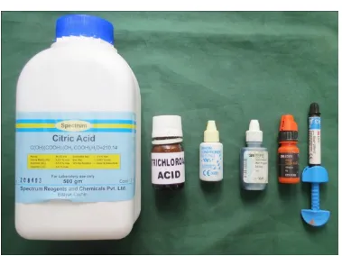
Materials used in the study
a) 35% Phosphoric acid (Scotchbond Multi-Purpose Etchant, 3M ESPE, St
Paul, MN, USA )
b) 10% Polyacrylic acid (GC Dentin conditioner, GC Corporation, Tokyo,
Japan)
c) 10% Citric acid (Spectrum Reagents and Chemicals Pvt Ltd , Kochi
India)
d) 35% Trichloroacetic acid (Spectrum Reagents and Chemicals Pvt Ltd ,
Kochi, India)
e) GC Fuji II LC Capsule (Radiopaque Light cured Reinforced Glass
ionomer restorative, GC Corporation, Tokyo, Japan)
f) Adper single bond 2 (3M ESPE, Dental Products, St Paul, MN, USA)
g) Resin composite (Filtek Z350 XT, 3M ESPE, Dental products, St.Paul,
MN, USA)
h) Distilled water (Nice chemicals Pvt Ltd, Kochi, India)
i) Stainless steel ring (Dentaurum Australia Pty Ltd, Mortlake, NSW)
j) 200, 400, 600 grit silicon carbide paper (Moyco Precision Abrasives,
Montgomeryville, PA, USA)
24
Equipments Used in the study
a) Composite light curing unit - DENTSPLY, Milford, Detroit, USA
b) Amalgamator (SYG 200) Hangzhou Sifang Medical Apparatus Co., Ltd,
Zhejiang, China
c) Universal Testing Machine- Model 3345; Instron corp, Canton, Mass,
USA
d) Gold Sputtering Machine-No E-1010 Ion sputter, Hitachi, Japan
e) Scanning electron microscope-No S-2400, Scanning electron microscope
Hitachi, Japan
METHODOLOGY
Specimen preparation
A total of 50 Glass ionomer discs were prepared using stainless steel molds,
6mm in diameter and 4mm in thickness. The stainless steel molds were prepared from
stainless steel orthodontic bands (Dentaurum Australia Pty Ltd). The molds were
filled with GC Fuji II LC (GC corporation, Tokyo, Japan), which was mixed
according to the manufacturer’s instructions in an amalgamator (Hangzhou Sifang
Medical Apparatus Co., Ltd , Zhejiang, China) for 2 seconds. The surface of the filled
25 unit (DENTSPLY, Milford, Detroit, USA) of intensity 500mw/sec. The discs were
polished with 200,400 and 600 grit carbide polishing papers (Moyco Precision
Abrasives, Montgomeryville, PA, USA) and were randomly divided into five groups
of 10 molds, out of which 7 molds were used for testing of bond strength and 3 molds
for Scanning electron microscope analysis.
The various conditioners used in the study were 35% Phosphoric acid, 10%
Polyacrylic acid, 10% Citric acid and 35% Trichloro acetic acid.
Preparation of conditioners
10 grams of citric acid powder (Spectrum reagents) was dissolved in 100 ml of
distilled water to make 10% citric acid solution.
35 grams of acetic acid powder (Spectrum reagents) was dissolved in 100 ml
of distilled water to make 35% trichloro acetic acid solution.
Conditioning Protocol and Composite placement
Group I was the control therefore no conditioner was used. In the seven
specimens of group I, Adper single bond 2 (3M ESPE, Dental Products, St.Paul, MN,
USA) was applied to the surface and light cured according to manufacturer’s
instructions. Stainless steel mold measuring 4 mm in internal diameter and 2 mm in
height was placed on the disc surface and Filtek Z350 XT light cured composite resin
26 placed inside the molds and light cured for 40 seconds using light curing unit
(DENTSPLY, Milford, Detroit ,USA)
Group II: In seven specimens, 35% Phosphoric acid (Scotchbond
Multi-Purpose Etchant, 3M ESPE, St Paul, MN, USA) was used as conditioner on glass
ionomer for 20 sec and rinsed with distilled water. Then Adper single bond 2 (3M
ESPE) was applied to the surface and light cured according to manufacturer’s
instructions. After which stainless steel molds measuring 4 mm in internal diameter
and 2 mm in height was placed on the disc surface and Filtek Z350 XT light cured
composite resin was carefully placed inside the molds and light cured for 40 seconds
using light curing unit (DENTSPLY, Milford, Detroit, USA).
Group III: In seven specimens, 10 % polyacrylic acid (GC Dentin conditioner,
GC Corporation, Tokyo, Japan) was used as conditioner for 20 sec and rinsed with
distilled water. Then Adper single bond 2(3M ESPE) was applied to the surface and
light cured according to manufacturer’s instructions. After which cylindrical stainless
steel molds measuring 4 mm in internal diameter and 2 mm in height was placed on
the disc surface and Filtek Z350 XT light cured composite resin was carefully placed
inside the molds and light cured for 40 seconds using light curing unit. (DENTSPLY,
Milford, Detroit, USA)
Group IV: In seven specimens, 10% citric acid (Spectrum Reagents) was used
27 2 (3M ESPE) was applied to the surface and light cured according to manufacturer’s
instructions. After which cylindrical stainless steel molds measuring 4 mm in internal
diameter and 2 mm in height was placed on the disc surface and Filtek Z350 XT light
cured composite resin was carefully placed inside the molds and light cured for 40
seconds using light curing unit (DENTSPLY, Milford, Detroit, USA).
Group V: In seven specimens, 35% trichloroacetic acid (Spectrum Reagents)
was used as conditioner for 20 seconds and rinsed with distilled water. Then Adper
single bond 2 (3M ESPE) was applied to the surface and light cured according to
manufacturer’s instructions. After which cylindrical stainless steel molds (4mm
internal diameter and 2mm height) was placed on the disc surface and Filtek Z350 XT
light cure composite was carefully placed inside the molds and light cured for 40
seconds using light curing unit (DENTSPLY, Milford, Detroit, USA).
Group I (control): Glass Ionomer without pretreatment + Composite
Group II: Glass Ionomer + 10% Polyacrylic acid + Composite
Group III: Glass Ionomer + 35% Phosphoric acid + Composite
Group IV: Glass Ionomer + 10% Citric acid + Composite
28
SHEAR BOND STRENGTH TESTING
The seven specimens of each of the five groups were stored in distilled water
for 24 hours at 37oC. In order to measure shear bond strength, the specimens were
placed in between the jigs of the universal testing machine and a pointed shearing rod
was placed on to the composite resin/glass ionomer interface and was subjected to
static loading at a rate of 1mm/min. The machine was interfaced with a computer
through which operation was controlled and shear bond strength was calculated.
Maximum load at failure was recorded in kilo Newton (KN). The shear bond strength
(SBS) in mega Pascals (MPa) was calculated by the formula SBS=F (N)/πr2, where F
is force in newton and r is the radius of the prepared composite resin block.
The values obtained were tabulated and statistically analysed using computer
software, Statistical Package for Social Sciences (SPSS) version 16.0. Data was
expressed in its mean and standard deviation. Analysis of variance (One way
ANOVA) was performed as parametric test to compare different variables. To
elucidate multiple comparisons between groups, Dunnett test with ANOVA 16.0 as
post hoc test. For all statistical evaluations, a two-tailed probability of value, <0.05
29
SEM analysis of etch pattern
Scanning electron microscope was used to visualize the effect on glass
ionomer surface of the various surface treatments used in the bonding study. The
surfaces of the remaining three glass ionomer disc of each group were conditioned
with four different conditioners.
Group II (a) - 20 sec application of 35% phosphoric acid rinsed with distilled water
and air dried
Group III (a) - 20 sec application of 10% polyarcylic acid, rinsed with distilled water
and air dried
Group IV (a) - 20 sec application of 10% citric acid, rinsed with distilled water and air
dried
Group V (a) - 20 sec application of 35% trichloroacetic acid, rinsed with distilled
water and air dried.
All specimens were gold sputtered and analyzed by Scanning Electron
30
Tables I-V Shows the shear bond strength values of all the samples in their respective
groups in megapascals (Mpa) calculated by the formulae SBS = F (N)/πr2.
Table- I Shows the shear bond strength values in MPa obtained for each sample which
was bonded to composite without any surface treatment.
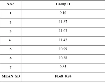
Table -II Shows the shear bond strength values in MPa obtained for each samples
which were bonded to composite following conditioning with 35% Phosphoric acid.
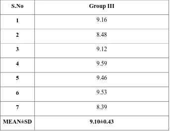
Table - III Shows the shear bond strength values in MPa obtained for each samples
which were bonded to composite following conditioning with 10 % Polyacrylic acid.
Table - IV Shows the shear bond strength values in MPa obtained for each samples
which were bonded to composite following conditioning with 10% citric acid.
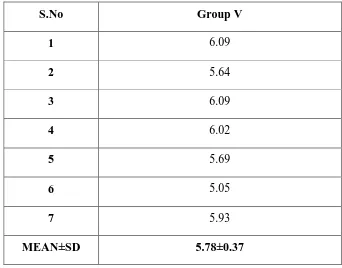
Table - V shows the shear bond strength values in MPa obtained for each samples
which were bonded to composite following conditioning with 35% Trichloroacetic
acid.
Table –VI shows the mean shear bond strength values of different groups.
Table VII- shows the multiple comparisons of mean shear bond strength (MPa) values
between the different groups.
As evident from statistical analysis high bond strength values were obtained
with Group II, the phosphoric acid etched group. Least bond strength values were
31 Group II specimens which were conditioned with 35% phosphoric acid showed
a mean bond strength value of 10.68MPa which is statistically significant than that of
group I, group IV and group V. Group II specimens showed slight increase in bond
strength value than that of group III (9.10 MPa) but were not statistically significant.
Group III specimens which were conditioned with 10% Polyacrylic acid showed
a mean bond strength value of 9.10 MPa which were statistically significant than that
of group I, group IV and group V. Group III specimens showed no significant
difference with group II.
Group IV specimens which were conditioned with 10% citric acid showed a mean
bond strength value of 7.19MPa which were statistically significant with group I,
group II, group III and group V.
Group V specimens which were conditioned with 35% trichloroacetic acid showed a
mean bond strength value of 5.78MPa which were statistically significant with group
II, group III and group IV.
SEM ANALYSIS
Specimens of groups II, III, IV and V were analysed by scanning electron microscopy
and following were the observations:
Figure 9 (Group II a) – Photomicrographs of 35% phosphoric acid treated specimens
32 Figure 10 (Group III a) - Photomicrograph of 10% polyacrylic acid treated specimens
with salt crumps formation greater than group II a i.e. 35% phosphoric acid treated
specimens.
Figure 11 (Group IV a)-Photomicrograph of 10% citric acid treated specimens with
more salt crumps than both Group II a and Group III a i.e. 35% phosphoric acid and
10% polyacrylic acid treated specimens.
Figure 12 (Group V a) - Photomicrograph of 35% Trichloroacetic acid treated
33
Table-1: Shear bond strength values of group I in MPa
S. No Group-I
1 4.62
2 4.02
3 3.81
4 3.60
5 4.83
6 4.98
7 4.11
[image:53.595.157.495.455.724.2]MEAN±SD 4.28±0.53
Table- 2: Shear bond strength values of group II in Mpa
S.No Group II
1 9.10
2 11.67
3 11.03
4 11.42
5 10.99
6 10.88
7 9.65
MEAN±SD 10.68±0.94
34
Table- 3: Shear bond strength values of group III in Mpa
S.No Group III
1 9.16
2 8.48
3 9.12
4 9.59
5 9.46
6 9.53
7 8.39
MEAN±SD 9.10±0.43
[image:54.595.157.499.116.382.2]
Table- 4: Shear bond strength values of group IV in Mpa
S.No Group IV
1 7.09
2 6.85
3 5.49
4 7.94
5 7.64
6 7.66
7 7.68
[image:54.595.156.497.456.724.2]35
Table- 5: Shear bond strength values of group V in Mpa
S.No Group V
1 6.09
2 5.64
3 6.09
4 6.02
5 5.69
6 5.05
7 5.93
[image:55.595.131.473.114.382.2]MEAN±SD 5.78±0.37
Table-6: Mean Shear bong strength (MPa) values of different groups
Groups Type of etching Shear Bond Strength
(MPa) (MEAN±SD)
Group-I Control group (without any
surface treatment)
4.28±0.53
Group-II 35% Phosphoric acid 10.68±0.94
Group-III 10 % Polyacrylic acid 9.10±0.43
Group-IV 10% Citric acid 7.19±0.84
36
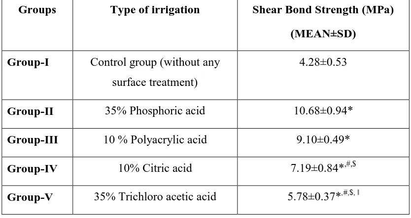
Table-7: Multiple comparisons of mean shear bond (MPa) values between the different groups
Groups Type of irrigation Shear Bond Strength (MPa)
(MEAN±SD)
Group-I Control group (without any
surface treatment)
4.28±0.53
Group-II 35% Phosphoric acid 10.68±0.94*
Group-III 10 % Polyacrylic acid 9.10±0.49*
Group-IV 10% Citric acid 7.19±0.84*,#,$
Group-V 35% Trichloro acetic acid 5.78±0.37*,#,$, ǁ
(*P<0.05 significant compared group-I with II, III, IV and V, #P<0.05 significant
compared group-II with I, IV and V, $P<0.05 significant compared group-III with I,
IV and V, ǁP<0.05 significant compared group-IV with I, II, III and IV, P<0.05 no
37 In this study it was observed that the application of all the four surface
conditioners to glass ionomer in sandwich restorations improved the bond strength.
This proves our hypothesis that surface conditioning enhances the bond strength of
Resin modified GIC to composite.
There is a continuous desire for novelties in dentistry originating from
changing professional perceptions, changing demands from the patient and progress
in industrial potentials. Today’s dentistry can be characterized by a shift from metallic
to non-metallic restorations. The patient attitude to treatment is mainly based on
concern for aesthetics and biocompatibility. In direct restorative dentistry this
correlates with a shift from traditional amalgam restorations to aesthetic composite
restorations.56
The resin composites were used to replace the missing tooth structure and
modify tooth colour and contour thus enhancing the aesthetic appearance of the
individual. Before the advent of composites, silicates were the first aesthetic direct
restorative material used. Although silicates provided an anti-cariogenic effect its use
has subsided due to its early clinical failure which was related to its dissolution in oral
fluids, loss of translucency, surface crazing and lack of adequate mechanical
38 lack of reinforcement potential. Another drawback of unfilled resins is its dimensional
instability leading to unsightly stains and recurrent caries.57
The traditional methacrylate based composites were first developed in mid
1960 as a replacement for silicate cements and unfilled resins. Since then these
materials had greatly improved in its properties and handling characteristics and now
it is considered as the primary restorative material. Over the years properties such as
lack of color stability and wear resistance of these materials are improved due to
changes made to the initiator, introduction of microfillers and hybridization of
manufacturing process.50
In order to overcome the drawbacks of traditional composites, such as surface
roughness and low translucency microfilled composites was introduced in which
colloidal silica particles are added as inorganic filler. The microfilled composites also
had their disadvantages mainly polymerization shrinkage, water sorption and thermal
expansion. 60
Hybrid composites were developed in an effort to obtain a smooth surface
provided by microfilled composites while maintaining the properties of small particle
composites. There are two kinds of filler particles colloidal silica and ground glass
39 over microfilled composite, but their surface smoothness and translucency are inferior
to microfilled composite resins. 60
To overcome the drawback of microfilled and hybrid composites, nano
composites were introduced which showed aesthetic properties similar to those of
microfilled composite while maintaining physical properties equivalent to those of
hybrid composites. This allows the clinician to use them for restoring both anterior
and posterior teeth. Nano composites have improved mechanical properties such as
compressive and tensile strength, higher fracture and wear resistance, reduced
polymerization shrinkage, high translucency, high polishability, retention and better
aesthetics.60 Hence in this study Filtek Z 350 XT nano composite was used.
Various improvements in mechanical properties of composite resins and
aesthetic need lead to an increased application of these materials by the clinicians.
Composite resins have undergone improvement in all areas, including aesthetics,
wear, and handling. However, high-polymerization shrinkage continues to be a major
disadvantage. Previous studies have shown polymerization shrinkage leading to bond
failure and micro-leakage of resin composite restorations. Micro-leakage is a matter
of concern because it leads to staining at the margins of restorations, recurrent caries,
hypersensitivity, and pulp pathology.12Another main drawback of composite is its
40 In order to overcome these drawbacks sandwich restoration was introduced.
The concept of this technique is to use two types of materials to form one restoration.
This technique made use of the chemical adhesion and fluoride release property of
glass ionomer and aesthetics and polishability of composite resins.40 The main
advantage of this technique is the adhesive property of glass ionomer which make it
an ideal restorative material for non carious cervical lesions. Another advantage of
sandwich technique is the fluoride releasing property of GICs, which has an inhibitory
effect on formation and progression of caries around the restoration.33
The bond strength between glass ionomer and composite is dependent on following
factors:
i) The tensile bond strength of glass ionomer cement
ii) Viscosity of bonding agent and ability to wet the surface of GIC
iii) Volumetric change in composite resin during polymerization
iv) Packing and adaptation of composite resin to glass ionomer without any
entrapment of voids.
v) Surface treatments used13
The drawback of conventional glass ionomer is its sensitivity to moisture and
41 ionomer does not effectively seal the dentin which is mainly attributed to dehydration
after setting leading to crazing and cracking.55
The main advantage of resin modified glass ionomer over conventional glass
ionomer cement is that it sets by an acid base reaction and exhibits command set
when activated by light via methacrylate group.35 Ismail et al in a study has shown
better bond strength of resin modified glass ionomer to composite resin than
conventional glass ionomer. Resin modified glass ionomer and resin composite are
polymerized by a free radical initiator system which is necessary for chemical
bonding between the two materials.39 The presence of hydroxyethyl methacrylate on
the glass ionomer surface enhances the surface wetting of bonding agent resulting in
increased bond strength.28
Several criterias are thought to be involved in the chemical adhesive bond
between resin modified glass ionomers and composite resins. Increased availability of
unsaturated double bonds in air inhibited layer of resin modified glass ionomer
cement may assist in chemical bonding to the resin bonding agent and resin
composite.28
Resin modified glass ionomer capsules were used in this study and was mixed
according to the manufacturer’s instructions in a standard amalgamator. Studies have
42 but the porosities in triturated cements are smaller and more uniform than hand mixed
specimens.28 Hence in this study GC Fuji II LC Capsule (Radiopaque Light cured
Reinforced Glass ionomer restorative, GC Corporation, Tokyo, Japan) was used.
The surfaces of the prepared glass ionomer samples were polished with 200,
400, 600 grit silicon carbide papers in order to create a flat surface for treatment and
bonding.41
Arora et al has confirmed in an in vitro study that conditioning of GIC
was necessary to improve its bonding with the composite resin. The matrix of the GIC
gets dissolved in the acid leading to rough and porous surface, so that the bonding
agent could easily penetrate into these irregularities and provide resin tags for bonding
with the composite.35
Thus in the study four conditioners were used namely 35% phosphoric acid,
10% polyacrylic acid, 10% citric acid and 35% trichloroacetic acid was used to
condition the surface of the resin modified glass ionomer cement to enhance it’s
bonding to resin composite.
Phosphoric acid (also known as orthophosphoric acid ) is a mineral (inorganic)
acid having the chemical formula H3PO4. Orthophosphoric acid molecules can
combine with themselves to form a variety of compounds which are also referred to as
43 Polyacrylic acid (PAA or Carbomer) is generic name for synthetic high
molecular weight polymers of acrylic acid. They may be homopolymers of acrylic
acid, crosslinked with an allyl ether pentaerythritol, allyl ether of sucrose or allyl ether
of propylene. In a water solution at neutral pH, PAA is an anionic polymer, i.e. many
of the side chains of PAA will lose their protons and acquire a negative charge. This
makes PAAs polyelectrolytes, with the ability to absorb and retain water and swell to
44 Citric acid is a commodity chemical, and more than a million tonnes are
produced every year by fermentation. It is used mainly as an acidifier, as a flavoring,
and as a chelating agent. Citric acid has the formula C6H8O7. It is a natural
preservative/conservative which occurs naturally in citrus fruits and is also used to
add an acidic or sour taste to foods and drinks. It consists of 3 carboxyl (R-COOH)
groups.
Trichloroacetic acid (TCA; also known as trichloroethanoic acid) is an
analogue of acetic acid in which the three hydrogen atoms of the methyl group have
all been replaced by chlorine atoms. It is prepared by the reaction of chlorine with