A STUDY ON THE MANAGEMENT OF BORDERLINE
SUB DURAL HEMATOMA.
Dissertation submitted in partial fulfillment of the requirements of
M.Ch BRANCH II NEUROSURGERY (3 YEARS) EXAMINATIONS AUGUST 2014
MADRAS INSTITUTE OF NEUROLOGY
MADRAS MEDICAL COLLEGE
&
RAJIV GANDHI GOVERNMENT GENERAL HOSPITAL
CHENNAI-600003.
THE TAMILNADU Dr.M.G.R MEDICAL UNIVERSITY
CERTIFICATE
This is to certify that this dissertation titled “A Study on the management of borderline Subdural Hematoma ” submitted by Dr.Ramkumar R.R., appearing for M.Ch (Neurosurgery) degree examination in August 2014, is an original bonafide record of work done by him from January 2012 to January 2014 under my guidance and supervision in partial fulfillment of requirement of the Tamil Nadu Dr.M.G.R. Medical University, Chennai. I forward this to the Tamil Nadu Dr.M.G.R. Medical University, Chennai, Tamil Nadu, India.
Prof.Dr.K.MAHESHWAR,MCh
Professor of Neurosurgery and GUIDE Head of department Institute of Neurology, Madras Medical College & Rajiv Gandhi Government General Hospital,
Chennai – 600 003.
THE DEAN
Madras Medical College & Rajiv Gandhi Government General Hospital,
DECLARATION
I, Dr.Ramkumar R.R. , solemnly declare that this dissertation “A Study on the
management of borderline Subdural Hematoma” was done by me at the
Madras Institute of Neurology, Madras Medical College and Rajiv Gandhi Government General Hospital, Chennai under the guidance and supervision of the Professor of Neurosurgery, Institute of Neurology, Madras Medical College and Rajiv Gandhi Government General Hospital, Chennai-3, from January 2012 and January 2014.
This dissertation is submitted to the Tamil Nadu Dr.M.G.R. Medical University, Chennai-600032 in partial fulfillment of the University requirements for the award of the degree of M.Ch. Neurosurgery.
Place: Chennai RAMKUMAR R.R.
ACKNOWLEDGEMENT
I thank the Dean Dr.Vimala.M.D and previous Deans of, Madras Medical College, Chennai from January 2012, for permitting me to utilize the facilities of the hospital for conducting this study. I extend my thanks to the members of Ethical Committee for their role in approval and guidance of the study.
I am extremely grateful to Prof.K.Maheshwar, M.Ch. Professor of Neurosurgery and Head of the Department, Madras Institute of Neurology, Madras Medical College and Rajiv Gandhi Government General Hospital, Chennai, who has also been my guide for this study, for his constant encouragement, expert advice, and guidance throughout the study. I thank my teachers, Prof. Ranganathan Jothi, Prof. G.S.Jegannarayana, Prof. J.V.Mahendran, Prof. S.Syamala, and Prof. D.Balasubramanian for sparing
their valuable time in guiding me during this study. I am indebted to all my Assistant Professors for their support, guidance and help to carry out this study. I thank the Anesthesia team including chiefs, assistant professors, and post graduates. I thank all my fellow Postgraduates for their support. I thank all the nursing staff who took excellent care of the patients. I thank all my patients and their relatives for their immense cooperation and consent to this study.
TABLE OF CONTENTS
1. Introduction 1
2. Aims and objectives 4
3. Review of literature 5
4. Materials and methods 9
5. Observations and results 14
6. Discussion 39
7. Analysis and statistics 42
8. Conclusion 56
9. Bibliography 57
ABBREVIATIONS USED
SAH-SUB ARACHNOID HEMORRHAGE
EDH-EPIDURAL HEMATOMA
SDH- SUB DURAL HEMATOMA
MLS- MIDLINE SHIFT
IVH-INTRA VENTRICULAR HEMORRHAGE
GCS –GLASGOW COMA SCALE
GOS- GLASGOW OUTCOME SCALE
CT- COMPUTERISED TOMOGRAPHY
B.P-BLOOD PRESSURE
PRE-OP- PRE OPERATIVE
POST –OP –POST OPERATIVE
BTF- BRAIN TRAUMA FOUNDATION
TBI- TRAUMATIC BRAIN INJURY
INTRODUCTION
Traumatic Brain Injury remains the major cause of death and severe disability among people, particularly the young. For Acute Subdural Hematomas and intra parenchymal lesions, the outcome is worse, with death or severe disability occurring in up to 60% of patients.
Some experts maintain that aggressive surgical intervention, though able to preserve life, will result in a very poor quality of life for the survivors.
The initial event in cranio cerebral trauma involves direct impact injury to the brain producing parenchymal contusion, EDH, SDH and shearing injury of axons in the white matter of cerebral hemisphere and brainstem producing DAI.
Under these loading conditions, the strain within the brain is concentrated along the outer margins where the Para sagittal bridging veins reside. If the vascular tolerance is suppressed, subdural bleeding occurs. SDH may also occur due to bleeding from a cortical vein or from a laceration in the brain surface. Due to its similar etio pathological mechanism, SDH may coexist with underlying DAI. This explains the frequency of cases in which the SDH is small, but the brain damage underlying it is more than what is expected because of the brain compression from the hematoma.
pathophysiological processes can produce changes in the intracranial pressure – volume relationship with resulting intracranial hypertension and brain herniation.
Borderline SDH is a Sub Dural Hematoma with 5-9 mm thickness and/or
Midline Shift 2-5 mm.
Current studies do not have adequate information regarding Borderline SDH and its subsequent management.
It is a known fact that most intra parenchymal mass lesions /SDH will enlarge with time requiring serial CT scanning, neurological monitoring and ICP monitoring.
Studies reveal 10% mortality in SDH less than 10mm thick7 (90 % in thickness greater than 30mm). In some studies, Midline Shift has been found to be a better predictor of failure of non-operative treatment.8
Despite available guidelines for surgical or conservative management of acute SDHs, management of the patients has to be individualized. Associated injuries have been reported to occur in 47%-57% of patients, accounting for a higher morbidity and mortality.
AIMS AND OBJECTIVES OF THE STUDY
1. To study the various parameters affecting the outcome in management of borderline Acute Sub Dural Hematoma.
3. To analyse if any significant correlation exists between Midline Shift (MLS), SDH thickness and the outcome and to compare with CT
findings, Glasgow Coma Scale (GCS) and outcome.
REVIEW OF LITERATURE
Authors have concluded that the operated and non-operated patients represent two different patient populations and that studies evaluating the conservative management of traumatic acute SDHs should only include patients conservatively treated.2,5
Review of literature indicates that:-
67% of the patients with acute SDH survive with conservative management. In a study by Feliciano, patients less than 65 years old had a favorable or functionally independent outcome in 85% of the cases. Patients with Glasgow Coma Scale scores greater than 8, had a functionally independent outcome in 78% of the cases. Patients with acute subdural hematomas with thicknesses ≤10 mm and midline shifts ≤ 5 mm showed functionally independent outcomes in
82% of the cases.3
Croce, et al. reported a functionally independent outcome in 93% of the patients conservatively managed presenting acute SDHs that measure 10 mm or less at the thickest diameter and a GCS>11.5
Zumkeller, et al. showed that the amount of midline shift is very important in survival rate with the survival function decreasing as the midline shift increases.11
Servadei, et al. recommended conservative treatment in comatose patients (GCS≤8) with SDHs less than 10 mm thick and midline shifts less than 5 mm
who have an improved or stable GCS score since the injury, show no pupillary abnormalities, and an intracranial pressure less than 20 mm Hg or between 20 and 30 mm Hg if the cerebral perfusion pressure is greater than 75 mm Hg.10
Haematoma thickness, midline shift, status of the basal cisterns and presence of SAH are related to outcome when identified on the initial (early) CT examination. However, early (within 3 h from injury) CT under-estimates the ultimate size of parenchymal contusions. Patients with SAH on early CT are those at highest risk for associated evolving contusions. The use of sequential
CT should be included in the routine management of head-injured patients.12
cerebral compensatory mechanism within 3 days of injury Mathew, et al. 4 recommended conservative management with a midline shift <10 mm in conscious patients, but Wong recommended it only on those patients with a GCS score of 15. Wong found that a midline shift >5 mm in patients with a GCS score <15 was significantly related to conservative management failure due to exhaustion of the cerebral compensatory mechanisms within three days of injury. Therefore, he recommended that the minimal hospital stay for patients with small SDHs under conservative management should be three days. He also found that the thickness of the hematoma was non-predictive of the outcome.1,2,7,8
It is found that patients with a TICH volume of less than 15 ml, a midline shift of less than 5 mm, an open peri mesencephalic cistern on CT scans, a Glasgow Coma Scale (GCS) score of 12 or more, and an absence of lateralizing signs may be treated conservatively and expected to make a good recovery.1,2,7,8
Lobato et al recommend ICP monitoring after admission in all patients and serial CT scanning at 2-4, 12, 24, 48 and 72 hours after injury with additional controls as indicated by clinical or ICP changes in all cases. Over 50% of the patients with initial lesions developed new CT changes and nearly 50% showed intracranial hypertension during the acute posttraumatic period.9
Small subsets of patients fail with non-operative management, and subsequently require surgical decompression for progression of a pre-existing lesion or delayed presentation of new lesions. Failure of non-operative management has been associated with the timing of initial post injury CT scans, hematoma location and volume, the presence of edema around the hematoma, and physiologic variables such as hypotension, hypoxia, and coagulopathy of the variables investigated, only anatomic location of injury was found to be predictive of early failure of non-operative management. Frontal intra parenchymal hematomas are particularly prone to early failure. Clinical examination and intracranial pressure monitoring are equally important in detecting failure and should be an integral part of non-operative
MATERIALS AND METHODS
The study was done at Department of Neurosurgery at the Madras Institute of Neurology, Madras Medical College and Rajiv Gandhi Government General Hospital, Chennai from January 2012 to January 2014. Patients who were admitted with head injury and acute SDH were included based on the following inclusion and exclusion criteria.
Inclusion Criteria
1. Age-adults (13 years and above)
2. SDH thickness 5-9 mm
3. Midline Shift (MLS) 2-5 mm
4. GCS score greater than 8
5. Time since injury within 6 hours of trauma
6. Volume of contusion less than 20 ml
Exclusion Criteria
1. Age less than 13 years
2. Midline shift greater than 5 mmin CT scan at admission.
3. Subdural Hematoma thickness more than 9 mm in CT scan at admission. 4. GCS score less than 9.
5. Loss of consciousness more than one hour duration. 6. Time between injury and admission more than 6 hours 7. Chronic alcoholics or abnormal LFT
8. Presence of Bradycardia. 9. Pupillary Asymmetry
10.Presence of paucity of movements on one side or hemiplegia 11.Associated Contusion size more than 20 ml.
12.SAH with Fisher grade 4
13.Severe life threatening musculo skeletal/spine/thoraco abdominal injuries 14.Evidence of severe brain stem dysfunction.
15.Systemic illness, metabolic disorder, endocrine abnormality, coagulopathy
Methodology
Head injury patients who were admitted at our hospital with borderline SDH as per the above inclusion and exclusion criteria were included in the study. Patient and attenders were explained in detail regarding the study. Written informed consent was obtained for inclusion in the study. Data regarding the various parameters such as age, sex, mode and time of injury, clinical examination pertaining to GCS, pupils, neurological deficit, pulse, Blood Pressure, respiration were noted. CT was done at regular intervals as per Proforma and earlier if needed. Findings were recorded.
Renal function tests, blood sugar, serum electrolytes, complete blood count, Coagulation profile and blood grouping was done.
Patients were monitored daily till discharge. After discharge patients were followed up at weekly intervals for one month. Patients were contacted regularly throughout the study period over mobile phones.
Patients were evaluated and treatment was done according to brain trauma foundation guidelines and as per the attached Proforma.
Brain Trauma Foundation Guidelines
2. A comatose patient (GCS less than 9) with an SDH less than 10mm thick and a midline shift less than 5 mm should undergo surgical evacuation of the lesion if the GCS score decreased between the time of injury and hospital admission by 2 or more points on the GCS and / or the patient presents with asymmetric or fixed and dilated pupils and / or the ICP exceeds 20 mm Hg.
Most patients were treated conservatively with antibiotics, antipyretics, anticonvulsants, anti edema measures and supportive treatment as required.
Serial CT scans of brain were taken at regular intervals as per the hospital protocol. Neurological status of the patient was monitored intensively. Treatment was altered according to changes in neurological status or CT scan.
In patients, improving with this treatment, conservative management was continued.
Patients who had deteriorating GCS and patients who had increase in SDH thickness or MLS radiologically were taken up for emergency decompressive craniectomy.
SURGERY:
Temporal decompression was done up to temporal base. Dura opened sinus based. SDH evacuated. Contusions or burst lobe if present are excised. After achieving haemostasis, dura was left open. Galea was closed with interrupted 1 vicryl sutures. Skin was closed with continuous 1-0 Nylon.
Patients with low GCS were managed with mechanical ventilator support.
Wound care was given appropriately. Suture removal was done on 7-9th POD.
Physiotherapy and respiratory exercises were given regularly. Outcome was assessed by Glasgow Outcome Scale on the 30th day.
OBSERVATIONS AND RESULTS
Age distribution:
In our study, the agewise distribution was from 13 to 74 years. 90% of patients were between 21 to 60 years with peak incidence in the 31 to 40 years group.
13 - 20
Yrs 21 - 30 31 - 40 41 - 50 51 - 60 61 - 70
Above 70
TOTAL NO. OF PATIENTS 4 21 30 20 19 5 1
0 5 10 15 20 25 30 35
SEX DISTRIBUTION: 93% of the patients with borderline SDH were males
and the remaining 7% were females.
MODE OF INJURY:
Majority of the patients in our study were admitted due to RTA (82%). Falls and assault accounted for 7% and 11% respectively.
93% 7%
SEX DISTRIBUTION
MALE FEMALE
0 10 20 30 40 50 60 70 80 90
RTA FALL ASSAULT
82
7 11
MODE OF INJURY
CLINICAL SYMPTOMS:
Among the 100 patients studied in this group, the most common clinical symptom observed was loss of consciousness (92%). The next common symptom was vomiting which occurred in 64% of patients. Ear and nasal bleed occurred in 28% of patients. The least common symptom was seizures (8%).
SEIZURE
VOMITING
ENT BLEED
LOC 8
64
28
92
GCS ON ADMISSION:
In the 100 patients studied, Mild head injury group with GCS 13 to 15 were the majority with 57 cases. Moderate head injury patients constituted second with 43 patients in GCS 9 to 12.
DISTRIBUTION OF GCS AND ASSOCIATED PATHOLOGY: Patients
with only SDH were predominantly in the mild head injury group with 80% 0
10 20 30 40 50 60
13 - 15 9 to 12
Series1 57 43
57
43
having GCS score 13-15 and the remaining 20% in the moderate head injury group.
In the patients having both SDH and contusion 48% had mild head injury, 52% had moderate head injury.
80% of patients with SDH and SAH had moderate head injury while 20% had mild head injury.
GCS COURSE DURING TREATMENT:
Of the 100 cases studied, majority (61%) had no gross change (more than 2) in
the GCS score during the course of treatment. There was improvement more 0 5 10 15 20 25 30 35
SDH only SDH with
Contusion
SDH with SAH
13 TO 15 33 21 3
9 to 12 8 23 12
33 21 3 8 23 12
than 2 in GCS score in 25 patients, while 14 patients had deterioration more than 2 in GCS score.
PUPIL REACTION: In our study, 93 patients had normal pupillary response on admission. Sluggish response was seen in 7 patients.
0 10 20 30 40 50 60 70
worsening static improved
14
61
25
GCS COURSE during treatment
93% 7%
PUPIL REACTION
PUPIL SIZE:
No Patient with borderline SDH had anisocoria at admission. Of the 100 patients only 3 had anisocoria. One patient developed anisocoria after 24 hours and two patients developed anisocoria after 48 hours.
In this study, pupillary asymmetry was not significant in influencing the outcome.
97% 3%
PUPIL SIZE
CT FINDINGS:
SDH: Among the 100 cases in this study, 83 had SDH with thickness 5 to 9mm.
There was SDH thickness less than 5mm in 17 cases.
MIDLINE SHIFT: In 55 patients, there was midline shift of 2-5 mm whereas in 45 patients, the midline shift was less than 2mm.
83% 17%
SDH THICKNESS
SDH thickness 5-9mm
SDH thickness less than 5mm
55% 45%
MIDLINE SHIFT
OUTCOME AT 30 DAYS:
Of the 100 patients, majority (78%) were alive at 30 days while 22% expired.
INDICATION FOR SURGERY:
The most common indication for surgical intervention was fall in GCS score after initial conservative therapy in this study among 12 cases.
Increase in MLS was the second most common indication and occurred in 7 cases.
The least common indication was increase in SDH thickness which occurred in only 4 patients.
78% 22%
30 DAY OUTCOME
CT FINDINGS:
In the study group, 83 patients had a SDH thickness 5-9mm. SDH thickness less than 5mm was found in 17 patients who had midline shift of 2-5mm.
The most common associated injury has been found to be an ipsilateral intra parenchymal contusion in 32 patients and contralateral contusions was found in 12 patients.
There were associated SAH in 15 patients 0
2 4 6 8 10 12
Increase in SDH Thickness
Increase in MLS
Fall in GCS
Series1 4 7 12
4
7
12
FINAL OUTCOME:
In this study, 100 patients were treated conservatively.
There was failure of non-operative treatment in 23 patients among the 100, requiring surgical evacuation.
Of these 23 patients, 9 patients were alive at 30 days and 14 patients had died. In the conservative group of 77 patients at 30 days, 69 were alive and 8 were dead. In the event of failure of conservative treatment, in those patients requiring surgery 39% survived and 61% expired.
0 10 20 30 40 50 60 70 80 90 SDH thickness 5 TO 9mm MID LINE SHIFT 2 to 5
mm SAH CONTUSIO N IPSILATERA L CONTUSIO N CONTRALA TERAL
Series1 83 45 15 32 12
CT AND OUTCOME:
In this study, among 41 patients with SDH only, death occurred in only 2 patients with 39 surviving patients at a survival rate of 95% and mortality of 5%
Of the 32 patients with SDH and ipsilateral contusions, 23 patients were alive and 9 patients expired with a survival rate of 72% and mortality of 28%
In 12 patients with SDH and contralateral contusion, there were 6 survivors and 6 deaths with a survival rate of 50% and mortality 50%
In 15 patients with SDH and SAH, 10 patients survived and 5 expired with a survival rate of 67% and mortality 33%.
0 10 20 30 40 50 60 70
CONSERVATIVE CONSERVATIVE
followed by SURGERY
Alive 69 9
Dead 8 14
69
9
8 14
Of all the cases, patients with SDH alone had a good prognosis and those with SDH and contralateral contusion had a relatively poor prognosis.
CONSERVATIVE TREATMENT FAILURE AND DEATHS:
Our study showed that in patients with only SDH failure of conservative treatment occurred in 5 out of 41 patients. In patients with SDH and contusion, 14 patients in a group of 44 had conservative treatment failure. There was treatment failure in 4 out of 15 patients in the SDH with SAH group.
So failure of non-operative treatment occurred in 12% of SDH only group.
In the SDH with contusion group, treatment failure occurred in 32% cases.
Treatment failure occurred in 27% cases in SDH with SAH group. 0
5 10 15 20 25 30 35 40
SDH alone SDH with
ipsilateral contusion
SDH with contralateral
contusion
SDH with SAH
Alive 39 23 6 10
Dead 2 9 6 5
No deaths were observed among patients in the SDH only group who were operated.
In the SDH with contusion group, of the 14 patients operated, there were 11 postoperative deaths with a survival rate of 21% and mortality 79%.
In the SDH with SAH group among the 4 patients operated 3 expired with a survival rate of 25% and mortality 75% . However these statistics were not significant on applying the chi square test.
In the conservatively managed groups, the SDH only, SDH with contusion and SDH with SAH had 2, 4 and 2 deaths respectively. The mortality rates were 6% in SDH only group, 13 % in SDH with contusion group and 18 % in SDH with SAH group. Thus mortality rates were 2 to 3 times higher when SDH had an associated contusion or SAH.
0 2 4 6 8 10 12 14
Operated Postop
deaths
Conservative Deaths
SDH only 5 0 2
SDH with Contusion 14 11 4
SDH with SAH 4 3 2
MANAGEMENT AND GOS RELATIONSHIP:
In the 77 patients treated conservatively, majority of patients scored GOS
4(32%) and GOS 5(39%) at 30 days.
In the 23 patients who had failure of conservative management, 61% were in GOS 1 score.
In this study, better GOS scores were observed in patients who were treated by conservative therapy successfully. But in case of non-operative treatment failure, GOS scores decreased drastically.
0 5 10 15 20 25 30
GOS 1 GOS 2 GOS 3 GOS 4 GOS 5
CONSERVATIVE 8 2 12 25 30
CONSERVATIVE followed by
SURGERY 14 2 4 2 1
8 2 12 25 30 14 2 4
2 1
Management and GOS
GOS VS SDH TYPE:
In SDH only group, there were 2 GOS I (5%), 1 GOS II (3%), 3 GOS III (7%), 9 GOS IV (22%) and 26 GOS V (63%) scores.
In the SDH with contusion group there were 15 GOS I (34%), 2 GOS II (5%), 9 GOS III (21%), 14 GOS IV (32%) and 4 GOS V (9%) scores.
In the SDH and SAH group, there were 5 GOS I (33%), 1 GOS II (7%), 4 GOS III (27%), 4 GOS IV (27%) and 1 GOS V (7%) scores.
0 5 10 15 20 25 30
GOS 1 GOS 2 GOS 3 GOS 4 GOS 5
SDH only 2 1 3 9 26
SDH with Contusion 15 2 9 14 4
SDH with SAH 5 1 4 4 1
SDH THICKNESS AND OUTCOME:
Comparing the SDH thickness and outcome, it was observed that out of 17 cases with SDH thickness less than 5 mm, 16 were alive and 1 case expired with a survival rate of 94%
In 83 patients with SDH thickness greater than 5mm, survival rate was 75% with 62 survivors and 21 deaths.
0 10 20 30 40 50 60 70
alive dead
<5mm 16 1
5to 9 mm 62 21
16
1 62
21
MIDLINE SHIFT AND OUTCOME:
In the group with MLS 2-5 mm and SDH thickness less than 5 mm, there was only one death among 17 patients with 16 survivors. Survival rate was 94% and mortality rate 6%.
In the group with MLS 2-5mm and SDH thickness 5-9 mm, there were 19 deaths with 9 survivors. The survival rate was 47% and mortality 53%.
0 2 4 6 8 10 12 14 16 18 20
mls with <5mm sdh mls with > 5 mm sdh
ALIVE 16 9
DEAD 1 19
16
9
1
19
SDH THICKNESS AND MLS:
Of the 55 cases of SDH 5-9mm thick with MLS less than 2mm, at 30 days 53 patients were alive with 2 deaths with a survival rate of 96%. The mortality rate was 4% only. In 28 cases of SDH 5-9mm thick with MLS 2-5 mm there were 9 survivors and 19 deaths. The survival rate was 32% and the mortality rate was 68%
In 17cases of SDH with less than 5mm thickness, MLS 2-5mm was present. In this group, at 30 days there were 16 survivors and one death with a survival rate of 94% and a mortality of 6%.
0 10 20 30 40 50 60
Alive Dead
MLS <2 mm 53 2
MLS 2-5mm 9 19
53
2 9
19
SDH 5 to 9 mm
94% 6%
SDH 2-5 mm
MIDLINE SHIFT AND SDH ONLY:
In the 41 patients who had only SDH, 33 patients with MLS less than 2mm were alive at 30 days with no deaths in this group.
In the remaining 8 patients who had MLS 2-5 mm, there were 6 survivors and 2 deaths at 30 days. The survival rate in this group was 75% with a mortality of 25%.
0 5 10 15 20 25 30 35
Alive Dead
MLS < 2mm 33 0
MLS 2-5 mm 6 2
33
0 6
2
MIDLINE SHIFT AND SDH WITH IPSILATERAL CONTUSION:
All patients in this group had MLS 2-5 mm. There were 23 survivors and 9 deaths with a survival of 72% and a mortality rate of 28%
MIDLINE SHIFT AND SDH WITH CONTRALATERAL CONTUSION
:
SDH with contralateral contusion consisted of 12 patients. Four patients had midline shift 2-5mm towards the side of the contusion, who did not survive. Of
0 5 10 15 20 25
Alive Dead
MLS 2-5 mm 23 9
23
9
the remaining 8 patients with no MLS, 6 were alive and 2 were dead at 30 days. Survival was 50%. The mortality in this group was 50%.
MIDLINE SHIFT AND SDH WITH SAH:
In this group, of the 11 cases with MLS less than 2mm, there were 9 survivors and 2 deaths. In the 4 cases with MLS 2-5 mm, there was one survivor and 3 deaths. The overall survival rate in this group was 65% with a mortality of 35%.
0 1 2 3 4 5 6 Alive Dead
MLS < 2mm 6 2
MLS 2-5 mm 0 4
6
2
0
4
SDH WITH CONTRALATERAL
CONTUSION AND MLS
0 2 4 6 8 10 Alive Dead
MLS < 2mm 9 2
MLS 2-5 mm 1 3
9
2 1
3
SDH with midline shift SDH with contusion
SDH with SAH SDH with Contusion
SDH with Contusion
SDH evacuated and Decompression done
DISCUSSION
Majority of the patients admitted with borderline SDH had mild and moderate head injury. The peak incidence was found in the 31 to 40 age group and predominantly in males (97%). This is accordance to the observations in literature regarding TBI.
RTA (82%) was the main cause of injury with LOC the main presenting symptom (92%) followed by vomiting (64%).
Admission GCS score was from 13 to 15 in SDH only patients (80%). In the presence of associated injury there was fall in the GCS score accordingly. 52% of SDH with contusion had GCS 9-12 and 80% of SDH with SAH had GCS score of 9-12.
During the course of treatment, there was improvement of more than 2 in GCS score in 25% of cases while no major change was observed in 61%. In 14% there was deterioration of more than 2 in GCS score.
Pupillary reaction, size or symmetry did not have a major impression in this study probably due to better GCS at admission.
The common intra parenchymal injury associated with borderline SDH was an ipsilateral contusion (32%). SAH (15%) and contralateral contusion (12%) were also present.
Of the 100 patients treated conservatively, failure of conservative treatment was observed in 23%. There was failure of non-operative treatment in 12% of SDH only group.
In the SDH with contusion group, treatment failure was seen in 32% cases.
In 27% cases in SDH with SAH group there was treatment failure.
The primary indication for surgery after initial conservative management was fall in GCS: 52%. Increase in midline shift caused failure of non-operative treatment in 31% and Increase in SDH size accounted for 17%
ANALYSIS OF MORTALITY IN RELATION TO TREATMENT:
Among the patients treated conservatively the mortality was 10%.
The mortality in the patients, who were taken up for surgery after deterioration, was 61%. This poor prognosis reflects the secondary brain damage which is inflicted after the primary trauma and in addition the operative insult also.
Mortality rate associated with fall in GCS (followed by surgery) – 67%
Mortality rate associated with increase in MLS (followed by surgery)- 57%
ANALYSIS OF MORTALITY IN RELATION TO CT FINDINGS:
Overall SDH only patients had a good prognosis with a mortality of 5%.
All Patients with SDH with ipsilateral contusion had a mortality of 28%. SDH with contralateral contusion cases had a mortality of 50%
SDH with SAH patients had a mortality of 33%
Among the cases which were operated after initial conservative treatment, there were no deaths in the SDH only group. In the SDH with contusion group there were 11 deaths with a post-op mortality of 79%.
In the SDH with SAH group, there were 3deaths with a post-op mortality of 75%. But this was found to be not significant when the chi square test was applied.
The GOS were better in the SDH only group. In the SDH with contusion and SDH with SAH groups the GOS were comparatively lower.
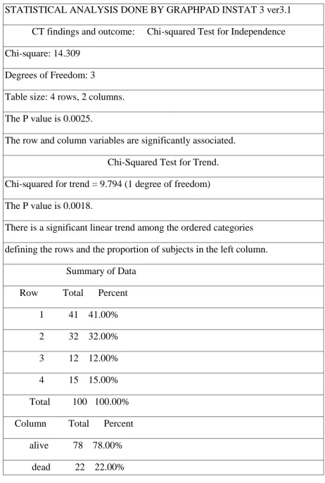
STATISTICAL ANALYSIS DONE BY GRAPHPAD INSTAT 3 ver3.1
CT findings and outcome: Chi-squared Test for Independence Chi-square: 14.309
[image:48.595.65.528.67.755.2]Degrees of Freedom: 3
Table size: 4 rows, 2 columns. The P value is 0.0025.
The row and column variables are significantly associated. Chi-Squared Test for Trend. Chi-squared for trend = 9.794 (1 degree of freedom) The P value is 0.0018.
There is a significant linear trend among the ordered categories defining the rows and the proportion of subjects in the left column. Summary of Data
Total 100 100.00% TOTAL OUTCOME
Fisher's Exact Test
The two-sided P value is < 0.0001, considered extremely significant. The row/column association is statistically significant.
Relative risk = 2.433
95% Confidence Interval: 1.391 to 4.253 (using the approximation of Katz.)
Difference between the two proportions Top row (Alive):
Fraction in the left column: 0.8846
95% Confidence Interval of that fraction: 0.7923 to 0.9459 Bottom row (Dead):
Fraction in the left column: 0.3636
95% Confidence Interval of that fraction: 0.1721 to 0.5933 Difference:
Difference between the fractions: 0.5210 Standard error of the difference: 0.1016
95% confidence interval of difference: 0.3218 to 0.7201 Data analyzed
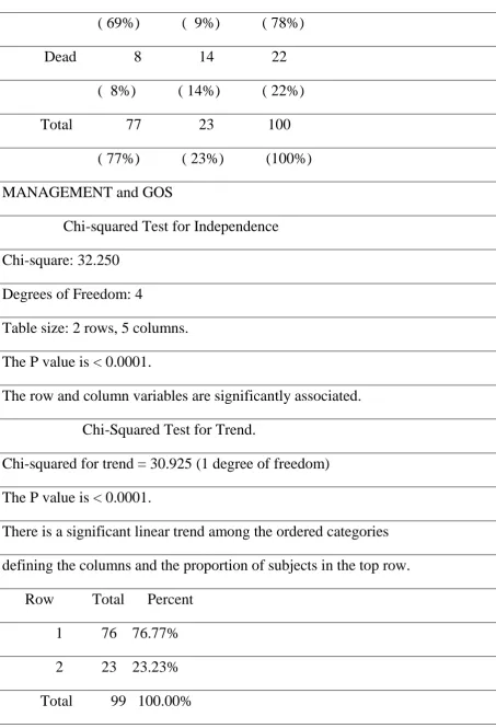
( 69%) ( 9%) ( 78%) Dead 8 14 22 ( 8%) ( 14%) ( 22%) Total 77 23 100 ( 77%) ( 23%) (100%) MANAGEMENT and GOS
Chi-squared Test for Independence Chi-square: 32.250
[image:50.595.70.524.61.727.2]Degrees of Freedom: 4
Table size: 2 rows, 5 columns. The P value is < 0.0001.
The row and column variables are significantly associated. Chi-Squared Test for Trend. Chi-squared for trend = 30.925 (1 degree of freedom) The P value is < 0.0001.
There is a significant linear trend among the ordered categories defining the columns and the proportion of subjects in the top row. Row Total Percent
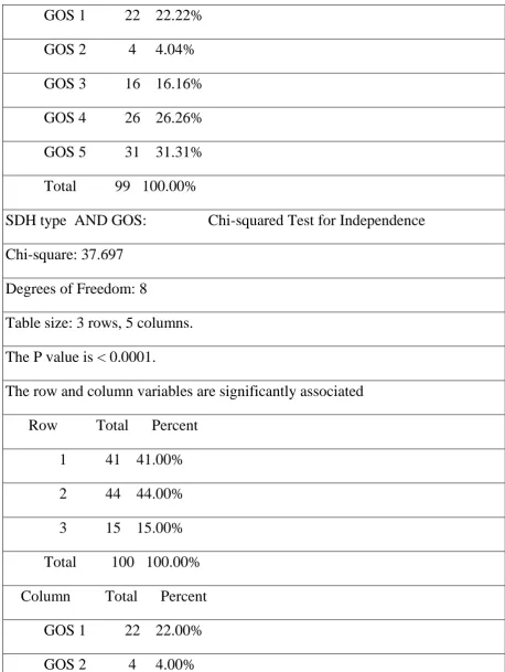
GOS 1 22 22.22% GOS 2 4 4.04% GOS 3 16 16.16% GOS 4 26 26.26% GOS 5 31 31.31% Total 99 100.00%
SDH type AND GOS: Chi-squared Test for Independence Chi-square: 37.697
[image:51.595.67.526.68.678.2]Degrees of Freedom: 8
Table size: 3 rows, 5 columns. The P value is < 0.0001.
The row and column variables are significantly associated Row Total Percent
GOS 5 31 31.00% Total 100 100.00%
SDH THICKNESS AND OUTCOME
Fisher's Exact Test
The two-sided P value is 0.1094, considered not significant. The row/column association is not statistically significant. Relative Risk
Relative risk = 4.513
95% Confidence Interval: 0.6328 to 32.185 (using the approximation of Katz.)
Difference between the two proportions Top row (alive):
Fraction in the left column: 0.2051
95% Confidence Interval of that fraction: 0.1222 to 0.3118 Bottom row (deaths):
Fraction in the left column: 0.04545 Difference:
Difference between the fractions: 0.1597 Data analyzed
SDH <5mm SDH >5mm Total alive 16 62 78
deaths 1 21 22 ( 1%) ( 21%) ( 22%) Total 17 83 100 ( 17%) ( 83%) (100%) SDH THICKNESS AND OUTCOME
Odds Ratio
Odds ratio= 5.419
95% Confidence Interval: 0.6767 to 43.402 (using the approximation of Woolf.)
Data analyzed
SDH <5mm SDH >5mm Total alive 16 62 78
( 16%) ( 62%) ( 78%) deaths 1 21 22 ( 1%) ( 21%) ( 22%) Total 17 83 100 ( 17%) ( 83%) (100%)
MLS AND OUTCOME Fisher's Exact Test
The two-sided P value is < 0.0001, considered extremely significant. The row/column association is statistically significant.
Variable Value 95% Confidence Interval Sensitivity 0.6400 0.4255 to 0.8204
Specificity 0.9500 0.7513 to 0.9987 Positive Predictive Value 0.9412 0.7133 to 0.9985 Negative Predictive Value 0.6786 0.4762 to 0.8414 Likelihood Ratio 12.800
Data analyzed
ALIVE DEAD Total SDH < 5MM 16 1 17 ( 36%) ( 2%) ( 38%) SDH 5 TO 9 MM 9 19 28 ( 20%) ( 42%) ( 62%) Total 25 20 45 ( 56%) ( 44%) (100%) Relative Risk
Relative risk = 2.928
95% Confidence Interval: 1.687 to 5.082 (using the approximation of Katz.)
Difference between the two proportions Top row (SDH < 5MM):
Fraction in the left column: 0.3214
95% Confidence Interval of that fraction: 0.1586 to 0.5238 Difference:
Difference between the fractions: 0.6197 Data analyzed
ALIVE DEAD Total SDH < 5MM 16 1 17 ( 36%) ( 2%) ( 38%) SDH 5 TO 9 MM 9 19 28 ( 20%) ( 42%) ( 62%) Total 25 20 45 ( 56%) ( 44%) (100%) SDH THICKNESS 5-9mm AND MLS
Fisher's Exact Test
The two-sided P value is < 0.0001, considered extremely significant. The row/column association is statistically significant.
Relative Risk
Relative risk = 2.998
95% Confidence Interval: 1.746 to 5.148 (using the approximation of Katz.)
Fraction in the left column: 0.9636 Bottom row (MLS 2-5 mm):
Fraction in the left column: 0.3214
95% Confidence Interval of that fraction: 0.1586 to 0.5238 Difference:
Difference between the fractions: 0.6422 Data analyzed
Alive Dead Total MLS <2mm 53 2 55 ( 64%) ( 2%) ( 66%) MLS 2-5 mm 9 19 28 ( 11%) ( 23%) ( 34%) Total 62 21 83 ( 75%) ( 25%) (100%) Odds Ratio
Odds ratio= 55.944
95% Confidence Interval: 11.074 to 282.62 (using the approximation of Woolf.)
Data analyzed
MLS 2-5 mm 9 19 28 ( 11%) ( 23%) ( 34%) Total 62 21 83 ( 75%) ( 25%) (100%) Sensitivity and specificity
Variable Value 95% Confidence Interval Sensitivity 0.8548 0.7423 to 0.9314
Specificity 0.9048 0.6963 to 0.9883 Positive Predictive Value 0.9636 0.8748 to 0.9956 Negative Predictive Value 0.6786 0.4762 to 0.8414 Likelihood Ratio 8.976
Data analyzed
Alive Dead Total MLS <2mm 53 2 55 ( 64%) ( 2%) ( 66%) SDH only and MLS
Fisher's Exact Test
The two-sided P value is 0.0341, considered significant. The row/column association is statistically significant. Relative Risk
Relative risk = 1.333
(using the approximation of Katz.) Difference between the two proportions Top row (MLS <2mm):
Fraction in the left column: 1.000 Bottom row (MLS 2-5 mm):
Fraction in the left column: 0.7500 Difference:
Difference between the fractions: 0.2500 Data analyzed
Alive Dead Total MLS <2mm 33 0 33 ( 80%) ( 0%) ( 80%) MLS 2-5 mm 6 2 8 ( 15%) ( 5%) ( 20%) Total 39 2 41 ( 95%) ( 5%) (100%) Sensitivity and specificity
Variable Value 95% Confidence Interval Sensitivity 0.8462 0.6945 to 0.9413
Data analyzed
Alive Dead Total MLS <2mm 33 0 33 ( 80%) ( 0%) ( 80%) SDH WITH CONTRALATERAL CONTUSION Fisher's Exact Test
The two-sided P value is 0.0606, considered not quite significant. The row/column association is not statistically significant.
Relative Risk
Relative risk = Infinity
95% Confidence Interval: -Infinity to Infinity (using the approximation of Katz.)
Difference between the two proportions Top row (MLS<2 mm):
Fraction in the left column: 0.7500 Bottom row (MLS 2-5mm):
Fraction in the left column: 0.000 Difference:
Difference between the fractions: 0.7500
ALIVE DEAD Total
MLS<2 mm 6 2 8
( 50%) ( 17%) ( 67%)
MLS 2-5mm 0 4 4
( 0%) ( 33%) ( 33%)
Total 6 6 12
( 50%) ( 50%) (100%) SDH with SAH and MLS
Fisher's Exact Test
The two-sided P value is 0.0769, considered not quite significant. The row/column association is not statistically significant.
Relative Risk
Relative risk = 3.273
95% Confidence Interval: 0.5857 to 18.286 (using the approximation of Katz.)
Difference between the two proportions Top row (MLS <2mm):
Fraction in the left column: 0.8182 Bottom row (MLS 2-5 mm):
Fraction in the left column: 0.2500 Difference:
STATISTICAL ANALYSIS DISCUSSION:
1. In cases of SDH, mortality is 12% if the SDH thickness is less than 5mm.
If the SDH thickness is greater than 5mm, the mortality becomes 24%.
On applying the Fischer test, the P value is 0.3484 which is not statistically significant.
2. On comparing the MLS, in the group with MLS and SDH less than 5mm the mortality was 6% while patients with MLS and SDH 5-9mm had a mortality of 53%. On applying the Fischer test, the P value is <0.0001 which is extremely significant with statistical significant row /column association
3. On comparing MLS with isolated SDH, overall mortality was 5% in the group with MLS less than 2mm. Mortality was significantly increased in the presence of MLS. On applying Fischer’s test, this was statistically significant.
4. Mortality was also increased in SDH with other associated injuries, in the presence of MLS. But this was found to be statistically not significant, probably because of the small sample size.
Conclusion
Our study had mainly focussed on borderline SDH. According to our study thickness of SDH alone, was not statistically significant in determining the outcome. Sdh when associated with midline shift had very significant influence on both mortality and functional outcome.
Patients with greater midline shift had worse outcome than those with a lesser midline shift.
SDH with associated contusion or SAH has a worse prognosis than SDH alone.
Early surgery in these cases may improve the prognosis. Further studies are needed to confirm this.
The mortality rate among those taken up for surgery after initial conservative management was significantly higher and thus emphasising the need for close monitoring of conservatively managed patients and also to take up for primary surgery at the slightest degree of suspicion regarding conservative management.
Bibliography
1. Brain Trauma Foundation Guidelines 2012
2. Youman’s Neurological Surgery sixth edition volume 4
3. Conservative Management of Acute Subdural Hematoma Feliciano CE, et al. PRHSJ Vol. 27 No. 3 September, 2008
4. Dent DL, Croce MA, Menke PG, et al. Prognostic factors after acute subdural hematoma. J Trauma 1995; 39:36-42.
5. Croce MA, Dent DL, Menke PG, et al. Acute subdural hematoma: Nonsurgical management of selected patients. J Trauma 1994; 36:820-827.
6. Subdural Hematoma in Childhood The Internet Journal of Paediatrics and Neonatology. 2004 Volume 5 Number 1 C Pereira, et al
7. Mathew P, Oluoch-Olunya DL, Condon BR, et al. Acute subdural haematoma in the conscious patient: Outcome with initial non-operative management. Acta Neurochir 1993; 121:100-108.
8. Wong CW. CT and Clinical Criteria for conservative treatment of supra tentorial acute subdural haematomas. Acta Neurochir 1995; 135:38-43. 9. Value of serial CT scanning and intracranial pressure monitoring for
10.Servadei F, Nasi MT, Cremonini AM, et al. Importance of a reliable admission Glasgow Coma Scale score for determining the need for evacuation of posttraumatic subdural hematomas. J Trauma 1998; 44:868-873.
11.Zumkeller M, Behrmann R, Heissler HE, et al. Computed tomographic criteria and survival rate for patients with acute subdural hematoma. Neurosurgery 1996;39:708-713.
12.Ct Prognostic Factors In Acute Subdural Haematomas: The Value Of The 'Worst' Ct Scan
BJS 2000, Vol. 14, No. 2, Pages 110-116 F. Servadei, M. T. Nasi, et al. 13.Traumatic Brain Injury: Patterns of Failure of Nonoperative Management
by Patel, Nirav Y. MD et al Journal of Trauma-Injury Infection & Critical Care: March 2000 - Volume 48 - Issue 3 - pp 367-375
Master chart
AGE SEX MOD E LO C VOMITIN G ENT BLEE D SEIZURE S AD GCS PUPIL REACTIO N SDH SIDE SDH SIZE ML S I/L CONT C/L
CONT SAH TREA T SDH INCREAS E MLS INCREAS E GCS DECREAS E GO S OUTCOM E
Abbreviations
SEIZURE
0 ABSENT SDH SIZE 0 <5mm
1 PRESENT
1 5-9mm
VOMITING
0 ABSENT
MLS 0 <2mm
1 PRESENT 1 2-5mm
ENT BLEED
0 ABSENT
I/L contusion 0 ABSENT
1 PRESENT 1 PRESENT
LOC
0 ABSENT
C/L contusion 0 ABSENT
1 PRESENT
1 PRESENT
GCS 0 09 TO 12 SAH 0 ABSENT
1 13 TO 15 1 PRESENT
PUPILS 0 SLUGGISH TREATMENT 0 CONSERVATIVE
1 REACTING 1 OPERATED
SDH SIDE 0 LEFT SDH increase 0 NO
1 RIGHT 1 YES
GCS fall 0 NO MLS increase 0 NO
1 YES 1 YES
OUTCOME 0 DEAD
Appendix II
Information sheet
Name of the Principal Investigator :
Name of the Participant :
Place of Study : Rajiv Gandhi Govt. General
Hospital, Chennai-3.
We are conducting a study of “A Study on the management of Borderline Subdural Hematoma ” at the Institute of Neurology, Rajiv Gandhi Govt. General Hospital, Chennai . The purpose is to study the correlation between various factors affecting management of borderline Sub Dural hematoma.
We study the demographic parameters, CT finding, Clinical Parameters and operative parameters for the patients.
The privacy of the patients in the research will be maintained throughout the study. In the event of any publication or presentation resulting from the research, no personally identifiable information will be shared.
Taking part in this study is voluntary. You are free to decide whether to participate in this study or to withdraw at any time. Your decision will not result in any loss of benefits to which you are otherwise entitled.
The results of the study may be intimated to you at the end of the study period or during the study if anything is found abnormal which may aid in the management or treatment.
Signature of the investigator Signature of the participant
Appendix III
PATIENT CONSENT FORM
Study Details
“A Study on the management of borderline Subdural Hematoma ” Study Centre : Institute of Neurology,
Madras Medical College and
Rajiv Gandhi Government General Hospital,
Chennai - 600 003.
Patient may check ( ) these boxes:
I confirm that I have understood the purpose of procedure for the above study. I have the opportunity to ask question and all my questions and doubts have been answered to my complete satisfaction.
I understand that my participation in the study is voluntary and that I am free to withdraw at any time without giving reason, without my legal rights being affected.
I understand that the investigator of the clinical study, others working on his behalf, the ethical committee and the regulatory authorities will not need my permission to look at my health records, both in respect of current study and any further research that may be conducted in relation to it, even if I withdraw from the study. However, I understand that my identity will not be revealed in any information released to third parties or published, unless as required under the law. I agree not to restrict the use of any data or results that arise from this study.
I agree to take part in the above study and to comply with the instructions given during the study and faithfully cooperate with the study team and to immediately inform the study staff if I suffer from any deterioration in my health or wellbeing or any unexpected or unusual symptoms.
I hereby give permission to undergo complete clinical examination and diagnostic tests including hematological, biochemical, radiological, EMG, EEG, NCS, Lumbar puncture and muscle biopsy, appropriate to the clinical diagnosis.
I hereby consent to participate in this study.
Signature / Thumb impression: Place : Date :
Patient Name and Address:
Signature of Investigator: Place : Date
Study Investigator’s Name :
APPENDIX VI
Proforma
Name: Age/Sex: IP No: DOA:
DOD:
Mode of Injury: Time of Injury:
History:
Smoker: Alcoholic:
DM: SHT: CAD: On oral Anticoagulants:
GCS less than 9: GCS more than 9:
Clinical findings:
ASSOCIATED INJURIES:
1. THORACIC: 2. ABDOMINAL:
3. LONG BONES
4. OTHERS
RADIOLOGY:
Initial Scan
1. THICKNESS OF SDH
2. MIDLINE SHIFT
3. BASAL CISTERNS
4. CONTUSION – Ipsilateral
5. CONTUSION- Contralateral
6. VOLUME OF CONTUSION
7. NUMBER OF CONTUSIONS
8. PRESENCE OF SAH
INVESTIGATIONS:
TC: DC: Hb: PCV: RBC: Platelet:
B.Sugar Urea: S.Creat:
S.Na+: S.K+:
BT: CT: PT: INR:
MANAGEMENT:
1. Conservative:
2. Surgery: Day of Surgery:
Initial:
Subsequent: Clinical Deterioration:
Radiological Increase:
OUTCOME:
1. MORTALITY: Date of Death:
Submission author: Assignment title:
Submission title: File name:
File size: Page count:
Word count: Character count:
Submission date: Submission ID:
Digital Receipt
This receipt acknowledges that Turnitin received your paper. Below you will find the receipt information
regarding your submission.
The first page of your submissions is displayed below.
17111501 RAMKUMAR Medical
ram dissert
ram_dissert_FINAL.pdf
1.74M 57
6,702 31,928
19-Apr-2014 01:39PM 418357319