i
Using TALENs to Knockout
H2A.Lap1 Function in Mice
Nur Diana Anuar
A thesis submitted for the degree of
Doctor of Philosophy
of the Australian National University
February 2018
ii
DECLARATION
The experimental work presented in this thesis constitutes original work by myself carried out at the John Curtin School of Medical Research under the supervision of Dr Tanya Soboleva and Prof David Tremethick unless otherwise stated in the methods, texts or figures.
This thesis conforms to the Australian National University guidelines and regulations. The work contained within has not been submitted for the purpose of obtaining any other degree at this or other universities.
iii
ACKNOWLEDGEMENTS
In the name of Allah, the Most Gracious Most Merciful.“The two most important days in your life are the day you were born and the day you find out why”- Mark Twain
I am still searching the reason why I am here on this earth, but one thing for sure is to complete this thesis.
Determination, perseverance, patience and humility.
Throughout this PhD journey, I have learnt how important it is to have and understand these 4 values in pursuit of learning new knowledge.
I would not be here finishing this project and thesis without these following individuals whom I owe a special debt of gratitude for their tremendous help and kind support in every aspect of my life.
To Dr Tanya Soboleva and Prof. David Tremethick
Thank you for accepting me to be in this well-regarded team and trusting me to work on this project. Thank you for teaching and guiding me in this scientific research world. Thank you for introducing me to the exciting world of epigenetics. Thank you for all the valuable knowledge that you gave me throughout these years. Above all, thank you for your continuous encouragement academically and emotionally. Your supports have been the major driving forces through my graduate years here at the JCSMR. Thank you for opening your door to me, which I will truly treasure for the rest of my life. May Allah bless you and your beautiful family and may this beautiful friendship last forever.
To my dear Sophie (Dr Qian Shen)
Like you always said, what would I do without you. Girl, thank you for always giving me a shoulder to cry on. Thank you for all of your help inside and outside the lab. Thank you for being the understanding friend that I always need. You are indeed a wonderful person. You know I love you no matter what happens and may you and your family continue to lead a happy life.
To Dr Daniel Ryan
Thank you for all of your scientific suggestions, feedback and academic advice throughout these years. I truly admire your wisdom and I truly enjoyed working with you. All the best at your new workplace.
iv To my family
Mom and Dad, thank you for all your prayers and thank you for letting me to pursue higher education. Thank you for the financial assistance throughout these years. The road was not always easy for all of us, but I promise you that one day we will look back and feel proud for what we have achieved so far. Forgive me for my absence in time of need and for all of my wrongdoings. Your continuous blessing is essential to my success in this journey. To my sisters, nephews and niece, you brought colours into my life. I love you all deeply.
To my dearest friends in Canberra
To my housemates, thank you for your continuous emotional support throughout these years. To the rest of my friends, thank you for all of your help and for bringing joy and happiness into my years in Canberra.
To my financial sponsor, Malaysian Government, Prof David Tremethick Research Grant
Thank you for providing the financial assistance. And special thanks to Prof David Tremethick, thank you for providing the stipend and visa for the final year of my study. This PhD is not possible without it.
To the JCSMR staff, The Australian Phenomics Facility (APF), the Bioinformatics and Statistical unit
Cathy, Mick, Dr Harpreet and Ann thank you for helping me with all the microscopy issues and thank you for making the MCRF an enjoyable place to be. To Barbara Burke and Phillipa Haas, thank you so much for all the mice handling training, and for all your service in handling our mice colony. I enjoyed and appreciate every single moment of it. To Dr Terry Neeman, thank you for dedicating your time for one-on-one R lesson to me. I never thought that coding can give me so much fun! I look forward to it in the future. To Dr Sebastian Kurscheid, thank you for helping me with chapter 4 and thank you for all the codes that you sent me.
To all collaborators at Sangamo, Koopman’s Lab, Khochbin lab and Arkell’s lab Thank you for all of the experiments outside our expertise, the plasmids, reagents and all of the scientific advice throughout this project. Every input is valuable to this project and this project is possible with all of your help.
To Dr Kathie Brown Thank you for editing my thesis in the requested time frame.
v
ABSTRACT
The Tremethick laboratory in 2012 discovered a new mouse histone variant, which was designated H2A.Lap1 (Lack of an acidic patch), which, in the adult, is uniquely expressed in the testis and the brain. It was proposed that in the testis, H2A.Lap1 is involved in a new mechanism of gene regulation whereby H2A.Lap1 co-ordinately activates the expression of many genes by being targeted to and directly opening the chromatin region encompassing their transcription start site (TSS). More recently, this laboratory also identified H2A.Lap1 as a component of the intron-exon boundary of active genes expressed in the testis and the brain. These observations suggested a role in transcription and splicing but all the experiments to date have been correlative in nature. Therefore, the major aim of this thesis was to generate an in vivo mouse knockout model in order to directly test the importance of H2A.Lap1.
However, the generation of a H2A.Lap1 knockout (KO) mouse was potentially a technically challenging feat because the H2A.Lap1 protein is expressed from three different genes (all located on the X chromosome). In this thesis, we report a simplified TALEN approach that achieved this goal whereby we used one pair of TALENs to simultaneously disrupt the three gene copies of H2A.Lap1. To our knowledge, this has not been done before. Importantly, bioinformatics analyses and wet-lab validation on exome sequencing data of our KO mice did not reveal any TALEN-induced off-target mutations, proving the reliability of this genome editing approach in generating a H2A.Lap1-specific KO model.
Since H2A.Lap1 is predominantly expressed in the testis, we first explored whether are H2A.Lap1 KO mice are fertile. Our results showed that the loss of H2A.Lap1 caused a mild subfertility phenotype that manifested itself in smaller litter sizes when the hemizygous KO males (H2A.Lap1 -/Y) were mated with wild type females
(H2A.Lap1 +/+). Given that these hemizygous KO mice are still able to reproduce despite
the absence of H2A.Lap1 suggested that H2A.Lap1 is important but not essential for fertility. Alternatively though, H2A.Lap1 may be critical but compensatory epigenetic mechanisms may come into play in its absence.
vi
cells, as well as investigating another H2A.Lap1 like histone variant, H2A.L.2 (expressed at latter spermatogenesis stages compared to H2A.Lap1) in H2A.Lap1 -/Y
mice and their wt siblings. The immunostaining analyses showed that, in comparison to the wt mice, H3K4me3 become more enriched in the euchromatin of round spermatids of KO mice, implying a possible involvement of this PTM in a compensation mechanism. In addition, and most interestingly, H2A.L.2 changed its timing of expression being expressed earlier in the round spermatid stage thus also possibly compensating for the loss of H2A.Lap1.
Another striking observation in this thesis is that, in the absence of H2A.Lap1, the nuclei of round spermatids become significantly compacted consistent with its in vitro ability to decompact chromatin. Further, the nuclei of round spermatids in KO males showed RNA Polymerase II (RNA Pol II) no longer occupies microscopically distinct transcription hubs. Significantly, RNA-Seq data revealed an increase in intron retention in the absence of H2A.Lap1. Therefore, despite possible compensatory mechanisms, clear changes to the functional organisation of the nucleus and splicing are observed.
During meiosis, sex chromosome becomes highly condensed and silenced to ensure that there is no recombination between the X and Y chromosomes. This inactivation continues post-meiotically, which is referred to post meiotic sex chromosome (PMSC). However, as early round spermatids (ERS) develop into late round spermatids (LRS), approximately 20 % of X-chromosome genes required for spermiogenesis become activated. In the absence of H2A.Lap1, the expression of several X-linked genes (as well as examined autosomal genes) is significantly reduced.
Another interesting observation arising from the immunostaining of active and repressive marks are the dynamic epigenetic changes between ERS and LRS, which has not been observed before. The repressive mark H3K9me3 becomes more enriched in LRS on PMSC which coincides with a depletion in the active marks H4K8Ac and Kcr. At euchromatin, we also observed other active marks (H3K36me3 and H3K4me3) become depleted in LRS in wt mice. This reduction of active marks as round spermatids differentiate suggests that these cells are preparing for a global transcription shut down, which occurs in subsequent developmental stages.
vii
an increased number of clogged tubules indicating an inefficient clearance of residual bodies (RBs) by Sertoli cells, which could also explain why H2A.Lap1 KO mice are subfertile.
viii
ABBREVIATIONS
Abbreviation Full name
Arc Animal Resource Centre
BLAST Basic Local Alignment Search Tool
CC Chromocentre
CS Condensing spermatids
ChIP Chromatin immunoprecipitation
CRISPR Clustered Regularly Interspaced Short
Palindromic Repeats
DNA Deoxyribonucleic acid
DLSSA Dual Luciferase Single Strand Annealing
Assay
DSB Double strand break
EC/EU Euchromatin
ERS Early round spermatid
FVB Friend Virus B-type
G1 1st generation
G2 2nd generation
G3 3rd generation
G4 4th generation
HC Heterochromatin
ICI Intracytoplasmic injection
Indel Insertion/deletion
JAX Jackson laboratory
Kcr Lysine crotonylation
KO Knock out
LRS Late round spermatid
NDR Nucleosome depleted region
NGS Next generation sequencing
NHEJ Non-homologous end joining
PCR Polymerase chain reaction
PMSC Post meiotic sex chromosome
ix
PTMs Post translational modifications
RGN RNA guide nuclease
RNA Ribonucleic acid
RNA Pol II RNA Polymerase II
RS Round spermatid
RVD Repeat variable di-residue
SNP Small nucleotide polymorphism
SNV Small nucleotide variation
SV Structural variant
TALEN Transcription activator-like effector
nuclease
T7EI T7 endonuclease I
TSS Transcription start site
VCF Variant call format
WT/wt Wild type
x
Conference and paper arising from this research
Nur D Anuar, David J Tremethick, Philip D Gregory, Lei Zhang, Josephine Bowles, Tara-Lynne Davidson, Peter Koopman, Ruth M Arkell and Tatiana A Soboleva (2013). Targeting a multicopy histone variant gene for knockout using TALEN technology in mice. Epigenetics 2013, Australian Scientific Conference, NSW, Australia.Poster presentation
Nur Diana Anuar, Matt Field, Sebastian Kurscheid, Lei Zhang, Edward Rebar, Philip Gregory, Josephine Bowles, Peter Koopman, David J. Tremethick and Tatiana Soboleva (2017). Gene editing of the multi-copy H2A.B gene family by a single pair of TALENS. Submitted as preprint;
xi
Table of contents
CHAPTER 1: INTRODUCTION….………... 1
1.1 Epigenetics and chromatin……….. 1
1.2 DNA methylation and hydroxymethylation……… 4
1.3 Post-translational modifications of histones……… 6
1.4 Non-coding RNA………... ……. 9
1.5 Nucleosome positioning………... 11
1.6 ATP remodelers……… 13
1.7 Histone variants: Structure and versatility of function………. 14
1.8 Histone H2A.Z variants: The structure and function……… 20
1.8.1 H2A.Bbd (human) and recently discovered mouse histone variant H2A.Lap1……….. 21
1.8.2 H2A.Lap1 is tissue specific histone ………. 25
1.8.2.1 H2A.Lap1 in spermatogenesis……….. 25
1.8.2.2 H2A.Lap1 in the brain………... 28
1.9 Targeting H2A.Lap1 encoded genes ………... 28
1.9.1 Genome editing approach to disrupt genes ………. 29
1.9.1.1 ZFN, TALEN or CRISPR………... 29
1.10 Project objectives and chapter summary……….. 37
CHAPTER 2: MATERIALS AND METHODS……….. 51
2.1 Reagents ……….. 51
2.2 Antibodies ………... 53
2.3 Methods ……….. 54
2.3.1 Purification of antibody……… 54
2.3.1.1 Thrombin digest of 6-His tagged protein ……… 54
2.3.1.2 Affinity purification of antibody ………. 54
2.3.2 Preparation of gDNA ... 55
2.3.2.1 Preparation of gDNA for exome sequencing………... 55
2.3.2.2 Preparation of gDNA for standard PCR ……….. 56
2.3.3 PCR ………. 56
2.3.3.1 Standard PCR for genotyping purpose ……… 56
2.3.3.2 Amplicon visualisation ……… 59
2.3.3.3 Purification of PCR product using Agencourt AMPure XP……… 61
2.3.3.4 Ligation of amplicons into pGEMT vector ………. 61
2.3.3.5 Transformation ………... 61
2.3.3.6 DNA amplification form E.coli colonies by PCR………... 62
2.3.3.7 Sanger sequencing ……….. 62
2.3.4 Preparation of cDNA ……….. 63
2.3.4.1 Extraction of RNA ……….. 62
2.3.4.2 DNAse treatment of purified RNA ………. 63
2.3.4.3 First strand cDNA synthesis ……… 65
2.3.5 Real time PCR ………. 65
xii
2.3.6.1 Preparation of total tissue lysate for Western blot ……….. 66
2.3.6.2 Polyacrylamide gel electrophoresis ………. 66
2.3.6.3 Western blot transfer ……… 66
2.3.6.4 Incubation with primary and secondary antibody ……… 68
2.3.6.5 Image development.………. 68
2.3.7 Preparation of protein for immunoprecipitation ..……… 68
2.3.7.1 Beads preparation ..……….. 68
2.3.7.2 Total cell lysate preparation ..……….. 69
2.3.7.3 Immunoprecipitation assay ………. 69
2.4 Extraction of mouse tissue……… 70
2.4.1 Extraction of germ cells from testes for hypotonic spread and fixation ….. 70
2.4.2 Sperm extraction from mouse cauda epididymis for sperm swim-up……... 71
2.5 Immunofluorescence staining ……….. 71
2.6 Mammalian cell culture and transfection ………. 71
2.6.1 Mouse Neuroblastoma 2a (Neuro 2a) cell culture ……… 71
2.6.2 Transfection with Lipofectamine 2000 ……… 72
2.6.3 CelI assay ………. 73
CHAPTER 3: TALEN PLASMID DESIGN AND THE ESTABLISHMENT OF H2A.LAP1 KNOCKOUT (KO) MOUSE COLONY……… 74
3.1 TALEN plasmid design and preliminary testing by Sangamo ……… 74
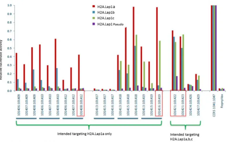
3.1.1 Pair 101421:101422 showed the highest activity in all 4 H2A.Lap1 genes 80 3.1.2 Surveyor (CelI) assay showed pair 101421:101422 have the highest activity…………...……… 83
3.2 Validation of TALEN activity in cultured Neuro 2a cells ………. 87
3.2.1 Optimisation of Neuro 2a transient transfection using CMV-driven GFP plasmid ………. 87
3.2.2 T7 endonuclease (T7EI) digests more efficiently than CelI surveyor nuclease in Neuro 2a cells ……… 92
3.2.3 H2A.Lap1 pseudogene Gm14904 is likely an assembly error ……… 93
3.2.4 Screening potential founder mice for single nucleotide polymorphism ….. 96
3.3 Production of H2A.Lap1KO founder pups by the delivery TALENs to one-cell embryo ……… 98
3.3.1 A modification to CelI surveyor nuclease assay to detect homozygous mutant progeny in mice ……… 103
3.3.2 Improvement in the screening method to characterise the mutation pattern method in TALEN-induced mice ………. 106
3.3.3 In-frame mutation in founder L74-5 may be as detrimental as frameshift mutation ……… 112
3.3.4 Choice of breeders to establish a H2A.Lap1 knockout colony ……… 115
3.3.5 TALENs induce stable and inheritable mutations in multi-copy genes but TALEN activity in retained for several embryonic divisions ……….. 119
xiii
3.3.7 No defect in sex ratio and genotype distribution between KO mice and wt
siblings ………. 127
3.4 Discussion ……… 131
CHAPTER 4: EXOME SEQUENCING TO DETERMINE WHETHER TALEN GENERATES OFF-TARGET MUTATIONS..……….. 135
4.1 Introduction ………. 135
4.2 Results ……….. 137
4.2.1 Preparation of sample for exome sequencing ……….. 137
4.2.2 Reference sequence used in bioinformatics analysis is of FVB/NJ, not FVB/NJArc ……….. 139
4.2.3 TALENs do not recognise off-target locations ……… 139
4.2.4 Higher coverage was obtained with our sample and higher number of variants detected relative to mm10 mouse reference genome ……… 141
4.2.5 Structural variants (SV) and indel analysis detected expected mutation in all three samples ……… 144
4.2.6 Further analysis to detect heterozygous and homozygous deletions ……… 150
4.2.7 Validation of 19 indels from sequencing data using PCR amplification to determine whether they are true indels or strain specific variations ……… 152
4.3 Discussion ……… 156
CHAPTER 5: CHARACTERISATION OF H2A.LAP1KO MICE …………. 162
5.1 Introduction ………. 162
5.2 Results and Discussion ……… 162
5.3 The litter size produced by hemizygous (H2A.Lap1-/Y) KO males is significantly reduced in comparison to their wild type litter mates ……… 162
5.4 The nuclei of round spermatids in H2A.Lap1 KO mice are significantly smaller in hypotonic conditions ………... 165
5.5 The active promoter mark, H3K4me3, accumulates in euchromatin of H2A.Lap1 KO spermatids ……… 169
5.6 H2A.L.2 histone variant displays an unusual behaviour in round and condensing spermatids in the absence of H2A.Lap1 ………... 175
5.7 Localisation of RNA Polymerase II within splicing speckles is affected in H2A.Lap1-/Y round spermatids ……….. 179
5.8 H3K36me3 enrichment in autosomal euchromatin of round spermatids is affected by the absence of H2A.Lap1 ……… 182
5.9 Effect of H2A.Lap1 absence in overall gene transcription and splicing efficiency ……… 185
5.10 H2A.Lap1 depletion affects the epigenetic landscape of post meiotic sex chromosome (PMSC) and the expression of X-chromosome genes that escape inactivation ……….. 189
xiv
CHAPTER 6: GENERAL DISCUSSION ……… 202
6.1 TALEN vs CRISPR ……….. 202
6.2 H2A.Lap1 KO mice are viable but subfertile suggesting a compensatory mechanism may exist ……… 203
6.3 H2A.Lap1 deficient mice display major changes to nuclear compaction and to the organisation of transcriptional hubs………. 204
6.4 RNA-seq analysis of 30-day mouse testes reveals no changes in gene
expression but alterations to intron retention……… 205
6.5 Loss of H2A.Lap1 alters the post-meiotic sex chromatin epigenetic
landscape……… 210
6.6 Dynamic epigenetic changes between early and late round spermatids…… 210
6.7 A possible role for H2A.Lap1 in somatic Sertoli cell function………. 211
xv
List of figures
Figure 1-1: Schematic diagram of chromatin……… 2
Figure 1-2: Nucleosome core particle schematic diagram ……… 16
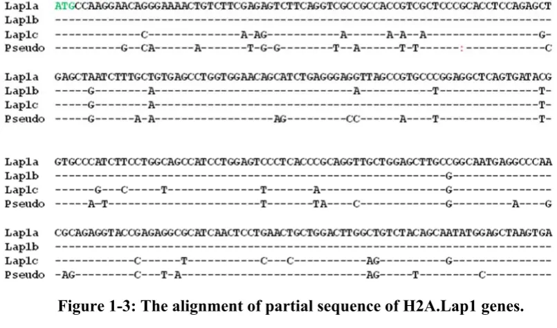
Figure 1-3: The alignment of partial sequence of H2A.Lap1 genes……….... 24
Figure 1-4: Stages of spermatogenesis in mammals……… 26
Figure 1-5: ZFN recognize nucleotide in triplet manner………. 31
Figure 1-6: FokI will only start the enzymatic cleavage activity when ZFNs bind to both sense and antisense strand………. 31
Figure 1-7: The design of TALEN is much more flexible than ZFN……….. 34
Figure 1-8: A mechanism of CRISPR-Cas9 action in bacterial cells………... 36
Figure 3-1: Schematic diagram of TALEN construct ………. 75
Figure 3-2: H2A.Lap1 encoded genes on the X-chromosome……….. 75
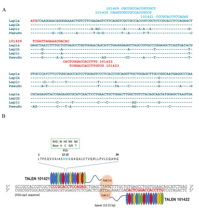
Figure 3-3: The alignment of H2A.Lap1 encoded genes………. 76
Figure 3-4: TALENs design are specific to target H2A.Lap1 genes……… 78
Figure 3-5: TALEN plasmid pair 101421:101422 showed a high enzyme activity on all H2A.Lap1 genes……….…. 82
Figure 3-6: CelI enzyme recognizes on heteroduplex strand……… 84
Figure 3-7: Three TALEN plasmid pair showed a visible CelI digestion product on all H2A.Lap1……… 85
Figure 3-8: TALEN plasmid pair 101421:101422 has the higher CelI enzyme cleavage in H2A.Lap1 genes………. 86
Figure 3-9: Optimised parameters with high transfection efficiency was used in the transfection of TALEN pair 101421:101422……….. 89
Figure 3-10: TALEN 101421:101422 works efficiently in our mouse Neuro 2a cell culture………. 91
Figure 3-11: T7 endonuclease I digest efficiently than CelI……… 93
Figure 3-12: The expression level of H2A.Lap1 mRNA in FVB/NJArc mouse…. 95 Figure 3-13: CelI assay on H2A.Lap1 genes from FVB/NJArc strain of female and male mouse gDNA………. 97
Figure 3-14: The summary of TALEN mice production……….. 99
xvi
Figure 3-16: CelI assay on H2A.Lap1a,b and c genes showed a positive digestion on male founder L74-5 and female founder L90-3 and L89-1……… 105 Figure 3-17: The workflow employed to characterise the genotype of TALEN
mouse………... 108 Figure 3-18: Sequence alignment of H2A.Lap1 genes from mutant founders relative to
H2A.Lap1 genes from wt FVB/NJArc mouse showed a successful
mutation created in coding region of H2A.Lap1 genes………110 Figure 3-19: Male founderL74-5 has all H2A.Lap1 genes mutated frameshift and
in-frame……….113 Figure 3-20: Structure of canonical H2A-H2B dimer as observed in the context of
nucleosome………. 114 Figure 3-21: Mutation is stably inherited by TALEN progeny………. 118 Figure 3-22: The gel image of three H2A.Lap1 genes amplified from pups with
recombination genotype………... 121 Figure 3-23: The TALEN pups have recombination genotype in H2A.Lap1b and
H2A.Lap1c………122 Figure 3-24: The loss of wt mRNA genes and protein………126 Figure 3-25: The sex ratio and genotype distribution of the KO mice progeny showed
no difference than the wt progeny………130 Figure 4-1: Purified gDNA from KO mice strain NM1 and NM4 that were sent for
exome enrichment and sequencing………...138 Figure 4-2: Workflow and filtering strategy used to identify off-target sites………..140 Figure 4-3: Exome sequencing allows to detect all 3 expected mutations in H2A.Lap1
genes……….148 Figure 4-4: Computational analysis predicted 19 putative heterozygous deletions…151 Figure 4-5: Interrogation of predicted off-target heterozygous deletions in KO
mice……….. 154 Figure 4-6: FokI will only cleave when TALEN binds to both sense and antisense
strand……….155 Figure 5-1: The litter size produced by hemizygous KO males is significantly reduced
in comparison to wild type litter mates……….164 Figure 5-2: The nuclei of round spermatids in H2A.Lap1 KO mice are significantly
smaller in hypotonic conditions………168 Figure 5-3: The active promoter mark, H3K4me3, accumulates in euchromatin of
H2A.Lap1 KO round spermatids………..172 Figure 5-4: H3K4me2 does not accumulate in euchromatin of round spermatids in KO
xvii
Figure 5-5: H2A.L.2 localisation in the nuclei of round and condensing spermatids in H2A.Lap1-/Y KO mice is different from the wild type……….. 178
Figure 5-6: Localisation of the active form of RNA Polymerase II within splicing speckles is affected in H2A.Lap1-/Y round spermatids………180
Figure 5-7: Splicing speckle morphology is not affected by the absence of
H2A.Lap1……….181 Figure 5-8: H3K36me3 accumulation in euchromatin of hemizygous KO mice reduced in early round spermatid………...184 Figure 5-9: Expression level of autosomal round spermatid-specific genes that
normally contain H2A.Lap1 nucleosome in their TSS in wt and
H2A.Lap1-/Y mice……….187
Figure 5-10: RNA-seq intron retention………188 Figure 5-11: Kcr intensity is reduced in PMSC of hemizygous KO round
spermatids……… 190 Figure 5-12: H3K4me2 was depleted at the PMSC in the late RS in the absence of
H2A.Lap1……….192 Figure 5-13: H4K8Ac displays no difference between wt and KO round spermatids but reduced in PMSC of late RS in both wt and hemizygous KO mice…….193 Figure 5-14: The intensity of repressive mark, H3K9me2, displays no difference
between wt and KO round spermatids but is significantly increased in PMSC of late RS in both wt and hemizygous KO mice……… 194 Figure 5-15: Round spermatid-specific X-linked genes that contain H2A.Lap1 in their
xviii
List of tables
Table 1-1: List of reported histone variants in eukaryotic organisms………...19
Table 2-1: List of PCR ingredients………58
Table 2-2: List of ingredients for native PAGE gels……….60
Table 2-3: List of lysis buffer used for protein extraction……….67
Table 3-1: Genomic coordinates for H2A.Lap1 encoding genes………..75
Table 3-2: List of TALEN plasmids designed by Zhang et al………..79
Table 3-3: The percentage of transfected Neuro 2a cells by pEGFP-N1………..89
Table 3-4: List of standard PCR primers for H2A.Lap1 encoding genes……….90
Table 3-5: List of microinjections performed by Koopman’s lab to produce viable founder ……… ..100
Table 3-6: The efficiency of TALEN-mediated microinjection into mouse oocytes..102
Table 3-7: The genotype and mutation pattern (by sequencing) of all 19 founders…111 Table 3-8: Breeding strategy of TALEN founders in order to produce H2A.Lap1KO colony………..116
Table 3-9: The percentage of offspring that inherit the TALEN founder mutation and offspring with recombination genotype………..123
Table 4-1: The total reads sequenced and number of reads aligned to exonic target regions per sample……… 143
Table 4-2: The raw variants detected from KO mice sequencing data………143
Table 4-3: Variants output after strain FVB/NJ filter was applied………..143
Table 4-4: Expected mutations detected from sequencing data………. 149
Table 4-5: Variants with heterozygous and homozygous deletions detected from sequencing data of KO mice……….. 149
Table 4-6: Common variants with deletions detected in all three KO mice that were sequenced………151
Table 4-7: List of primers used to amplify 19 indels from TALEN mutant mice….. 153
1
CHAPTER 1
INTRODUCTION
1.1 Epigenetics and chromatin
In 1942, C.H. Waddington introduced the term ‘epigenetics’, which he defined as changes in phenotype without changes in genotype. Today, epigenetics is defined as a number of mechanisms that ensure the inheritance of patterns gene expression without changes occurring in the corresponding DNA sequence. Epigenetic-based mechanisms influence the structure and compaction of chromatin, a complex of DNA, histones and many other associated proteins. Chromatin serves as a reversible structure for complex gene regulation and governs diverse processes including gene transcription, DNA replication, and DNA repair.
2
3
The states of chromatin compaction can be simplistically categorized as active euchromatin (EC) (‘open’ state) and inactive heterochromatin (HC) (‘closed’ state) with each of these states marked by a distinct epigenetic modification (Kouzarides 2007; Woodcock & Ghosh 2010). Euchromatin, or ‘active’ state, consists primarily of coding and regulatory sequences (e.g., enhancers and promoters), which only account for a small fraction (less than 4%) of the genome, in mammals (Allis et al. 2007). Heterochromatin tends to be located at the nuclear periphery, where specific interactions with the envelope may occur. It often forms blocks surrounding the nucleolus and is usually associated with repression of transcription. Heterochromatin may be further classified as constitutive or facultative. Constitutive heterochromatin typically remains condensed throughout the cell cycle and tends to be enriched in repetitive, gene-poor and late replicating DNA sequences. Examples of constitutive heterochromatin are telomeric, centromeric and pericentromeric regions. In contrast, facultative heterochromatin can undergo reversible transition from a more open transcriptionally active state to a compact, transcriptionally inactive state, as occurs during differentiation (Bell et al. 2011; Woodcock & Ghosh 2010).
Berger et al. (2009) stated that there are three categories of signals that culminate in the establishment of a given heritable epigenetic state of chromatin. Firstly, the "Epigenator", which originates from changes in the environment of the cell (e.g., temperature) and triggers an intracellular pathway (either protein-protein or modification-based). The signal is short-lived, but sufficient for the epigenetic phenotype to occur. Secondly, the Epigenetic Initiator signal which responds to the Epigenator and is necessary to define the precise location of the epigenetic chromatin environment (e.g., a DNA-binding protein, non-coding RNA (ncRNA) or any molecule(s) that can define the coordinates of chromatin to be assembled). It should be able to self-reintroduce and self-renew through positive feedback mechanisms. Thirdly, the Epigenetic Maintainer signal which sustains the chromatin environment throughout the generations by a variety of different epigenetic processes as outlined below (Berger et al. 2009).
ATP-4
dependent remodelers, nucleosome positioning and histone variants (Berger et al. 2009), which will be further discussed in the following section.
1.2 DNA methylation and hydroxymethylation
DNA methylation is an epigenetic mechanism that is widely studied in animals, plants and fungi (Feng et al. 2010). It occurs by the addition of a methyl (CH3) group
at the carbon 5 of cytosine, resulting in 5-methylcytosine (5mC), and informally known as the "fifth base" of DNA. In mammals, methylation predominantly occurs at CG dinucleotides. CG dinucleotides are overall depleted in the mammalian genome except at short DNA sequences (on average 1 kb), termed ‘CpG' islands (which in mammals is globally depleted in methylation). CpG islands typically contain around 5-10 CpGs per 100 bp and often co-localise with gene promoters and regulatory regions (Auclair & Weber 2012; Jones 2012). The methyl groups of 5mC project into the major groove of DNA and thus can either inhibit transcription directly or indirectly by the recruitment of methyl DNA binding complexes (Guibert & Weber 2013; Rose & Klose 2014; Clouaire & Stancheva 2008). Gene silencing by DNA methylation is important in developmental processes such as cell differentiation, tumour suppression and reprogramming in plants and mammals (Feng et al. 2010).
According to Song et al. (2016), 5-methylcytosine is generated by DNA methyltransferases (DNMTs) of which there are three conserved in mammals that regulate methylation. DNMT1 plays a role in the maintenance of DNA methylation and restoration of 5mC, after replication (Goll & Bestor 2005). DNMT3A and DNMT3B are de novo DNA methyltransferases and are essential during early development in mammals (Okano et al. 1999). DNMT1 is ubiquitously expressed in proliferating cells where it localises at replication foci and interacts with the proliferating cell nuclear antigen (PCNA) (Chuang et al. 1997; Leonhardt et al. 1992). Studies by Song et al. (2011) revealed that DNMT1 methylates hemi methylated cytosines at an initial rate 5- to 30-fold greater than unmethylated residues.
5
(Borgel et al. 2010; Okano et al. 1999). It has been shown by Chen et al. (2004) that the conserved PWWP domain of DNMT3A and DNMT3B, characterised by the presence of the highly conserved proline-tryptophan-tryptophan-proline motif, is involved in the functional specialisation of these enzymes. The disruption of this domain prevents their association with pericentric heterochromatin and abolishes their ability to methylate major satellite repeats (Chen et al. 2004). Double knockouts of DNMT3A and DNMT3B in ES cells have revealed a complete lack of de novo methylation activity which supports our understanding of their functional importance and redundancy in the process (Okano et al. 1999).
Hydroxymethylation (5hmC) results from the conversion of 5mC to 5hmC by the ten-eleven translocation (TET) family of dioxygenases and is reported to be mostly enriched in ES cells (Stroud et al. 2011) and in the brain (Tahiliani et al. 2009; Kriaucionis & Heintz 2009; Khare et al. 2012). Analysis of chromosomal distribution in ES cells has shown that 5hmC is enriched at gene-rich genomic regions and in enhancers (Stroud et al. 2011). Further, 5hmC also has the tendency to localise at other protein-DNA binding sites, such as transcription factor binding sites (TFBs), and with the insulator binding protein, CTCF. 5hmC is also reported to be associated with GC-skew, (where Gs residues are enriched over Cs residues at 5' ends and vice versa at 3 ends) leading to the suggestion that sequence composition serves as a signal for the deposition of this epigenetic mark (Stroud et al. 2011).
6
The lack of significant intronic 5hmC differences between cell lines, coupled with the observation that in prostate cancer, intronic regions are the most likely to gain 5hmC (Kamdar et al. 2016) suggests that intronic hydroxymethylation is both tightly regulated and potentially critical for basic cellular function (Kamdar et al. 2016). Similarly, the proportional gain of intergenic 5hmC and its status as the most likely feature to retain 5hmC in cancer may indicate either a lack of importance for intergenic 5hmC marks or, conversely, a key regulatory function in oncogenic formation (Kamdar et al. 2016). 5hmC alterations are generally reported as being associated with various neurological disorders. This includes autism (5hmC enrichments are detected on autism related genes) (Wang et al. 2012; Khare et al. 2012; Zhubi et al. 2014); Rett syndrome (5hmC is globally decreased in the genome) (Szulwach et al. 2011); Angelman syndrome (5hmC is globally increased in the genome) (Szulwach et al. 2011); Fragile X syndrome (5hmC is enriched in FXS related genes) (Wang et al. 2012), Alzheimer’s disease (5hmC is increased or decreased in the genome) (Villar-Menéndez et al. 2013; Chouliaras et al. 2013; Condliffe et al. 2014; Coppieters et al. 2014; Bradley-Whitman & Lovell 2013); and Huntington’s disease (5hmC is globally decreased in the genome) (Wang et al. 2013).
1.3 Post-translational modifications of histones (PTMs)
7
the guanidinium groups of arginine contribute to protein stability by forming ionic interactions and hydrogen bonds with proteins and DNA, in addition to acting as a general base in catalysis (Azevedo & Saiardi 2016; Patel et al. 2011). Although both lysine and arginine have side chains of similar reactivity, lysine is more prone to PTMs. The guanidinium group of arginine undergoes three-dimensional ionic interactions that allow arginine to drive protein folding and stability. Lysine polarity contributes to protein structure by way of its amine group that can form a single ionic interaction. Lysine residues are thus more flexible and more easily modified. This unusual chemical plasticity within the lysine residue eliminates steric hindrance and allows histone-modifying enzymes, central to transcriptional regulation, to perform acetylation and methylation as well as subsequent deacetylation and demethylation (Patel et al. 2011). Over 100 different modification sites have been described to date on histones, and depending on the type of modification, they can function as docking sites for trans-acting factors or influence the structural organization of chromatin and thus affect the accessibility of DNA (Rando & Chang 2009; Tan et al. 2011). PTMs of histones are essential for changing the dynamic state of chromatin in order to allow access of DNA binding complexes to the binding site. These dynamic changes include open/active states (associated with transcriptional activation) and closed/inactive states (associated with transcriptional repression). These modifications correlate with cellular processes such as transcription (Rando & Chang 2009). The elimination of histone-modifying enzymes can affect transcription rates, chromosome stability, and other chromosomal processes. Thus, mapping histone modifications has become an important way to characterize genome function and regulation (Rando & Chang 2009).
8
of these two sites is executed by lysine-specific demethylase 4A (KDM4A) (for H3K36me3) and lysine demethylase five families (KDM5) (for H3K4me3) (Kooistra & Helin 2012). The ‘reader' recognizes methylated histones, including PHD, chromo, WD40, Tudor, double/tandem Tudor, MBT, Ankyrin Repeats, zf-CW and PWWP (Yun et al. 2011). Li (2013) demonstrated the mechanism whereby H3K36me3 interacts with the PWWP domain, in vivo and in vitro, by examining the regulation of human DNA mismatch repair (MMR) with MutSα. It was shown that the epigenetic mark H3K36me3 is required (in vivo) to recruit the mismatch recognition protein hMutSα onto chromatin, through direct interaction with the PWWP domain of hMutSα. The recruitment of hMutSα is cell cycle-dependent, such that H3k36me3 and the histone methyltransferase SETD2 are required for MMR, in vivo. The abundance of H3K36me3, in G1 and early S phase, ensures that hMutSα is enriched in chromatin before mis-pairs are introduced during DNA replication. They showed that cells with depleted SETD2 displayed microsatellite instability and elevated spontaneous mutation frequency, characteristics of MMR-deficient cells. Further, cells with depleted H3K36me3 fail to recruit hMutSα, and the restoration of H3K36me3 in SETD-depleted cells restored the localization of hMutSα to the chromatin, via its PWWP domain. Therefore, H3K36me3 and the PWWP domain regulate human MMR in vivo.
Tan et al. (2011) recently identified histone crotonylation as another example of histone modification and a mechanism for epigenetic regulation. Lysine crotonylation (Kcr) marked active promoters and potentially enhancers in both mouse somatic and male germ cell genome (Tan et al. 2011). A report by (Li et al. 2016) showed that Kcr has high-affinity binding to AF9 YEATS domain via an extended aromatic sandwich pocket. Also, AF9 co-localises with crotonylation of H3 and positively regulates gene expression in YEATS domain-dependent manner. They proposed the YEATS domain as a family of a crotonyllysine readers and AF9 YEATS domain directly links Kcr to activate transcription.
9
pericentric heterochromatin through direct interaction with chromodomain-containing protein (HP1) (Bannister et al. 2001). Meanwhile, the Polycomb repressive complex 2 (PRC2) ensures trimethylation of H3K27 (Young et al. 2011). In ES cells, H3K27me3 is enriched at the promoter region of the Polycomb group (PcG) protein associated genes, which includes the Hox genes as well as the inactive X chromosome (Boyer et al. 2006; Escamilla-Del-Arenal et al. 2013; Zhang et al. 2015). There is evidence that shows that in some instances a crosstalk through PRC2 is necessary for the coexistence of H3K9me3 and H3K27me3 in order for gene repression to occur (Boros et al. 2014; Cooper et al. 2014). For example, Boros et al. (2014) showed that binding of HP1α to H3K9me3 is significantly increased in the presence of H3K27me3 and that this is dependent on PRC2. In addition, they have shown that PRC2 is an indirect binder of H3K9me3. The knockdown of two PRC2 subunits, EZH2 and SUZ12, leads to the depletion of H3K27me3, a decrease in the total cellular level of HP1α and a slight reduction of H3K9me3. This is most likely due to a reduced ability to recruit SUV39H1/2 H3K9me methyltransferase to chromatin in response to HP1α loss.
1.4 Non-coding RNA
The developments in next-generation sequencing have increased our ability to sequence cDNA to an unprecedented scale. Transcriptome sequencing, such as RNA-sequencing, coupled with computational analyses allowed researchers to obtain in-depth sequence information that led to the identification and mapping of novel alternatively spliced transcripts and of non-coding transcripts. Protein coding genes account for approximately 1.5% of the genome, while more than 80% is transcribed into non-coding RNA (ncRNA) (Greco & Condorelli 2015). Non-coding RNA is classified according to its sequence length. There are two main categories that differentiate ncRNAs, small (< 200 bp) and long ncRNA (>200 bp). Small ncRNAs include microRNA (miRNA), piwi-interacting RNA (piRNA), and endogenous short-interfering RNA (siRNA), while long ncRNAs (lncRNA) form a highly diverse class with less protein-coding potential (Wang & Chang 2011).
10
transcription or through splicing, which fold into stem-loop structures to form imperfect double-stranded RNA (dsRNA) molecules. These are then processed by RNase III endoribonuclease (generally Dicer) before being denatured. One of the RNA strands binds to the RNA-induced silencing complex (RISC), which later binds to a specific target of mRNA, containing a sequence complementary to the miRNA. The association induces either cleavage, degradation or blocks translation to protein (Collins et al. 2011). siRNAs, on the other hand, are produced as dsRNA and can enter the post-transcriptional gene-silencing (PTGS) pathway, which leads to mRNA degradation in the cytoplasm. Alternatively, siRNAs enter the transcriptional gene-silencing (TGS) pathway, which involves chromatin modification. piRNAs and ssRNAs are produced in clusters and cleaved to individual mRNA by means of a yet unidentified processing mechanism(s) (Collins et al. 2011).
Long non-coding RNAs (lncRNAs) are heterogeneous transcripts produced predominantly by RNA polymerase II. They are defined as RNA molecules of more than 200 bases in length with no protein-coding capacity (Aliperti & Donizetti 2016). lncRNA have been further categorized according to the anatomical properties of their gene loci. For instance, antisense lncRNA overlap with known protein-coding genes; intronic lncRNA are encoded within intronic regions of protein-coding genes; overlapping transcripts are those that overlap protein-coding genes; and lincRNA are encoded completely within the intergenic genomic space between protein-coding loci (Rinn & Chang 2012). In humans, estimates for the number of different types of lncRNA have ranged from 5,400 to 53,000, with only a small fraction being found at levels high enough to suggest evidence of functionality (Rinn & Chang 2012). The association of ncRNA with cellular development has been reported across many mammalian cell types and tissues, including human neuronal cells (Aliperti & Donizetti 2016), mouse cardiomyocytes (Wang et al. 2014), human B-cells (Petri et al. 2015) and human keratinocytes (Kretz et al. 2012).
11
Xist allele in cis (even as cell differentiation signals trigger XCI on the selected X chromosome) by directing euchromatic modifications to the Xist locus; and secondly, Txis associates with Dnmt3a at the Xist promoter and facilitates de novo CpG methylation and stable silencing of active X chromosome Xist allele (Collins et al. 2011; Lee 2009).
In addition to gene silencing, lncRNA has been associated with regulation of the mitochondrial network (Wang et al. 2014). Specifically, a cardiac apoptosis-related lncRNA (CARL) was shown to suppress mitochondrial fission and apoptosis, by targeting miR-539 and PHB2. PHB2 is an oestrogen receptor (ER)-binding protein, which represses transcriptional activation and is negatively regulated by miR-539. CARL binds to miR-539 directly and participates in the regulation of mitochondrial network and apoptosis, through the miR-539/PHB2 pathway (Wang et al. 2014). One approach that has been used to predict lncRNA function was to co-express it with well-characterized protein-coding genes (Petri et al. 2015). Co-expression alone may not be sufficient to assign a function to lncRNA, but the information obtained from embedding lncRNA in a transcriptional network associated with cellular development, provides a starting point for functional studies. To further determine the function of lncRNA, it will be necessary to perform knock out experiments (Palazzo & Lee 2015).
1.5 Nucleosome positioning
Before the era of genome-wide, next-generation sequencing, mapping of nucleosome occupancy through the whole genome was not feasible, and the question of whether nucleosomes are positioned randomly or at specific DNA sequences, remained an open question. In recent years, technologies like MNase-ChIP-seq, Hydroxyl-radical-seq and MNase exoII-seq have allowed us to map nucleosome positions to a base-pair resolution first in S. cerevisiae and then in D. melanogaster and to determine their influence(s) in gene regulation (Jiang & Pugh 2009). Given the large size of the mammalian genome, such base pair resolution has been challenging (Soboleva et al. 2014)
12
sequencing-based mapping approaches have been able to identify the positions of individual nucleosomes as a cell population average (Barski et al. 2007) and more recently in single cells at a specific time (Struhl & Segal 2013).
Genome-wide studies have provided a view of the nucleosome positions in a typical gene (Radman-Livaja & Rando 2010). Promoters and other functional regions, such as enhancers and replication origins, are depleted of nucleosomes relative to transcribed genes (Lieleg et al. 2015). These regions are known as "nucleosome-depleted regions" (NDR) and are localized upstream of transcription start sites (TSS) (Radman-Livaja & Rando 2010). However, as shown by this laboratory, rather than being nucleosome depleted, NDRs may be occupied by unstable or fragile nucleosomes (Soboleva et al. 2014)
Genome-wide mapping of the nucleosome has also provided insights into the organization of nucleosomes around protein-coding genes. The organization of nucleosomes is well documented in S.cerevisiae, which provides the clearest example of a consensus pattern of organization. The first predominant nucleosome located upstream of the TSS (designated -1) covers a region from -300 to -150 (relative to the TSS) and can regulate the accessibility of the promoter in that region (Jiang & Pugh 2009). During a transcription cycle, the -1 nucleosome experiences many changes that affect its stability, including histone replacement, acetylation, and methylation, as well as translational positioning and ultimately eviction after pre-initiation complex (PIC) formation to RNA polymerase II such as TFIID and SAGA. Further, in mammals, it has been reported that nucleosomes occupy exons more frequently than introns, providing that the exon has a higher guanine-cytosine (GC) content than its neighbouring introns. The nucleosomes positioned over exon sequences produce so-called “speed bumps” that are believed to slow the rate of RNA pol II, promoting exon inclusion during pre-mRNA splicing. In addition to the -1 nucleosome, there exists a highly positioned nucleosome downstream of the TSS (the +1 nucleosome), which may also act as a barrier to the initial movement of RNA Pol II (Soboleva et al. 2014)
13
in mouse C2C12 myoblast cells that appear around transcription factor binding sites which can be used to predict gene expression (Maehara & Ohkawa 2016). This study also showed that the 5’ nucleosome positioning patterns act as functional units on chromatin and describe the transcriptional state of the transcription factors. This indicates that transcription factors are a major determinant of nucleosome positioning.
Nucleosome positioning is strongly affected by DNA sequence (Struhl & Segal 2013). There are two major sequences that determine nucleosome positioning. First, the intrinsic sequence property that refers to the variability of dinucleotide bending properties. Optimal nucleosome formation occurs when bendable nucleotides (AT and TA) occur on the face of the helix that directly interacts with histones (Struhl & Segal 2013). Other than DNA sequence, nucleosome positioning is also determined by ATP-dependent nucleosome remodelling enzymes. For instance, the combined action of nucleosome remodelling enzymes, ISW1 and CHD1 in yeast, are required to maintain the positioning of nucleosomes within the coding regions of eukaryotic genes and in alignment with transcriptional start sites (TSS) (Gkikopoulos et al. 2011). In addition, a recent study also showed these two nucleosome spacing enzymes compete to set the spacing on most genes, such that CHD1 dominates genes with shorter spacing, and ISW1 dominates genes with longer spacing. In contrast, ISW2 (another yeast nucleosome enzyme) plays a minor role, limited to transcriptionally inactive genes (Ocampo et al. 2016).
1.6 ATP remodelers
14
There are four major classes of remodelers, namely, i) Swi/Snf (switching defective/sucrose nonfermenting); ii) INO80 (inositol requiring 80); iii) CHD (chromodomain, helicase, DNA binding); and iv) ISW1 (imitation switch) (Clapier & Cairns 2009). They are considered derivatives of the SNF2 ATPase family which is evolutionarily conserved across eukaryotic organisms (all have a conserved ATPase domain), reflecting their conserved function and importance in gene regulation (Runge et al. 2016). Swi/snf are well known remodelling complexes, composed of 8 to 14 subunits (Clapier & Cairns 2009), and were initially identified in yeast (Winston & Carlson 1992). They are identified by their ability to activate transcription of the HO and SUC2 genes. Their mechanism of action, similar to many others that are regulated by this complex in yeast, involves binding in proximity to the promoter in order to assist the subsequent binding of specific transcription factors (Masliah-Planchon et al. 2015). In mammals there are 15 subunits that make up the Swi/Snf complex. Among these subunits, only two ATPases, Brg1 or Brm, are sufficient to remodel nucleosomes
in vitro (Phelan et al. 1999), while the others are likely providing specificity in vivo. In addition, there are reports suggesting that mutations in Swi/Snf subunits may contribute to various human malignancies, as evidenced by the high frequency of mutations, within genes encoding BAF subunit (mammalian Swi/Snf complex), occurring in >20% of human cancers (Kadoch & Crabtree 2015).
1.7 Histone variants: Structure and versatility of function
15
16
17
Histone variants, unlike the core histones, are expressed and incorporated into chromatin throughout the cell cycle. The replication-independent gene and mRNA structure of histone variants also differ from core histones. The histone variant genes often contain introns, and the transcripts are often polyadenylated; features that are thought to be important in the post-transcriptional regulation (Kamakaka & Biggins 2005). The most prominent sequence divergence is found in H2A and H2B, with H2A having the most variants. In contrast, H4 has no reported variants, and H3 variants have only minimal sequence variations (Mattiroli et al. 2015).
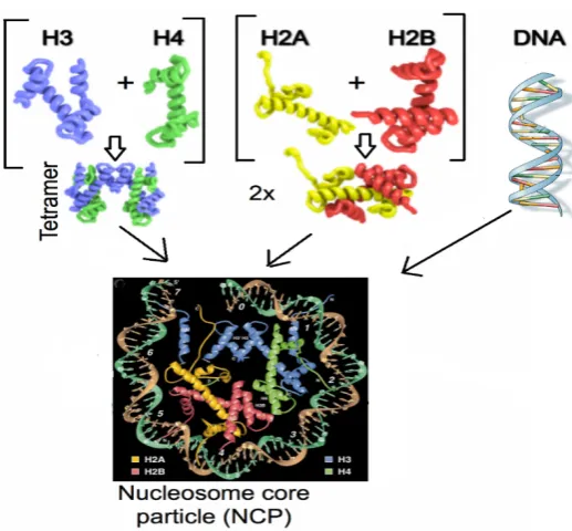
Histone variants have evolved to contribute to the complexity of chromatin and have specialized functions in regulating chromatin dynamics. Their sequence divergence can alter chromatin states and the fate of cell decisions (Weber & Henikoff 2014). The role of histone variants in regulating transcription has been widely reported. It was initially understood that in order for transcription to take place, a nucleosome-depleted region (NDR) must form at the TSS, to allow binding of transcription machinery. Based on experimental evidence, the accessibility of micrococcal nuclease supported this notion, however, this has been disputed upon finding that nucleosomes containing labile H2A variants flanked the NDR. Jin et al. (2009) used chromatin immunoprecipitation (ChIP) and re-ChIP, followed by high throughput Solexa sequencing on HeLa cells expressing Flag-tagged H3.3 histone to reveal that hybrids of H3.3-H2A.Z nucleosome core particle (NCP) localized to TSS of active genes. Subsequently, this laboratory has shown that heterotypic H2A.Z-H2A nucleosomes occupy the TSS in stem cells thus raising the possibility that H3.3-H2A.Z nucleosomes are in fact heterotypic with respect to H2A.Z (Nekrasov et al. 2012; Soboleva et al. 2014). As mentioned above, we also found that H2A.Lap1-containing nucleosomes are found at the TSS of active genes in the testis (Soboleva et al. 2012; Soboleva et al. 2014). These unstable nucleosomes may serve as a ‘place holder’ to prevent the region from being covered by adjacent stable NCP and/or nonspecific factors, when the region is nucleosome-free. Moreover, these fragile nucleosomes could be more easily displaced by transcription factors because of its instability (Jin et al. 2009).
18
heterotypic. This nucleosome persisted as heterotypic until M phase (Nekrasov et al. 2012). The depleted expression of H2A.Z at promoter, after DNA replication, is possibly due to its redistribution to constitutive heterochromatin and/or at the centromere. This cell cycle-dependent possible dynamic movement of H2A.Z suggests its functional importance in providing transcriptional memory, that is, active marks established at the G1 phase and remaining at M phase may facilitate the reestablishment of gene transcription after cell division (Nekrasov et al. 2012).
19
Table 1-1: List of reported functions of histone variants in eukaryotic organisms (Yuan & Zhu 2012)
Histone variants Function
CENP-A or cenH3 Epigenetic marker of the centromere
H3.3 Involved in transcription
Enriched at transcriptionally active regions, telomerase, and pericentric regions
H3.Z Regulation of cellular response to outside stimuli H3.Y Regulation of cellular response to outside stimuli H2A.Z Transcriptional control rheostat
Heterochromatin formation and maintenance
H2A.X Double strand break repair
Meiotic remodelling of sex chromosome
macroH2A Gene silencing
X-chromosome inactivation H2A.Bbd/H2A.B or
H2A.Lap1
Epigenetic mark of active chromatin. Found at the TSS and intron-exon boundaries.
20
1.8 Histone H2A.Z variants: The structure and function
H2A.Z is the most widely studied of the histone variants, with respect to structure and function (Chakravarthy et al. 2005; Sarma & Reinberg 2005; Yuan & Zhu 2012; Suto et al. 2000). It is an essential histone found in all higher eukaryotes and there is approximately 90 % sequence homology between species (Suto et al. 2000; Chakravarthy et al. 2004; Yuan & Zhu 2012). The H2A.Z sequence differs by approximately 60 % from canonical H2A and the other H2A variants, mainly in its “docking” domain in the C-terminus and in the L1 loop where two H2A molecules contact each other at the back face of the nucleosome (Suto et al. 2000). The docking domain of the H2A.Z histone variant contains several negatively charged amino acid residues that come together on the surface of the nucleosome to create an ‘acidic patch’ (Luger et al. 2012). The larger acidic patch of H2A.Z-H2B displays higher affinity with H4 N-terminal tail than that of the H2A-H2B dimer, resulting in the compaction of chromatin by facilitating intra-nucleosome-nucleosome interactions. Specifically, the acidic patch of a H2A.Z-containing nucleosome interacts better with the basic histone H4 tail originating from a neighbouring nucleosome thus driving intra-nucleosome interactions (Fan et al. 2004; Luger et al. 2012). Hence, DNA accessibility is suppressed and transcription is inhibited in vitro from a chromatin template that is enriched with H2A.Z to a greater extent than a H2A-containing chromatin template (Zhou et al. 2007)
21
contribute to the specialised 3-D organization of a centromere at metaphase (Greaves et al. 2007). H2A.Z also has a unique and specific role in assembling the X chromosome into a silenced heterochromatic state, following meiosis (Greaves et al. 2006)
In both yeast and mammals, H2A.Z has been shown to associate with transcription activation. Genome-wide chromatin immunoprecipitation (ChIP) showed that H2A.Z localizes primarily to the promoter region (Zhang et al. 2005; Barski et al. 2007) Formaldehyde crosslinking followed by ChIP experiments on budding yeast S. cerevisiae showed that H2A.Z is enriched at intergenic regions upstream of PHO5 and
GAL1, even under repressed conditions (Santisteban et al. 2000). Subsequent genome-wide studies showed that H2A.Z is enriched at the promoter-proximal +1 and -1 nucleosome (Albert et al. 2007). The presence of H2A.Z nucleosome surrounding most yeast promoters, in the absence of transcription, led to the proposal that H2A.Z-containing nucleosomes helped poise genes for transcription (reviewed by Luk et al. 2010).
Evidence is accumulating to associate H2A.Z with cancers, such as melanoma (Vardabasso et al. 2015), prostate (Baptista et al. 2013), bladder (Kim et al. 2013) and breast (Hua et al. 2008; Svotelis et al. 2010). It was observed by Svotelis et al. (2010) that H2A.Z mRNA is elevated in breast cancer biopsies. This correlates with increased levels of oestrogen receptor gene α (ERα) in low-grade breast tumours, and elevated levels of H2A.Z in high-grade tumours. ERα was shown to regulate H2A.Z in breast cancer by way of a positive feedback loop that appears important for ERα signalling under normal and pathological conditions. This study correlates elevation of H2A.Z expression with cellular proliferation, when E2 (ligand for oestrogen receptor) levels are low and during tamoxifen (drug used widely to treat breast cancer due to its capacity bind to ERα) treatment. H2A.Z therefore could be an important factor in the transformation of breast cancer cells and in the establishment of endocrine resistance, which is of major clinical importance in breast cancer treatment.
1.8.1 H2A.Bbd (human) and the recently discovered mouse histone H2A.Lap1
22
arrays cannot compact and de-represses chromatin mediated repression in vitro (Zhou et al. 2007). Further, restoration of the acidic residues in H2A.Bbd was shown to restore the intranucleosomal interactions, needed for the folding of a nucleosomal array, and repression of transcription (Zhou et al. 2007). In addition, H2A.Bbd has a truncated C-terminus/docking domain making this nucleosome highly unstable leading to unwrapping of the DNA from the octamer surface (Bao et al. 2004; Doyen et al. 2006). Specifically, this domain is responsible for the interaction with the N-terminus H3, which guides the DNA at the entry and exit points. Therefore, micrococcal nuclease digestion of H2A.Bbd-NCP (nucleosome core particle) revealed that only 118 ± 2 bp of DNA, compared with 145 bp for the wild type nucleotide, was protected (Bao et al. 2004).
H2A.Bbd was first discovered in humans and shown to be excluded from the female X-chromosome (Chadwick & Willard 2001), a feature that gave this variant its name, Barr body deficient (Bbd). H2A.Bbd protein consists of 115 amino acids with a molecular weight of 12.7 kDa and is only 48 % identical to the core H2A histone, making it the most evolutionary divergent histone variant known to date (Chadwick & Willard 2001; González-Romero et al. 2008). It has been proposed that the major hallmarks of Bbd, compared to core H2A (Bao et al. 2004; Doyen et al. 2006), are: i) the presence of a continuous stretch six arginine residues and the conspicuous absence of lysine in the N-terminal tail; ii) the absence of a C-terminus including the last segment of the docking domain that is responsible for the interaction with H3 in major NCP; iii) major sequence differences in the docking domain of H2A (residues 81-119); iv) the presence of only one lysine in H2A.Bbd (compared to 14 in major H2A); and v) the absence of ‘acidic patch as discussed above.
23
24
25
1.8.2 H2A.Lap1 is a tissue-specific histone 1.8.2.1H2A.Lap1 in spermatogenesis
In mammals, germ cells move from the periphery to the lumen of a seminiferous tubule and are subsequently transported to the epididymis, a long tubule that stores mature sperm. Mammalian spermatogenesis takes place in the seminiferous tubules of the testes.
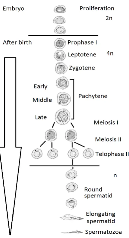
Spermatogenesis consists of three stages (Figure 1-4), i) somatic diploid cell, or spermatogonia (2n): establishment of primordial germ cell (PGC) in the embryo, and mitosis of spermatogonial cells; ii) tetraploid cells (4n) or spermatocytes: meiosis of diploid cells which involves reductional divisions in primary and secondary spermatocytes to produce haploid cells. The primary spermatocytes undergo differentiation through preleptotene (in Prophase I)leptotenezygoteneearly pachytene mid-pachytenelate pachytene. Secondary spermatocytes undergo further meiosis divisions to produce haploid spermatids. In the final stage, iii) haploid (n) spermatids that are represented by round, elongating and condensing stage of differentiation resulting in final stage, mature haploid spermatozoa (Clermont 1972).
26
27
Many histone variants of H2A and H2B are specifically expressed in the testis. For instance, TH2A and TH2B have been detected in post-meiotic spermatids. During the condensation of spermatid nuclei, TH2A and TH2B gradually decrease, before being degraded during DNA compaction (Moss et al. 1989). An increase in histone acetylation in elongating spermatids serves as a signal for the recruitment of specific machinery acting on acetylated histones needed for the histone-protamine exchange (Govin et al. 2004). Investigation of nucleoproteins on condensing spermatids has led to the identification of five more H2A and H2B histone variants that are expressed mainly in testes, namely H2A.L1 (H2A.Lap2), H2A.L2 (H2A.Lap3), H2A.L3 (H2A.Lap4), H2B.L1 (of which mRNA is strongly enriched at round and elongating spermatids) and H2B.L2 (which has very low mRNA present at meiotic and post-meiotic stages). Immunostaining using antibodies that recognize H2A.L1 and H2A.L2 (H2A.L1/H2A.L2) or H2B.L1 showed an accumulation of H2A.L1/L2 and H2B.L1 in condensing spermatids, similar to that of transition proteins and protamines (Govin et al. 2007).
Further investigation to identify testis-specific histones has led to the in-depth characterization of the function of H2A.Lap1 variant in vivo, by Soboleva and colleagues (2012) in our laboratory. Mechanistically, and similar to H2A.Bbd, H2A.Lap1 inhibited chromatin compaction in vitro (Soboleva et al. 2012). In vivo, H2A.Lap1 expression begins at the pachytene stage and peaks in the round spermatid stage coincident with the highest levels of transcription. In elongating spermatids, H2A.Lap1 is exported out of the nucleus (Soboleva et al. 2012). ChIP-Seq and RNA-Seq analysis revealed that H2A.Lap1 is targeted to the TSS of active genes including those few genes on the X chromosome that escape the X chromosome inactivation process (Soboleva et al. 2012).
28
1.8.2.2H2A.Lap1 in the brain.
The role of H2A.Lap1 in the mouse brain remains unknown, but similar to the testis, H2A.Lap1 ChIP-Seq and RNA-Seq in the hippocampus revealed that H2A.Lap1 is also found at the TSS and intron-exon boundaries of active genes (Soboleva et al. 2017). An immunohistochemical staining of mouse brain tissues showed that H2A.Lap1 is expressed in neurons throughout the adult mouse brain, including the hippocampal areas of dentate gyrus (DG), CA1 and CA3 (unpublished data). In cerebellum, H2A.Lap1 localized specifically in the Purkinje neurons, while H2A.Z is expressed in the granular layer (unpublished data). Further, H2A.Lap1 expression was found to coincide with the maturation of the Purkinje neurons, implicating H2A.Lap1 in Purkinje neuron development (unpublished data).
In spite of these successes in characterizing many aspects of H2A.Lap1 function in mouse brain and testes, the ultimate approach to deciphering a function of a gene is to create and characterize a biological system where the function of the gene of interest is disrupted. Therefore, the overall aim of this thesis was to generate a mouse model in which all three H2A.Lap1-coding genes are knocked-out.
1.9 Targeting H2A.Lap1 encoded genes
The purpose of gene targeting is to discern a particular gene’s biological function. There are two ways to achieve this, either by overexpression of the gene of interest, or by down regulation of its expression. Overexpressing a gene can be employed by transgenic technology, whereas, homologous recombination is typically employed to create a mutation that leads to ‘loss-of-function' (Hall et al. 2009). Where overexpression can violate balanced gene dosage, affecting protein folding, complex assembly and downstream regulation (Gibson et al. 2013), most researchers opt for a knockdown or knockout targeting strategy to disrupt a gene’s open reading frame and block its expression, as a direct means to delineate its function (Hall et al. 2009).
29
1.9.1 Genome editing approach to disrupt genes 1.9.1.1ZFN, TALE or CRISPR
Zinc finger nuclease (ZFN)
Specificity of targeting remains the focus in disrupting a gene of interest. A recent approach called ‘genome-editing' has enabled scientists to target virtually any sequence. This approach uses engineered nucleases which consist of a specific sequence of DNA binding domain fused to a non-specific DNA cleavage motif (Gaj et al. 2013; Boettcher & McManus 2015; Cermak et al. 2011). The first genome editing introduced was zinc finger nucleases (ZFN). A zinc finger nuclease contains a DNA-binding domain composed of at least three tandem arrays of Cys2His2 zinc fingers, commonly found in eukaryotes and representing the second most frequently encoded protein domain in the human genome. This motif is fused with a nonspecific DNA cleavage domain derived from the endonuclease FokI which requires dimerization for enzymatic activity (Gutschner et al. 2011; Gupta et al. 2011) (Figure 1-5). The specificity of targeting lies in the DNA binding domain that can be customized by the researcher. One zinc finger recognizes three base-pairs on one DNA strand. For example, a protein comprising of three zinc fingers recognizes a 9- to 10-base-pair segment. But the zinc-finger triplet also means you have less flexibility in how the recognition sequence must be read i.e. although there are hundreds of zinc-finger modules, not every possible sequence is represented (DeFrancesco 2011).
30
31
Figure 1-6: FokI will only start the enzymatic cleavage activity when ZFNs bind to both sense and antisense strand. This creates double-strand breaks (DSB) in the genomic strand which will be naturally repaired by NHEJ or HR.