International Journal of Innovative Technology and Exploring Engineering (IJITEE) ISSN: 2278-3075, Volume-8 Issue-10, August 2019
Abstract: Brain tumor image segmentation is a play a vital role in the medical field or medical processing. Patient treatment with brain tumors is the significant level determine on early-stage detection of these tumors. Early stage detection of Brain Tumors will enhance the patient lives. The disease of brain tumors by a neurologist frequently uses a manual image segmentation that is a hard and time-consuming process, because of necessary automatic image segmentation. Nowadays, automatic image segmentation is very popular and can solve the issue of tumor brain image segmentation with better performance. The main motive of this research work is to provide a survey of MRI image based brain tumor segmentation techniques. There are various existing study papers, focusing on new techniques for Reasonable Magnetic Image-based brain tumor image segmentation. The main problem is considered a complicated process, because of the variability of tumor area of the complexity of determining the tumor position, size, shape and texture. In this research work, mainly worked on interference method, feature extraction, morphological operators, edge detection methods of gray level and Swarm Ant Lion Optimization based on brain tumor shape growing segmentation to optimize the image complexity and enhance the performance. In new algorithm implemented an inspiring nature method for segmentation of brain tumor image using hybridization of PSOA and ALO is also called a Swarm Ant Lion method. Evaluate the performance metrics with image quality factor (PSNR), Error Rate (MSE), and Exact value (Accuracy Rate). In research work, improve the performance metrics with PSNR and Accuracy Rate and reduce the error rates and compared with the existing method (PNN).
Index Terms: Brain Image Segmentation, Probabilistic Neural Network, Ant Lion and Particle Swarm Optimization method.
I. INTRODUCTION
The brain is the central portion of the nervous system that has control of the whole body system.. Brain is the most complex organ of the body [1]. The brain tumor is the development of a mass of anomalous cells of the brain. Various type of brain tumors is found among individuals. A brain tumor may be cancerous or non-cancerous tumor [2]. The tumor disease starts as the main tumor and may spread to other parts of the brain. Generally, the maximum number of people affected by a cancerous brain tumor [3] [4]. It has been researched by America cancer organization that estimation amount of the 24000 fresh cases has been gradually increasing with maximum death rate up 17000
Revised Manuscript Received on August 05, 2019
Sonali Bansal, CEC, CGC, Landran,Mohali, India.
Dr. Shubpreet Kaur, CEC, CGC, Landran,Mohali, India.
Navdeep Kaur, CEC, CGC, Landran, Mohali,India.
[image:1.595.307.550.360.449.2]cases [5]. It has been investigated by world health organisation that brain tumor based on several stages which are –stage 1 is the stage where there is slow growth rate known as pilocytic astrocytoma , in stage 2, there is production of the tissues that are affected by other tissues and known as low-grade astrocytoma , Stage 3 tumor is cancerous tumor where there is reproduction of cells and the cells are affected by the tissues which called as anaplastic astrocytoma stage 4 is cancerous tumor and at last stage where the reproduction of cell tissue takes place at faster rate and also affects the nearest brain tissue is called as glioblastoma tumor [6] [7] . Segmentation of brain tumor is a technique for division of a picture into a different area like as the pixel in the area have the same features [8] [9].
Fig 1 Tissue In Human Brain (A)White Matter (B)Gray Matter (C) Cerebrospinal Fluid
During magnetic resonance brain imaging, departure of various tumor tissue from normal tissue is marked as a segmentation technique [10]. Practically, the segmentation process of brain tumor is prepared through the manual method [11] [12]. This method may provide incorrect experimental results with a maximum processing time period. Hence, wide research has been done by different scientists and researchers based on diagnosed techniques of brain tumor [13]. Though, segmenting of brain tumor is a challenging issue, so, there is a need to do investigation on various segmentation methods of brain tumor. Medical imaging plays an essential role in the diagnosis of the patients having brain tumor [14] [15] . Various new methods developed for the diagnosis of the brain tumor disease which are described as ultrasonography, magnetic resonance imaging, computerized tomograpghy [16].
In existing research, focused on the noise elimination method by extracting the features of gray level reoccurrence characteristics. In this research, a discrete wavelet transform depends on brain tumor area development segmentation method to remove the complex nature. This research emphasis on the removal of
Enhancement in Brain Image Segmentation
using Swarm Ant Lion Algorithm
the interference through segmenting technique. The possible neural network method(PNN) utilized for the training and testing purpose to recognize the position of brain tumor magnetic resonance pictures. Performance metrics acquire maximum accuracy rate for identification of irregular tissue from the brain magnetic resonance pictures. In the proposed approach, proposed work implemented a novel method which is Particle Swarm and Ant Lion optimization to find the region and selected features with the help of fitness function. Evaluate the performance metrics with PSNR, MSE, and Accuracy Rate. Compared with the existing performance parameters.
Section I described that the digital image processing and brain tumor image segmentation. Section II defined that the lots of paper research surveyed, flow and methods. III and IV, Section defined that the presented work with proposed methods and performance metrics (PSNR, MSE and ACCURACY). Section V described that the conclusion means conclude the research methods and Future scope will further enhance.
II. LITERATURESURVEY
Hasan, S. K and Ahmad, M. et al., 2018 [17] proposed research on two-stage authenticated technique for detection of brain tumor. In this research, the proposed technique is authenticated scheme for detection of brain tumor, called a watershed matched approach. The segmentation of brain tumor area was done through classification approach of the watershed algorithm. In addition, scale invariant feature transform technique was also used to extract feature regions and matching divided area of brain tumor with an actual picture. Along with that, compute dimensions of the tumor using various approaches. They implemented an approach through free surfer system software to search the width of cortical and the compute variance among benign and malignant. Kilic, H., Yuzgec, U and Karakuzu, C et al.,
2018 [18] implemented a novel technique for the
development of ant lion optimization (ALO) that encouraged by hunting method of ant lion. The main issue of the used method is maximum time consuming, so proposed a new randomized walk technique to overcome this issue. In this research, the ant lion optimized selection method is proposed that depends on event choice and compared with an investigative approach for benchmarks variables. In addition, testing and training of the ant lion optimized selection method were done. Experimental analysis proved that the planned method gives a better presentation as compared to existing ant lion optimization algorithm.
Kurnar, M., Sinha, A. and Bansode, N. V et al., 2018[19]
proposed research on the detection and visualization of the tissues of the brain. Detect the dimension of the tumor of the brain image, different stages of the brain can be determined. The step may be cancerous or non-cancerous. To recognize the size of the brain, a tumor of the brain can be detected using a k mean clustering algorithm based on the separation and morphological technique. The morphological method used for the extraction of the features of the parts of the body through MRI scanning. Experimental analysis on calculating the extracted features of the portion of the brain disease.
Akter, L. A., and Kwon, G. R et al., 2018 [20] proposed research on novel distributed picture segmentation technique
for magnetic resonance brain pictures. In this technique, the different conversion is called a contour transformation that is the through canny edge detection. In this research work, the distribution technique acquired for the improved method on contour constants through the canny edge detection approach. Experimental analysis was done on improvement of contourlet conversion through the segmentation process
using canny edge detection technique. Bai, X., Chen, Z.,
Liu, M. and Zhang, Y et al.,2015 [21] presented a novel fuzzy c mean clustering approach that is also known as possible fuzzy c mean clustering technique. They separate of the database through middle free fuzzy c mean clustering into fuzzy c means clustering technique. The proposed method is less prone to interference. In this research, it was analysed that the proposed method helps in the segmentation of the magnetic resonance imaging brain pictures along with intervention.
III. RESEARCHMETHODOLOGY
In this section, elaborate the image segmentation problem statement in brain images (MRI). Surveyed the various paper regarding brain image segmentation and found a lot of issues on this topic. After the problem found, design research objectives with new methods to improve the performance of the brain images. Research methodology defines the flow of the research proposal in brain image segmentation. In this research work, implement a PSO and ALO optimization to segment the brain images.
The main problems are whether it is possible and, if yes, how to choose an acceptable threshold or several inceptions to separate one or more desired objects from their background. In many applied cases the simple thresholding is unable to segment objects of interest. The problem of this research work is to enhance the accuracy and acceptance rate as well as to reduce the rejection rate of the detection using hybridization of nature-inspired techniques named as Particle Swarm Optimization and Ant Lion Optimization Algorithm. Lastly, we calculate the Performance Parameters like Mean Square Error, Peak signal to noise ratio, and Accuracy rate.
[image:2.595.330.543.597.838.2]First of all, upload the MRI image from the dataset folder. Resize the brain images to reduce the image size or dimensionality of the image. Find the distortion in the uploaded image (MRI). Filtration method implemented to remove the noisy data in the uploaded image.
International Journal of Innovative Technology and Exploring Engineering (IJITEE) ISSN: 2278-3075, Volume-8 Issue-10, August 2019
Morphological operations used are:
(i) Dilate and
(ii) Inerode
These operators are used to process image based on the shape and area. The edge detection methods are used:
(i) Sobel and
(ii) Canny
In Sobel edge detector or operation detects the edges on by one. Canny edge detector extracts the regions based on multiple sides, one at a time.
Feature extraction phase, extract the unique properties in the form of eigenvalues and vectors. After that, proposed work implemented a novel method which is Particle Swarm and Ant Lion optimization to find the region and selected features with the help of the fitness function. Evaluate the performance metrics with PSNR, MSE, and Accuracy Rate. Compared with the existing performance parameters.
IV. RESULTDISCUSSIONS
In this section, describe the dataset in brain MRI images. Mathematical equation elaborates with formula and arithmetic equations. Research result and discussion with a new optimization method based. Detect the brain image segmented image. After that, all performance metrics show in graph format. Comparative analysis with various methods and parameters.
Figure 3. Image Uploading
[image:3.595.391.458.52.129.2]Fig 3 show that the uploading image from the database folder. Uploading image calculates the histogram of the image concerning frequency count and input data. After that, histogram creation resizes the brain images as well as 256 to 256 size.
Figure 4. Image Conversion
Above figure 4. describe that the check the noisy data in the resize image. In case noise present in the image then filtration method implements to resolve these issues in the brain images. Filter image calculated then evaluate the histogram of the smooth image.
Figure 5. Binary Image
Above figure 5 show after the filtration process, it converts the Binarize image. In the binary image is a digital image that is two possible values for individual pixel. Usually, the two-colors used for a binary image are black and white. All colors used for the object in the brain image is the foreground color, while the left of the MRI image is background color. It is also called Bi-level and 2-level. It means that individual image pixel is stoed as a single bit that is 1 and 0 the name is black and white.
Figure 5 (i) Dilate Image and (ii) In rode the grayscale image
Figure 5 (i) defines that the dilate image calculated with the morphological operators. Dilate image amount of output image pixel is the higher value 1 of all image pixels in the close. In the binary image, image pixel is a morphological dilated creates objects shift visible and fills in small circles or holes in image objects. 5 (ii) shows that the in rode image amount of output image pixel is the lower value 0 of all image pixels in the close. In the binary image, image pixel is morphological in rode creates and small image objects, so that essential purposes continue.
Figure 6. Edge Detection, Image (i) Sobel and (ii) Canny
[image:3.595.313.540.277.404.2] [image:3.595.47.293.380.474.2] [image:3.595.53.282.563.649.2] [image:3.595.310.542.580.706.2]image refers to calculating the gradient magnitude (GM) of a brain MRI image using the 3*3 filter. Where GM is for individual image pixel, several giving the absolute value of the rate of modifying the intensity of light in the direction that improves. Canny edge image is going a bit further by deleting noise with a low-pass filter first, then implemented sobel methods, and then doing non-maximum suppression to pick out the best brain image pixel for edges when there are various outcomes in the local neighborhood. Feature extraction method using the principal component analysis method to extract the features based on image pixel data. It derives the unique properties of the MRI images in the form of eigenvalues and vector. All features are saving in workspace and feature set created based on the brain image properties.
Figure 7. Optimization Process and Curve
[image:4.595.48.253.244.401.2]The above figure 7 shows that the optimization process and convergence curve with PSO and ALO optimization algorithm used. In swarm optimization algorithm is a metaheuristic method, as it creates few or no assumptions about the issue being filtered and can find a considerable amount of spaced of CSs (Candidate Solutions). It selects the features with the fit value from the extracted properties. After that, implemented that Ant Lion Optimization method. It is a new meta-heuristic that numeric structures the interaction of features and selected features in nature. It has been applied to solve the issues considering the random calculation of features, constructing traps, entrapment of features , finding preys, and re-constructing traps are developed.
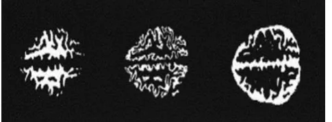
Figure 8 Segmentation Image
[image:4.595.315.556.474.669.2]Above figure 8 shows that the segmented image is an outcome with swarm antlion optimization algorithm based. It is a procedure of division a DIs into various segments. The main motive of segmentation is easy or alter the representation of an image. It is an usually to locate objects and curves, etc. in MRI images. Its more precise brain image segmentation is the procedure of evaluating the image label to all image pixels. Image pixels has a similar name shared specific properties.
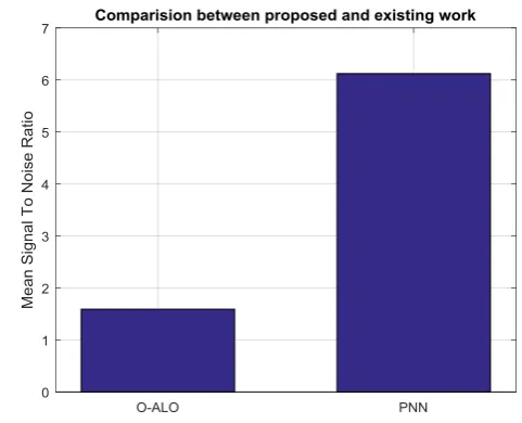
Figure 9 Comparison Between Proposed And Existing Work – PSNR
The comparison shows that the swarm lion optimization method and PNN classifier in PSNR. Peak Signal to Noise ratio is defined as the segmented image quality. In proposed work improve the performance parameters as compared with the existing classifier.
Figure 10 Comparison Between Proposed And Existing Work – MSE
[image:4.595.104.226.608.751.2]International Journal of Innovative Technology and Exploring Engineering (IJITEE) ISSN: 2278-3075, Volume-8 Issue-10, August 2019
[image:5.595.47.268.97.311.2]total sum of error rate in the segmented image. In proposed work MSE value is reduce the performance parameters as compared with the existing classifier.
Figure 11. Comparison Between Proposed And Existing Work – Accuracy Rate
[image:5.595.46.297.455.726.2]The comparison shows that the swarm lion optimization method and PNN classifier in ACCURACY rate parameter. Accuracy Rate is defined as the exact classifier value in the segmented image. In proposed work, Accuracy value enhanced the performance parameters as compared with the existing classifier.
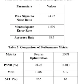
Table 1: Performance Analysis In Proposed Work
Parameters Values
Peak Signal to Noise Ratio
24.22
Means Square Error Rate
1.509
Accuracy Rate 98.5
Table 2: Comparison of Performance Metric
Metrics Swarm
Optimization
PNN
PSNR (%) 24.22 14.011
MSE 1.509 6.12
ACC (%) 98.5 65
Table 1 shows that the proposed parameter PSNR value is 24.22, the MSE value is 1.509, and Accuracy Rate value is 98.5. Table 2 shows the comparison between swarm ant lion optimization method with PNN classifier. It proposed method has implemented in brain image segmentation
improve the PSNR rate, Accuracy Rate, and reduce the value is error rate.
V. CONCLUSIONANDFUTURESCOPE
In this conclusion, brain image plays an essential role in the recognition of segmented brain tumor area. Brain recognition and segmentation help in presenting data related to functional arrangement and possible abnormal tissue in the brain, which is compulsory for diagnosis of brain disease. The primary objective of the segmentation of brain tumor is to locate the digital pixel in the estimated module, that may be standard or non-standard tissues. Several performance metrics also describes that the research method gives better result by enhancing particular parameters like Image Quality (PSNR), Error Rate (MSE) and Accuracy Rate (ACC). Experimental analysis shows that the research work can aid in the accurate and timely detection of brain tumor along with the verification of its feature extract position. The proposed method is essential for brain image segmentation from MRI image. Hybridization of Particle swarm optimization (PSOA) algorithm and Ant lion optimization (ALO) or Swarm Ant Lion method implemented to improve the PSNR value. Experimental analysis emphasis on performance metrics which are peak to signal noise ratio (PSNR), Error Rate (MSE) and Exact value (Accuracy Rate). The evaluated results from parameters are image quality factor (PSNR) value with 24.4, Error Rate (MSE) value with 1.509, and Exact value (Accuracy Rate) value with 98.58 %. The final result improves the performance metrics with PSNR and Accuracy Rate and reduce the error rates and compared with the existing method (PNN). In research work conclusion is suitable for verifying clinical decision support systems for primary screening and disease finding by the radiologists experts. In future , extraction of features will be based on the energy , compactness and regularity to determine the shape and location of brain tumor area. Although , most of the brain tumors are cancerous, some novel algorithms must be developed based on deep learning to detect and segment brain tumor area image.
REFERENCES
1.Bousselham, A., Bouattane, O., Youssfi, M., & Raihani, A. (2019). Towards Reinforced Brain Tumor Segmentation on MRI Images Based on Temperature Changes on Pathologic Area. International Journal of Biomedical Imaging, 2019.
2.Calabrese, C., Poppleton, H., Kocak, M., Hogg, T. L., Fuller, C., Hamner, B., ... & Frank, A. (2007). A perivascular niche for brain tumor stem cells. Cancer cell, vol. 11(1), pp.69-82.
3.Koestner, A., & Higgins, R. J. (2002). Tumors of the nervous system. Tumors in domestic animals, vol. 2(3),pp. 697-738.
4.Prastawa, M., Bullitt, E., Ho, S., & Gerig, G. (2004). A brain tumor segmentation framework based on outlier detection. Medical image analysis, 8(3), 275-283.
5.Rajasekaran, K. A., & Gounder, C. C. (2018). Advanced Brain Tumour Segmentation from MRI Images. In High-Resolution Neuroimaging-Basic Physical Principles and Clinical Applications. IntechOpen.
7. Howe, F. A., & Opstad, K. S. (2003). 1H MR spectroscopy of brain tumours and masses. NMR in Biomedicine, 16(3), 123-131.
8. Patil, R. C., & Bhalchandra, A. S. (2012). Brain tumour extraction from MRI images using MATLAB. International journal of electronics, communication & soft computing science and engineering, vol. 2(1),pp. 1-4.
9. Angulakshmi, M., & Lakshmi Priya, G. G. (2017). Automated brain tumour segmentation techniques—A review. International Journal of Imaging Systems and Technology, vol. 27(1), pp. 66-77.
10. Foroglou, N., Zamani, A., & Black, P. (2009). Intra-operative MRI (iop-MR) for brain tumour surgery. British journal of neurosurgery, vol. 23(1),pp. 14-22.
11. Nasir, M., Baig, A., & Khanum, A. (2014, June). Brain tumor classification in MRI scans using sparse representation. In International Conference on Image and Signal Processing (pp. 629-637). Springer, Cham.
12. Logeswari, T., & Karnan, M. (2010). An improved implementation of brain tumor detection using segmentation based on soft computing. Journal of Cancer Research and Experimental Oncology,vol. 2(1),pp. 006-014.
13. Selvakumar, J., Lakshmi, A., & Arivoli, T. (2012, March). Brain tumor segmentation and its area calculation in brain MR images using K-mean clustering and Fuzzy C-mean algorithm. In IEEE-International Conference on Advances in Engineering, Science and Management (ICAESM-2012) (pp. 186-190). IEEE.
14. Wu, M. N., Lin, C. C., & Chang, C. C. (2007, November). Brain tumor detection using color-based k-means clustering segmentation. In Third International Conference on Intelligent Information Hiding and Multimedia Signal Processing (IIH-MSP 2007) (Vol. 2, pp. 245-250). IEEE.
15. Xuan, X., & Liao, Q. (2007, August). Statistical structure analysis in MRI brain tumor segmentation. In Fourth International Conference on Image and Graphics (ICIG 2007)(pp. 421-426). IEEE.
16. Hasan, S. K., & Ahmad, M. (2018). Two-step verification of brain tumor segmentation using watershed-matching algorithm. Brain informatics,vol. 5(2), 8.
17. Kilic, H., Yuzgec, U., & Karakuzu, C. (2018). A novel improved antlion optimizer algorithm and its comparative performance. Neural C. 18. Kurnar, M., Sinha, A., and Bansode, N. V. (2018). Detection of Brain
Tumor in MRI Images by Applying Segmentation and Area Calculation Method Using SCILAB. In 2018 Fourth International Conference on Computing Communication Control and Automation (ICCUBEA) (pp. 1-5). IEEE.
19. Akter, L. A., and Kwon, G. R. (2018). Integration of Contourlet Transform and Canny Edge Detector for Brain Image Segmentation. In 2018 Tenth International Conference on Ubiquitous and Future Networks (ICUFN) (pp. 798-800). IEEE.
20. Bai, X., Chen, Z., Liu, M., and Zhang, Y. (2015). Center-free PFCM for MRI brain image segmentation. In 2015 IEEE International Conference on Image Processing (ICIP)(pp. 656-660). IEEE.
AUTHORSPROFILE
Sonali Bansal, received my B.Tech degree in Computer Science And Engineeering From Quest Group of Institutions, Jhanjeri, India 2017. I m currently M.Tech student at Chandigarh Engineering College Landran in the department of Computer Science & Engineering Since 2017. My area of intrest are Image Processing.
Dr.Shubpreet kaur is curently working as Associate Professor in Department of Computer Science & Engineering (CSE) in Chandigarh Group of Colleges, Landran. She holds Ph.D degree in CSE from Punjabi University,Patiala. M.Tech in CSE from Thapar University Patiala and received her B. Tech. in CSE from Chitkara University in 2008 and M.tech in 2011from Thapar University, Patiala. Her research interests include Data mining, and Expert Systems. She has published many research papers in journals and conferences of reputed publishers. Her Email I'd - [email protected]
Navdeep Kaur, received her B.Tech degree in computer Science and Engineering from Regional Institute of Management and Technology (RIMT), Mandi Gobindgarh, Punjab, India and M.Tech degree in Computer Science and Engineering from Punjabi