National
University
A Genetic Analysis of
Escherichia coli
Using Bioinformatic Methods
Phataraporn Khumpbai
May 2014
A thesis submitted for the degree of Doctor of Philosophy of
The Australian National University
I hereby certify that results presented in this thesis are my original work except where reference has been provided. Chapter 4 had a co-author but, in all cases, I am the primary contributor to the work. None of the work in this thesis has been submitted for the award of any other degree.
Phataraporn Khumphai
Acknowledgments
First of all, I would like to express my sincere gratitude and deep appreciation to my wonderful supervisor, David Gordon, for his excellent and valuable advice, continuous guidance, constructive criticisms, understanding and especially kindness to me throughout the study which was enable me to carry out my work successfully. I am also very grateful to the member of my supervisory panel, Rod Peakall and John Truman for helpful suggestions for my thesis
I am indebted to Division of Evolution, Ecology & Genetics, Research School of Biology for all facilities and to Ministry of Science and Technology, Thailand for financial supports. My sincere thanks are extended to all members in Gordon lab for
their helps with sincerity, kindness, encouragement andfriendship.
Finally, I wish to express my deepest appreciation to my parents, Na 'Na, P 'Tong, all lovely friends, and Knt for their love, understanding and encouragement throughout my study.
Phataraporn Khumphai
ABSTRACT
Escherichia coli is the best-known species of Enterobacteriaceae. The species is genetically
diverse and includes both commensal pathogenic strains and plays a significant role in veterinarian, environmental, and medical science. Despite the significance of E. coli, many aspects of the species biology, such as its genetic diversity and the pathogenesis are yet to
be truly understood. To understand the diversity of E. coli as a whole, genome data of
E. coli derived from variety of sources: humans, animals, and environment are essential. The aims of the research are to take the outstanding opportunity provided by the availability of many new E. coli genomes and to make use of a variety of bioinformatics tools to investigate the genetic diversity and reconstruct the evolutionary history of E. coli based on a genetic analysis and a comparative genomic approach. The thesis includes three main
themes. Firstly, "Distribution of extra-intestinal virulence traits among E. coli isolated from native Australian vertebrates with t~ose isolated from humans living in Australia".
The frequency and distribution of extra-intestinal virulence traits in a collection of E. coli
isolated from native Australian vertebrates as compared to E. coli isolated from humans living in Australia were reported. The result shows that the freque_ncy and distribution of
some traits varies with the source of isolation, human versus animal, and that there are traits typically associated with pathogenicity islands that are absent or very rare in animal isolates. The detected high rates of recombination in phylo-group B2 strains suggest
that this is an important evolutionary adaptation for attaining virulence. Secondly,
"Investigation of the evolution of conjugative plasmids in E. coli and their changing role in
E. coli ecology". Conjugative plasmids: key agents in the adaptation of E. coli populations were investigated. Comparing between Inell and IncF plasmids, Inell plasmids were
found to be more homogeneous and genetically conserved than IncF plasmids. These
plasmids have changed their role as mediators of intra- and interspecies interactions to become associated with E. coli virulence. Lastly, "Genetic and metabolic characteristics of phylo-group F". In this study, phylo-group F: a recently described group of E. coli strains was investigated. Strains belonging to phylo-group F were found to be closely related to phylo-group D strains known to be responsible for extra-intestinal infection. Whilst a high
degree of strain-specific genome differences were identified among F strains, some of genes
shared by F strains (absent in D, B2, and H299) were also present in other phylo-groups
(A, B 1, and E). All together the outcomes of this project lead to significant advances in our
TABLE OF CONTENTS
Page
ABSTRACT................................... 111
FIGURES AND TABLES.......................................... Vl CHAPTER 1: General Introduction and Research Significance 1 1.1 General Introduction... 1
1.2 Research Significance... 1
CHAPTER 2: Literature Reviews 3 2.1 Escherichia coli and the genus Escherichia.................. 3
2.2 Typing methods for E. coli... 3
2.2.1 Phenotypic methods... 3
2.2.2 Multi-locus enzyme electrophoresis (MLEE).. ... ... 4
2.2.3 DNA-based finger-printing methods... 4
2.2.4 Genome-based methods... 5
2.3 The diversity and phylo-group structure of E. coli... 5
2.3.1 Genome size and genomic diversity of E. coli... 6
2.3.2 Ecological niches and life-history characteristics of E. coli 7 2.3.3 Propensity to cause diseases of E. coli... 7
2.4 Conjugative plasmids in E. coli............. 10
2.5 Applications of bioinformatics in the genomic research of E. coli... 11
CHAPTER 3: Distribution of extra-intestinal virulence traits among E. coli isolated from native Australian vertebrates with those isolated from humans living in Australia 13 3 .1 Introduction... 13
3.2 Materials and Methods... 15
Page
CHAPTER 4: Investigation of the evolution of conjugative plasmids in
E. coli and their changing role in E. coli ecology 29
4.1 Introduction... 29
4.2 Materials and Methods... 32
4.3 Results... 42
4.4 Discussion and Conclusion... 70
CHAPTER 5: Genetic and metabolic characteristics of phylo-group F strains 77 5 .1 Introduction... 77
5.2 Materials and Methods ... ~... 79
5.3 Results... 86
5 .4 Discussion and Conclusion... 104
CHAPTER 6: Conclusion and Future Directions...................... 107
BIBLIOGRAPHY............................................................ 111
FIGURES AND TABLES
CHAPTER 2: Literature Reviews
Table I. Major virulence factors (VFs) encoded by
th . tr .
pa ogen1c s a1ns ... .
CHAPTER 3: Distribution of extra-intestinal virulence traits among
E. coli isolated from native Australian vertebrates with
those isolated from humans living in Australia
Table I. The distribution of 24 frequently detected traits
Page
8
among strains of the 4 genetic groups... 18
Figure 1. Phy lo genetic distribution of 24 virulence factors of
114 phylo-group B2 strains... 20 Figure 2. Phylogenetic distribution of 24 virulence factors of
55 phylo-group D and 2 phylo-group F (B093 and
H038) strains... 21
Table 2. Frequency(%) with which virulence-associated
traits was detected in E. coli strains with respect to
the phylo-group (B2 and D) membership of the
strains... 22
Table 3. Recombination in phylo-group B2 strains using the
PHI test, LDhat and ClonalFrame.. ... ... .. ... .. ... ... 24
CHAPTER 4: Investigation of the evolution of conjugative plasmids in
E. coli and their changing role in E. coli ecology
Table 1. Plasmids used in this study... 33 Table 2. Conjugative plasmids extracted from the E. coli
Antibiotic Resistance Database
(http://www.broadinstitute.org/)... 37 Figure 1. Phylogenetic relationships of the 19 plasmids in the
Rep I 1 group... 44
Figure 2. The transfer genes of 19 plasmids belonging to the
Repil (Inell) group... 45 Figure 3. Genotypic characteristics of 19 plasmids belonging
FIGURES AND TABLES (cont.)
Page
CHAPTER 4 (cont.)
Figure 4. Genes present in each unique genomic region of
conjugative plasmids in the Repll group... 4 7
Table 3. List of unique genes within unique genomic regions of
each conjugative plasmid in the Repll group... 48 Table 4. List of genes with known functions on conserved
segments sharing by all conjugative plasmids in the
Repll group based on the core genome data... 55 Table 5. List of genes with known functions on conserved
segments shares by conjugative plasmids in cluster A... 56
Table 6. List of genes with known functions on conserved
segments shares by conjugative plasmids in cluster B... 57 Figure 5. Phylogenetic relationships of the 55 plasmids
belonging to RepFIB/RepFIIA group... 59 Figure 6. The transfer genes of plasmids belonging to
RepFIB/RepFIIA (IncF) group... 62 Figure 7. Genotypic characteristics of 55 plasmids m the
RepFIB/RepFIIA (IncF) group in E. coli................. 65
Table 7. List of genes with known functions on conserved
segments sharing by conjugative plasmids in
the group (i).. ... ... .... ... ... 67
Table 8. List of genes with known functions on conserved
segments s?aring by conjugative plasmids in
the group (ii)... 68 Table 9. List of genes with known functions on conserved
segments sharing by conjugative plasmids in
FIGURES AND TABLES (cont.)
Page
CHAPTER 5: Genetic and metabolic characteristics of phylo-group F strains
Table 1. Principal characteristics of 23 Escherichia coli
genomes used in this study... 80
Figure 1. Maximum-likelihood tree of E.coli El227 and known phylo-group E. coli strains based on the
complete sequences of 22 housekeeping genes... 82
Figure 2. Maximum likelihood tree of 23 E.coli strains (F, D, and B2 strains) inferred from the core genome data
based on 3.52 Mb of nucleotide sequence... 87
Figure 3. The phylogeny of the 23 E. coli genomes inferred from the core genome data (3.52 Mb) implemented
using GenoPlast.... .. .. . . 88
Figure 4. The phylogeny of the 23 E. coli genomes inferred from the gene content data implemented using
GenoPlast... .. . . .. . . .. . . 89
Table 2. The list of genes on conserved segments (alignment
blocks greater than 500 nt) shares by strains in
cluster A (phylo-group F, D, and unknown
phylo-group H299 strains)... 91
Table 3. The list of genes on conserved segments (alignment
blocks greater than 500 nt) shares by strains in
cluster B (phylo-group B2 strains)... 94
Table 4. The list of genes on conserved segments (alignment
blocks greater than 500 nt) shares by phylo-group F
strains._ ... .-... 95
Table 5. The list of genes on conserved segments (alignment
blocks greater than 500 nt) shares by phylo-group D
FIGURES AND TABLES (cont.)
Page CHAPTER 5 (cont.)
Table 6. The number of enzymes (distinct ECs) for pathway x
in a given organism/total number of enzymes ( distinct
ECs) in the same pathway x defined in the KEGG
database... 97 Figure 5. The hierarchical clustering of metabolic profiles of 23
CHAPTER 1
General Introduction and Research Significance
1.1 General Introduction
Escherichia coli is the best-known species of Enterobacteriaceae and represents one of
the most important model organisms, especially E. coli K-12. E. coli is the most numerous facultative anaerobe presenting in the lower intestinal tract of birds and
mammals. The species is very genetically diverse and includes both commensal strains
with little ability to cause disease and pathogenic strains, which are able to cause
intestinal or extra-intestinal infections. In addition to its significance as a pathogen,
E. coli also plays a significant role in veterinarian, environmental, and medical science.
To understand the diversity of E. coli as a whole, the investigation of genome data of
E. coli derived from variety of sources: humans, animals, and environment is essential.
Thanks to high throughput sequencing technology, a number of complete and draft
E. coli genomes are available at the GenBank database. However, most of the E. coli
strains in the database are pathogenic strains isolated from humans. This limited information may lead to a biased assessment of the diversity to be found in E. coli.
However, there is a growing body of genome sequence data for E. coli, for example, the
Escherichia genome sequencing project recently undertaken by the Broad Institute of
MIT and Harvard. Consequently, there is a more genome database representing the diversity of E. coli derived from the variety of sources. The availability of these
genome sequence data will enable researchers to address a range of questions
concerning studies of E. coli.
1.2 Research Significance
Strains of E. coli species play a significant role m several aspects: veterinarian,
environmental, and medical science. Focusing on its ability to cause a variety of
diseases, E. coli is considered to be a major cause of human morbidity and mortality around the world. Despite the significance of E. coli, however, many aspects of the
.
.Therefore the aims of the research are to take advantage of the outstanding opportunity
provided by the availability of many E. coli genomes and to make use of a variety of bioinformatics tools to investigate the genetic diversity and reconstruct the evolutionary
history of E. coli. To understand the diversity within the species, strains from a variety of sources: human, animal, and environment were investigated using a comparative
genomic approach. The outcomes of this project will lead to significant advances in our
understanding presented in E. coli species. Consequently, the thesis includes three main themes with a specific objective for each chapter as follows:
Chapter 3: Distribution of Extra-intestinal Virulence Traits among E. coli Isolated
from Native Australian Vertebrates with Those Isolated from Humans
Living in Australia
Objective: To examine the frequency and distribution of extra-intestinal virulence traits in a collection of E. coli isolated from native Australian vertebrates as compared to E. coli isolated from humans living in Australia.
Chapter 4: Investigation of the Evolution of Conjugative Plasmids in E. coli and
Their Changing Role in E. coli Ecology
Objective: To investigate the evolution of two of the most common types of conjugative plasmids to be found in E. coli: IncF and Inell, and their changing role in E. coli ecology.
Chapter 5: Genetic and Metabolic Characteristics of Phylo-group F Strains
CHAPTER2
Literature Reviews
2.1 Escherichia coli and the genus Escherichia
E. coli is the best-known species of Enterobacteriaceae and represents one of the most important model organisms, especially E. coli K-12. E. coli is the most numerous facultative anaerobe presenting in the lower intestinal tract of birds and mammals.
The species is very genetically diverse. E. coli strains include both commensal variants
with little ability to cause disease and various pathogenic strains that are able to cause
intestinal or extra-intestinal infections. In addition to its significance as a pathogen, E. coli also plays a significant role in veterinarian, environmental, and medical science.
Besides E. coli, there are other closely related species within the genus Escherichia such
as E. fergusonii, E. albertii, and the 5 novel clades of the genus Escherichia including
Clade I - Clade V (Walk et al., 2009). The Escherichia spec-ies and novel clades
differed in their rates of evolution and E. fergusonii has evolved at an accelerated rate
under selection conditions (Walk et al., 2009). Among 5 novel clades, Clade I was the most closely related clade to E. coli, whilst Clade V was the most distantly related clade. These 5 novel clades can be distinguished from typical E. coli using a gene
sequence analysis, however they were not discriminated from E. coli by a traditional biochemic·a1 profiling with the exception for Clade III (Walk et al., 2009).
2.2 Typing methods for E. coli
The highly genetic diversity is found among E.coli isolates and is what enable the species to exhibit such a variety of life style (Gordon, 20 l 0). Various typing methods have been developed to differentiate among isolates of E. coli species.
2.2.1 Phenotypic methods
Serotyping: Serotyping involves the determination of strains by the somatic (0),
capsular (K), or flagellar (H) antigen present on a strain (Kauffmann, 1947).
However the method cannot be used to discriminate the phylogenetic relationships
among stains when genetically distinct E. coli genotype have been found to have the
same serotype (Caugant et al., 1985).
Biochemical and antibiotic resistance profiling: Biochemical and antibiotic
resistance profiling are inexpensive and provide a reasonable degree of discrimination
among strains, as they are so phenotypically diverse. However, both techniques are
very dependent on specific conditions: incubation time for biochemical profiling and the
background level of resistance of a strain for antibiotic resistance profiling (Gordon,
2010). In addition, both techniques cannot be used to assess the genetic relatedness of E. coli strains.
2.2.2 Multi-locus enzyme electrophoresis (lvfLEE)
The technique analyses the genetic differences between E. coli isolates by discovering
variants of a range of constitutively expressed enzymes in a species based on their
electrophoretic mobility (Selander et al., 1986). H_owever the method is no longer in
common use as it is expensive and time consuming as compare with many of new DNA-based typing methods.
2.2.3 DNA-based methods
There is an abundance of DNA typing methods including random amplification of
polymorphic DNA (RAPD), ribotyping, amplification fragment length polymorphism
(AFLP), plused-field gel electrophoresis (PFGE), repetitive extragenic palindromic
PCR (rep-PCR), and multi-locus sequence typing (MLST). The methods all have an advantage but also a disadvantage for each technique.
Of these techniques, PFGE is thought to be the sensitive discriminating method.
However the technique is extremely labor intensive· making it unsuitable for large scale studies (Gordon, 2010). Consequently, many studies currently employ rep-PCR as the
.
.utilizes oligonucleotide primer complementary to particular repetitive sequences within the E. coli genome (Versalovic et al., 1991). However the degree of discrimination of rep-PCR obtained with the choice of primer used (Mohapatra et al., 2007).
The multi-locus sequence typing (MLST) uses the allele profile data of selected housekeeping genes (usually 7 genes), which the nucleotide sequence of a 300-700 bp
region of each gene is determined, to assign a strain to a sequence type (ST) (Maiden
et al., 1998, Gordon, 2010). The extensive collections of STs found in E. coli are available, for example the largest MLST database at the ERI, University College Cork available online at http://mlst.ucc.ie/mlst/dbs/Ecoli. MLST is also the method used to assign an unknown isolate to a phylo-group of E. coli. Although MLST is more discriminating as compare to PFGE, the method is expensive and time consuming.
In addition, a PCR-based method is shown to be more sensitive and discriminating than
MLST (Clermont et al., 2013).
2.2.4 Genome-based methods
The growing body of E. coli genome data has provided evidence of the extent of
substructure in E coli. Based on whole-genome scale analysis, the genomic data are informative and also more reliably reflect the phylogenetic relationships among strains. In addition, the available E. coli genomes were used to develop primers of PCR-based methods resulting in the improvement of specificity and detection of a new phylo-group
(Clermont et al., 2013).
2.3 The diversity and phylo-group structure of E. coli
E. coli has a largely clonality which enable E. coli strains to be classified into a number of distinct phylo-group. The existence of extensive substructure within the species has been demonstrated using various typing methods. Based on methods such as multi
-locus enzyme electrophoresis (MLEE) (Selander et al., 1986) and multi-locus sequence typing (MLST) (Gordon et al., 2008), E. coli can be subdivided into 5 main phylo-groups known as A, Bl, B2, D, and E. Among these phylo-groups, Dis described as
to phylo-group E are rare, and are largely enterohaemorrhagic E. coli (EHEC)
(Gordon et al., 2008). However, due to the diversity of the strains and the growing body
of multi-locus sequence data and genome data for E. coli, the additional phylo-groups
have been recently delineated. Based on the recent method: the extended quadruplex PCR phylo-group assignment, E. coli are now assigned to 8 phylo-groups including A,
Bl, 82, C, D, E, F, and Escherichia cryptic clade I (Clermont et al., 2013). A
phylo-group C is described as a phylo-group of strains closely related to the B 1 phylo-group (Clermont et
al., 2011). While a phylo-group F has been suggested for a sister group to phylo-group
82 (Jaureguy et al., 2008, Clermont et al., 2011 ).
E.coli strains have phylogenetic cohesiveness; however among strains of the various
phylo-groups, they differ in their phenotypic characteristics (Gordon, 2004), genome
size (Touchon et al., 2009), and propensity to cause intestinal or extra-intestinal infections. Strains of the different phylo-groups are also associated with certain
ecological niches and life-history characteristics (Gordon and Cowling, 2003). The phenotypic differences of the phylo-groups of E. coli comprise differing growth rates, antibiotic resistance, and biochemical profiles (Gordon, 2004). E.coli strains
are phenotypically heterogeneous (Touchon et al., 2009). However, evidence suggested
that the environmentally adapted E. coli lineages were found to be phenotypically and taxonomically indistinguishable from typical E. coli based on traditional phenotypic tests (Luo et al., 2011 ).
2.3.1 Genome size and genomic diversity ofE. coli
Genomes within the E. coli species can differ in size by more than 1 Mb. Among sttains of the different phyla-groups, strains belonging to phylo-groups A and
B 1 have smaller genomes than 82 or D strains (Bergthorsson and Ochman, 1998).
The investigation of 20 E. c;oli genomes from 5 main phylo-groups (A, Bl, B2, D, and
E) showed that the average size of an E. coli genome was around 5 Mb and represented about 4700 protein-coding genes (Touchon ~t al., 2009). However, only 46% were
common to all genomes investigated. Of about 18,000 total genes for the pan-genome, there were about 11,000 genes with no strong relation of homology and about 10,000
unique genes after removing all transposable elements and prophage (Touchon et al., . .
at least 18 genomes, whereas 26% were present in 4 or fewer genomes (Touchon et al., 2009). These indicated the enormous genetic diversity present in the species and they are what enable E. coli strains to exhibit such a variety of life styles.
2.3.2 Ecological niches and life-history characteristics of E. coli
The ecological niches of E. coli strains have been found to correlate with phylo-group membership. According to the genetic diversity of the species, strains belonging to phylo-groups B2 and D are less frequently isolated from the environment (Walk et al.,
2007) or fish, frogs, and reptiles than A or Bl strains (Gordon and Cowling, 2003). However, B2 and D strains are more frequently recovered from extra-intestinal body sites than A or B 1 strains (Gordon, 2004). In addition, B2 strains are rarely isolated from water samples; however, the strains are more frequently isolated from mammal
hosts possessing hindgut modifications for microbial fermentation than other strains of E. coli (Gordon and Cowling, 2003). Moreover, B2 strains have been shown to persist for longer periods in infants than strains of the other phylo-groups (Nowrouzian et al.,
2006).
Generally, E. coli depends on the presence of a vertebrate host population to persist as
the primary habitat. However, E. coli can transit in water, sediment, and soil, where
represent the species' secondary habitat (Savageau, 1983). There is evidence that some strains may be essentially free living in the environment independent of warm-blooded
hosts (Power et al., 2005). For example, two environmental strains belonging to phylo-group F: E. coli SMS-3-5 (Fricke et al., 2008) and E. coli E1227 (this study). In addition, the strains belonging to novel five Escherichia clades (Clade I to Clade V)
were isolated primarily from environmental sources (Luo et al., 2011 ).
2.3.3 Propensity to cause diseases ofE. coli
Escherichia coli is genetically diverse and includes both commensal and vanous
pathogenic strains. The ability of a strain to cause disease is due to the presence of a range of traits thought to enhance the ability to cause disease of a strain. Many of
the most significant virulence genes: adhesins, extracellular protein secretion systems,
pathogenicity-associated islands (P Als) which are known to normally present in pathogens. Although
the majority of traits associated with P Als are located on the chromosome, many are
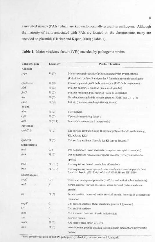
encoded on plasmids (Hacker and Kaper, 2000) (Table 1 ).
Table 1. Major virulence factors (VFs) encoded by pathogenic strains
Category/ gene
Adhesins papA sfa lfocDE sfaS focG iha eaeA Toxins hlyA cnfl astA Protectios kpsMTII kpsMTKI
Sideropho_res
iutA fyuA iroN ireA Miscellaneous cvaC traT iss ompT H7 ibeA she ma/X irp2 Location*
PI (C)
Pl (C) Pl (C) PI (C) PJ (C) PI (C)
Pl (C) Pl (C) PI (C, P)
PI (C)
PI (C)
C,P
PI (C)
PI (C, P)
PI (C, P)
C, p
p
PI (P)
C
C
C
C
PI (C)
PI (C)
Product/ function
Major structural subunit of pilus associated with pyelonephritis
(P fimbriae); defines F antigen the P fimbrial structural subunit gene Central region of sfa (S fimbriae) and foe (FI C fimbriae) operons
Pilus tip adhesin, S fimbriae (sialic acid specific)
Pilus tip molecule, FIC fimbriae (sialic acid specific)
Novel nonhemagglutinin adhesin (from Ol57:H7 and CFT073)
lntimin (mediates attaching/effacing lesions)
a-Hemolysin
Cytotoxic necrotizing factor I heat-stable enterotoxin 1 ( enterotoxin)
Cell surface attribute: Group II capsular polysaccharide synthesis (e.g.,
Kl, KS, and K12)
Cell surface attribute: Specific for KI (group II) kpsMT
Iron acquisition: Ferric aerobactin receptor (iron uptake: transport)
Iron acquisition: Yersinia siderophore receptor (ferric yersiniabactin
uptake)
Iron acquisition: Novel catecholate siderophore
Iron acquisition: iron-regulated outer membrane virulence protein (also
found in plasmid pEl 1210pl of E.coli 0104:1-14 str. El 12/10)
Colicin V; conjugative plasmids (traT, iss, and antimicrobial resistance)
Serum survival: Surface exclusion, serum survival (outer membrane protein)
Serum survival: increased serum survival protein; involved in complement resistance
Cell surface attribute: Outer membrane protein T (protease) Cell surface attribute
Cell invasion: Invasion of brain endothelium Secreted protein
PAI marker from strain CFT073
non-ribosomal peptide synthase (yersiniabactin siderophore biosynthetic protein)
[image:18.579.15.550.20.852.2]The pathogenic E. coli can be divided into strains causing intestinal diseases and other
strains causing extra-intestinal infections (Kaper et al., 2004). These lead to categories
of the pathotypes of E. coli into intestinal and extra-intestinal pathotypes. The intestinal
pathotypes consist of enterotoxigenic E. coli (ETEC), enteropathogenic E. coli (EPEC),
enterohemorrhagic E. coli (EHEC), enteroaggregative E. coli (EAEC), enteroinvasive
E. coli (EIEC), and diffusely adherent E. coli (DAEC) (Nataro and Kaper, 1998, Nataro, 2005, Kaper et al., 2004). The extra-intestinal pathotype includes extra-intestinal
pathogenic E. coli (ExPEC) which can be subdivide into uropathogenic E. coli (UPEC),
neonatal meningitis-associated E. coli (NMEC), necrotoxigenic E. coli (NTEC), and
avian pathogenic E. coli (APEC) (Kaper et al., 2004).
The ability of a strain to cause diverse diseases varies among strains of various
phylo-groups. As phylo-group B2 and to a lesser extent phylo-group D strains are frequently
isolated from intestinal body sites, they are most likely to be responsible for
extra-intestinal infections. In contrast, phylo-group A strains are less likely and phlyo-group
B 1 strains the least likely to cause such infections (Johnson and Russo, 2002, Jaureguy
et al., 2008). Strains belonging to phylo-groups A and Bl are typi-cally only known to
cause opportunistic infections in compromised hosts (Moreno et al., 2005).
The distribution of extra-intestinal disease associated traits varies among strains of the different phylo-groups (Johnson et al., 2001) and numerous studies have shown that
most extra-intestinal virulence genes are concentrated in phylo-group B2 strains and
to a lesser extent phylo-group D strains (Johnson et al., 2001, Gordon et al., 2005).
Strains belonging to phylo-groups B2 and D express a number of putative virulence
traits that are proposed to play roles in the process of causing disease (Bahrani-Mougeot
et al., 2002). These traits include fimbriae, capsule, hemolysins, membrane-bound and
secreted proteins, lipopolysaccharides, and iron- acquisition systems, which appear
more frequently in uropathogenic E.coli (UPEC) (Johnson, 1991). Most UPEC strains
belonging to phylo-groups B2 and D carry large blocks of genes encoding virulence
associated traits, called pathogenicity-associated islands (PAis), not found in fecal
isolates (Bahrani-Mougeot et al., 2002, Johnson and Russo, 2002, Johnson, 1991 ).
For example, papA gene encodes a structural subunit of P fimbriae which mediate
attachment to intestinal and urinary epithelium, and iutA gene encodes an aerobactin
.
.enable B2 and D strains to cause extra-intestinal infections beyond fecal E. coli such as
strains in phylo-groups A and B 1 that are most likely to cause intestinal infections
(Bahrani-Mougeot et al., 2002, Johnson et al., 2006). However available evidence has
revealed that E. coli strains belonging to phylo-group B 1 can also often cause extra-intestinal infections in birds (Barbieri et al., 2012). These virulence factors are
considered to be one of the molecular factors that sustain the diversity of adaptive paths
and complexity of E. coli niches (Tenaillon et al., 2010).
2.4 Conjugative plasmids in E. coli
Plasmids are extrachromosomal replicons that are prevalent symbionts of bacteria.
Generally, plasmids are not essential for normal bacterial growth. However they often
code for genes involved in antibiotic and heavy metal resistance, virulence, and
ecological interactions. That is traits that encode for their host's adaptation to the
environment. Plasmids can propagate themselves vertically via cell division and many
can propagate themselves horizontally usually by infectious transfer via mobilization or conjugation (Summers, 1996, Bergstrom et al., 2000).
Based on mobility systems of plasmids, they can be categorized into three groups;
conjugative ( self-transmissable ), mobilizable (transmissible), and nonmobilizable
plasmids (Smillie et al., 2010). A mechanism of conjugation of conjugative plasmids involves four components of a conjugative machinery needed for self-transfer that
includes an origin of transfer ( oriT), a relaxase gene, a type IV coupling protein (T4CP),
and a type IV secretion system (T4SS) (Smillie et al., 2010). All conjugative plasmids
possess core components of a plasmid backbone including replication genes, stability
genes, and a conjugative transfer (tra) region ensuring their self-transmissibility.
Conjugation encoded by plasmids involves a donor bacterium carrying a conjugative
plasmid and a recipient cell without a conjugative plasmid. Conjugation between donor and recipient cells of some conjugative plasmids can occur between different species, genera, or kingdoms (Amabile-Cuevas and Chicurel, 1992). Thanks to these properties
In E. coli, many plasmids types comprising a number of plasmid incompatibility (Inc)
groups (or plasmid replicon (Rep) groups which are used interchangeably) are known to occur among E. coli strains and they play an important role in the "adaptation" of
bacterial populations (Frost et al., 2005). Of these Inc groups, almost conjugative
plasmids belonging to IncF group carrying a fertility factor (F factor) transfer region for
their self-transmissibility were associated with E. coli virulence (Johnson and Nolan, 2009). Although a number of other Inc (Rep) groups have been identified, only a few of them (i.e., IncB/O and Inell) have .been found to be associated with E. coli virulence (Johnson and Nolan, 2009).
However these E. coli plasmids originally encoded traits thought to mediate competitive
interactions among strains, traits known as bacteriocins: key agents mediating a strain's
adaptation to local environmental niches (Eberhard, 1990). Bacteriocins are defined as antimicrobial proteins with a narrow killing range, that are toxic only to bacteria closely related to the producing strain (Riley and Wertz, 2002). These substances play
a significant role in maintaining microbial biodiversity by acting as important mediators
of intra- and interspecies interactions (Kirkup and Riley, 2004, Czaran et al., 2002). In E. coli, the production of multiple bacteriocins by a single strain is a common
phenomenon (Gordon and O'Brien, 2006). Several bacteriocins have been found to be encoded on the same large conjugative plasmid in E. coli strains more often than
expected by chance (Gordon and O'Brien, 2006, Gordon et al., 2007).
2.5 Applications of bioinformatics in the genomic research of E. coli
Advances in innovative high-throughput biotechnologies, especially in a high
throughput sequencing technology, are resulting in the exponential growth of
high-dimensional data. Consequently, the huge amounts of biological data have created
an enormous challenge to optimize and make effective use of accumulated information.
Thanks to modem computational tools in bioinformatics, they have revolutionized biological research by providing powerful approaches for managing biological
databases and investigating biological data systematically.
The National Center for Biotechnology Information (NCBI) in the United States
(http://www.ncbi.nlm.nih.gov/) and the European Bioinformatics Institute (EBI) in
England (http://www.ebi.ac.uk/) are two classic life science servers maintaining
databases and software tools in the life sciences. The usefulness of these and other
centralized databases allow scientific community to make effective use of the databanks
as free resources of the structure and function of genes and proteins in biology and
medical research.
Furthermore, advances in computational methodologies have generated a variety of bioinformatic tools for investigating biological data ranging from the individual nucleic
acid sequence to systems biology of the living cell. These include tools for nucleotide
and protein sequence analysis, phylogenetic analysis, genomics, proteomics,
transcriptomics, metabolomics, systems biology, etc. Several bioinformatic tools are
freely-accessed on-line for scientists to make use of them as a set of scientific tools to
address a range of research.
Thanks to a high throughput sequencing technology, a number of E. coli genomes are
being sequenced to serve as resources of the diversity within E. coli and the genus
Escherichia. The power of the sequencing technology, i.e., the Genome Sequencer
FLX System ( 454 Life Sciences, Roche) has also been applied to generate enormous
sequence information for studying the genomic content in a complex mixture of microorganisms so called Metagenomics (Harkins and Jarvie, 2007). To manage E. coli
genome sequences systematically, many special databases of E. coli have been
established by several institutes. The database, such as the Escherichia coli Antibiotic
Resistance Database hosted by the Broad Institute of MIT and Harvard brings together the E. coli' genome sequences and their annotation with bioinformatic tools providing
users with BLAST search and several comparative analysis tools. The other database,
ASAP, the database hosted by University of Wisconsin-Madison also brings together
the E. coli genome annotation and bioinformatic tools with other bacterial strains.
In addition to the database for E. coli genomes, there are databases for Multi-locus sequence typing (MLST) of E. coli (EcMLST, Escherichia coli MLST Database, etc.)
CHAPTER]
Distribution of extra-intestinal virulence traits among E. coli isolated from native
Australian vertebrates with those isolated from humans living in Australia
3.1 Introduction
Escherichia coli is the most numerous facultative anaerobe present in the lower
gastrointestinal tract of humans and other warm-blooded species. Although normally a commensal, strains of the species can cause a variety of intestinal and extra-intestinal infections. Methods such as multi-locus enzyme electrophoresis (MLEE) (Och.man and Selander, 1984) and multi-locus sequence typing (MLST) (Gordon et al., 2008) have shown that E. coli can be subdivided into five main phylo-groups known as A, B 1, B2, D and E. Besides these phylo-groups, an additional phylo-group called F has been delineated (Jaureguy et al., 2008). Strains of the various phylo-groups may differ in
many aspects, including their phenotypic characteristics (Gordon, 2004), genome size (Touchon et al., 2009), ecological niches, and life-history characteristics (Gordon and Cowling, 2003). Strains of the different phylo-groups also vary in their propensity to cause extra-intestinal infections. Phylo-group B2 and to a lesser extent phylo-group D
strains are most likely to be responsible for extra-intestinal infections in humans, whilst phylo-group A are less likely, and phylo-group B 1 strains the least likely to cause such infections (Jaureguy et al., 2008; Johnson, 2002). However, available evidence has revealed that E. coli strains belonging to phylo-group B 1 can also often cause extra-intestinal infections in birds (Barbieri et al., 2012).
According to MLST and several PCR-based assays for the identification of highly virulent clonal group, these methods have suggested that some particular sequence types (STs) and clonal groups may be clinically significance as they are found to be common and responsible for extra-intestinal infection in E. coli virulent strains (Bonacorsi et al.,
been identified (Johnson and Russo, 2002). It is well known that the distribution
of these extra-intestinal disease associated traits vanes among strains of
the four main phylo-groups (A, Bl, B2 and D) (Johnson et al., 2001) and numerous
studies have shown that many of these traits are concentrated in phylo-group B2 strains
and to a lesser extent phylo-group D strains (Johnson et al., 2001, Gordon et al., 2005).
Many of the most significant virulence genes ( adhesins, extracellular protein secretion
systems, and toxins) for several E. coli pathotypes are clustered together in
pathogenicity-associated islands (PAis). For example, the High Pathogenicity Island
(HPI) that encodes the virulence genes fyuA and irp2 (Schubert et al., 2002). This and
other E. coli pathogenicity-associated islands (PAI) are concentrated in phylo-groups
B2 and D, and these islands are only occasionally found in phylo-groups A and B 1
(Schubert et al., 2009, Clermont et al., 2001, Boyd and Hartl, 1998). Conjugative
transfer and homologous DNA recombination are reported to play a major role in
horizontal transfer of PAI within E. coli (Schubert et al., 2009).
Previous study has demonstrated that phylo-group B2 strains known to be responsible
for extra-intestinal infections in humans are more frequently isolated- from mammal host
possessing hindgut modifications for microbial fermentation than other strains' of
E. coli (Gordon and Cowling, 2003). Phylo-group B2 strains have been also capable of
persisting in infants than strains of the other phylo-group (Nowrouzian et al., 2006).
There is some controversy as to whether the propensity of phylo-group B2 strains to
cause disease is a consequence of the concentration of known and suspected virulence
factors among phylo-group B2 strains (Johnson et al., 2006) or an ancestral
characteristic of this phylo-group (Le Gall et al., 2007). Johnson et al. (2006) showed
that it is the presence of specific virulence factors in a strain that are the best predictor
of a straih's virulence in a mouse lethality model rather than its phylo-group
membership. By contrast, Le Gall-et al. (2007) argued that the virulence of phylo-group
B2 strains in the mouse lethality assay is an ancestral trait, and extra-intestinal virulence
is a coincidental by-product of these strains' enhanced ability to persist in the
gastro-intestinal tract compared to strains of the other phylo-groups. However the exact
answer for this controversy is still unclear. Additional investigations might be required
to deal with the fact that B2 strains are competitively dominate in the gut leading to the
numberical abundant of the strains. B2 strains ~lso show the ability of persisting for
dominate and persisting for longer period in the gut) should be taken into account as these make it likely that it will be B2 strains responsible for the contamination event.
The majority of studies examining the distribution of extra-intestinal virulence traits
among strains of the different E. coli phylo-groups have been clinical isolates or isolates
from humans. Few studies have examined the distribution of the traits in strains
isolated from native animals. Here we report on the frequency and distribution of
extra-intestinal virulence traits in a collection of E. coli isolated from native Australian
vertebrates (Gordon & Cowling, 2003) as compared to E. coli isolated from humans
living in Australia (Gordon et al., 2005). We show that the frequency and distribution
of some traits varies with the source of isolation, human versus animal, and that there
are traits typically associated with pathogenicity-associated islands (P Als) that are
absent or very rare in animal isolates.
3.2 Materials and Methods
3.2.1 Strain collection
The strains used in this study were 266 E. coli isolated from faecal samples taken from
people living in the Canberra region of Australia and 690 faecal isolates from
native non-human vertebrates living in Australia. Further details of the collection and
identification of the strains were described in Gordon et al. (2005) for the isolates from
humans and· in Gordon & Cowling (2003) for the isolates from animals. All isolates
were previously assigned to one of the four main E. coli phylo-groups (A, B 1, B2, and
D) (Ochman & Selander, 1984; Herzer et al., 1990) using a PCR based technique
(Clermont et al., 2000). Sample sizes are as follows: isolates from humans; A (n = 52),
Bl (n = 33), B2 (n = 120), D (n = 61): animals; A (n = 93), Bl (n = 272), B2 (n = 213),
D (n = 120). Strain are labeled with a letter prefix indicating host group: H, human;
MIT A, mammal (non-human); B, bird; R, reptile; and E, environmental.
3.2.2 Virulence gene detection
The 956 E. coli isolates (266 human and 690 animal isolates) were previously screened
disease including malX, irp2, afa/draBC, eaeA, jimH, focG, gajD, papAH, sfa/focDE,
ibeA, H7 fliC, kpsMT.II, kpsmMT.Kl, ompT,fyuA, iha, ireA, iroN, iutA, iss, traT, astA,
cnfl, hylD, stxl, stx2, she, eaag, and l 3jb. All 29 virulence genes vvere detected using
a PCR method as described by Gordon et al. (2005). The information of detected
virulence factors (VFs) were used for the investigation of the distribution of VFs in this
collection of E. coli strains.
3.2.3 MLST analysis of E. coli strains belonging to phylo-groups B2 and D
Strain selection for lvfLST: Due to their clinical significance, a subset of 171
phylo-group B2 and D strains were selected from David Gordon's E. coli strain collection
for MLST characterization. Included in the study were clinical and faecal human
isolates (B2 = 33, D = 19, and F = 1), faecal isolates from native non-human vertebrates
(B2 = 65, D = 30, and F = 1 ), and isolates from soil and water (B2 = 16, D = 6)
collected in Australia. The environmental isolates were PCR-screened for the 29
virulence factors described in the previous section. The MLST data subsequently
revealed that 2 of the D strains were actually members of phylo-group F.
lvfLST Sequencing: The 171 E. coli isolates (114 phylo-group B2, 55 phylo-group D,
and 2 phylo-group F strains) were characterized using the MLST scheme (German)
described by Wirth
et
al., (2006). This scheme examines the 7 housekeeping genes:adk, fumC, gyrB, icd, mdh, purA, and recA. Sequence data for the 7 genes were
concatenated (3,422 bp) and analyzed to construct phylogenetic relationships among strains for each phyla-group using MEGA5 program (neighbor joining (NJ) method;
p-distance model) (Tamura et al., 2007). Sequence data for the 7 genes were also
submitted to the MLST database (http://mlst.ucc.ie/mlst/dbs/Ecoli) in order to obtain
a sequence type (ST) for each of the strains.
3.2.4 Analysis of recombination rates in phylo-group B2 strains
The concatenated sequences (3,422 bp) of 114 phylo-group B2 strains were partitioned
into 4 input sample sets based on their source (humans, . mammals, birds, and
environment). The duplicate STs in each input s~mple set were removed. The number
used for the investigation of putative recombination rates usmg three methods to compare the consistency of recombination rates derived from each method.
The first method used was the PHI statistic ( <pw) (Bruen et al., 2006), as implemented in SplitsTree4 (Huson and Bryant, 2006). The second method, was Clonal Frame,
a model-based approach for determining bacterial microevolution (Didelot and Falush,
2007). The last method was, a coalescent-based likelihood permutation test, as implemented in LDhat 2.1 (McVean et al., 2002).
3.2.5 Statistical analyses
Statistical analyses were performed usmg the package JMP® (SAS Institute).
The Fisher's Exact test was used to determine if there are nonrandom associations between the distribution of virulence-associated traits and the phyla-group membership of the strains. Effects were not considered to be significant unless the probability values were less than 0.05.
3.3. Results
3.3.1 Distribution of virulence factors (VFs) among
E.
coli strains belonging to4 main phylo-groups
The distribution of 29 VFs for human- and animal-source isolates from the 4 main phyla-groups, A, B 1, B2, and D was determined. Among the 956 isolates, the virulence-associated trait I 3jb was not detected, whilst stxl, stx2, eaag, and gajD were detected 2, 3, 4, and 6 times, respectively (data not shown).
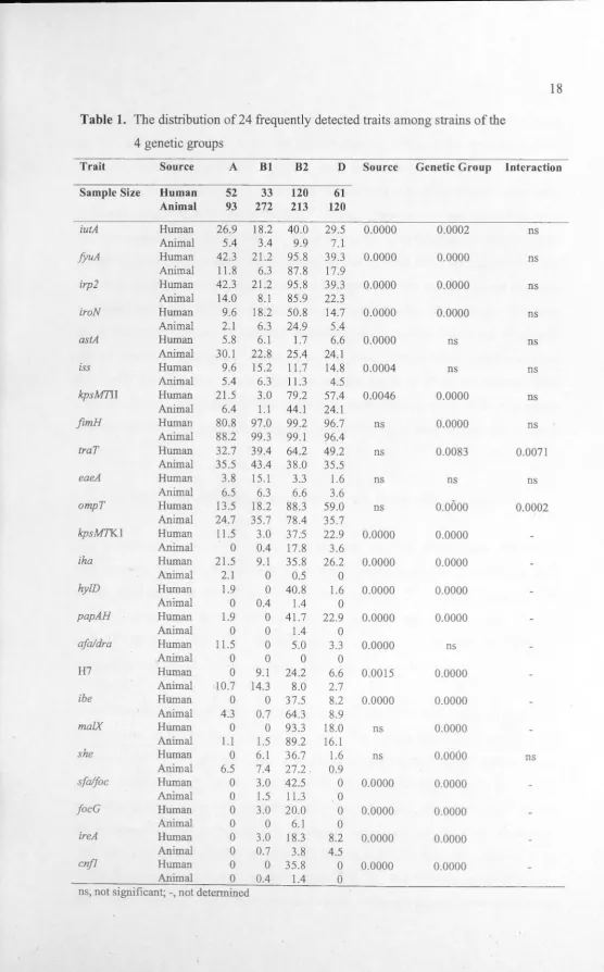
Table 1. The distribution of 24 frequently detected traits among strains of the
4 genetic groups
Trait Source A Bl B2 D Source Genetic Group Interaction
Sample Size Human 52 33 120 61
Animal 93 272 213 120
iutA Human 26.9 18.2 40.0 29.5 0.0000 0.0002 ns
Animal 5.4 3.4 9.9 7.1
fyuA Human 42.3 21.2 95.8 39.3 0.0000 0.0000 ns
Animal 11.8 6.3 87.8 17.9
irp2 Human 42.3 21.2 95.8 39.3 0.0000 0.0000 ns
Animal 14.0 8.1 85.9 22.3
iroN Human 9.6 18.2 50.8 14.7 0.0000 0.0000 ns
Animal 2.1 6.3 24.9 5.4
astA Human 5.8 6.1 1.7 6.6 0.0000 ns ns
Animal 30.1 22.8 25.4 24.1
iss Human 9.6 15.2 11.7 14.8 0.0004 ns ns
Animal 5.4 6.3 11.3 4.5
kpsM71I Human 21.5 3.0 79.2 57.4 0.0046 0.0000 ns
Animal 6.4 1.1 44.1 24.1
fimH Human 80.8 97.0 99.2 96.7 ns 0.0000 ns
Animal 88.2 99.3 99.1 96.4
traT Human 32.7 39.4 64.2 49.2 ns 0.0083 0.0071
Animal 35.5 43.4 38.0 35.5
eaeA Human 3.8 15.1 3.3 1.6 ns ns ns
Animal 6.5 6.3 6.6 3.6
ompT Human 13.5 18.2 88.3 59.0 ns 0.0000 0.0002
Animal 24.7 35.7 78.4 35.7
kpsMTKl Human 11.5 3.0 37.5 22.9 0.0000 0.0000
Animal 0 0.4 17.8 3.6
iha Human 21.5 9.1 35.8 26.2 0.0000 0.0000
Animal 2.1 0 0.5 0
hy!D Human 1.9 0 40.8 1.6 0.0000 0.0000
Animal 0 0.4 1.4 0
papAH Human 1.9 0 41.7 22.9 0.0000 0.0000
Animal 0 0 1.4 0
afa/dra Human 11.5 0 5.0 3.3 0.0000 ns
Animal 0 0 0 0
H7 Human 0 9.1 24.2 6.6 0.0015 0.0000
Animal 10.7 14.3 8.0 2.7
ibe Human 0 0 37.5 8.2 0.0000 0.0000
Animal 4.3 0.7 64.3 8.9
malX Human 0 0 93.3 18.0 ns 0.0000
Animal 1.1 1.5 89.2 16.1
she Human 0 6.1 36.7 1.6 ns 0.0000 ns
Animal 6.5 7.4 27.2 0.9
sfa/foc Human 0 3.0 42.5 0 0.0000 0.0000
Animal 0 1.5 11.3 0
focG Human 0 3.0 20.0 0 0.0000 0.0000
Animal 0 0 6.1 0
ireA Human 0 3.0 18.3 8.2 0.0000 0.0000
Animal 0 0.7 3.8 4.5
cnfl Human 0 0 35.8 0 0.0000 0.0000
Animal 0 0.4 1.4 0
[image:28.579.11.568.0.895.2]were absent or virtually absent in the animal isolates, and these traits include iha, hy!D,
papAH, afaldraBC, sfalfoc, focG, and cnfl (Table 1). Other traits, iutA, fyuA, irp2,
iroN, iss, kpsMT.11, kpsMT.KI, and ireA were less common in the animal isolates
compared to the human isolates. Two traits, ibe and astA were detected more frequently
in animal isolates than in human isolates. Although there was no overall difference in the frequency of traT positive strains between isolates from humans and from animals,
for the isolates from animals, the frequency of traT did not differ among strain of the 4 genetic groups. However, in isolates from humans, traT was more prevalent in strains belonging to groups B2 and D as compared to strains from the other groups. The gene
ompT was more frequently detected in isolates from animals belonging to genetic
groups A and Bl compared to A and Bl isolates from humans, whilst ompTwas more
frequently detected in D strains from humans compared to D strains isolated from animals. Overall, the virulence associated traits were more prevalent in human-source
and animal-source isolates belonging to phylo-group B2 as compared to phylo-group D strains.
3.3.2 MLST and distribution of virulence traits (VFs) with phylogeny
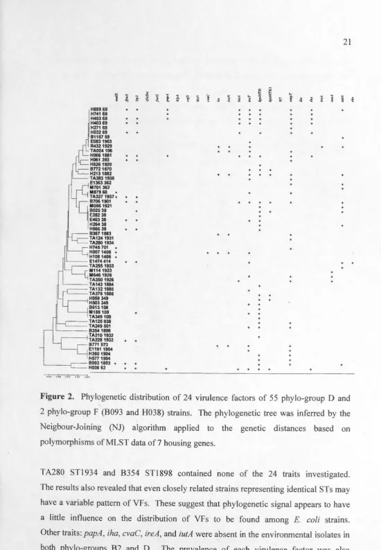
The results of the MLST analysis revealed that the 114 phylo-group B2 strains represented 83 sequence types (STs), whilst the 55 phylo-group D and 2 phylo-group F strains represented 38 STs (Fig. I and 2, respectively). Some strains isolated from humans, animals, and environmental sources were found to represent the same ST, such as ST127 (B2) and ST38 (D). Although other STs, such as ST 95 (B2) and ST69 (D) were predominately observed in humans (Fig. 1 and 2, respectively).
The virulence profiles of the 114 phylo-group B2 strains revealed that all contained at least one of the 24 traits studied (Fig. 1). The human B2 isolate, H411 ST73, contained 16 VF traits, whilst only a single trait was detected in the reptile isolate (Rl38 ST1909) and environmental isolate (E211 ST1910); ompT and ma!X,
respectively. Of the 24 VFs examined, the traits ma!X, fyuA, irp2, and ompT showed the substantial prevalence among 114 B2 strains. Some traits (sfa(foc,focG, hlyA, cnfl,
rl
H&a9141 H707141 + +d
'
H223141 H2B4 998 + +'
+=1~:
1116561!127 +~i1~fsa ! HZ83 1917 +
H5781855 +
-
c____,
+:1~m~:
i
e
E2111910 •L- ~~~;~:
- -H588126 + ~c1ills~:
- - - -E112&589 - - - -TA130638 +
. 6 86791900 +
E10091900 +
111992 1860 +
- 8339 9711 ♦
TA2061386 +
H3241919 +
E5351861 +
TA115 491 +
~
~ TA149 491 +. 1112481824 +
TA3511862 +
--H178144 +
r - -TA201105 +
q
i~~~:r :
8266127 +8288127 +
11361127 +
E1197127 H744127 +
9045 883
D
E15151908 E5H18831
86711865 + · - -TAOM 1864 +. - - -9144 1866 + ' r '---1E15181909 + j I j ~~~~~ •
! i
~~~::2:
n
H8301867 +H41173 +
H580 73 +
H37873 +
' B108 73 + ,---i L H320 1918 + :--, - TA014104 +
LJ -
-
98701868 + r H1461916 +~
~-
~=~~:
H217648 + TA046 646 9264646 +, - - H5041871 +
;__ IH14495 H347 95
- t«.1395 + R527 95 +
'---C=:=
~rr mi
:
-- -TA2921935 +
, - --__ _ TA32618n +
I . ,---j 9433 1873 +
.I E11201873 +
: --1f~J~~~4 :
~ E1193190!i +
._ E1360 1906 + - ----H0831915 +
~
HD90131 +
H186131 + B980 131 +
8127131 +
-c=:
:1ns1m
6 : - H246357 +- TA0931170 +
- TA422110 +
.
--j
~rn:
:
s - 81031894 + , -M6051876 +
~~ H021 91 H333 91 +
H468 91 + " H655 91 +
· - - - - --- -910861895 +
- - - - - -H51518n
---
~=
~m91
1~ :- - - -- 812671897 +
---E536 1879 +
--j B311 681 E1149681 + + H001 681 +
{
'---c:::=
~ig:i1~:~ : - - -- -- - -- E14911907 +. · 1116581928
- --- - --c
:~mg
+ + + + + + + + + + + + + + + + + + + + + + + + + + ♦ ♦ ♦ ♦ ♦ + + ♦ ♦ + + • + + + ♦ + + + ♦ + + + + + + + • + • + + • + + + + + + + + + + + + + + + ♦ + + + + + + + + + + + ♦ + + + + + + + + + + • + + + • + • + + • + • • + • + + + + + + + + • ♦ ♦ + + • ♦ • + + + ♦ ♦ ♦ ♦ ♦ ♦ ♦ ♦ ♦ ♦ • ♦ ♦ ♦ ♦ + + ♦ + • + + + + • ♦ • ♦ + + + + ♦ • + + + + + + + + • + • + + + + + + • + + + + • • ♦ + • ♦ ♦ + + + ♦ + + • • + + + + • + • + + ♦ + + + • + • ♦ + ♦ + + + • • • + + • + + • • + + + + + + + ♦ + + + + + + + + + + + + .e • + + + + + + + + + + + + + + + + + + + + + • + + • + • • + ♦ ♦ ♦ ♦ • • + • • + + • • • + + + + + + + + + + + + + + + + + + 'S
·
•
♦ + + + ♦ + + • + + + + + + + + • + + ♦ + + • + ♦ ♦ + ♦ ♦ + + + + + + + + + + + + + + + + + + + + + ♦ ♦ ♦ + + + ♦ ♦ • + • + • + • • + • • + • • • + + + • • + + + + + + + + + + + + + + + + • + + + + + + + + + + + + + + + + + • + ♦ • ♦ + + + + + + + + + + + + + + + ♦ ♦ + + + + + + + + + + + + + • + + ♦ + + + + • • + • • + + + • • • + • • + + • • + + + + + + + + + + • • + + • + • • + • + + • + + + + + + + • + • + + + + + + + + + + + + • + + + + + + + + + + + + + + + + + • + • + + ♦ ♦ • + + + ♦ + + + + + + + + + + + ♦ ♦ + + + + + + + + + + + + + + + + + + + + + + + + + + + + + + + + + + + + + + + + + + • + + + ♦ ♦ + + + + + • + + + + • • + ♦ + + + + + + ♦ + + + + + + + +Figure 1. Phylogenetic distribution of 24 virulence factors of 1 14 phylo-group 82
strains. The phylogenetic tree was inferred by the Neighbour-Joining (NJ) algorithm
applied to the genetic distances based on polymorphisms of MLST data of 7 housing
H699 69 + +
H741 69
H453 69 + +
H403 69 + +
H271 69
H032 69 + +
B1167 69 E083 1903 R4321929 TA024106
H0061881 + +
H061 393 + +
H6261920 Bn21670 H2131882 TA3831938 E1363 362 M701 362 M879 68 +
TA337 1937 + + +
B706 1901 + + M0S& 1921
B025 38 +
E282 38
E463 38 + +
H26438
H566 38 + +
B3671883 TA1241931 TA2801934 H745 701 +
H0071406 +
H1081406 +
~ -- E1474 414 + +
TA2551933 M1141923
M6461926
TA3501926 '---- - TA1431B84 ~ -TA132 1885 ' - - - TA376 1886 HOSS 349 H503 349 B613108
M188 108 +
TA349108
r - - -TA125 938 c___- -TA249 501 ~ - - - B354 1898
r -- ---<TA210 1932
TA228 1932 + + BTT1 973
E11911904 H3B0 1904 HSTT 1904
B093 1893 + + + H038 62 + +
+ + + + + + + + + + + + + + + ♦ + + + ♦ + + + + ♦ ♦ ♦ + ♦ ♦ ♦ + + ♦ + ♦ + + + + + + + + + + + + ♦ ♦ ♦ + + ♦ + + + + + + + ♦ ♦ + + + + + + + + + + + + + + + ♦ + + + + + + + ♦ + + + + + + + + + + + + • + + + + + + + + + + + + + + + + + + + ♦ + + + + +
Figure 2. Phylogenetic distribution of 24 virulence factors of 55 phylo-group D and 2 phylo-grol}p F (B093 and H038) strains. The phylogenetic tree was inferred by the
Neigbour-Joining (NJ) algorithm applied to the genetic distances based on
polymorphisms of MLST data of 7 housing genes.
TA280 STl 934 and B354 STl 898 contained none of the 24 traits investigated.
The results also revealed that even closely related strains representing identical STs may
have a variable pattern of VFs. These suggest that phylogenetic signal appears to have
a little influence on the distribution of VFs to be found among E. coli strains. Other traits: papA, iha, cvaC, ireA, and iutA were absent in the environmental isolates in
both phyla-groups B2 and D. The prevalence of each virulence factor was. also
[image:31.579.11.562.25.819.2]a particular ST was positive for the trait, all members of the ST were scored as positive
for the trait. This analysis reveals that the frequency of many traits is significantly less
Table 2. Frequency(%) with which virulence-associated traits was detected in E. coli
strains with respect to the phylo-group (B2 and D) membership of the strainsa.
VFs
ma!X fyuA
irp2 sfa/foc focG
papAH
hlyA
cnfl H47 cvaC iss iroN iutA traT kpsM11I kpsMTKl H7 ompT ibe iha ireA
eaeA astA she
Phylo-group B2
(no. of STs = 83)
%VF+
90.4 b
83.1 b
84.3 b
16.9 b
8.4
8.4
9.6b
8.4
8.4
6.0
19.3
34.9 b
13.3
39.8
47.0
18.1
]4.5 b
84.3 b
66.3 b
6.0
7.2
8.4
20.5
24.1 b
Phylo-group D Fisher's Exact Test
(no. of STs = 38)
%VF+ P value c
13.2 <0.0001
26.3 <0.0001
29.0 <0.0001
0.0 0.0048
0.0 ns
7.9 ns
0.0 ns
0.0 ns
0.0 ns
2.6 ns
15.8 ns
15.8 0.0330
13.2 ns
55.3 ns
47.4 ns
18.4 ns
0.0 0.0174
47.4 <0.0001
0.0 <0.0001
7.9 ns
2.6 ns
5.3 ns
26.3 ns
2.6 0.0035
a Produced/grouped at least one example of duplicate STs ( e.g. ST95) was positive for a trait the ST
was scored as +.
b Significant P values for Fisher's Exact Test
c Two-tailed Fisher's Exact Test P values for frequency differences across phylo-groups
at the ST level than at the isolate level. For example,papAHwas detected in 16% of the
B2 isolates, but only in 8% of B2 STs. Similarly, papAH is detected in about 42% on
phylo-group B2 isolates and 23% of phylo-group D isolates (Table 1), but at the ST
level is equally frequent in B2 and D STs (Table 2).
3.3.3 Recombination rates in phylo-group B2 strains
Several different techniques were used to assess the extent of recombination in
phylo-group B2 sequence types (STs) isolated from different sources (humans, mammals,
birds, and environment) using the concatenated sequence data collected for the MLST
characterization of the isolates.
The PH] statistic indicates significant levels of recombination among phylo-group
B2 strains regardless of their source (Table 3). The LDhat and ClonalFrame estimates
of the relative importance of recombination versus mutation are highly correlated
(Table 3). The magnitude of the estimates also suggests that the relative importance of
recombination versus mutation is greatest in B2 STs from humans and least important in
B2 strains from the environment. However, the 95 % confidence intervals estimated by
ClonalFrame reveal that estimates for each of the populations overlap. Further with the
exception of the estimates for the environmentally derived STs the confidence intervals
provide little evidence that recombination is a more important force than mutation in