This is a repository copy of
Associations Between Serum Bone Biomarkers in Early Breast
Cancer and Development of Bone Metastasis: Results From the AZURE (BIG01/04) Trial
.
White Rose Research Online URL for this paper:
http://eprints.whiterose.ac.uk/124922/
Version: Published Version
Article:
Brown, J, Rathbone, E, Hinsley, S orcid.org/0000-0001-6903-4688 et al. (13 more authors)
(2018) Associations Between Serum Bone Biomarkers in Early Breast Cancer and
Development of Bone Metastasis: Results From the AZURE (BIG01/04) Trial. Journal of
the National Cancer Institute, 110 (8). ISSN 0027-8874
https://doi.org/10.1093/jnci/djx280
© The Author(s) 2018. Published by Oxford University Press. This is an open access
article distributed under the terms of the Creative Commons CC BY license, which permits
unrestricted use, distribution, and reproduction in any medium, provided the original work
is properly cited.
eprints@whiterose.ac.uk
https://eprints.whiterose.ac.uk/
Reuse
Unless indicated otherwise, fulltext items are protected by copyright with all rights reserved. The copyright
exception in section 29 of the Copyright, Designs and Patents Act 1988 allows the making of a single copy
solely for the purpose of non-commercial research or private study within the limits of fair dealing. The
publisher or other rights-holder may allow further reproduction and re-use of this version - refer to the White
Rose Research Online record for this item. Where records identify the publisher as the copyright holder,
users can verify any specific terms of use on the publisher’s website.
Takedown
If you consider content in White Rose Research Online to be in breach of UK law, please notify us by
A R T I C L E
Associations Between Serum Bone Biomarkers in Early Breast
Cancer and Development of Bone Metastasis: Results From
the AZURE (BIG01/04) Trial
Janet Brown*, Emma Rathbone*, Samantha Hinsley, Walter Gregory, Fatma Gossiel, Helen
Marshall, Roger Burkinshaw, Helen Shulver, Hasina Thandar, Gianfilippo Bertelli, Keane
Maccon, Angela Bowman, Andrew Hanby, Richard Bell, David Cameron, Robert Coleman
Affiliations of authors:Academic Unit of Clinical Oncology and Sheffield ECMC, University of Sheffield, Weston Park Hospital, Sheffield, UK (JB, ER, RB, HS, RC); Leeds Institute of Cancer and Pathology, University of Leeds, Leeds, UK (JB, ER, AH); Calderdale and Huddersfield NHS Foundation Trust, Huddersfield, UK (ER); Clinical Trials Research Unit, Leeds Institute of Clinical Trials Research, University of Leeds, Leeds, UK (SH, WG, HM); Academic Unit of Bone Metabolism, Metabolic Bone Centre, University of Sheffield, Northern General Hospital, Sheffield, UK (FG); Royal Surrey County Hospital, Guildford, UK (HT); Singleton Hospital, Swansea, UK (GB); Cancer Trials Ireland, University College Hospital, Galway, Ireland (KM); University of Edinburgh Cancer Research Centre, Western General Hospital, Edinburgh, UK (AB, DC); Deakin University, Geelong, Australia (RB).
*Authors contributed equally to this work.
Correspondence to:Janet E. Brown, MD, MSc, MBBS, Academic Unit of Clinical Oncology, University of Sheffield, Weston Park Hospital, Sheffield, S10 2SJ, UK (e-mail j.e.brown@sheffield.ac.uk).
Abstract
Background:Adjuvant therapies can prevent/delay bone metastasis development in breast cancer. We investigated whether serum bone turnover markers in early disease have clinical utility in identifying patients with a high risk of developing bone metastasis.
Methods:Markers of bone formation (N-terminal propeptide of type-1 collagen [P1NP]) and bone resorption (C-telopeptide of type-1 collagen [CTX], pyridinoline cross-linked carboxy-terminal telopeptide of type-1 collagen [1-CTP]) were measured in baseline (pretreatment blood samples from 872 patients from a large randomized trial of adjuvant zoledronic acid (AZURE-ISRCTN79831382) in early breast cancer. Cox proportional hazards regression and cumulative incidence functions (adjusted for factors having a statistically significant effect on outcome) were used to investigate prognostic and predictive associations between recurrence events, bone marker levels, and clinical variables. All statistical tests were two-sided.
Results:When considered as continuous variables (log transformed), P1NP, CTX, and 1-CTP were each prognostic for future bone recurrence at any time (P¼.006,P¼.009,P¼.008, respectively). Harrell’s c-indices were a P1NP of 0.57 (95% confidence interval [CI]¼0.51 to 0.63), CTX of 0.57 (95% CI¼0.51 to 0.62), and 1-CTP of 0.57 (95% CI¼0.52 to 0.63). In categorical analyses based on the normal range, high baseline P1NP (>70 ng/mL) and CTX (>0.299 ng/mL), but not 1-CTP (>4.2 ng/mL), were also
prognostic for future bone recurrence (P¼.03,P¼.03,P¼.10, respectively). None of the markers were prognostic for overall distant recurrence; that is, they were bone metastasis specific, and none of the markers were predictive of treatment benefit from zoledronic acid.
Conclusions:Serum P1NP, CTX, and 1-CTP are clinically useful, easily measured markers that show good prognostic ability (though low-to-moderate discrimination) for bone-specific recurrence and are worthy of further study.
More than 40 000 women die from breast cancer annually in the United States, mainly from distant relapse, which often occurs years after initial breast cancer diagnosis (1). Bone metastases
ultimately affect more than two-thirds of patients with ad-vanced disease (2). Breast cancer cells can remain dormant for many years in the bone microenvironment, escaping the effects
ARTI
CLE
Received:April 11, 2017;Revised:October 26, 2017;Accepted:December 6, 2017
©The Author(s) 2018. Published by Oxford University Press.
This is an Open Access article distributed under the terms of the Creative Commons Attribution License (http://creativecommons.org/licenses/by/4.0/), which permits unrestricted reuse, distribution, and reproduction in any medium, provided the original work is properly cited.
doi: 10.1093/jnci/djx280
of adjuvant systemic therapies and retaining the potential for future activation and proliferation, resulting in metastasis in bone and/or other distant sites.
Because breast cancer cells display this affinity for bone, there is a sound rationale for targeting the bone in the adjuvant setting. Randomized trials of adjuvant bisphosphonates, con-firmed by a meta-analysis of all available data (n¼18 766), have indeed shown that development of bone metastases and death from breast cancer can be reduced. However, the benefits are confined to postmenopausal patients at the time of bisphosph-onate initiation (3–7), strongly suggesting that the postmeno-pausal bone (marrow) microenvironment has a specific interaction with tumor cell homing to bone and/or tumor dormancy.
In the AZURE trial (ISRCTN79831382) in early breast cancer, 3360 women with stage II/III breast cancer were randomized to standard adjuvant treatment alone or with the addition of zole-dronic acid (zoledronate, administered over five years (3,6). With a median of 84.2 months (interquartile range [IQR]¼66–93 months) of follow-up, zoledronate improved invasive disease– free survival (IDFS) in women who were more than five years postmenopausal at diagnosis (n¼1041, adjusted hazard ratio [HR]¼0.77, 95% confidence interval [CI]¼0.63 to 0.96). Baseline (pretreatment) serum samples were collected in a subset of patients, and these provide the opportunity for prespecified analyses of relationships between bone metabolism, as deter-mined by serum bone turnover markers and disease outcomes with or without zoledronate. The role of bone turnover markers has been extensively studied in established bone metastasis (8,9). In the current study, our aims were to determine whether, in early breast cancer, levels of bone turnover markers predicted either the risk of disease relapse (both in and outside bone) or the treatment benefits from zoledronate.
Methods
Patients
In the AZURE trial (3,6), following written informed consent, women with histologically confirmed breast cancer and either lymph node metastasis or T3/T4 primary tumor were randomly assigned to either standard adjuvant therapy (control) or stan-dard adjuvant therapy plus intravenous zoledronate 4 mg (19 doses over five years).
At UK centers, ethics approval was obtained for this study, and participants gave additional consent for blood donation at study entry to be used for biomarker assessment. Serum sam-ples were collected and stored under strict standard operating procedures temporarily at –20C or –80C at local centers before regular transfer to Sheffield for storage at –80C until central batch analysis.
Laboratory Assays
Bone biomarkers were measured against reference standards in a fully accredited central laboratory (Metabolic Bone Unit, University of Sheffield) according to strict standard operating procedures. Personnel performing and reporting the analyses were blinded to clinical data.
Bone Biomarker Analysis
We measured two biomarkers of bone resorption, C-telopeptide of type-1 collagen (CTX), a measure of cathepsin-K-linked
collagen breakdown, and pyridinoline cross-linked carboxy-ter-minal telopeptide of type-1 collagen (1-CTP), which is liberated by matrix metalloproteinases during degradation of mature type-1 collagen. Because 1-CTP is not produced through cathep-sin-K-mediated bone resorption, its concentration is less af-fected by menopause (10). N-terminal propeptide of type-1 collagen (P1NP), released during collagen formation, is a robust and reliable measure of bone formation and was selected for this study (11). P1NP and CTX were measured using Cobas e411 automated immunoassays (Roche Diagnostic, Mannheim, Germany), and 1-CTP was measured by manual enzyme immu-noassay (Orion Diagnostica UniQ ICTP EIA, Espoo, Finland).
The P1NP assay has a lower detection limit of 5 ng/mL and interassay coefficient of variation (CV) of 4.1%. Values were categorized as “high” if greater than 70 ng/mL based on advice from Roche Diagnostics. Results of 70 ng/mL or less were categorized as normal. The CTX assay has a measurement range of 0.010 to 6.00 ng/mL and an interassay CV of 4.0%. The upper limit of normal for premenopausal women (0.299 ng/mL) was used to categorize results as either “high” (>0.299 ng/mL) or
“normal” (0.299 ng/mL). We also performed additional analy-ses with a higher threshold (high>0.556 ng/mL) to allow closer
comparison with an earlier study by Lipton and colleagues (12). The 1-CTP assay was conducted manually with a lower detec-tion limit of 0.3 ng/mL and an upper limit of normal of 4.2 ng/ mL. The intraassay CV was 8.1% at 5.6 ng/mL, and the interas-say CV was 7.6% at 4.8 ng/mL.
Statistical Analysis
Statistical analysis (consistent with REMARK guidelines) was performed on the final AZURE analysis datalock with a median of 84.2 months (IQR ¼ 66–93 months) of follow-up and 966 disease-free survival events (6). All analyses were performed on the intention-to-treat population using SAS version 9.2 or 9.4. Hypothesis testing was performed at the two-sided 5% level.
Cumulative incidence function (CIF) curves were used to in-vestigate time to recurrence, as defined below. The Cox propor-tional hazards (PH) model was used to assess the relationships between the bone biomarkers and prognosis and treatment ef-fect with zoledronate. The proportional hazards assumption was verified by assessing the statistical significance of the inter-action of the relevant bone biomarker and time via an interac-tion term within the Cox model, as well as by a manual review of the CIF curves. Bone marker data were analyzed both as con-tinuous variables (log transformed) and as categorical variables, using the prespecified high vs normal cut-points for both prog-nostic and predictive relationships.
The prespecified end points in the statistical analysis plan were: 1) time to bone recurrence, whether or not bone was the first recurrence (with deaths without prior bone recurrence cen-sored in the Cox PH models and considered competing-risk events in CIF curves); 2) time to first recurrence in bone, includ-ing first recurrence beinclud-ing in bone only or concurrently with recur-rence in another distant site (with deaths without prior recurrence and nonbone (only) first recurrences censored in the Cox PH models and considered competing-risk events in CIF curves); 3) time to first distant recurrence (with deaths without prior distant recurrence censored in the Cox PH models and con-sidered competing-risk events in CIF curves).
Analyses were performed for all participants combined and according to menopausal status and were adjusted for mini-mization factors found to be statistically significant for disease
ARTICLE
outcomes in the main AZURE analyses (ie, lymph node involve-ment, estrogen receptor [ER] status, tumor stage, and type/timing of systemic therapy for each end point), as well as treatment allocation, where this was statistically significant in the main AZURE subgroup analyses. Analyses were also adjusted for treat-ment allocation when assessing the interaction of biomarkers with treatment (predictive analyses), where the interaction term is used to test for heterogeneity between the different biomarker levels. Exploratory analyses were carried out with a composite P1NP/CTX biomarker, in terms of both markers high vs not both markers high.
Harrell’s c-index was used to assess the discriminatory abil-ity of the markers, with a value of 1 representing perfect dis-crimination and 0.5 being no better than chance. Confidence intervals for Harrell’s c-index were calculated as suggested by Newson (13).
Results
Patient Demographics and Baseline Data
Serum samples from 872 UK AZURE participants (441 control arm, 431 treatment arm) were analyzed, with a median follow-up of 84.2 months (IQR¼71.1–92.1 months). Baseline patient demographics (age, lymph node involvement, ER, progesterone receptor [PR], and human epidermal growth factor receptor 2 (HER2) status, menopausal status, systemic therapy, chemo-therapy and statin use) in this test subpopulation were similar to the overall AZURE patient population (Table 1).
Comparison of IDFS outcomes for the biomarker population with the whole AZURE population showed that both the propor-tions of patients with an event and the hazard ratios were simi-lar, though the confidence intervals were wider in the biomarker population due to the smaller number of patients. This similarity also applied when broken down into post- and nonpostmenopausal subgroups (Supplementary Figure 1, avail-able online).
Baseline data for the three biomarkers (also broken down into menopausal status) revealed that the proportion of patients in each category who fall above the normal ranges for P1NP, CTX, and 1-CTP for the whole population were 27.3%, 30.0%, and 50.5%, respectively (Table 2), confirming that the data were appropriate to test the relationship between acceler-ated baseline bone turnover and subsequent distant recurrence events.
Bone Biomarker Prognostic Analyses
Figure 1andTable 3display key data for prognostic analyses in the three prespecified recurrence categories. The proportional hazards assumption was also investigated for each Cox propor-tional hazards model applied in our study. The majority of markers and end points were not close to violating this assump-tion (ie, suggesting no difference in the effects of the markers as time elapses). For the 1-CTP marker, the assumption was only borderline valid, suggesting that the impact of 1-CTP may differ as time elapses, although the Cox proportional hazards model was still appropriate.
Bone Recurrence at Any Time
In adjusted continuous log-transformed analyses (Figure 1), increases in all three markers were strongly associated with sta-tistically significantly increased risk of development of bone
metastasis (P1NP:P¼.006; CTX:P¼.009; 1-CTP:P¼.008). In cat-egorical analyses, P1NP greater than 70 ng/mL (P¼.03) and CTX greater than 0.299 ng/mL (P¼.03), but not CTX greater than 0.566 ng/mL (P¼.12) or 1-CTP greater than 4.2 ng/mL (P¼.10), were statistically significantly prognostic for recurrence in bone at any time (Table 3). Cumulative incidence plots for categorical analysis of time to bone metastasis at any time are shown, ex-emplified for P1NP, inFigure 2for both control and treatment arms.
Taking P1NP, on the basis of the above data, as the likely most sensitive prognostic factor, we tested the role of meno-pausal status in P1NP analyses (data not shown). However, we detected no statistically significant prognostic effect of P1NP on bone recurrence in either postmenopausal or non-postmenopausal patients, when analyzed with P1NP as either a categorical or continuous variable.
Harrell’s c-index values (when coded as [log] continuous var-iables) were similar for all three markers: P1NP c-index was 0.57 (95% CI¼0.51 to 0.63); CTX c-index was 0.57 (95% CI¼0.51 to 0.62); and 1-CTP c-index was 0.57 (95% CI¼0.52 to 0.63).
First Recurrence in Bone (1/2Concurrent Recurrence Elsewhere) In the adjusted continuous analyses, both P1NP (P¼.03) and 1-CTP (P¼.045), appeared statistically significantly prognostic for first recurrence in bone (Figure 1,Table 3). However, in ad-justed categorical analyses, although the hazard ratios for each marker were similar to bone recurrence at any time, the 95% confidence intervals were wide, and no statistically significant relationships between higher marker values and first disease recurrence in bone were seen. The number of bone-only first re-currence events was too small to justify separate analysis of this potential end point of interest.
First Distant Recurrence (Whatever the Site)
There were no associations in either continuous or categorical analyses between baseline P1NP, CTX, or 1-CTP and develop-ment of distant recurrence at any site (Figure 1,Table 3), clearly demonstrating that, in contrast to recurrence specifically in bone, the markers were not prognostic for distant metastasis taken as a whole. Categorical data for IDFS were statistically nonsignificant.
Composite P1NP and CTX Biomarker Analysis
Adjusted analyses were performed to assess risks of recurrence for patients where both P1NP and CTX (using the 0.299 ng/mL cut-point) were high compared with all other patients; details are displayed inTable 4. No statistically significant relation-ships were identified between the composite marker and subse-quent recurrence, although there was an increased risk for bone recurrence at any time in the patients with elevation of both biomarkers (HR ¼ 1.60, 95% CI ¼ 0.99 to 2.48, P ¼ .06). Consideration was given to a joint Cox model containing all three markers, but there were insufficient events to make this meaningful. Further, 1-CTP levels represent a different aspect of the bone turnover process than P1NP and CTX and are largely unaffected by inhibitors of osteoclast function such as bisphosphonates (10).
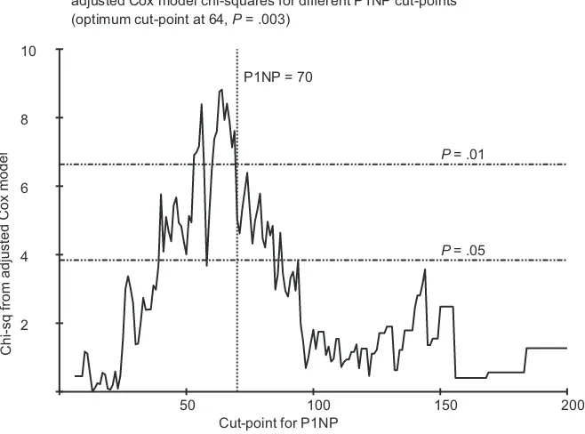
Sensitivity Analyses Assessing Optimum Cut-Points
We explored the effects of different cut-points for categorical prognostic analysis of P1NP and bone metastasis at any time. This analysis (Figure 3) showed that the optimal cut-point for
ARTI
P1NP was approximately 64 nmol/mL, which we judged was sufficiently close to the prespecified value of 70 nmol/mL, bear-ing in mind that the number of events was not sufficient to gen-erate a smooth relationship. For 1-CTP and CTX, similar exploration yielded no clearly optimal cut-point or improve-ment to those preselected (Supplementary Figures 2 and 3, available online).
Analyses for Treatment Effect—Test for Predictive Biomarkers
Although P1NP is higher in postmenopausal women and the benefits of zoledronate are largely restricted to this subset of
patients (6,7), baseline P1NP did not predict benefit from zoledr-onate when assessed against bone metastasis at any time out-come. For example, in categorical analyses considering the effect of P1NP on bone recurrence at any time, there were no statistically significant differences in outcome between the zoledronate and control arms for either high P1NP (HR¼0.99, 95% CI¼0.52 to 1.90) or normal P1NP (HR¼0.84, 95% CI¼0.52 to 1.37) with a nonsignificantPinteractionvalue for the interaction of P1NP and treatment (P¼.69) (Figure 1B).
We also found no statistically significant interaction with treatment allocation for either of the other defined, less frequent outcome categories with any of the bone markers, or with the P1NP/CTX composite bone marker. However, consider-ing the complex inter-relationships between treatment effect and menopausal status in the main AZURE study, the numbers of events are likely insufficient for definitive analysis.
Corresponding continuous (log-transformed) analyses for bone metastases at any time found no statistically significant interaction with treatment allocation for any of the markers analyzed (P1NP:P¼.74; CTX:P¼.47; 1-CTP:P¼.31), confirming that these baseline markers are not predictive of the treatment benefits of zoledronate.
Discussion
Our study showed that patients with high serum levels of P1NP, CTX, or 1-CTP shortly after diagnosis of early breast cancer were associated with a higher risk of developing bone metastasis dur-ing the course of their disease. P1NP appeared to be the most sensitive of the markers studied, but was not predictive of bene-fit from zoledronate. Using CTX and P1NP as a composite bio-marker did not add to the sensitivity of the individual bio-markers. This may be partially because the markers are not independent, reporting on essentially linked metabolic processes, but may also be due to the relatively small number of events in the com-bined group.
It has been long-established that the rate of bone loss accel-erates relatively rapidly in perimenopause, across the meno-pausal transition, with consequent increase in bone turnover markers, associated with an accelerated decrease in measured bone mineral density (BMD) (14,15). The inverse relationship be-tween loss of BMD and increase in bone turnover markers (in-cluding P1NP and CTX) from premenopause through perimenopause to postmenopause is well established (16). As anticipated, our data reflect this pattern, with baseline bone marker values all increasing progressively from premenopause, through the early years following cessation of menses, to more than five years postmenopause.
Although all three markers and especially P1NP are good pre-dictors of bone-specific recurrence, the calculated values of Harrell’s c-index (each around 0.57) suggest that they have only low-to-moderate discrimination, although this is statistically dif-ferent from a chance finding as shown by the lower limit in the 95% confidence intervals being greater than 0.5. From a clinical per-spective, however, it should be borne in mind that even for the two key prognostic indicators in everyday use in breast cancer (lymph nodes and stage), c-indices are 0.62 and 0.63, respectively (W. Gregory, personal communication), only marginally greater than those values reported for the three bone markers in this study.
[image:5.595.55.286.84.559.2]Our findings are consistent with a bone microenvironment with increased bone turnover, providing a fertile “soil” for the development of skeletal metastasis. By contrast with this clear association between baseline bone turnover markers and
Table 1.Baseline demographics of patients in the biomarker
subpop-ulation and overall AZURE popsubpop-ulation*
Parameter
Biomarker population
Overall study population
(n¼872) (n¼3359)
No. (%) No. (%)
Mean age, y 51.4 51.5
Lymph node status
0 16 (1.8) 62 (1.8)
1–3 534 (61.2) 2075 (61.8)
4 320 (36.7) 1211 (36.1)
Unknown 2 (0.2) 11 (0.3)
T stage
T1 285 (32.7) 1065 (31.7)
T2 427 (49.0) 1717 (51.1)
T3 131 (15.0) 456 (13.6)
T4 29 (3.3) 117 (3.5)
Histological grade
1 66(7.6) 285 (8.5)
2 361 (41.4) 1439 (42.8)
3 428 (49.1) 1552 (46.2)
ER status
Positive 676 (77.5) 2634 (78.4)
Negative 192 (22.0) 705 (21.0)
Unknown 4 (0.5) 20 (0.6)
PR status
Positive 361 (41.4) 1423 (42.4)
Negative 205 (23.5) 806 (24.0)
Unknown 304 (34.9) 1119 (33.3)
HER2 status
Positive 108 (12.4) 415 (12.4)
Negative 318 (36.5) 1251 (37.2)
Unknown/not measured 442 (50.6) 1672 (49.8)
Neo-adjuvant therapy intended 52 (6.0) 212 (6.3)
Systemic therapy
Endocrine therapy alone 33 (3.8) 152 (4.5)
Chemotherapy alone 190 (21.8) 719 (21.4)
Endocrine therapy and chemotherapy
649 (74.4) 2488 (74.1)
Use of statins 43 (4.9) 197 (5.9)
Type of chemotherapy
Anthracyclines 819 (93.9) 3132 (93.2)
Taxanes 178 (20.4) 775 (23.1)
Menopausal status
Premenopausal 409 (46.9) 1504 (44.8)
5 y since menopause 123 (14.1) 490 (14.6)
>5 y since menopause 266 (30.5) 1041 (31.0)
Unknown 74 (8.5) 324 (9.6)
*ER¼estrogen receptor; HER2¼human epidermal growth factor receptor; PR¼progesterone receptor.
ARTICLE
recurrence in bone, there was no association detectable between bone turnover markers and distant recurrence taken as a whole, indicating that bone turnover markers specifically provide prog-nostic information for future recurrence in bone and not for me-tastasis more generally (17–19). We acknowledge that, in some cases, elevation of baseline bone markers may be linked with ac-tive, but as yet undetected, bone metastases. However, the rela-tively long follow-up (median¼84 months) and few bone events in the first two years (<5%), when the cumulative incidence
curves diverge, makes it unlikely that the raised markers are sim-ply an early diagnostic indication of bone metastases.
There is important literature evidence supporting our study. In particular, Lipton et al. (12) investigatedb-CTX in 621 postme-nopausal early breast cancer patients in a five-year phase III trial of tamoxifenþ/octreotide. Over 7.9 years (median) of fol-low-up, 19 (3.1%) patients developed bone-only recurrence as first event, 47 (7.5%) developed bone and concurrent other re-lapse as first event, and 57 (9.2%) developed first recurrence in sites excluding bone. Using a categorical analysis (cut-point¼
0.71 ng/mL), higher pretreatmentb-CTX was associated with shorter bone-only recurrence-free survival (HR¼2.8, 95% CI¼
[image:6.595.54.543.73.228.2]1.05 to 7.48, P ¼ .03). However, there was no statistically
Table 2.Distribution of patients with high/normal bone marker values according to menopausal status*
Biomarker Whole population Premenopausal 0–5 y postmenopausal >5 y postmenopausal
P1NP
Assay, median (IQR), ng/mL 55.1 (41.2–72.7) 49.1 (37.3–64.3) 58.4 (42.8–76.1) 64.8 (48.1–84.4)
No. of patients 867 409 121 263
%>70 ng/mL 27.3 18.3 30.1 38.7
CTX
Assay, median (IQR), ng/mL 0.23 (0.15–0.32) 0.18 (0.13–0.26) 0.25 (0.18–0.37) 0.29 (0.21–0.41)
No. of patients 863 408 120 262
%>0.299 ng/mL 30.0 17.4 37.4 45.9
%>0.556 ng/mL 4.2 1.0 7.3 7.9
1CTP
Assay, median (IQR), ng/mL 4.25 (3.26–5.15) 3.99 (3.12–4.95) 4.26 (3.20–5.02) 4.60 (3.77–5.41)
No. of patients 861 408 118 265
%>4.2 ng/mL 50.5 44.5 49.6 61.7
*This table does not include data for the patients whose menopausal status was unknown, included in the whole study population. 1-CTP¼pyridinoline cross-linked carboxy-terminal telopeptide of type-1 collagen; CTX¼C-telopeptide of type-1 collagen; IQR¼interquartile range; P1NP¼N-terminal propeptide of type-1 collagen.
Figure 1.Hazard ratios and 95% confidence intervals for adjusted continuous analyses of log-transformed data for baseline N-terminal propeptide of type-1 collagen, C-telopeptide of type-1 collagen, and pyridinoline cross-linked carboxy-terminal telopeptide of type-1 collagen and disease outcomes.Pvalues were calculated using the likelihood ratiov2test statistic, and tests were two-sided. 1-CTP¼pyridinoline cross-linked carboxy-terminal telopeptide of type-1 collagen; CI¼confidence inter-val; CTX¼C-telopeptide of type-1 collagen; HR¼hazard ratio; P1NP¼N-terminal propeptide of type-1 collagen.
ARTI
[image:6.595.59.536.287.558.2]significant association with first event in the bone plus concur-rent relapse elsewhere or with first recurrence at other distant sites. It should be noted that there were differences in the pa-tient populations and administration of bone-targeted therapy between the Lipton et al. study and our data (the former in-cluded only postmenopausal patients whose tumors were mostly ER positive with consequent lower-risk disease).
A limitation of our study is that only baseline biomarker measurements were available, although because the propor-tional hazards assumption was not violated, this suggests no difference in the effect of the markers as time elapses. While our data suggest that the rate of bone turnover at this early stage of disease when tumor cells may be homing to potential metastatic sites is a statistically significant contributing factor to development of bone metastasis, changes in subsequent bone turnover may also play a role. There is evidence that this might be the case in a study that assessed paired serum sam-ples at baseline and one year within a large, placebo-controlled, randomized study of oral clodronate in early breast cancer (20). Although baseline P1NP was not prognostic for developing bone metastasis after five years of follow up, the incidence of bone metastasis was statistically significantly higher in women whose P1NP value increased by more than 20% in the first year (P<.02).
A further possible limitation of our analysis is that the bio-marker population comprised slightly more than 25% of the total AZURE population. Although we have shown that both baseline demographics and outcomes of the biomarker population and the total trial population are similar, this cannot completely ex-clude the possibility of bias in the population analyzed.
Because the treatment benefits of adjuvant zoledronate in postmenopausal women might be related to inhibition of the increased bone turnover associated with menopause, our find-ing that baseline P1NP levels were not predictive for benefit from zoledronate was initially surprising. However, a number of factors (in addition to low event numbers in some analyses) may contribute to this result. Administration of multiple doses of a potent bisphosphonate can confidently be assumed to sup-press bone turnover throughout the five-year treatment period. This could render baseline marker values less relevant in analy-ses of association. Also, it should be noted that, although zoledronate only produced a benefit in overall invasive relapse in patients who were five or more years postmenopause, it was associated with a reduction in first and subsequent metastasis to bone across all menopausal groups (6). Additionally, bone turnover markers reflect activity across the skeleton as a whole, whereas the amount of bone associated with disseminated tu-mor cells likely comprises only a very small fraction of the total skeletal metabolic activity. Finally, there is the intriguing possi-bility that the efficacy of zoledronate in the adjuvant setting may be due to a direct toxic effect on tumor cells in the bone mi-croenvironment and independent of its action on bone turnover.
Other recent studies have also addressed the need for prognos-tic/predictive biomarkers relating to adjuvant bone-targeted treatment in early breast cancer. Using primary tumor tissue from patients in the AZURE study, we showed that a novel composite biomarker comprising the proteins CAPG and GIPC1 was prognostic for developing bone metastasis (HR¼4.5, 95% CI¼2.1 to 9.8,P<
[image:7.595.53.537.75.346.2].001) and predicted response to zoledronate (P ¼ .008) (21).
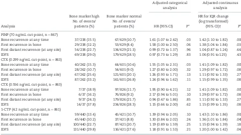
Table 3.Adjusted analyses for high vs normal values of bone markers and analyses for interquartile range change for continuous analyses
Analysis
Bone marker high No. of events/
patients (%)
Bone marker normal No. of events/
patients (%)
Adjusted categorical analysis
Adjusted continuous analysis
HR (95% CI) P*
HR for IQR change (log transformed)
(95% CI)† P*
PINP (70 ng/mL cut-point, n¼867)
Bone recurrence at any time 37/238 (15.5) 67/629 (10.7) 1.61 (1.07 to 2.42) .03 1.42 (1.10 to 1.82) .006
First recurrence in bone 29/238 (12.2) 53/629 (8.4) 1.58 (1.00 to 2.50) .06 1.38 (1.04 to 1.84) .03
First distant recurrence (at any site) 54/238 (22.7) 134/629 (21.3) 0.99 (0.72 to 1.37) .96 1.04 (0.87 to 1.24) .64
IDFS 69/238 (29.0) 179/629 (28.5) 0.97 (0.73 to 1.29) .83 1.06 (0.91 to1.25) .43
CTX (0.299 ng/mL cut-point, n¼863)
Bone recurrence at any time 40/262 (15.3) 64/601 (10.6) 1.55 (1.05 to 2.31) .03 1.41 (1.09 to 1.82) .009
First recurrence in bone 28/262 (10.7) 54/601 (9.0) 1.27 (0.80 to 2.00) .32 1.29 (0.97 to 1.72) .08
First distant recurrence (at any site) 67/262 (25.6) 121/601 (20.1) 1.26 (0.93 to 1.71) .13 1.11 (0.92 to 1.33) .27
IDFS 87/262 (33.2) 161/601 (26.8) 1.24 (0.96 to 1.62) .11 1.15 (0.99 to 1.35) .08
CTX (0.556 ng/mL cut-point, n¼863)
Bone recurrence at any time 7/37 (18.9) 97/826 (11.7) 1.95 (0.90 to 4.21) .12 1.41 (1.09 to 1.82) .009
First recurrence in bone 6/37 (16.2) 76/826 (9.2) 2.17 (0.94 to 5.01) .10 1.29 (0.97 to 1.72) .08
First distant recurrence (at any site) 9/37 (24.3) 179/826 (21.7) 0.94 (0.47 to 1.86) .85 1.11 (0.92 to 1.33) .27
IDFS 14/37 (37.8) 234/826 (28.3) 1.15 (0.66 to 2.00) .62 1.15 (0.99 to 1.35) .08
1-CTP (4.2 ng/mL cut-point, n¼861)
Bone recurrence at any time 59/440 (13.4) 45/421 (10.7) 1.39 (0.94 to 2.05) .10 1.43 (1.10 to 1.86) .008
First recurrence in bone 45/440 (10.2) 37/421 (8.8) 1.30 (0.84 to 2.02) .24 1.36 (1.01 to 1.84) .045
First distant recurrence (at any site) 100/440 (22.7) 87/421 (20.7) 1.19 (0.89 to 1.59) .25 1.15 (0.94 to 1.39) .18
IDFS 131/440 (29.8) 116/421 (27.6) 1.18 (0.91 to 1.53) .21 1.20 (1.00 to 1.42) .04
*Pvalues were calculated using the likelihood ratiov2test statistic, and tests were performed at the two-sided 5% significance level. 1-CTP¼pyridinoline cross-linked carboxy-terminal telopeptide of type-1 collagen; CI¼confidence interval; CTX¼C-telopeptide of type-1 collagen; HR¼hazard ratio; IDFS¼invasive disease–free sur-vival; IQR¼interquartile range; P1NP¼N-terminal propeptide of type-1 collagen.
†These results show the hazard ratio for an interquartile range increase in the log10(P1NP) or ln (CTX, 1-CTP) transformed variables. ThePvalue of these analyses is
unchanged from the adjusted continuous analyses shown inFigure 1.
ARTICLE
No. at risk
Normal High
629 605 565 535 507 479 442 318 77 0
238 229 212 199 188 179 160 115 22 0
Cumulative incidence
0.00 0.05 0.10 0.15 0.20 0.25 0.30 0.35 0.40 0.45 0.50 0.55 0.60 0.65 0.70 0.75 0.80 0.85 0.90 0.95 1.00
Time from random assignment, mo
0 12 24 36 48 60 72 84 96 108
A
No. at risk
No ZOL ZOL
125 119 111 105 100 95 84 61 9 0
113 110 101 94 88 84 76 54 13 0
Cumulative incidence
0.00 0.05 0.10 0.15 0.20 0.25 0.30 0.35 0.40 0.45 0.50 0.55 0.60 0.65 0.70 0.75 0.80 0.85 0.90 0.95 1.00
Time from random assignment, mo
0 12 24 36 48 60 72 84 96 108
B
Figure 2. A)Cumulative incidence function for time to bone metastasis at any time for categorical analysis of N-terminal propeptide of type-1 collagen (P1NP) level or<70 ng/mL (hazard ratio [HR] for adjusted analyses¼1.61, 95% confidence interval [CI]¼1.07 to 2.42,P¼.03).B)Cumulative incidence function for time to bone me-tastasis at any time by treatment arm for participants with high P1NP (70 ng/mL; HR for adjusted analyses¼0.989, 95% CI¼0.517 to 1.895,Pinteraction¼.69 for the inter-action between P1NP and treatment; ie, to assess for differing effects of treatment within the two groups of high or normal P1NP).Pvalues were calculated using the likelihood ratiov2test statistic, and tests were two-sided. 1-CTP¼pyridinoline cross-linked carboxy-terminal telopeptide of type-1 collagen; CTX¼C-telopeptide of
ARTI
[image:8.595.90.513.61.695.2]In another study, amplification of the 16q23 chromosomal region, including amplification of the MAF gene (22), was predictive of breast cancer metastasis to bone (23). However, there remains a need for a simple blood-based test in early breast cancer that can identify patients with a high risk for development of bone metasta-sis. Bone turnover markers are easily measured and are worthy of additional investigation in helping to meet this need.
Funding
This work was supported by Cancer Research UK (through the awards of a Research Studentship to ER, a Clinician Scientist Fellowship to JEB, and support to the Sheffield Experimental Cancer Medicine Centre) and a grant from Novartis Pharmaceuticals.
Notes
The funders had no role in the design of the study; the collec-tion, analysis, or interpretation of the data; the writing of the
manuscript; or the decision to submit the manuscript for publi-cation. Novartis provided academic grant support and supplies of zoledronic acid (Zometa) for the AZURE trial. The authors dis-close the following: JB received fees from Novartis and Amgen for Advisory Boards and Speakers Bureaux; WG received fees from Celgene for statistical consultancy and honoraria from Janssen; DC received nonfinancial support from Novartis; RC re-ceived institutional research grants from Amgen and Bayer, fees from Novartis for expert testimony, and lecture fees from Amgen. All other authors declared no conflicts.
We wish to thank the AZURE trial patients who provided blood samples to support this research.
References
1. US Breast Cancer Statistics. 2017. http://www.breastcancer.org/symptoms/ understand_bc/statistics.
2. Coleman RE. Metastatic bone disease: Clinical features, pathophysiology and treatment strategies.Cancer Treat Rev.2001;27:165–76.
[image:9.595.57.540.75.165.2]3. Coleman RE, Marshall H, Cameron D, et al. Breast-cancer adjuvant therapy with zoledronic acid.N Engl J Med.2011;365:1396–1405.
Table 4.Adjusted prognostic categorical analyses according to a composite P1NP-CTX marker for both P1NP and CTX high vs not both high
Analysis
P1NP, CTX Adjusted analysis
Both high events/censored (%)
Not both high
events/censored (%) HR (95% CI) P*
Bone recurrence at any time 24/118 (16.9) 80/641 (11.1) 1.60 (0.99 to 2.48) .06
First recurrence being in bone 18/124 (12.7) 64/657 (8.9) 1.50 (0.89 to 2.54) .14
First distant recurrence (at any site) 34/108 (23.9) 154/567 (21.4) 1.00 (0.68 to 1.45) .99
First recurrence being in bone only 11/131 (7.7) 47/674 (6.5) 1.26 (0.65 to 2.44) .50
*Pvalues were calculated using the likelihood ratiov2test statistic, and tests were performed at the two-sided 5% significance level. 1-CTP¼pyridinoline cross-linked carboxy-terminal telopeptide of type-1 collagen; CI¼confidence interval; CTX¼C-telopeptide of type-1 collagen; HR¼hazard ratio; IDFS¼invasive disease–free sur-vival; P1NP¼N-terminal propeptide of type-1 collagen.
50 100 150 200
2 4 6 8 10
Cut-point for P1NP
Prognostic analysis - for bone recurrence at any time: adjusted Cox model chi-squares for different P1NP cut-points (optimum cut-point at 64, P = .003)
P = .01
P = .05 P1NP = 70
Figure 3.v2values from adjusted Cox proportional hazards model, analyzing bone metastasis at any time by N-terminal propeptide of type-1 collagen (P1NP), with dif-fering high vs normal P1NP cut-points. Optimum cut-point observed at 64 ng/mL with a correspondingPvalue of .003.Pvalues were calculated using the likelihood ra-tiov2test statistic, and tests were two-sided. P1NP¼N-terminal propeptide of type-1 collagen.
ARTICLE
[image:9.595.134.463.229.473.2]4. Gnant M, Mlineritsch B, Stoeger H, et al. Adjuvant endocrine therapy plus zoledronic acid in premenopausal women with early-stage breast cancer: 62-month follow-up from the ABCSG-12 randomized trial.Lancet Oncol.2011;12: 631–641.
5. Paterson AH, Anderson SJ, Lembersky BC, et al. Oral clodronate for adjuvant treatment of operable breast cancer (National Surgical Adjuvant Breast and Bowel Project protocol B-34): A multicentre, placebo-controlled, randomized trial.Lancet Oncol.2012;13:734–742.
6. Coleman RE, Cameron D, Dodwell D, et al. Adjuvant zoledronic acid in patients with early breast cancer: Final efficacy analysis of the AZURE (BIG 01/04) randomized open-label phase 3 trial.Lancet Oncol.2014;15: 997–1006.
7. Coleman RE, Powles T, Paterson A, et al; Early Breast Cancer Clinical Trials Collaborative Group (EBCTCG). Adjuvant bisphosphonate treatment in early breast cancer: Meta-analyses of individual patient data from randomised tri-als.Lancet.2015;386(10001):1353–1361.
8. Brown JE, Cook RJ, Major P, et al. Bone turnover markers as predictors of skel-etal complications in prostate cancer, lung cancer, and other solid tumors.J Natl Cancer Inst.2005;97:59–69.
9. Coleman RE, Major P, Lipton A, et al. The predictive value of bone resorption and formation markers in cancer patients with bone metastases receiving the bisphosphonate zoledronic acid.J Clin Oncol.2005;23:4925–4935. 10. Maemura M, Iino Y, Yokoe T, et al. Serum concentration of pyridinoline
cross-linked carboxyterminal telopeptide of type I collagen in patients with metastatic breast cancer.Oncol Rep. 2000;7:1333–1338.
11. Lee J, Vasikaran S. Current recommendations for laboratory testing and use of bone turnover markers in management of osteoporosis.Ann Lab Med. 2012; 32:105–112.
12. Lipton A, Chapman J-AW, Demers L, et al. Elevated bone turnover predicts for bone metastasis in postmenopausal breast cancer: Results of NCIC CTG MA.14.J Clin Oncol.2011;29:3605–3610.
13. Newson RB. Comparing the predictive power of survival models using Harrell’s c or Somers’ D.Stata J. 2010;10:339–358.
14. Ebeling PR, Atley L, Guthrie JR et al. Bone turnover markers and bone density across the menopausal transition.J Clin Endocrinol Metab. 1996;81:3336–3371. 15. Riggs BL, Melton LJ. Involutional osteoporosis. N Engl J Med. 1986;314:
1676–1686.
16. Botella S, Restituto P, Monreal I, et al. Traditional and novel bone remodelling markers in premenopausal and postmenopausal women.J Clin Endocrinol Metab.2013;98:E1740–E1748.
17. Ottewell PD, Wang N, Meek J, et al. Castration-induced bone loss triggers growth of disseminated prostate cancer cells in bone.Endocr Relat Cancer.
2014;21:769–781.
18. Wang N, Reeves KJ, Brown HK, et al. The frequency of osteolytic bone metas-tasis is determined by conditions of the soil, not the number of seeds; evi-dence from in vivo models of breast and prostate cancer.J Exptl Clin Cancer Res.2015;34:124.
19. Wang N, Docherty FE, Brown HK, et al. Prostate cancer cells preferentially home to osteoblast-rich areas in the early stages of bone metastasis: Evidence from in vivo models.J Bone Miner Res.2014;29:2688–2696.
20. McCloskey E, Paterson A, Kanis J, et al. Effect of oral clodronate on bone mass, bone turnover and subsequent metastases in women with primary breast cancer.Eur J Can.2010;46:558–565.
21. Westbrook JA, Cairns DA, Peng J, et al. CAPG and GIPC1: Breast cancer bio-markers for bone metastasis development and treatment.J Natl Cancer Inst.
2016;108:djv360.
22. Pavlovic M, Arnal-Estape´ A, Rojo F, et al. Enhanced MAF oncogene expression and breast cancer bone metastasis.J Natl Cancer Inst. 2015;107:djv256. 23. Coleman R, Hall A, Albanell J. et al. Effect of MAF amplification on treatment
outcomes with adjuvant zoledronic acid in early breast cancer: a secondary analysis of the international, open-label, randomised, controlled, phase 3 AZURE (BIG 01/04) trial.Lancet Oncol. 2017;18:1543–1552.
ARTI